effect of green tea extract on the rat liver ... · india and lipton-unilever, englewood cliffs,...
TRANSCRIPT
) 5(7;1120, Journal of American Science org.americanscience.www://http
http://www.sciencepub.net/life [email protected] 65
Effect of Green Tea Extract on the Rat Liver; Histoarchitectural, Histochemical and Ultrastructural Studies
Amal A.A. El Daly
Department of Zoology, Faculty of science, Benha University, Benha, Egypt
com.ahooml_eldaly@y Abstract: Green tea consumed worldwide since earliest time considered beneficial to human health due to its specific metabolic activity along with antioxidant effect. This study was headed for investigate the effect of green tea on histoarchitecture and histochemistry as well as the fine structure of rat liver. For this purpose, male albino rats (Rattus norvegicus); 3 months age weighing between 100 –120 g were used. The animals group-housed six for each in wire mesh cages fed ad libitum divided into two groups: control and experimental group. The latter was divided into three subgroups; 1%, 1.5% and 3% green tea extract feeding animals. Green tea was received instead of drinking water for 25 days using feeding bottles. After the experimental period, the animals were sacrificed and liver pieces were prepared for both light and electron microscopic examination. The results depicted hypertrophied hepatocytes associated with cloudy swelling. There were some pyknotic and karyorrhectic hepatic nuclei in comparison to the control. Blood vessels appear congested and Blood sinusoids contracted. There was an indication of few collagen fibrils in the hepatic stroma. Hepatocytes had PAS positive deposits in their cytoplasm. Furthermore, hyalinization of the hepatocytes was distinct in the animal's liver feeding on higher doses. The ultrastructural results revealed destructed hepatocytic organelles as well as hypertrophied and irregular contoured hepatocytic nuclei. Moreover, many lipid droplets, few profiles of granular endoplasmic reticulum and destructed mitochondria in the hepatocytes cytoplasm were apparent especially after higher doses of treatment. Though, it was fulfilled that green tea consumption induced an alteration in the liver tissues and its fine structure as well as carbohydrate metabolism. Consequently, another aspect was providing into the cellular response of rat liver toward green tea extract property. It must be carefully using for it's harmfully outcome on long term.
[Amal A.A. El Daly. Effect of Green Tea Extract on the Rat Liver; Histoarchitectural, Histochemical and Ultrastructural Studies. Journal of American Science 2011;7(5):65-73]. (ISSN: 1545-1003). http://www.americanscience.org. Keywords: Green tea extract, Histoarchitecture, Histochemistry, Ultrastructure, Liver, Rat 1. Introduction
Tea is one of the most widely consumed beverages in the world made from the leaves of Camellia sinensis L. species. Green tea is the non-oxidized/non fermented product contains high quantities of several polyphenolic flavonoid compounds such as epicatechin, epicatechin gallate, epigallocatechin, and epigallocatechin gallate (EGCG) which reported to have antioxidant properties (Graham, 1992; Rice-Evens et al., 1996) well thought-out to maintain and improve health. It contains high quantities of an active compounds (catechin and caffeine) so as to might act to exert thermogenic and so an antiobesity action (Chantre and Lairon, 2002). It has anticarcinogenic activity (Stoner and Mukhtar, 1995; Zhen, 2002). Moreover, polyphenols because an active ingredients exhibit antioxidant and free radical scavenging properties showed cardioprotective, neuroprotective, antidiabetic, and antimicrobial properties (Liao et al., 2001; Alschuler et al., 1998; Augustyniak et al., 2005). Besides, EGCG prevents the ethanol induced
hepatotoxicity and inhibits the development of the fatty liver and renal failure (Yokozawa et al., 1996 & 2005; Yun et al., 2007). At the level of green tea extract, many researches reported that it have a benefit for human and protective against the diseases resembling treatment of arthritis, infection, and impaired immune function (Van et al., 1999). Moreover, it reduce cholesterol synthesis in liver (Bursill et al., 2007) and traditional decrease the severity of liver injury in association with lower concentrations of lipid peroxidation (Chen et al., 2004; Duh et al., 2004). In addition, Kan et al. (2009) demonstrated that green tea extract have a protective against nephrotoxicity and oxidative damage in rat kidney induced by gentamicin. In the period in-between, few researchers (McCormick et al., 1999; Stratton et al., 2000; Chang et al., 2003) have investigate the potential toxicity of green tea and polyphenols when administered at high doses, as a concentrated product. Although the potential biological effects and powerful antioxidant properties of green tea extracts (Vial et al., 2003 and

) 5(7;1120, Journal of American Science org.americanscience.www://http
http://www.sciencepub.net/life [email protected] 66
Bonkovsky, 2006) data regarding the hepatotoxicity effect are limited in the literature. There are no longer doubt that ingestion of concentrated extracts of Chinese green tea (C. sinensis) poses a real and growing risk to liver health (Jimenez-Saenz and Martinez-Sanchez, 2006) of which we require to be attentive. Additionally, the mechanisms of hepatotoxicity of that C. sinensis ought to have further investigation and its wide-spread consumption requires an increase attention by doctors and pharmacovigilance authorities (Javaid and Bonkovsky, 2006). However, the data available are still not definitive for cellular and in vivo hepatotoxicity caused by green tea phenolic acids and catechins (Galati et al., 2006). It was reported that green tea, like any active principle, needs to be used at the exact composition and the accurate posology to exert a pharmacologically significant effect (Chantre and Lairon, 2002). Therefore, the aim of the current study was to investigate the effect of green tea on the liver of rats, is it has a side effects at any hazard doses, especially it used in a further attenuated studies as a protective antioxidant with no harmful material. 2. Materials and Methods Green tea extract:
Green tea (Kangra, Himanchal Pradesh, India and Lipton-Unilever, Englewood Cliffs, NJ, USA) was purchased locally packaged by the Egypt National Native Product sources. Green tea extract (GTE) was prepared according to Khan et al, (2009) by adding green tea (30 g) to 500ml of boiling water, steeped for 15–20 min. Infusion was cooled to room temperature and then filtered. The tea leaves were extracted a second time with 500ml of boiling water and filtered, and the two filtrates were combined to obtain 3% green tea extract (3 g tea leaves/100 ml water).This dietary treatment reflected a daily consumption of 3 cups of green tea by an adult weighing 70 kg. The resulting clear solution is similar to tea brews consumed by humans. The used extract poured into the animals feeding bottles. The rats supplied with freshly prepared tea every morning. Animals and experimental design:
Male albino rats (Rattus norvegicus) obtained from the National Research Center in Dukki, Cairo (N.R.C.), 3-4 months age weighing between 100 –120 g were used. The animals were housed in wire mesh cages fed ad libitum and allowed to adjust to the new environment for one week before starting the experiment. The rats were housed at 25 ± 2oC dark/light cycle. Animals were
randomly divided into two experimental groups each of six animals as follows: 1) Group I (control): Animals given standard diet and
tap water. 2) Group II (green tea extract): Animals given
standard diet and green tea extract (GTE). This group divided into three subgroups so as to received GTE in three concentrations 1% , 1.5% and 3% ) instead of drinking water within a repeated dose for 25 days to determine the dose dependent effect.
After the experimental period, 24 hrs after the previous administration the animals were anesthesized under light ether, livers were removed and subjected to different studies. Preparation of Specimens:
Small pieces of liver were fixed separately in 10% buffered neutral formalin for 24hrs. Tissues were processed and embedded in paraffin blocks. Paraffin sections were cut with at 5μ thickness, stained by routine hematoxylin and eosin stain and Crossman's Trichrome stain for evaluation of collagen fibers. Other sections prepared for periodic acid Schiff reaction intended for carbohydrates ( Bancroft and Stevens, 1990). The ready sections examined under a Zeiss light microscope. For electron microscopy small pieces of liver were fixed in 2% glutaraldehyde fixative in 0.1M Na- cacodylate buffer, pH 7.2, followed by three washes in the buffer, then post fixed during 1 h in 1% OsO4 in the same buffer, dehydrated, cleared and infiltrated within Resin. Ultra thin sections were stained with 2% uranyl acetate, followed by Reynold's lead citrate and examined with a Siemens ELMISKOP I or Zeizz M-109 Turbo electron microscope. 3. Results:
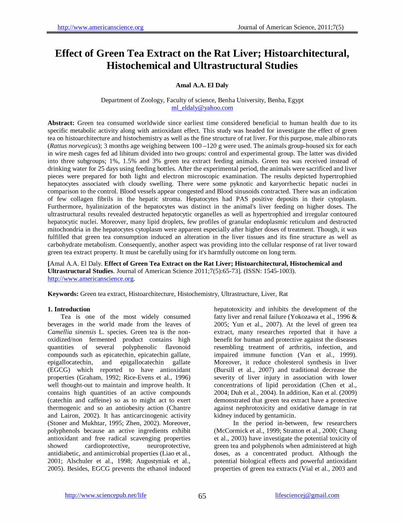
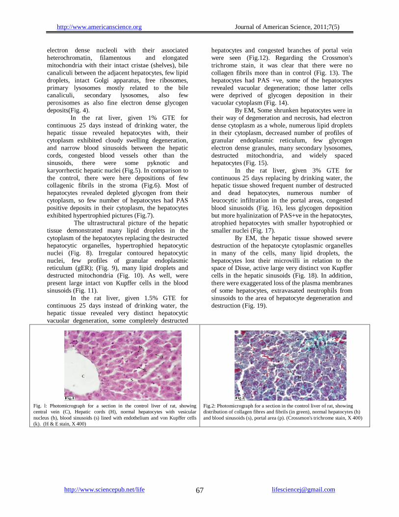
In control liver of rats, the hepatic tissue is formed of hepatic cords of normal healthy polyhedral hepatocytes having one or two vesicular nuclei, every with distinct one or more nucleoli. The blood sinusoids in between the hepatic cords are lined with flat endothelial cells and von Kupffer cells (Fig. 1). The distribution of collagenic fibers and fibrils could be distinguished with Crossmon's trichrome stain, while the hepatocytes took a reddish colour and the blood erythrocytes were orangiophilic (Fig. 2). The distribution of glycogen in the hepatocytes took the magenta colour after Periodic acid Schiff technique (PAS) (Fig. 3). The hepatic tissue by the electron microscope (EM) revealed the ideal hepatocytes with normal mostly euchromatic nuclei and fewer nuclear heterochromatic contents with apparent more
) 5(7;1120, Journal of American Science org.americanscience.www://http
http://www.sciencepub.net/life [email protected] 67
electron dense nucleoli with their associated heterochromatin, filamentous and elongated mitochondria with their intact cristae (shelves), bile canaliculi between the adjacent hepatocytes, few lipid droplets, intact Golgi apparatus, free ribosomes, primary lysosomes mostly related to the bile canaliculi, secondary lysosomes, also few peroxisomes as also fine electron dense glycogen deposits(Fig. 4).
In the rat liver, given 1% GTE for continuous 25 days instead of drinking water, the hepatic tissue revealed hepatocytes with, their cytoplasm exhibited cloudy swelling degeneration, and narrow blood sinusoids between the hepatic cords, congested blood vessels other than the sinusoids, there were some pyknotic and karyorrhectic hepatic nuclei (Fig.5). In comparison to the control, there were here depositions of few collagenic fibrils in the stroma (Fig.6). Most of hepatocytes revealed depleted glycogen from their cytoplasm, so few number of hepatocytes had PAS positive deposits in their cytoplasm, the hepatocytes exhibited hypertrophied pictures (Fig.7). The ultrastructural picture of the hepatic tissue demonstrated many lipid droplets in the cytoplasm of the hepatocytes replacing the destructed hepatocytic organelles, hypertrophied hepatocytic nuclei (Fig. 8). Irregular contoured hepatocytic nuclei, few profiles of granular endoplasmic reticulum (gER); (Fig. 9), many lipid droplets and destructed mitochondria (Fig. 10). As well, were present large intact von Kupffer cells in the blood sinusoids (Fig. 11). In the rat liver, given 1.5% GTE for continuous 25 days instead of drinking water, the hepatic tissue revealed very distinct hepatocytic vacuolar degeneration, some completely destructed
hepatocytes and congested branches of portal vein were seen (Fig.12). Regarding the Crossmon's trichrome stain, it was clear that there were no collagen fibrils more than in control (Fig. 13). The hepatocytes had PAS +ve, some of the hepatocytes revealed vacuolar degeneration; those latter cells were deprived of glycogen deposition in their vacuolar cytoplasm (Fig. 14). By EM, Some shrunken hepatocytes were in their way of degeneration and necrosis, had electron dense cytoplasm as a whole, numerous lipid droplets in their cytoplasm, decreased number of profiles of granular endoplasmic reticulum, few glycogen electron dense granules, many secondary lysosomes, destructed mitochondria, and widely spaced hepatocytes (Fig. 15).
In the rat liver, given 3% GTE for continuous 25 days replacing by drinking water, the hepatic tissue showed frequent number of destructed and dead hepatocytes, numerous number of leucocytic infiltration in the portal areas, congested blood sinusoids (Fig. 16), less glycogen deposition but more hyalinization of PAS+ve in the hepatocytes, atrophied hepatocytes with smaller hypotrophied or smaller nuclei (Fig. 17). By EM, the hepatic tissue showed severe destruction of the hepatocyte cytoplasmic organelles in many of the cells, many lipid droplets, the hepatocytes lost their microvilli in relation to the space of Disse, active large very distinct von Kupffer cells in the hepatic sinusoids (Fig. 18). In addition, there were exaggerated loss of the plasma membranes of some hepatocytes, extravasated neutrophils from sinusoids to the area of hepatocyte degeneration and destruction (Fig. 19).
Fig. l: Photomicrograph for a section in the control liver of rat, showing central vein (C), Hepatic cords (H), normal hepatocytes with vesicular nucleus (h), blood sinusoids (s) lined with endothelium and von Kupffer cells (k). (H & E stain, X 400)
Fig.2: Photomicrograph for a section in the control liver of rat, showing distribution of collagen fibres and fibrils (in green), normal hepatocytes (h) and blood sinusoids (s), portal area (p). (Crossmon's trichrome stain, X 400)

) 5(7;1120, Journal of American Science org.americanscience.www://http
http://www.sciencepub.net/life [email protected] 68
Fig.3: Photomicrograph for a section in the control liver of rat, showing normal distribution of glycogen in the hepatocytes (in magenta colour), blood arteriole (ar), blood sinusoids (s). (PAS-technique, X 400)
Fig.4: Electron micrograph for a section in the liver of control rat showing normal hepatocyte nucleus (N) and 2 nucleoli (nu), mitochondria (m), bile canaliculi (c ) and related lysosomes (s), granular endoplasmic reticulum (ER) in many profiles (ER), Golgi apparatus (G), few lipid droplets (P), free ribosomes ( r ), secoundary lysosomes (sc) and peroxisomes (ox).(X 6000)
Fig.5: Photomicrograph for a section in the liver of rat given 1% GTE for continuous 25 days instead of drinking water, showing cloudy swelling of hepatocytes and their nuclei, the cells are suffered from slight hydropic degeneration, narrow blood sinusoids (s) congested arteriole (ar), note pyknotic nucleus (p) and karyorrhectic nuclei (k) .( H & E stain, X 400).
Fig.6 : Photomicrograph for a section in the liver of rat given 1% GTE for continuous 25 days instead of drinking water showing, more deposition of collagen fibrils than control (in green), leucocytic infiltration in the portal area (in), bile ducts (b), branches of hepatic arterioles (ar). (Crossmon's trichrome stain, X 400).
Fig.7: Histochemical detection of liver section of rat given 1% GTE for continuous 25 days instead in drinking water, showing distribution of glycogen in the hypertrophied hepatocytes in few amounts (in magenta colour), and the PAS positive reacted basal lamina of the branch of the hepatic arteriole (arrow). (PAS technique, X 400).
Fig.8: Electron micrograph for a section in the liver of rat given 1% GTE for continuous 25 days instead of drinking water, showing many lipid droplets of variable sizes (P) in the hepatocytes, replacing the destructed some organelles, hypertrophied hepatocytic nucleus (n) that is deviated to one side, few number of lysosomes (l) some in groups (d) were poured to the adjacent blood sinusoid, endothelial cell of blood sinusoid (e), (X 6000).
Fig.9 : Electron micrograph for a section in the liver of rat given 1% GTE for continuous 25 days instead of drinking water, showing secondary lysosomes (l) many lipid droplets, irregular contour of hepatocytic nucleus (n), few granular ER profiles (ER), cross sectioned profiles of the endoplasmic reticulum cisternae (c ), (X 8000).
Fig.10: Electron micrograph for a section in the liver of rat given 1% GTE for continuous 25 days instead of drinking water, showing destructed mitochondria with many lipid droplets ( p ) at the region of the destructed mitochondria, distorted other mitochondria (m), (X 8000).

) 5(7;1120, Journal of American Science org.americanscience.www://http
http://www.sciencepub.net/life [email protected] 69
Fig.11: Electron micrograph for a section in the liver of rat given 1% GTE for continuous 25 days instead of drinking water, showing active intact von kupffer cell (k)in the hepatic sinusoid (s), distorted mitochondria (m) and many lipid droplets (p), (X 6000).
Fig.12: Photomicrograph of hepatic histopathology in rat liver given 1.5% Green tea extract for continuous 25 days instead of drinking water, displaying very distinct vacuolar degeneration of many hepatocytes (arrows), congested branches of portal vein (co) in the portal area, some degenerated hepatocytes and completely destructed or obscured (d), (H & E stain, X400).
Fig.13: Photomicrograph of hepatic histopathology in rat liver given 1.5% Green tea extract for continuous 25 days instead of drinking water ,screening congested blood sinusoids and branch of hepatic arteriole (ar), degenerated hepatocytes (d), (Crossmon's trichrome stain, X 400).
Fig.14: Histochemical examination of rat liver section given 1.5% Green tea extract for continuous 25 days instead of drinking water, viewing accumulation of hypertrophied hepatocytic glycogen in their cytoplasm(in magenta colour), some hepatocytes were suffering from vacuolar degeneration and had no glycogen deposits, (PAS-technique, X 400)
Fig.15: Electron micrograph for a section in the liver of rat given 1.5% GTE for continuous 25 days instead of drinking water ,performance some affected shrunken hepatocytes (1 & 2) having electron dense cytoplasm with many lipid droplets (P), the latter were also present in the still active hepatocytes, mitochondria with destructed cristae (m), primary lysosomes (l), bile canaliculi (c), decreased profiles of granular endoplasmic reticulum (ER), secondary lysosomes (sc), widely spaced hepatocytes, many delicate electron dense glycogen deposits (g), (X 8000).
Fig.16: Photomicrograph for a section in the liver of rat given 3% GTE for continuous 25 days instead of drinking water, showing numerous number of leucocytic infiltration in the portal area of the hepatic tissue, hypotrophied nuclei, dead hepatocytes (d), congested blood sinusoids(s), (Crossmon's trichrome stain, X 400).
Fig.17: Histochemical analysis of hepatocellular distribution of carbohydrates using periodic acid Schiff reaction in liver of rat given 3% GTE for continuous 25 days instead of drinking water; showing less storage of glycogen(in magenta colour) in the hepatocytes than in those of 1.5% GTE given, atrophied hepatocytes with smaller hypotrophied nuclei, many dead hepatocytes(d), (PAS-technique, X 400).
Fig.18: Electron micrograph for a section in the liver of rat given 3% GTE for continuous 25 days instead of drinking water, showing severe destruction for the cytoplasmic organelles of many hepatocytes 1,2,3 & 4 that evinced also many lipid dropletrs (p), the hepatocytes lost ther microvilli in the space of Disse (d), the blood sinusoids evinced intact active von kupffer cells(k),endothelium of the sinusoids (e), a lymphocyte (ly) in the sinusoid , (X 6000)

) 5(7;1120, Journal of American Science org.americanscience.www://http
http://www.sciencepub.net/life [email protected] 70
Fig.19: Electron photomicrograph corresponds to hepatocytes of rat given 3% GTE for continuous 25 days in drinking water ,showing exaggerated destructed hepatocytes (h) that lost its cell membrane, the neighboring 3 hepatocytes 1,2 & 3 had electron dense cytoplasm , many lipid droplets(p), extravasated neutrophil (n) to the area of hepatocyte degeneration, blood vessel (bl), (X 8000)
4. Discussion Green tea is a natural antioxidant that have been used in the most enduring of food cultures-Chinese and Japanese tea and to the safety concerns about nutrient supplements such as vitamin C., Vit. E and B-carotenes. Although these concerns still exist, consumption of naturally derived antioxidants such as green tea beverages and extract may a safer alternative and effective means of increasing the intake of antioxidants, since each cup of green tea (100 ml) is reported to contain 50- 100 mg of polyphenols ( Luo et al.,1997). However, an ancient knowledge in traditional remedies does not always guarantee safety. There have been a number of available adverse events, including hepatic failure associated with human use of green tea extracts intended for its antioxidant (Gloro et al., 2005; Javaid and Bonkovsky, 2006; Jimenez-Saenz and Martinez-Sanchez, 2006; Bjornsson and Olsson, 2007). Moreover, consumption of natural remedies which considered as food supplements need increase attention to prevent liver hepatotoxicity (Javaid and Bonkovsky, 2006). It was reported that in vitro treatment of rat hepatocytes with high concentration of epigallocatechin gallate (EGCG) reduced cell viability (Schmidt et al., 2005; Galati et al., 2006) and hence caused liver, kidney and gastrointestinal toxicity (Isbrucker et al. ,2006). So, concerning the findings of the latter authors, the current work is coincident with their results, where the hepatocytes of rats showed hydropic degeneration and the cells appear larger than the hepatocytes of control due to cloudy swelling after 1% GTE, also congested hepatic arterioles, and deprived hepatocytes from deposition of glycogen in their cytoplasm, leucocytic infiltration in the portal areas of the hepatic tissue. Moreover, the ultrastructural study revealed numerous secondary lysossomes, numerous lipid droplets; fewer profiles of granular endoplasmic reticulum, large intact and
active von Kupffer cells and some distorted mitochondria, some of the hepatocytes nuclei were deviated to one side, few numbers of primary lysosomes. In vivo studies, Isbrucker et al., (2006) and Galati (2006) suggested that administration of high doses of the tea catechin, EGCG resulted in hepatotoxicity, so disagreeing with the finding of Bonkovsky (2006), Federico et al., (2007) and Sarma et al., (2008) who considered green tea is safe in a wide range of doses, but hepatotxicity was related to consumption of high does of tea-based dietary supplements. Consequently, the results of this work, after giving GTE by a concentration of 1.5%, the hepatic tissue evinced hepatocyte vacuolar degeneration followed by destructed some hepatocytes, congestion of hepatic arterioles and blood sinusoids apparent intact well-developed active Kupffer cells and few fibrin deposits in the hepatic stroma, more glycogen deposition in the hepatocytes cytoplasm in comparison to control. Other effects of similarly doses that tested in this study have been found by Elasrag (2010). The research focused his demonstration on mutation, chromosomal damage and necrosis in the liver and kidney. Furthermore, Lambert et al. (2007) added that gastrointestinal irritation associated with consumption of high doses of tea preparations. From the view of the portal areas of the hepatic tissue which was also considered in this study, more leucocytic infiltrations and congested blood sinusoids were present. It might be due to inflammatory reaction and thrombosed capillaries a accompanied the necrosis of the hepatocytes (Cotran et al., 1999). In the other hand, the atrophied hepatocytes with smaller hypertrophied nuclei apparent in this study probably due to vascular damaged after ingestion of higher doses as reported by the same author. Moreover, at the level of tea ingredients, liver necrosis was associated with treatment of high concentrations of high doses of epigallocatechin

) 5(7;1120, Journal of American Science org.americanscience.www://http
http://www.sciencepub.net/life [email protected] 71
gallate (Schmidt et al., 2005; Goodin and Rosengren,2003) and catechin preparation up to 2000 mg/kg induced chromosomal aberration (Ogura et al., 2008). In converse, Manna et al. (2009) reported that tea polyphenol diminish the inflammatory response, proliferation and apoptosis in lung lesions on the 9th week of benzo[a] pyrene exposure. According to this work, ingestion of the higher dose (3% GTE) revealed depletion of glycogen in hepatocytes cytoplasm but lesser than lower dose i.e. dose dependant. It may possibly induced instability of metabolic enzymes related to glycogen storage. Hence, it previously reported that the polyphenols of GTE such as flavonoids and pyrogallol are potent inhebetors of the detoxifation enzyme COMT (Kdowakiet al., 2005) and catechin gallate, the major constituent in GTE were found to inhebits human COMT-mediate O-methyl translation of catechol estrogen with high potency (Nagai,2004). The mechanism by which green tea extract induces trouble in carbohydrate metabolism may by attributed to mitochondrial toxicity and reactive oxygen species (ROS) formation by tea catechins which induced cytotoxic effects (Galati et al., 2006). The present data agreed with other work by Broadhurst et al. (2000) and Kao et al. (2000) who found that glucose production inhibited by green tea leading to lower blood glucose level that is related to glycogen (PAS-positive) of the hepatocytes. Some authors reported that GTE polyphenols, epigalleatechi-3-gallate was mimetic in that lowered blood glucose, while others approved that glucose-carrier transport through cell membrane (Waltner-Law et al., 2002). Electron microscopic examination of rats liver cells after supplemented green tea in this study, revealed destruction of the mitochondrial cristae and somewhat decreased profiles of the granular ER besides wider blood sinusoids that carried neutrophils to the areas of destructed hepatocytes and their organelles. These changes were recognized especially after higher dose treatment. Hence, might be due to the bioavailability and oxidative stress induced by administration of high doses of the tea catechin EGCG (Maeta et al. (2007; Schmidt et al., 2005). Moreover, Arakawa et al. (2004) and Nakagawa et al. (2004) added that EGCG promotes apoptosis and has bactericidal activity which is qualified to its ability to reduce O2 to yield H2O2 The present data revealed trouble in several hepatocytes which denoted within electron dense cytoplasm and rich lipid droplets in accordance with Kan et al (2009) who reported significant increase of phospholipids after GT consumption. Furthermore, abundant of secondary lysosomes were apparent because of the attack of primary lysosomes to the degenerated cytoplasmic organelles of the
hepatocytes. Kumar et al. (2005) reported that the increases in the number of autophagic vacuoles within the cell that contain fragments of cell components like mitochondria and endoplasmic reticulum at which the lysosomes discharge their hydrolytic contents This thought to connected to potential toxicity when green tea administered at high dose that previously confirmed by Change et al. (2003) and Stratton et al. (2000). Since each model has its reverse, this work can be finish with the intention of GTE must supplement by one or two cups daily to be beneficially for human. In the main time, it was determined that polyphenols of GTE are exhibit antioxidant and free radical scavenging properties (Scott et al., 1993) but other searchers found to have potent inhibitors of the detoxification enzyme (Kadowaki et al., 2005). Others sure those catechins are known to be unstable at neutral and basic pH and undergo oxidation leading to production of reactive oxygen species. Besides, EGCG has been reported to have both antioxidant and pro-oxidative activities (Sang et al., 2005a) under certain conditions and may induce oxidative stress in vivo (Lambert et al., 2007 and Sang et al., 2005b). Conversely, Bonkovsky (2006), Federico et al. (2007) and Sarma et al. (2008) found that green tea is safe in a wide range of doses. In this regard, Isbrucker et al. (2006) found that oral administration of green tea extract (Teavigo) to Bangle dogs for 13 weeks resulted in dose dependent toxicity and death, conditions that are supported by the findings of this work. 5. Conclusion:
The repeated ingestion of highly concentrated up to 3% green tea extracts has its harmful effect on the liver tissues that are subjected to variable hazards due to hepatotoxicity from tea extract polyphenols, so it can be allowed to ingest GTE beverage in a low concentration and for one or two times a day, every one a cup of 100ml containing 50-100mg of poly phenols, so to keep well the hepatic tissue. Corresponding author: Amal A.A. El Daly Department of Zoology, Faculty of science, Benha University, Benha, Egypt [email protected] References: Alschuler, L. 1998: Green tea: Healing tonic. Am J
Nat Med, 5: 28 –31. Arakawa, H., Maeda, M., Okubo, S. and
Shimamura,T. 2004: Role of hydrogen peroxide in

) 5(7;1120, Journal of American Science org.americanscience.www://http
http://www.sciencepub.net/life [email protected] 72
bactericidal action of catechin. Biol. Pharm. Bull., 27: 277-281.
Augustyniak, A, Waskiewicz, E., Skrzydlewska, E. 2005: Preventive action of green tea from changes in the liver antioxidant abilities of different aged rats intoxicated with ethanol. Nutrition, 21: 925 –32.
Bancroft, J.D., Stevens, A., 1990. Theory and Practice of Histological Techniques. 3rded. Churchill Livigstone, Londo, Torento.
Bursill, C. A., Abbey, M. and Roach, P. D. 2007: A green tea extract lowers plasma cholesterol by inhibiting cholesterol synthesis and upregulating the LDL receptor in the cholesterol-fed rabbit. Atherosclerosis, 193: 86-93.
Bjornsson, E., Olsson, R. 2007: Serious adverse liver reactions associated with herbal weight-loss supplements. J Hepatol, 47: 295 –297.
Bonkovsky, H. L. 2006: Hepatotoxicity associated with supplements containing Chinese green tea (Camellia sinensis). Ann Intern Med, 144: 68–71.
Broadhurst, C. L., Polansky M. M. and Anderson, R. A. 2000: Insulin-like biological activity of culinary and medicinal plant aqueous extracts in vitro. J Agric Food Chem; 48: 849 –52.
Chantre, P. and Lairon, D. 2002: Recent findings of green tea extract AR25 (Exolise) and its activity for the treatment of obesity. Phytomedicine, 9 : 3 – 8.
Chang, P. Y., Mirsalis, J., Riccio, E. S., Bakke, J. P., Lee, P. S., Shimon, J., Phillips, S., Fairchild, D., Hara, Y. and Crowell, J. A. 2003: Genotoxicity and toxicity of the potential cancer-preventive agent polyphenon E. Environmental and Molecular Mutagenesis, 41 : 43 –54.
Chen, J. H. ,Tipoe, G. L. , Liong, E. C., So, H. S., Leung, K. M. and Tom, W. M., 2004: Green tea polyphenols prevent toxin-induced hepatotoxicity in mice by down-regulating inducible nitric oxide-derived prooxidants. Am J Clin Nutr, 80: 742 –51.
Cotran, R. S., Kumar, V. and Collains, T. 1999: Robbins Pathologic Basis of Disease.6th ed.Saunders.
Duh, P. D., Yen, G. C., Yen, W. J., Wang, B. S. and Chang, L. W. 2004: Effects of pu-erh tea on oxidative damage and nitric oxide scavenging. Journal of Agricultural and Food Chemistry, 52: 8169–8176.
Elasrag, M. A. 2010: protective effect of some medical plants against probable genotoxic effects of certain antibiotic on certain mammal. M. Sci. Thesis, department of zoology, Faculty of Science, Benha University, P: 189-201.
Federico, A., Tiso, A. and Loguercio, C. 2007: A case of hepatotoxicity caused by green tea. Free Radic. Biol. Med, 43: 474.
Galati, G., Lin, A., Sultan, A. M. and O’Brien, P.J. 2006: Cellular and in vivo hepatotoxicity caused by green tea phenolic acids and catechins. Free Radic. Biol. Med., 40: 570–580.
Gloro, R., Hourmand-Ollivier, I., Mosquet, B., Mosquet, L., Rousselot, P. and Salame, E. 2005: Fulminant hepatitis during self-medication with hydroalcoholic extract of green tea. Eur J Gastroenterol Hepatol., 17: 1135–1137.
Goodin, M. G. and Rosengren, R. J. 2003: Epigallocatechin gallate modulates CYP450 isoforms in the female Swiss–Webster mouse. Toxicol Sci, 76:262–270.
Graham, H. N. 1992: Green tea composition, consumption and polyphenol chemistry. Prev Med, 21: 334 –50.
Isbrucker, R. A., Edwards, J. A., Wolz, E., Davidovich, A. and Bausch, J. 2006: Safety studies on epigallocatechin gallate (EGCG) preparations. Part 2: dermal, acute and
short-term toxicity studies. Food Chem. Toxicol., 44: 636 – 650.
Javaid, A., Bonkovsky, H. L., 2006: Hepatotoxicity due to extracts of Chinese green tea (Camellia sinensis): A growing concern. Journal of Hepatology, 45: 334–336.
Jimenez-Saenz, M. and Martinez-Sanchez, M. D. .C. 2006: Acute hepatitis associated with ingestion of green tea infusions. J Hepatology, 44: 616–617.
Kao, Y. H., Hiipakka, R. A. and Lio, S. 2000: Modulation of endocrine systems and food intake by green tea epigallocatechin gallate. Endocrinology, 141: 980-7.
Kadowaki, M., Ootani, E., Sugihara, N. and Furuno, K., 2005: Inhibitory effects of catechin gallates on o-methyltranslation of protocatechuic acid in rat liver cytosolic preparations and cultured hepatocytes. Biol. Pharm. Bull. , 28: 1509–1513.
Khan, S. A., Priyamvada,S., Farooq, N., Khan, S., Khan, M. W., Yusufi, A. N. K. 2009: Protective effect of green tea extract on gentamicin-induced nephrotoxicity and oxidative damage in rat kidney. Pharmacological Research, 59 : 254 –262.
Kumar, V., Abbas, A. K. and Fausto, N. 2005: Robbens and Catron Pathologic Basis of Disease. 7thed Elsevier Saunders. P: 6-7.
Luo, M., Kannar, K, Wahlqvist, M. L. and Brien, R. C. O. 1997: Inhibition of LDL oxidation by green tea extract. THE LANCET, 349: 360-631.
Lambert, J. D., Sang, S. and Yang, C.S. 2007: Possible controversy over dietary polyphenols: benefits vs risks. Chem. Res. Toxicol., 20: 583–585.
Liao S. 2001: The medicinal action of androgens and green tea epigallocatechin gallate. Hong Kong Med, 7: 369 –74.

) 5(7;1120, Journal of American Science org.americanscience.www://http
http://www.sciencepub.net/life [email protected] 73
Maeta, K., Nomura, W., Takatsume,Y., Izawa, S., and Inoue, Y. 2007: Green Tea Polyphenols Function as Prooxidants To Activate Oxidative-Stress-Responsive Transcription Factors in Yeasts. Applied and Environmental Microbiology, 73 (2): p. 572-580.
Manna, S., Mukherjee, S., Roy, A., Das, S., Panda, C. K. 2009: Tea polyphenols can restrict benzo[a]pyrene-induced lung carcinogenesis by altered expression of p53-associated genes and H-ras, c-myc and cyclin D1. Journal of Nutritional Biochemistry 20: 337 –349.
McCormick, D. L., Johnson, W. D., Morrissey, R. L. and Crowell, J. A., 1999: Subchronic oral toxicity of epigallocatechin gallate (EGCG) in rats and dogs. Toxicological Sciences, 48: 57 (Abstract 270).
Nakagawa, H., K. Hasumi, J. T. Woo, K. Nagai, and M. Wachi. 2004: Generation of hydrogen peroxide primarily contributes to the induction of Fe(II)-dependent apoptosis in Jurkat cells by (–)-epigallocatechin gallate. Carcinogenesis, 25:1567-1574.
Ogura,R.,Ikeda,N.,Yuki,K.,Morita,O.,Saigo,K.,Blackstock,C.,Nishiyama,N.andKasamatsu,T. 2008: Genotoxicity studies on green tea catechin, Food and Chemical Toxicology, 46 : 2190 – 220.
Rice-Evans, C. A., Miller, N. J. and Paganga, G. 1996: Structure-antioxidant activity relationships of flavonoids and phenolic acids. Free Radic. Biol Med, 20 :933–564.
Scott, B. C., Butler, J., Halliwell, B. and Aruoma, O. I. 1993: Evaluation of the antioxidant action of ferulic acid and catechin. Free Radic Res Commun, 19: 241–53.
Sang, S., Hou, Z., Lambert, J. D. and Yang, C.S. 2005a: Redox properties of tea polyphenols and related biological activities. Antioxid. Redox Signal., 7 : 1704–1714.
Sang, S., Lambert, J. D., Hong, J., Tian, S., Lee, M. J., Stark, R. E., Ho, C. T. and Yang, C.S. 2005b: Synthesis and structure identification of thiol conjugates of (−)-epigallocatechin gallate and their urinary levels in mice. Chem. Res. Toxicol., 18: 1762–1769.
Sarma, D. N., Barrett, M. L., Chavez, M. L., Gardiner, P., Ko, R., Mahady, G. B., Marles, R. J., Pellicore, L. S., Giancaspro, G. I. and Low Dog, T. 2008: Safety of green tea extracts: a systematic
review by the US Pharmacopeia. Drug Saf., 31: 469 – 484.
Schmidt, M., Schmitz, H. J., Baumgart, A., Guedon, D., Netsch, M. I., Kreuter, M. H., Schmidlin, C. B. and Schrenk, D. 2005: Toxicity of green tea extracts and their constituents in rat hepatocytes in primary culture. Food Chem. Toxicol., 43: 307 – 314.
Stoner, G. D and Mukhtar, H. 1995: Polyphenols as cancer chemopreventive agents. J Cell Biochem, 22(suppl): 169A – 80A.
Stratton, S. P., Bangert, J. L., Alberts, D. S. and Dorr, R.T. 2000: Dermal toxicity of topical (-)-epigallocatechin-3-gallate in BALB/c and SKH1 mice. Cancer Letters, 158: 47–52.
Waltner-Law, M. E., Wang, X. L., Law, B. K., Hall, R. K. and Nawano, M. 2002: Epigallocatechin gallate, a constituent of green tea represses hepatic glucose production. J Biol. Chem., 277 : 34933 - 40.
Vial, T., Bernard, G., Lewden, B., Dumortier, J. and Descotes, J. 2003: Acute hepatitis due to Exolise, a Camellia sinensis-derived drug. Gastroenterol Clin Biol, 27: 1166 –1167.
Van het, Hof, K. H., Wiseman, S. A. , Yang, C. S. and Tijburg, L. B. 1999: Plasma and lipoprotein levels of tea catechins following repeated tea consumption. Proc Soc Exp Biol. Med, 220: 203 – 9.
Yokozawa, T., Chung, H. Y., He, L. Q. and Oura, H. 1996: Effectiveness of green tea tannin on rats with chronic renal failure. Biosci Biotechnol Biochem , 60 : 1000 –5.
Yokozawa, T., Nakagawa, T., Ova, T., Okubo, T. and Juneja, L. R. 2005: Green tea polyphenols and dietary fiber protect against kidney damage in rats with diabetic nephropathy. J Pharm Pharmacol, 57:773 – 80.
Yun, J. W., Kim, Y. K., Lee, B. S., Kim, C. W., Hyun, J. S. and Baik, J. H. , 2007: Effect of dietary epigallocatechin-3-gallate on cytochrome P450 2E1-dependent alcoholic liver damage: enhancement of fatty acid oxidation. Biosci Biotechnol Biochem, 71:2999 –3006.
Zhen, Y.S. (Ed.), 2002: Tea-Bioactivity and Therapeutic Potential. Taylor & Francis, London, pp. 23 –58.
3/15/2011