effect of inorganic or organic selenium supplementation on ... · pdf fileeffect of inorganic...
TRANSCRIPT

505
Czech J. Anim. Sci., 55, 2010 (11): 505–519 Original Paper
Effect of inorganic or organic selenium supplementation on productive performance, egg quality and some physiological traits of dual-purpose breeding hens
Y.A. Attia1, A.A. Abdalah2, H.S. Zeweil3, F. Bovera4, A.A. Tag El-Din1, M.A. Araft1
1Department of Animal and Poultry Production, Faculty of Agriculture, Damanhour University, Egypt
2Department of Poultry Nutrition, Animal Production Research Institute, ARC, Ministry of Agriculture and Land Reclamation, Egypt
3Department of Animal and Fish Production, Faculty of Agriculture-Saba Basha, Alexandria University, Egypt
4Department of Animal Science and Food Conrol, Faculty of Veterinary Medicine, University of Napoli Federico II, Italy
ABSTRACT: One hundred and twenty (100♀ + 20♂) 30-weeks-old dual-purpose breeding hens of Gimmi-zah strain were housed in individual cages in a semi-open house. Birds were distributed randomly into five treatments of 20♀ + 4♂. The 1st treatment was fed a control (unsupplemented) diet (17.5% CP and 11.4 MJ per kg diet) containing 0.10 mg Se/kg (low level). The 2nd, 3rd, 4th and 5th treatments were fed the control diet supplemented with 0.15 and 0.30 mg Se/kg from inorganic (sodium selenite) and organic (selenomethionine, as Se-yeast Selplex® Alltech, Nicholasville, USA) sources, respectively. The total concentration of Se in experi-mental diets was 0.25 (medium level) and 0.40 ppm (high level). Feed and water were provided ad libitum throughout the experimental period (30–50 weeks of age). Different Se levels of the organic and inorganic form and their interaction did not significantly (P > 0.05) affect egg production percentage, and most of egg quality traits. Egg weight and egg mass significantly (P < 0.002) increased and the feed conversion ratio (FCR) significantly (P < 0.04) improved due to Se supplementation compared with hens fed the control diet. Piped embryos and spleen percentage significantly (P < 0.05) decreased due to Se supplementation. In addition, the level of organic and inorganic Se and their interaction significantly (P < 0.0001) decreased the plasma cholesterol concentration. Tibia Ca and P percentages and yolk selenium concentration significantly (P < 0.03; P < 0.0001 and P < 0.0001, respectively) increased due to Se supplementation and the greatest increase was recorded by a group fed diet with the high level (0.40) of organic Se. The duodenal and intestinal mucosa of the ileum was negatively affected by the high level of inorganic Se while chickens fed the organic form showed less toxic effects in hepatic and splenic tissues than those receiving the inorganic form. In conclusion, the organic and inorganic Se supplementation at 0.15 and 0.30 mg/kg diet, which corresponded to a dietary level of 0.25 and 0.40 mg/kg diet, improved the productive and reproductive performance of Gimmizah breeding hens. A decrease in plasma total cholesterol and an improvement in the bone mineralization were observed. The level of 0.25 mg/kg diet of organic Se was adequate to enrich eggs, which may be recommended for practical application and which would improve the consumer health benefit.
Keywords: selenium; egg production; fertility; hatchability; egg quality

506
Original Paper Czech J. Anim. Sci., 55, 2010 (11): 505–519
The role of trace elements to increase poultry productivity is very important in the high-stress production environment. Nutritionists realize that both the level and the source of trace elements play an important role in ration formulations and op-timizing production level, product quality, health status of birds and economic returns. Using chicken eggs in the human diet leads to additional benefits that can be derived from modifying the egg nutri-tional profile, particularly egg fats and antioxidants. In this regard, Kucharzewski et al. (2003) suggested that the low concentrations of zinc and selenium in the thyroid tissue confirm their participation in the carcinogenic process.
Nowadays, there is a considerable interest in re-placing inorganic trace minerals by the metal amino acid complex that can meet the requirements of animals for both the 1st or the 2nd limiting amino acid and simultaneously improve the availability of the metal to animals. The aim behind the applica-tion of this new biotechnology in animal nutrition is to protect the metals from dietary antagonists such as phytate, non-starch polysaccharides, crude fibre and interaction among minerals as well as to reduce its negative effect on the availability of vita-mins. It is clear that in some minerals, i.e. selenium, chromium and iron, organic forms were more ef-ficient utilized than inorganic forms (Hallberg and Rossander-Hulthen, 1993; Skřivan et al., 2006).
Functional foods are nowadays of great interest and producing poultry product modified foods may be possible with the applications of the biotechnol-ogy in trace mineral nutrition (Surai, 2000, 2006; Bobcek et al., 2004; Skřivan et al., 2006; Gajčević et al., 2009). The aim of this work was to study the effect of different dietary levels and/or sources of Se on productive and reproductive performance, blood biochemical constituents and histopathology of breeding hens as well as a means of producing functional foods such as low cholesterol eggs and Se-enriched eggs.
MATERIAL AND METHODS
Birds, management and experimental design
A total number of 120, 30-weeks-old dual-pur-pose breeding chickens of Gimmizah (Gallus gallus f. domestica), a crossbred originated by crossing of Dokki4 (Fayoumi × White Plymouth Rock) and
Barred Plymouth Rock) strain (100♀ + 20♂) were weighed individually and housed in individual wire laying cages until the end of the experiment (50 weeks of age). Birds were randomly divided into five groups, each consisting of 20 pullets and 4 cocks. Males were kept separately from females for artificial insemination and fed the crosspend-ing female experimental diets. The first group was used as a control and fed the basal diet without Se supplementation containing 0.1% Se by analy-sis (Table 1). The second and the third treatment groups were fed the basal diet supplemented with 0.15 and 0.30 inorganic Se mg/kg diet as sodium
Table 1. The composition and analysis of experimental basal diet fed to Gimmizah laying hens during 30 to 50 weeks of age
Ingredient profiles (%)
Yellow maize 63.14
Soybean meal 44% CP 27.10
Limestone 7.60
Dicalcium phosphate 1.50
Sodium chloride (NaCl) 0.30
Vitamin + mineral premix1 0.30
DL-methionine 0.06
Total 100.00
Calculated and determined values2
ME MJ/kg diet 11.40
Lysine (%) 0.87
Calcium (%) 3.00
Av. phosphorus (%) 0.42
Chemical analyses
Se (ppm) 0.10
CP (%)3 17.45
1vitamin-mineral premix supplied per 1 kg of diet: vita- min A 12 000 IU; vitamin D3 2 200 ICU; vitamin E10 mg; vitamin K3 2 mg; vitamin B1 1 mg; vitamin B2 4 mg; vitamin B6 1.5 mg; vitamin B12 10 Ug; nicotinic acid 20 mg; folic acid 1 mg; pantothenic acid 10 mg; biotin 50 Ug; choline chloride 500 mg; copper 10 mg; iron 30 mg; manganese 55 mg; zinc 50 mg; iodine 1 mg; selenium 0.1 mg supplemented as sodium selenite “Na2SeO3”,2calculated values (NRC, 1994);3determined values (AOAC, 1995)

507
Czech J. Anim. Sci., 55, 2010 (11): 505–519 Original Paper
selenite (Na2SeO3), respectively. The fourth and the fifth groups were fed the basal diet supple-mented with 0.15 and 0.30 organic Se mg/kg diet as selenomethionine (Se-yeast Selplex® Alltech, Nicholasville, USA), respectively.
Feed and water were provided ad libitum throughout the experimental period (30–50 weeks of age). Birds were illuminated with a 15:9 h light-dark cycle. Vaccination and medical care were done according to common veterinary care under vet-erinarian’s supervision.
Measurements
Daily egg production and egg weight were re-corded in each hen, and egg mass and hen day egg production percentage were calculated. Feed intake (FI) was recorded weekly. Egg production criteria were calculated for monthly intervals during the production period, then FCR was calculated as g of feed required per each g of egg.
Eggs were collected for a 7-day period at 38, 42 and 46 weeks of age and were incubated in automatic incubators. The removed eggs and eggs not hatched on day 21 were broken to differentiate infertile eggs from those containing dead embryos. Fertility was calculated as the number of fertile eggs as relative to the total number of eggs set while hatchability was calculated as the number of hatched chicks as relative to the total number of egg set.
Eggs laid on two successive days, from each treatment at 40 and 48 weeks of age, were used for measuring egg quality traits. Egg shell (SW), yolk and albumen were weighed to the nearest 0.1 g (egg shells were washed, the inner egg shell membrane was separated and air-dried for 72 h before weighing). The following parameters were also measured: egg shape index (ESI) according to Ramanoff and Ramanoff (1949); yolk index (YI%) as reported by Funk (1948); albumen height (AH); Haugh unit score (HU) according to Haugh (1937); shell weight per unit of surface area (SWUSA) ac-cording to Attia et al. (1995); egg surface area ac-cording to Carter (1975). SWUSA was calculated by dividing the shell weight by the egg surface area and presented as mg/cm2 of egg.
The content of Se in the basal diet and in egg yolk was determined as follows: two grams of basal diet or yolk mixture were digested by H2SO4 and hydrogen peroxide H2O2 according to Black (1982). Se was measured by the inductively coupled argon
plasma spectroscopy (ICP) in Agriculture Research Center, Giza, Laboratories according to the method of Cottenie et al. (1982). Feeds samples of experi-mental diets were chemically analyzed according to the official methods of AOAC (1995).
At the end of the experiment, blood samples (3 ml) were collected from the brachial vein into heparinized tubes of three birds/treatment. Plasma was immediately separated by centrifugation for 10 minutes at 3 200 rpm. Plasma triglyceride (TG) and total cholesterol (TC) were determined by colorimetric methods using available commercial kits (Diamond Diagnostics).
Two grams of yolk mixture (n = 5 per treatment) was extracted with a mixture of chloroform: metha-nol (2:1 v/v) using the procedure described by Folch et al. (1957). Cholesterol determination was done in triplicate using a commercial test kit for cholesterol analysis (Sigma diagnostic cholesterol reagent proce-dure No. 352, Sigma Chemical Co., St Louis, MO).
At the end of experiment, 5 females (50 weeks of age) were randomly chosen, weighed after 12 h fasting, slaughtered, feather picked and the total inedible parts (head, legs and inedible viscera) were taken aside and the remaining carcass (dressed weight) was weighed. Liver, spleen, abdominal fat, pancreas, spleen, intestine and ovary were sepa-rated and individually weighed. Percentages of in-ternal organs to live body weight were calculated. The right tibia of slaughtered hens was removed, cleaned from tissues, set in hexane for 48 h to re-move fat and dried in an oven for 24 h until constant weight (g), then ashed. Tibia Ca and phosphorus contents were determined according to the meth-ods of AOAC (1995).
Histopathological study
Tissue specimens were collected from the liver, spleen and small intestine (duodenum, jejunum and ileum) of both treated and control hens. Fixation was done in bovine solution for specimens of the intestine as well as in 10% neutral buffered formalin solution for the other specimens. After washing in tap water, specimens passed through the steps of a routine paraffin embedding technique (dehydration in series of alcohol, clearing in xylol and embedding in melted paraffin wax). After blocking, paraffin sections of 3–5 microns in thickness were obtained with a microtome and later they were stained with haematoxylin and eosin according to Bancroft and

508
Original Paper Czech J. Anim. Sci., 55, 2010 (11): 505–519
Stevens (1990), and subjected to light microscopy to record histopathological changes in the various examined organs.
Statistical analysis
Data were analyzed by ANOVA using a two-way model (SAS, 1990). The main effects were Se levels and sources. The interaction between the two main factors was tested. A 0.05 level of significance of Student-Newman-Keuls test was used to test mean differences. All the percentages were converted as log 10 to normalize data distribution.
RESULTS AND DISCUSSION
Selenium concentration in basal diet
The chemical composition of basal diet (control) showed that the Se concentration was 0.10 ppm
(Table 1). This value is within the acceptable range of the recommendation for laying hens cited by NRC (1994). Standing this result, the total concen-tration of selenium in experimental diets was 0.10, 0.25 and 0.40 mg/kg diet both for either organic or inorganic source.
Productive, quality and reproductive traits
Table 2 shows the effect of Se level and/or source on egg production traits. The egg production per-centage for the whole experimental period was not significantly affected by the level and source of Se and the interaction between these variables was not significant either. The increasing Se level up to 0.40 ppm significantly increased both egg weight and egg mass compared with those in hens fed the control diet. A similar response was observed due to Se supplementation when the inorganic or or-ganic form was compared with the control diet. The results of the interaction indicated that the high
Table 2. Effects of different Se sources and/or levels on egg production traits in Gimmizah hens from 30 to 50 weeks of age (mean ± SD)
Level/source of of selenium
Egg production (%)
Egg weight (g)
Egg mass (g)
Feed intake (g/hen/day)
FCR (g feed/g egg)
Se level (ppm)
0.10 56.4 ± 8.94 54.3b ± 2.61 30.6b ± 4.72 134.5a ± 6.39 4.52a ± 0.78
0.25 61.8 ± 4.39 54.7b ± 2.30 33.8a ± 2.79 128.9c ± 6.71 3.84b ± 0.37
0.40 62.4 ± 6.33 56.0a ± 1.35 34.9a ± 3.71 131.9b ± 7.29 3.82b ± 0.48
P-value NS 0.002 0.002 0.008 0.04
Se source
Control 56.4 ± 8.94 54.3b ± 2.61 30.6b ± 4.72 134.5a ± 6.39 4.52a ± 0.78
Inorg 62.1 ± 6.86 55.4a ± 2.56 34.4a ± 4.23 126.1b ± 4.96 3.73b ± 0.51
Org 62.1 ± 3.53 55.4a ± 1.91 34.4a ± 2.10 134.7a ± 6.35 3.93b ± 0.29
P-value NS 0.002 0.002 0.0001 0.04
Se level × Se source
Control (0.10) 56.4 ± 8.94 54.3c ± 2.61 30.6c ± 4.72 134.5 ± 6.39 4.52a+ 0.78
Inorg (0.25) 62.9 ± 4.85 53.9c ± 2.82 33.9b ± 3.23 124.4 ± 3.53 3.70b ± 0.37
Inorg (0.40) 61.3 ± 8.47 56.8a ± 1.11 34.8ab ± 5.08 127.8 ± 5.63 3.76b ± 0.63
Org (0.25) 60.7 ± 3.68 55.5b ± 1.28 33.7b ± 2.36 133.4 ± 6.08 3.98b ± 0.33
Org (0.40) 63.6 ± 2.76 55.3b ± 1.12 35.1a ± 1.55 135.9 ± 6.50 3.88b ± 0.25
P-value NS 0.0003 0.002 NS 0.04
a,b,cmeans in the same column within similar treatments bearing different superscripts are significantly different at P ≤ 0.05; NS = not significant
509
Czech J. Anim. Sci., 55, 2010 (11): 505–519 Original Paper
Tabl
e 3.
Effe
cts
of d
iffer
ent S
e so
urce
s an
d/or
leve
ls o
n th
e qu
ality
of e
gg a
nd s
hell
in G
imm
izah
hen
s fr
om 3
0 to
50
wee
ks o
f age
(mea
n ±
SD)
Leve
l/sou
rce
of
sele
nium
Yolk
(%
)Yo
lk in
dex
Alb
umen
(%
)H
USh
ell
(%)
SWU
SA
(mg/
cm2 )
Shel
l thi
ckne
ss
(mm
)Sh
ape
inde
x
Se le
vel
0.10
33.9
4 ±
1.69
41.7
4 ±
3.12
53.4
2 ±
1.63
74.
16 ±
7.1
1 1
2.63
± 0
.90
105
.5 ±
8.7
90.
334
± 2.
88 7
6.76
± 5
.88
0.25
37.3
1 ±
3.72
42.2
2 ±
2.70
50.1
3 ±
4.17
70
.98
± 11
.22
12.5
6 ±
1.30
103
.9 ±
9.9
60.
319
± 3.
81 7
8.88
± 5
.85
0.40
35.8
4 ±
3.76
42.0
7 ±
3.11
52.1
4 ±
3.93
68.
14 ±
8.9
012
.04
± 0.
91 1
00.4
± 7
.34
0.32
5 ±
2.66
77.
11 ±
2.9
5
P-va
lue
NS
NS
NS
NS
NS
NS
NS
NS
Se s
ourc
e
Con
trol
33.9
4 ±
1.69
41.7
4 ±
3.12
53.4
2 ±
1.63
74.1
6 ±
7.11
12.6
3 ±
0.90
105
.5 ±
8.7
90.
334
± 2.
88 7
6.76
b ± 5
.88
Inor
g36
.44
± 3.
0841
.62
± 2.
5851
.15
± 3.
31
67.6
5 ±
11.0
312
.43
± 1.
21 1
03.9
± 9
.96
0.30
5 ±
3.81
79.
58a ±
5.6
0
Org
36.7
1 ±
4.43
42.6
8 ±
3.12
51.1
2 ±
4.89
71.4
6 ±
8.96
12.1
7 ±
1.08
100
.4 ±
7.3
40.
323
± 2.
6676
.41b ±
2.8
2
P-va
lue
NS
NS
NS
NS
NS
NS
NS
0.02
Se le
vel ×
Se
sour
ce
Con
trol
(0.1
0)33
.94
± 1.
6941
.74
± 3.
1253
.42
± 1.
6374
.16
± 7.
1112
.63
± 0.
90 1
05.5
± 8
.79
0.33
4 ±
2.88
76.
76b
± 5.
88
Inor
g (0
.25)
36.9
1 ±
2.73
41.7
5 ±
2.38
50.0
9 ±
2.28
68.
82 ±
13.
0813
.00
± 1.
23 1
07.4
± 9
.91
0.31
7 ±
3.61
82.
14a
± 6.
42
Inor
g (0
.40)
35.9
6 ±
3.46
41.4
9 ±
2.87
52.2
0 ±
3.91
66.4
7 ±
9.01
11.8
5 ±
0.91
9
9.4
± 7.
220.
324
± 2.
80 7
7.02
b ±
3.20
Org
(0.2
5)37
.72
± 4.
6142
.70
± 3.
0150
.16
± 5.
5973
.13
± 9.
1212
.13
± 1.
27 1
00.4
± 9
.13
0.29
2 ±
3.74
75.
63b
± 2.
72
Org
(0.4
0)35
.71
± 4.
2042
.65
± 3.
3752
.08
± 4.
1369
.80
± 8.
8912
.22
± 0.
91 1
01.5
± 7
.65
0.32
0 ±
2.66
77.
19b
± 2.
82
P-va
lue
NS
NS
NS
NS
NS
NS
NS
0.01
a,b,
c mea
ns in
the
sam
e co
lum
n w
ithin
sim
ilar t
reat
men
ts b
eari
ng d
iffer
ent s
uper
scri
pts a
re si
gnifi
cant
ly d
iffer
ent a
t P ≤
0.0
5; N
S =
not s
igni
fican
t; co
n =
cont
rol;
inor
g =
inor
gani
c;
org
= or
gani
c

510
Original Paper Czech J. Anim. Sci., 55, 2010 (11): 505–519
level (0.40 ppm) of inorganic Se or the medium level (0.25) and the high level (0.40 ppm) of or-ganic Se significantly increased egg weight and egg mass compared with those in hens fed the control diet (Table 2). However, the high level (0.40 ppm) of inorganic Se had the best effect on egg weight and both the inorganic and organic form at the high level (0.40 ppm) of Se positively affected egg mass.
The increasing Se level to 0.25 and 0.40 ppm sig-nificantly decreased FI and significantly improved the FCR by 15 and 15.5%, respectively, compared with those in hens fed the control diet, showing no differences between the medium and the high level. However, the inclusion of inorganic Se signifi-cantly decreased FI by 6.7% and 6.8%, respectively, compared with FI in hens fed the control diet and organic Se. The FCR was significantly improved by 17.5 and 13.1%, respectively, in hens receiving organic or inorganic Se supplementations com-
pared with those fed the control diet. The interac-tion between the Se level and source did not affect FI, while it significantly affected the FCR. The lat-ter indicates that Se supplementation at 0.25 or 0.40 ppm of inorganic or organic Se significantly improved the FCR by 22.2, 16.8, 11.9 and 14.2%, respectively, compared to the control diet.
In the literature, the effect of Se supplementation on egg production traits is not clear and it is based on Se content of the basal diet. Results reported by Jiakui and Xialong (2004), Utterback et al. (2005) and Leeson et al. (2008) indicated no differences in egg production, egg weight and FI of laying hens due to inorganic and organic Se supplementation. Ganpule and Manjunatha (2003) demonstrated that selenium yeast supplementation significantly im-proved the FCR of laying hens compared with that of hens fed the control diet. On the other hand, Ševčíková et al. (2006) reported that the FCR was not affected by Se supplementation. The above-
Table 4. Effects of different Se sources and/or levels on reproductive traits and tibia characteristics of Gimmizah hens from 30 to 50 weeks of age (mean ± SD)
Level/source of selenium
Fertility (%)
Hatchability (%)
Embryonic mortality (%)
Piped embryos (%)
Tibia
tibia (%) Ca (%) iP (%)
Se level
0.10 93.02 ± 2.77 76.02b ± 6.8 2.92 ± 3.56 14.09a ± 6.6 0.52 ± 0.03 13.55b ± 1.5 7.80 ± 0.73
0.25 92.81 ± 3.80 89.12a ± 2.5 0.00 ± 0.00 3.69b ± 4.5 0.50 ± 0.06 21.06a ± 4.3 6.90 ± 2.42
0.40 95.04 ± 4.40 88.94a ± 6.2 1.08 ± 2.76 5.02b ± 4.4 0.50 ± 0.03 21.23a ± 5.9 7.01 ± 1.13
P-value NS 0.02 NS 0.05 NS 0.03 NS
Se source
Control 93.02 ± 2.77 7 6.02c ± 6.8 2.92 ± 3.56 14.09a ± 6.6 0.52 ± 0.03 13.55c ± 1.5 7.80b ± 0.73
Inorg 94.05 ± 4.39 91.09a ± 4.1 0.00 ± 0.00 2.96b ± 3.1 0.50 ± 0.05 20.30a ± 4.9 8.58a ± 0.63
Org 93.80 ± 4.16 86.97b ± 4.4 1.08 ± 2.76 5.74b ± 5.1 0.50 ± 0.05 21.99a ± 5.4 5.33c ± 0.85
P-value NS 0.02 NS 0.05 NS 0.03 0.0001
Se level × Se source
Control (0.10) 93.02ab ± 2.77 76.02c ± 6.8 2.92 ± 3.56 14.09a ± 6.6 0.52 ± 0.03 13.55c ± 1.5 7.80b ± 0.73
Inorg (0.25) 90.66b ± 2.71 89.44ab ± 1.2 0.00 ± 0.00 1.22c ± 2.2 0.51 ± 0.05 24.90a ± 0.1 9.15a ± 0.23
Inorg (0.40) 97.44a ± 2.70 92.74a ± 5.4 0.00 ± 0.00 4.70bc ± 3.1 0.49 ± 0.05 15.70b ± 0.5 8.00b ± 0.08
Org (0.25) 94.96ab ± 3.68 88.80b ± 3.6 0.00 ± 0.01 6.16b ± 5.0 0.49 ± 0.07 17.22b ± 2.2 4.66d ± 0.37
Org (0.40) 92.64ab + 4.69 85.14b ± 4.7 2.17 ± 3.77 5.33b ± 5.7 0.51 ± 0.03 26.76a ± 1.1 6.01c± 0.58
P-value 0.007 0.02 NS 0.05 NS 0.0001 0.0001
a,b,cmeans in the same column within similar treatments bearing different superscripts are significantly different at P ≤ 0.05; NS = not significantl; con = control; inorg = inorganic; org = organic

511
Czech J. Anim. Sci., 55, 2010 (11): 505–519 Original Paper
mentioned literature may support the results of the present study regarding the positive effect of Se on egg weight, egg mass and FCR and the lack of effect on egg production percentage.
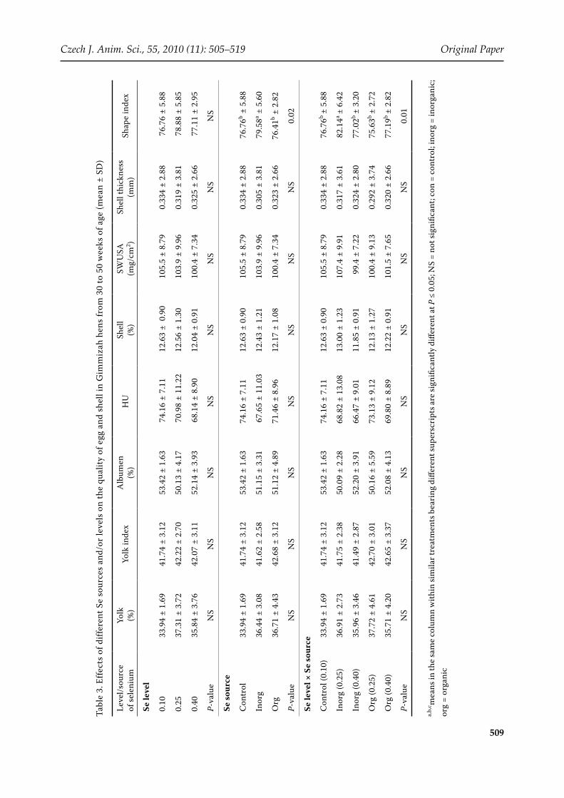
The level and source of Se and their interaction had no significant effect on any traits of egg qual-ity (Table 3), showing that Se content of the basal diet was adequate to support egg production of good quality. However, recent evidences by Payne et al. (2005) and Gajčević et al. (2009) indicated that eggs produced by hens fed a diet with organic Se had higher HU values than eggs of hens fed the recommended level of Se. Furthermore, Gajčević et al. (2009) reported that the dietary supplementa-tion of a higher amount of Se resulted in increased GSH-Px activity in the hens’ blood (P < 0.05).
The level and source of Se had no significant ef-fect on fertility and embryonic mortality percent-age compared with those in hens fed the control diet. On the other hand, the hatchability percent-age improved significantly and the piped embryos significantly decreased. The improvement in the hatchability percentage was ~ 17% for the medium (0.25) and high (0.40 ppm) level (Table 4). The re-sults of the interaction indicated a significant ef-fect with the greatest fertility percentage (97.4%) in hens fed the high level (0.40 ppm) of inorganic Se and the least significant one (90.7%) in hens fed the medium (0.25 ppm) level of inorganic Se. The organic Se groups exhibited intermediate values. In addition, the medium (0.25) and the high (0.40 ppm) levels of inorganic and organic Se in layer diets significantly improved the hatchability percentage while the embryonic mortality percentages signifi-cantly decreased compared with those in hens fed the basal diet (0.10 ppm of Se). On the other hand, the interaction between the level and the source of Se did not significantly affect the embryonic mor-tality percentage compared with that in hens fed the control diet. Surai (2006) indicated that Se has an important role in improved fertility, embryonic development and hatchability of poultry. Moreover, Se is deposited in the egg and distributed among the developing tissues during embryogenesis (Surai, 2000; Paton et al., 2002). However, the present results showed that sodium selenite had better hatchability than the Selplex®, which needs further verification. The improvement quoted herein could be due to the ability of Se to remove oxygen free radicals and lipid peroxidase and to alleviate the degree of tissue damage caused by the oxygen and the free radicals caused by hatching stress (Surai, 2006).
Tibia characteristics
Selenium level, source and the interaction be-tween the level and the source of Se had no signifi-cant effect on the tibia weight percentage compared with that in hens fed the control diet (Table 4). The increasing level of Se in the inorganic and organic source significantly increased the calcium percent-age in tibia compared with that in hens fed the control diet. These results indicated that Se sup-plementation improved the bone mineralization of laying hens during 30–50 weeks of age (Table 4). The Se level in layer diets had no significant effect on the inorganic phosphorus percentage in tibia. The phosphorus percentages in tibia significantly decreased by organic Se but significantly increased by inorganic Se compared with those in hens fed the control diet. Although inorganic Se increased tibia phosphorus, tibia Ca percentage and tibia weight percentage did not differ between the two sources.
It is worth noting that the medium level (0.25 ppm) of inorganic Se resulted in the greatest Ca and phos-phorus in tibia, nonetheless, the relative weight of tibia was not changed. The mechanism by which Se affects the bone formation is not known at present, for example the Se presence in human bones is about 16% of total body Se (Zachara et al., 2001). A relationship between active vitamin D metabolites and the Se-dependent enzyme thioredoxin reduct-ase was established (Schutze et al., 1999). An effect of thioredoxin reductase activity on 1.25 (OH)2D3 would be expected and this could be an important link between Se and bone metabolism. On the other hand, antioxidant properties of various selenopro-teins could also be of importance in maintaining the antioxidant protection of the oviduct during eggshell formation (Surai, 2006).
Organs
The level and/or the source of selenium had no significant effect on most of the organs except the relative weight of spleen which significantly de-creased compared with that in hens fed the control diet. In addition, the relative weight of pancreas significantly increased at both the medium (0.25) and high (0.40 ppm) level of Se in respect of the control group (Table 5). In agreement with the present results, EL-Sebai (2000) and Ševčíková et al. (2006) found that no significant effect due to

512
Original Paper Czech J. Anim. Sci., 55, 2010 (11): 505–519
supplementation of Se on liver weight of broiler chickens.
Biochemical constituents
The level and the source of Se did not signifi-cantly affect the albumin and triglyceride concen-tration of the plasma compared with the control treatment (Table 6). Similarly, Abaza (2002) found that the plasma triglyceride was insignificantly affected by Se or by the combination of Se and vitamin E. However, Attia et al. (2006) found that vitamin E and/or Se supplementation significantly decreased triglycerides. The low dosage of inorgan-ic Se and the high dosage of organic Se significantly decreased the plasma triglyceride concentration compared with the other Se treatments showing a significant interaction between the level and the source of Se.
Both the level and source of Se significantly decreased the plasma cholesterol concentration compared with that in hens fed the control diet (Table 6). In accordance with the present obser-vations Abd-El-Latif et al. (2004) reported that additions of Zn, Se and vitamin E significantly al-leviated the concentration of cholesterol in quails fed dietary ochratoxin compared with quails fed dietary ochratoxin without such feed additives. On the other hand, Abaza (2002) observed that the plasma cholesterol concentration was significantly increased by Se or by the combination of Se and vitamin E. However, Ljubic et al. (2006) suggested that the organic Se supplementation influences cholesterol metabolism in adipose tissue by de-creasing the total cholesterol concentration during the fattening period and increasing the free cho-lesterol concentration after 48 h feed deprivation. Changes in enzymes responsible for regulating cho-lesterol synthesis, oxidation or elimination may be
Table 5. Effects of different Se sources and/or levels on the percentage of some organs in Gimmizah hens at 50 weeks of age (mean ± SD)
Level/source of selenium
Spleen (%)Abdominal
fat (%)Ovary (%) Oviduct (%) Liver (%) Pancreas (%)
Intestinal weight (%)
Se level
0.10 0.162a ± 0.03 3.21 ± 1.82 1.72 ± 0.68 2.98 ± 0.34 0.52 ± 0.03 13.55b ± 1.5 7.80 ± 0.73
0.25 0.107b ± 0.03 5.09 ± 1.28 2.41 ± 0.83 2.58 ± 0.25 0.50 ± 0.06 21.06a ± 4.3 6.90 ± 2.42
0.40 0.081b ± 0.03 7.27 ± 1.69 2.13 ± 0.75 2.61 ± 0.18 0.50 ± 0.03 21.23a ± 5.9 7.01 ± 1.13
P-value 0.05 NS NS NS NS 0.03 NS
Se source
Control 0.162a ± 0.03 3.21 ± 1.82 1.72 ± 0.68 2.98 ± 0.34 2.20 ± 0.37 0.21 ± 0.04 2.02 ± 0.55
Inorg 0.090b ± 0.03 5.77 ± 1.97 1.96 ± 0.69 2.55 ± 0.20 1.86 ± 0.44 0.22 ± 0.02 2.15 ± 0.33
Org 0.098b ± 0.04 6.59 ± 1.71 2.57 ± 0.79 2.64 ± 0.23 2.09 ± 0.12 0.21 ± 0.01 2.44 ± 0.54
P-value 0.05 NS NS NS NS NS NS
Se level × Se source
Control (0.10) 0.162a ± 0.03 3.21 ± 1.82 1.72 ± 0.68 2.98 ± 0.34 2.20 ± 0.37 0.21 ± 0.04 2.02 ± 0.55
Inorg (0.25) 0.088c ± 0.01 4.66 ± 1.45 1.92 ± 0.02 2.52 ± 0.22 1.80 ± 0.22 0.21 ± 0.02 2.15 ± 0.11
Inorg (0.40) 0.092c ± 0.04 6.89 ± 1.91 2.01 ± 1.05 2.57 ± 0.20 1.93 ± 0.62 0.23 ± 0.02 2.16 ± 0.50
Org (0.25) 0.126b ± 0.03 5.52 ± 1.11 2.90 ± 0.99 2.64 ± 0.30 2.08 ± 0.17 0.21 ± 0.01 2.38 ± 0.82
Org (0.40) 0.070c ± 0.01 7.65 ± 1.61 2.24 ± 0.41 2.65 ± 0.18 2.10 ± 0.08 0.20 ± 0.02 2.50 ± 0.08
P-value 0.05 NS NS NS NS NS NS
a,b,cmeans in the same column within similar treatments bearing different superscripts are significantly different at P ≤ 0.05; NS = not significant; con = control; inorg = inorganic; org = organic

513
Czech J. Anim. Sci., 55, 2010 (11): 505–519 Original Paper
Tabl
e 6.
Effe
cts
of d
iffer
ent S
e so
urce
s an
d/or
leve
ls o
n pl
asm
a to
tal p
rote
in, a
lbum
in, g
lobu
lin, A
lb/G
lb, t
rigl
ycer
ide,
cho
lest
erol
, HD
L an
d se
leni
um y
olk
conc
en-
trat
ions
of G
imm
izah
hen
s fr
om 3
0 to
50w
eek
of a
ge (m
ean
± SD
)
Leve
l/sou
rce
of
sele
nium
To
tal p
rote
in
(g/d
l)A
lbum
in
(g/d
l)G
lobu
lin
(g/d
l)A
lb/G
lbTr
igly
ceri
de
(mg/
dl)
Cho
lest
erol
(m
g/dl
)H
DL
(m
g/dl
)Yo
lk S
e (p
pm
fres
h yo
lk)
Se le
vel
0.10
5.4
7 ±
0.4
4.06
± 0
.1 1
.41
± 0.
43.
25 ±
1.3
537.
2 ±
39.5
195.
0a ±
8.6
403.
8b ±
71.0
0.20
9c ±
0.01
0.25
5.2
4 ±
0.7
3.73
± 0
.6 1
.51
± 0.
63.
03 ±
1.6
457.
1 ±
99.9
131
.6c
± 44
.9 5
73.2
a ±
45.1
0.22
3b ±
0.01
0.40
5.9
7 ±
1.4
3.81
± 0
.4 2
.16
± 1.
73.
62 ±
2.8
464.
8 ±
79.9
163
.1b
± 12
.442
6.4b
± 43
.20.
356a
± 0.
06
P-va
lue
NS
NS
NS
NS
NS
0.00
010.
0001
0.00
01
Se s
ourc
e
Con
trol
5.47
b ±
0.4
4.06
± 0
.11.
41b ±
0.4
3.25
ab ±
1.3
537.
2 ±
39.5
195.
0a ±
8.6
403.
8b ±
71.0
0.20
9c ±
0.01
Inor
g6.
17a
± 1.
23.
58 ±
0.3
2.60
a ± 1
.31.
83b
± 0.
944
8.3
± 97
.5 1
69.6
b ±
12.1
40
3.8b
± 17
0.9
0.26
1b ±
0.05
Org
5.03
b ±
0.7
3.96
± 0
.61.
07b ±
0.5
4.82
a ± 2
.247
3.6
± 80
.9 1
25.1
c ±
38.2
595
.9a
± 36
.6 0
.318
a ±
0.09
P-va
lue
0.02
NS
0.00
30.
003
NS
0.00
010.
0001
0.00
01
Se le
vel ×
Se
sour
ce
Con
trol
(0.1
0)5.
47b
± 0.
44.
06 ±
0.1
1.41
b ± 0
.43.
25 ±
1.3
537.
2a ± 3
9.5
195.
0a ±
8.6
403
.8b
± 71
.0 0
.209
e ±
0.01
Inor
g (0
.25)
5.47
b ±
0.8
3.60
± 0
.41.
87b ±
0.5
2.03
± 0
.537
3.1b
± 63
.817
3.5b
± 2.
5 5
61.3
a ±
44.7
0.21
9d ±
0.01
Inor
g (0
.40)
6.87
a ±
1.3
3.55
± 0
.33.
32a ±
1.6
1.63
± 1
.252
3.5a
± 20
.9 1
65.8
b ±
17.1
246.
2c ±
1.8
0.3
03b
± 0.
01
Org
(0.2
5)5.
00b
± 0.
63.
86 ±
0.9
1.15
b ± 0
.54.
03 ±
1.8
541.
1a ±
20.9
89.
7c ±
5.2
585
.2a
± 48
.7 0
.227
c ±
0.01
Org
(0.4
0)5.
06b
± 0.
94.
07 ±
0.4
0.99
b ± 0
.65.
61 ±
2.5
406.
0b ±
51.7
160.
5b ±
6.7
606
.7a
± 20
.9 0
.408
a ±
0.01
P-va
lue
0.02
NS
0.00
3N
S0.
0001
0.00
010.
0001
0.00
01
a,b m
eans
in th
e sa
me
colu
mn
with
in si
mila
r tre
atm
ents
bea
ring
diff
eren
t sup
ersc
ript
s are
sign
ifica
ntly
diff
eren
t at P
≤ 0
.05;
NS
= no
t sig
nific
ant;
con
= co
ntro
l; in
org
= in
orga
nic;
or
g =
orga
nic

514
Original Paper Czech J. Anim. Sci., 55, 2010 (11): 505–519
responsible for lowering the cholesterol synthesis in mature as well as immature chickens (Konjufca et al., 1997).
Regardless of Se source and the interaction be-tween Se level and source, the Se level of 0.25 ppm significantly increased the plasma HDL concentra-tion compared with the other levels (Table 6). A decrease in plasma cholesterol is associated with an increase in plasma HDL. Organic Se significantly increased plasma HDL compared with the other treatments, regardless of the Se level and the in-teraction between Se level and source. Indeed, the decrease in plasma cholesterol of the organic Se group may be a reflection of the increase in plasma HDL. The interaction between Se level and source significantly affected HDL that was increased due to supplementation of 0.25 ppm of the inorganic and organic Se source as well as the high dosage of organic Se (0.40 ppm).
Yolk selenium
Table 6 shows the effect of Se level and/or source on yolk selenium content. The increasing dietary Se level significantly increased the yolk selenium content linearly. The increases were 6.7 and 70.3% due to diets containing the medium (0.25) and the high (0.40 ppm) level of Se, respectively. The in-clusion of inorganic and organic Se significantly increased the yolk selenium content compared with that in hens fed the control diet. However, hens fed organic Se deposited significantly more Se in yolks than those fed the inorganic source. This result indicated that organic Se was more potent than the inorganic source. Payne et al. (2005), Skřivan et al. (2006) and Leeson et al. (2008) reported similar results. The interaction results indicated that all supplemented levels and both sources significantly increased yolk Se content compared to the control group. Moreover, there was a linear increase within each source with increasing Se level. Furthermore, both levels of organic Se had greater values than the corresponding levels of inorganic source.
Research to produce Se enriched eggs for human health benefits shows that organic Se supplemen-tation (e.g. selenomethionine or Se-yeast) from 0.1 mg to 0.5 ppm increases the concentration of Se in whole egg from ~ 0.1 to 0.4 mg Se/kg (Paton et al., 2002) and in other animal tissues (Bobček et al., 2004). Payne et al. (2005), Utterback et al. (2005) and Skřivan et al. (2006) stated that the use of Se
yeast in laying hen diets was very effective from the aspect of increasing the Se content of eggs. As a result of dietary Se supplementation, the Se con-centration in egg yolk was significantly increased (Surai, 2006) included that in the albumen and yolk (Gajčević et al., 2009). The reported Se yeast (selenomethionine) improved product quality (re-duced carcass drip loss, Se enrichment of meat and eggs) and decreased intensity of lipid peroxidation in yolks as shown by thiobarbituric acid reactive substances (Gajčević et al., 2009) may be due to a significant increase in GSH-Px activity in the blood of hens (Gajčević et al., 2009). Paton et al. (2002) concluded that a possible reason for the elevated level of Se in yolk, albumen and egg contents after supplementation of dietary Se-yeast can be due to the fact that hens have additional metabolic path-ways by which Se is transferred into the egg. For example, increasing Se level in the egg albumen of hens fed Se-yeast may be due to the incorpora-tion of greater amounts of Se as selenomethionine during albumen synthesis, selenomethionine could replace methionine, thereby providing additional Se. Surai (2006) reported that the Se-enriched quail eggs (865.2 ng/g Se/egg yolk DM) could supply 50% of the recommended daily allowance (RDA) of Se. Previous studies showed that the production of Se-enriched chicken eggs were a valuable option for improving the Se status of the general population in various countries (Yaroshenko et al., 2003). This technology was successfully applied commercially and Se-enriched eggs found their way to the super-market shelves in more than 25 countries world-wide (Surai, 2006). In addition, two eggs from the group fed a diet containing 0.40 ppm of Se could supply 100% of RDA according FAO/WHO (2002) requirements.
Histopathological study
The administration of various levels of Se in different forms, organic or inorganic ones, led to nearly similar hepatic as well as splenic changes of the treated chickens but with dose dependent ef-fects that were smaller or milder in the low levels of especially organic forms. The hepatic changes in se-vere cases were characterized by fatty vacuolations with some variable degrees that may be advanced and somewhat serious and may led to the necrosis of hepatic tissue in the case of administration at high levels (Figures 1, 2, 3, 4 and 5).

515
Czech J. Anim. Sci., 55, 2010 (11): 505–519 Original Paper
Figure 1. Liver of a control chicken shows normal hepato- cytic acini (arrows); H and EX400
Figure 2. Liver of a chicken treated with low levels of inorganic selenium shows one focus of severe hepatocytic fatty vacuolations (asterisk) surrounded by mild or less degenerated hepatocytic acini (arrows); H and EX250
Figure 3. Liver of a chicken treated with high levels of inorganic selenium shows an area of severe fatty vacuolations (asterisk) surrounded by either small hepatocytic fatty vacuolations or necrosis (thick arrow) and another area of lymphocytic infiltration (thin arrow); H and EX250
Figure 4. Liver of a chicken treated with low levels of organic selenium shows an advanced degree of fatty vacuolations in excess numbers of hepatocytic (arrows); H and EX400
Figure 5. Liver of a chicken treated with high levels of organic selenium shows diffuse and severe sharply edged fatty vacuolations (arrows); H and EX400
Figure 6. Spleen of a control chicken: normal splenic tissue with small-sized germinal centres (arrows) surrounded by various numbers of small lymphocytes; H and EX250
The spleen of the control group showed a nor-mal histological structure where some small-sized germinal centres appeared normally surrounded by variable numbers of small lymphocytes (Figure 6). On the other hand, microscopic splenic changes
were detected due to treatment with both levels and sources of selenium (Figures 7, 8. 9 and 10).
The microscopic changes in the small intestine were variable from part to another and from the ad-ministered form to another. The duodenal mucosa

516
Original Paper Czech J. Anim. Sci., 55, 2010 (11): 505–519
Figure 7. Spleen of a chicken treated with low levels of inorganic selenium: small-sized follicular arterioles (arrow) surrounded by areas of hyperplasia and congestion (asterisk); H and EX400
Figure 8. Spleen of a chicken treated with high levels of inorganic selenium: area of severe congestion (asterisk), medium-sized follicular artery (arrow) surrounded by an excess of mature small lymphocytes H and EX400
Figure 9. Spleen of a chicken treated with low levels of organic selenium shows hyperplastic splenic tissue with an excess of newly formed hyperplastic germinal centres (arrows); H and EX400
Figure 10. Spleen of a chicken treated with high levels of organic selenium shows well formed lymphoid follicles (asterisk) and expanded germinal centre (artery); H and EX400
Figure 11. Intestine of a control chicken shows the normal duodenal wall with normal crypts of intestinal glands (arrows); H and EX250
Figure 12. Duodenum of a chicken treated with a high level of inorganic selenium shows vacuolar and hydropic degeneration of the epithelial cells lining the intestinal crypts (arrows); H and EX400
appeared to be affected only by the high levels of treat-ment with the inorganic form of Se. It was noticeable that the intestinal mucosa of the ileum was affected by administration of the high levels of Se. The micro-scopic ileal changes were similarly and obviously seen
at both high levels of organic and inorganic forms of Se but with somewhat small differences in their degrees (Figures 11, 12, 13, 14 and 15).
Although the nutritional supplements of Se are beneficial for feed performance and digestion, the

517
Czech J. Anim. Sci., 55, 2010 (11): 505–519 Original Paper
high levels of administration of especially inorgan-ic forms may lead to some adverse effects on the liver functions as well as the absorption and FCR in treated chickens.
CONCLUSIONS
It is possible to produce Se enriched eggs of crossbred breeding hens by feeding a diet supple-mented with organic Se at a medium (0.25) and high (0.40 ppm) level with expected improvement in the consumer health benefit. In addition, organic Se improved the productive and reproductive per-formance of laying hens, decreased the cholesterol concentration, improved the tibia mineralization and was less toxic than the inorganic form when the histopathology of liver, spleen and intestine was considered. Furthermore, the level and source of Se in layer diets decreased total plasma choles-terol. Moreover, Se increased percentage of Ca and
P in tibia compared to that in hens fed the control diet. However, the medium level (0.25 ppm) of or-ganic Se was adequate and can be recommended to enrich eggs with less harmful effects on tissues than the high level (0.40 ppm).
REFERENCES
Abaza M. (2002): Immune system and some physiologi-cal aspects in Japanese quail affected by antioxidants. Egyptian Poultry Science Journal, 22, 259–276.
Abd-El-Latif F.S.A., El-Ghamry A.A., El-yamany A.T. (2004): Effect of using zinc, selenium and vitamin E supplemen-tation on performance and metabolic re-sponses of growing Japanese quail fed diets contami-nated with ochratoxin. Egyptian Poultry Science Journal, 24, 447–463.
Association of Official Analytical Chemists (1995): Of-ficial Methods of Analysis. 16th ed. AOAC, Arlington, USA.
Figure 13. Ileum of a control chicken shows normal thickened villi with lymphocytic elements (asterisk); H and E. X160
Figure 14. Ileum of a chicken treated with high levels of inorganic selenium shows an excess of mononuclear cell infiltration and aggregation (asterisk) in between degenerated and necrotic intestinal glands (arrows); H and E X250
Figure 15. Ileum of a chicken treated with high levels of organic selenium shows periglandular oedema and glandular atrophy (arrows); H and E X250

518
Original Paper Czech J. Anim. Sci., 55, 2010 (11): 505–519
Attia Y.A., Burke W.H., Yamani K.A., Jensen L.S. (1995): Energy allotments and performance of broiler breeders. 2- Females. Poultry Science, 74, 261–270.
Attia Y.A., Abd-El-Hamid A.E., Abd El-Ghany F.A., Ha-biba H.I. (2006): Effect of oil source and antioxidant supplementation on growth performance and meat quality of Japanese quail males. In: Proceeding of XII. European Poultry Conference. Verona, Italy, 10–14 September. Abstract: Wolds Poultry Journal, 62, 405.
Bancroft J.D., Stevens A. (1990): Theory and Practice of Histological Technique. 3rd ed. New York, USA.
Black C.A. (1982): Method of soil analysis. Soil Science of America, Inc. Published Matison Wisconsin, USA.
Bobček B., Lahučký R., Mrázová J., Bobček R., Novotná K., Vašiček D. (2004): Effects of dietary organic sele-nium supplementation on selenium content, antioxida-tive status of muscles and meat quality of pigs. Czech Journal Animal Science, 49, 411–417.
Carter T.C. (1975): The hen’s egg. Estimation of shell superficial area and egg volume using measure- ments of fresh egg weight and shell length and breadth alone or in combination. British Poultry Science, 16, 541–543.
Cottenie A., Verloo M., Kaikan L., Vlghe G., Comerlynch R. (1982): Chemical analysis of plants and soils. Lab, Anal Agrochem. State University, Ghent, Belgium.
EL-Sebei A. (2000): Influence of selenium and vitamin E as antioxidants on immune system and some physio-logical aspects in broiler chickens. Egyptian Poultry Science Journal, 20, 1065–1082.
FAO/WHO (2002): Selenium. In: Human Vitamin and Mineral Requirements. Report of a Joint FAO/WHO Expert Consulation. FAO, Rome, Italy, 235–255.
Folch J., Lees M., Sloane-Slanley G.S. (1957): A simple method for the isolation and purification of total lipids from animal tissues. Journal of Biological Chemistry, 226, 497–509.
Funk E.M. (1948): The relation of yolk index determined in natural position to the yolk index as determined after separating the yolk from the albumen. Poultry Science, 27, 367.
Gajčević Z., Kralik G., Has-Schön Elizabeta, Pavić V. (2009): Effects of organic selenium supplemented to layer diet on table egg freshness and selenium content. Italian Journal Animal Science 8, 189–199.
Ganpule S.P., Manjunatha B.P. (2003): Antioxidant nutri-tion starts with breeders. Feed Mix 11, 14–16.
Hallberg L., Rossander-Hulthen L. (1993): Factors influ-encing the bioavailability of iron in man. In: Schlemmer U. (ed.): Bioavailability ‘93-Nutritional, Chemical and Food Processing Implications of Nutrient Availability. Ettlingen, Germany, 23–32.
Haugh R.R. (1937): The haugh units for measuring egg quality. US Egg and Poultry Magazine, 43, 552–555.
Jiakui L., Xiaolong W. (2004): Effect of dietary organic versus inorganic selenium in laying hens on the pro-ductivity, selenium distribution in egg and selenium content in blood, liver and kidney. Journal of Trace Elements in Medicine and Biology, 18, 65–68.
Konjufca V.H., Pesti G.M., Bakalli R.I. (1997): Modulation of cholesterol levels in broiler meat by dietary garlic and copper. Poultry Science, 76, 1264–1274.
Kucharzewski M., Braziewicz J., Majewska U., Gozdz S. (2003): Copper, Zinc and Selenium in whole blood and thyroid tissue of people with various thyroid diseases. Biological Trace Element Research, 93, 9–18.
Leeson S., Namkung H., Caston L., Durosoy S., Schlegel P. (2008): Comparison of selenium levels and sources and dietary fat quality in diets for broiler breeders and layer hens. Poultry Science, 87, 2605–2612.
Ljubic B., Milinkovic-Tur S., Pirsljin J., Zdelar-tuk M., Filipovic N. (2006): Effect of organic selenium food supplementation and fasting on adipose tissue lipid concentration and lipoprotein lipase activity in broiler chickens. In: Proceeding of European Poultry Confer-ence. Verona, Italy.
NCR (1994): National Research Council. Nutrient re-quirements of poultry. 9th ed. National Academy Press. Washington, USA.
Paton N.D., Cantor A.H., Pescatore A.J., Ford M.J., Smith C.A. (2002): The effect of dietary selenium source and level on the uptake of selenium by developing chick embryos. Poultry Science, 81, 1548–1554.
Payne R.L, Lavergne T.K., Southern L.L. (2005): Effect of inorganic versus organic selenium on hen production and egg selenium concentration. Poultry Science, 84, 232–237.
Ramanoff A.I., Ramanoff A.J. (1949): The Avian Egg. John Wiley and Sons, Inc. New York, USA.
SAS Institute (1990): SAS/STAT User’s Guide: Statistics. Version 6, Cary, NC, USA.
Schutze N., Fritsche J., Ebert-Dumig R., Schneider D., Kohrle J., Andreesen R., Kreutz M., Jkob F. (1999): The selenoprotein thioredoxin reductase is expressed in peripheral blood monocytes and THP1 human myeloid leukemia cells-regulation by 1.25-dihydroxyvitamin D3 and selenite. Biofactors, 10, 329–338.
Ševčíková S., Skřivan M., Dlouhá G., Koucký M. (2006): The effect of selenium source on the performance and meat quality of broiler chickens. Czech Journal of Animal Science, 51, 449–457.
Skřivan M., Simane J., Dlouhá G., Doucha J. (2006): Effect of dietary sodium selenite, Se-enriched yeast and Se-enriched Chlorella on egg Se concentration, physical

519
Czech J. Anim. Sci., 55, 2010 (11): 505–519 Original Paper
parameters of eggs and laying hen production. Czech Journal of Animal Science, 51, 163–167.
Surai P.F. (2000): Effect of selenium and vitamin E content of the maternal diet on the antioxidant system of the yolk and the developing chick. British Poultry Science, 41, 235–243.
Surai P.F. (2006): Selenium in Nutrition and health. Not-tingham University Press, Nottingham, UK.
Utterback P.L., Parasons C.M., Yoon I., Butler J. (2005): Effect of supplementing selenium yeast in diets of lay-ing hens on egg selenium content. Poultry Science, 84, 1899–1901.
Yaroshenko F.A., Dvorska J.E., Surai P.F., Sparks N.H.C. (2003): Selenium-enriched eggs as a source of selenium for human consumption. Applied Biotechnology, Food Science and Policy, 1, 13–23.
Zachara B.A., Pawluk H., Bloch-Boguslawska E., Sliwka K.M., Korenkiewicz J., Skok Z., Ryc K. (2001): Tissue level, distribution, and total body selenium content in healthy and diseased humans in Poland. Archives of Environmental Health, 56,461–466.
Received: 2009–07–07Accepted after corrections: 2010–01–06
Corresponding Author
Youssef A. Attia, Faculty of Agriculture, Damanhour University, EgyptTel. +0020 103 753 095, e-mail: [email protected]