effects of combined 5-fluorouracil and zno nps on human...
TRANSCRIPT

Nanomed. J. 6(3):232-240, Summer 2019
RESEARCH PAPER
Effects of combined 5-Fluorouracil and ZnO NPs on human breast cancer MCF-7 Cells: P53 gene expression, Bcl-2 signaling
pathway, and invasion activitySafoura Hoseinzadeh1, Elham Raeisi 2, Yves Lemoigne 3, Esfandiar Heidarian 1*
1 Clinical Biochemistry Research Center, Basic Health Sciences Institute, Shahrekord University of Medical Sciences, Shahrekord, Iran
2 Department of Medical Physics and Radiology, Shahrekord University of Medical Sciences, Shahrekord, Iran 3 Institute for Medical Physics, Ambilly, France
* Corresponding Author Email: [email protected]. This manuscript was submitted on January 3, 2019;approved on March 30, 2019
How to cite this articleHoseinzadeh S, Raeisi E, Lemoigne Y, Heidarian E. Effects of Combined 5-Fluorouracil and ZnO NPs on Human Breast Cancer MCF-7 Cells: P53 Gene Expression, Bcl-2 Signaling Pathway, and Invasion Activity. Nanomed J. 2019; 6(3):232-240 DOI: 10.22038/nmj.2019.06.000010
ABSTRACTObjective(s): The significant contribution of nanoparticles to cancer treatment has attracted therapeutic attention. The present study aimed to evaluate the synergistic effects of 5-fluorouracil (5-FU) and zinc oxide nanoparticles (ZnO NPs) as multimodal drug delivery on human breast cancer MCF-7 cells.Materials and Methods: In this in-vitro study, the impact of 5-FU and ZnO NPs in the single or combined forms was evaluated on cell viability, colony formation, apoptosis, p53 gene expression, and Bcl-2 signaling protein in MCF-7 breast cancer cell line using several techniques, such as MTT, clonogenic assay, flow cytometry, real-time quantitative polymerase chain reaction, and Western blot.Results: In this study, 5-FU combined with ZnO NPs showed synergistic effects against MCF-7 within 48 hours. In addition, the combination of 5-FU and ZnO NPs at the respective concentrations of 1 µM and 45 µg/ml exhibited significant apoptosis (79.53%), p53 gene expression (3.6 folds), reduction of cell invasion (9.82%), and plating efficiency (5%), thereby leading to the significant reduction of cell viability (40±0.9%) and decreased Bcl-2 anti-apoptotic protein relative to untreated control cells. Conclusion: According to the results, the synergistic effects of combined ZnO NPs and 5-FU on MCF-7 human breast cancer cells were exerted via Bcl-2 inhibition and the up-regulation of p53 expression.
Keywords: Bcl-2, p53 expression, Zinc oxide naoparticles, 5-Fluorouracil
INTRODUCTIONBreast cancer is the second most prevalent
cancer among women across the world, accounting for 25% of the diagnosed human malignancies and 15% of cancer-related deaths [1, 2]”ISBN” : “00079235”, “ISSN” : “1542-4863 (Electronic. The incidence of invasive breast cancer was expected to reach 266,120 new diagnosed cases in 2018 [3]. Breast cancer is more prevalent in women aged 35-70 years. The prevalence of breast cancer in Asia is reported to be four-fold compared to Western countries. The known risk factors for breast cancer include family history, pregnancy at old age, late menopause, early menstruation, long-term use of female hormones (e.g., contraceptives),
and alcohol consumption [4]. Some of the main approaches to the treatment of breast cancer include radiotherapy, chemotherapy, hormone therapy (i.e., endocrine therapy), and targeted therapy. The main intervening therapeutic decisional indices are considered to be the cancer stage, grade of invasiveness, patient’s age, and underlying health status [5].
Chemotherapy is often used as an adjuvant or neoadjuvant treatment for breast cancer. Nevertheless, the complications of chemotherapy have paved the way for the investigation of new drug combination strategies [6]. Reducing the complications of chemotherapy in terms of the synergistic impact of drug combination is based on decreasing the required therapeutic dose of classical chemotherapy agents, such as 5-fluorouracil (5-FU) [7]. This agent exerts anti-

233Nanomed. J. 6(3):232-240, Summer 2019
S. Hoseinzadeh et al. / Cytotoxic effects of 5-Fluorouracil and ZnO NPs
cancer effects by inhibiting and disrupting the DNA synthesis and functions. However, 5-FU resistance remains a major therapeutic challenge [8].
Various nanoparticles has been developed in order to provide new therapeutic alternatives with enhanced clinical effectiveness [9]surface coating, and cosmetics, and recently, they have been explored in biologic and biomedical applications. Therefore, this study was undertaken to investigate the effect of ZnO NPs on cytotoxicity, apoptosis, and autophagy in human ovarian cancer cells (SKOV3. Zinc oxide nanoparticles (ZnO NPs) have been reported to have promising anti-cancer properties through superior cancer cell penetration, targeting the salient cell cancer proliferation cycles, covering a large area of malignant cell membranes, and enhancing the availability of chemotherapy drugs [10]. ZnO NPs alter the production of reactive oxygen species (ROS), p53 gene expression, caspase-3 enzyme synthesis, Bcl-2 signaling pathway, and cell death.
The present study aimed to investigate the effects of separate and combined treatments with 5-FU and ZnO NPs on MCF-7 human breast cancer cells on cell growth and proliferation, p53 gene expression, and Bcl-2 signaling activity [11].
MATERIALS AND METHODS Cell culture
In this in-vitro study, human breast adenocarcinoma (MCF-7) cells were obtained from Pasteur Institute (Tehran, Iran). The cells were cultured in Dulbecco’s modified Eagle’s medium (DMEM; Gibco-Invitrogen) supplemented with 10% fetal bovine serum (Gibco-Invitrogen), 100 U/ml of penicillin/streptomycin Gibco (Rockville, MD, USA), and 1% L-glutamine in standard culture conditions (5% CO2, 98% humidity, and temperature of 37ºC) [12]recurrence, and metastasis formation. Salinomycin (SAL.
The study protocol was approved by the Ethics Committee of Shahrekord University of Medical Sciences in Shahrekord, Iran (code: IR.SKUMS.REC.1396.144).
Preparation of 5-FU and ZnO NPsInitially, 5-FU was purchased from Haupt
Pharma (Wolfratshausen GmbH Company, Germany) and diluted in the DMEM medium in order to obtain the concentrations of 0-11 µM [13]. ZnO NPs (10-30 nm, surface area: 20-60 m2/g) were purchased in the form of powder from Iranian
Nanomaterials Pioneers Company (Mashhad, Iran) and suspended in phosphate-buffered saline (PBS) in order to achieve the ZnO NP stock solution of 1,000 µg/ml. The stock solution was used to obtain the desired concentrations (0-110 µg/ml).
MTT assayAt this stage, 3-(4, 5-dimethylthiazol-2yl)-2,
5-diphenyltetrazolium bromide (MTT) assay was applied to evaluate MCF-7 cell viability. To this end, 5×10³ cells per well (1.8×10⁶ cells/ml) were seeded in 96-well plates, incubated overnight, and treated with various concentrations of 5-FU (0-11 µM) or ZnONPs (0-110 µg/ml) for 48 hours. As for combined therapy, the cells were treated with four selected concentrations (Table 1).
At the next stage, the MTT solution (5 mg/ml) was added to each well, which were incubated for four hours following incubation with DMSO (150 µL) for 20 minutes. The absorbance of each well was measured using a microplate reader (model: Stat Fax-2100, Spain) at the wavelengths of 490-570 nanometers. The rate of cell viability was expressed as the absorbance of the treated cells to the untreated control cells. The experiments were performed at least three times on three different days [14].
Clonogenic assayMCF-7 cells were seeded in six-well plates and
incubated overnight in six replicates. Afterwards, the cultured cells were treated with 1 µM of 5-FU, 45 µg/ml of ZnO NPs, and the combination of 5-FU and ZnO NPs for 48 hours (Table 1). After the treatment, the cells were incubated at the temperature of 37ºC in 5% CO2 for 14 days for colony formation, and the medium was changed every 48 hours. Visible colonies were fixed with 70% ethanol and stained with 0.5% crystal violet. In addition, the plating efficiency was measured [15].
Annexin V-PI staining for apoptosis assayMCF-7 cells were seeded in six-well plates at
the density of 2×10⁵ cell/well and incubated for 24 hours. When the cells reached 70% confluence, they were treated with 1 µM of 5-FU, 45 µg/ml of ZnO NPs, and the combination of 5-FU and ZnO NPs for 48 hours. Following that, all the cells (dead and viable) were collected by trypsination, washed with PBS, and stained with Annexin V (BD Bioscience California, USA) for 30 minutes at room
234
S. Hoseinzadeh et al. / Cytotoxic effects of 5-Fluorouracil and ZnO NPs
Nanomed. J. 6(3):232-240, Summer 2019
temperature. The cells were analyzed using the CYFLOW space (PARTEK, Germany) in accordance with the instructions of the manufacturer [16, 17].
Real-time polymerase chain reaction (PCR)MCF-7 cells were seeded at the density of 6×10⁵
cell/well for 24 hours and exposed to the IC40 of ZnO NPs, IC10 of 5-FU, and their combination (1 µM of 5-FU and 45 µg/ml of ZnO NPs). Afterwards, the cells were harvested and lysed with Roti®ZOL (Carl Roth GmbH, Germany) based on the instructions of the manufacturer. In each treatment, the concentration of mRNA was determined using Nanodrop (Thermo-USA).
At the next stage, cDNA was synthesized from one microgram of total mRNA using reverse transcriptase (Takara Bio Inc., Japan) in accordance with the instructions of manufacturer. Quantitative real-time polymerase chain reaction (RT-PCR) was performed using SYBR® green PCR master mix (Qiagen, Germany). During the RT-qPCR, cDNA was amplified using Rotor-Gene3000 (Corbett, Australia) in the presence of SYBR® green PCR master mix, the sequences of specific p53 primer sets (forward: 5’-CCCATCCTCACCATCATCACAC-3’, reverse:5’-GCACAAACACGCACCTCAAAG-3’),and glyceraldehyde-3-phosphate dehydrogenase (GAPDH; forward: 5’- ACACCCACTCCTCCACCCTTTG-3’,reverse:5’-GTCCACCACCCTGTTGCTGTA-3’)in each reaction. The primers were designed using the Oligo 7.0 software (Molecular Biology Insights, Cascade, Co.) and confirmed by blast (NCBI). The primers were purchased from Eurogentec (Seraing, Belgium).
The temperature profile of each reaction was adjusted. Initially, denaturation was performed at the temperature of 95°C for 10 minutes in 40 cycles based on a three-step program (10 seconds at 95°C, 15 seconds at 60°C, and 20 seconds at 72°C). GAPDH was used as the housekeeping gene to normalize the gene expression data [18].
Western blot assayMCF-7 cells were seeded at the density of
6×10⁵ cell/well for 24 hours and exposed to the IC40 of ZnO NPs, IC10 of 5-FU, and their combination (1 µM of 5-FU and 45 µg/ml of ZnO NPs). Following that, the cells were harvested and subsequently were lysed in ice-cold RIPA buffer (50 mM of Tris-HCl, pH of 8.0, 150 mM of NaCl, 0.5% sodium deoxycholate, and 0.1% sodium dodecyl sulfate [SDS]) containing protease inhibitors, phosphatase
inhibitor, and phenylmethylsulfonyl fluoride. The protein content was evaluated using the Bradford protein assay [19].
At this stage, 40 micrograms of protein was loaded in 10% SDS-PAGE and transferred to the polyvinylidene fluoride membrane. The membranes were incubated at the temperature of 4°C overnight using Bcl-2 and β-actin primary antibodies (Elabscience Biotechnology Co., Wuhan, China). Immunoblots were detected using horseradish peroxidase conjugated secondary antibody [20]. Band intensity was evaluated using chemiluminescent reagents (ECL; Thermo Fisher Scientific, USA) and analyzed using the Image J software.
Invasion assayMCF-7 cells were seeded in six-well plates at
the density of 2×10⁵ cell/well with 1 µM of 5-FU, 45 µg/ml of ZnO NPs, and their combination (1 µM of 5-FU and 45 µg/ml of ZnO NPs) for 48 hours. Afterwards, the invasion assay was performed as previously described [20].
Statistical analysisData analysis was performed using SPSS version
20.0 (SPSS, Chicago, IL, USA) and GraphPad Prism 6 (GraphPad Software Inc., San. Diego, CA, USA). The experimental data were expressed as mean and standard deviation of the mean from a minimum of three independent experiments. Group comparison of the means was carried out using Kruskal-Wallis test and Dunn’s test, and the P-value of less than 0.05 was considered significant.
The analysis of the data on the relative gene expression was performed using the 2-∆∆Ct method. Melting curves were generated in order to ensure the purity of the amplification product of each reaction. Moreover, the Western blot experiments were carried out in triplicate. The combination index (CI) was calculated using the CompuSyn software (Combo SynInc, City, State, USA), and CI of less than one, one, and more than one indicated synergism, additive, and antagonism effects, respectively.
RESULTS Cytotoxicity of combined 5-FU and ZnO NPs on MCF-7 cell proliferation
MCF-7 cells were treated with various concentrations of ZnO NPs (0-110 µg/ml) and 5-FU
235Nanomed. J. 6(3):232-240, Summer 2019
S. Hoseinzadeh et al. / Cytotoxic effects of 5-Fluorouracil and ZnO NPs
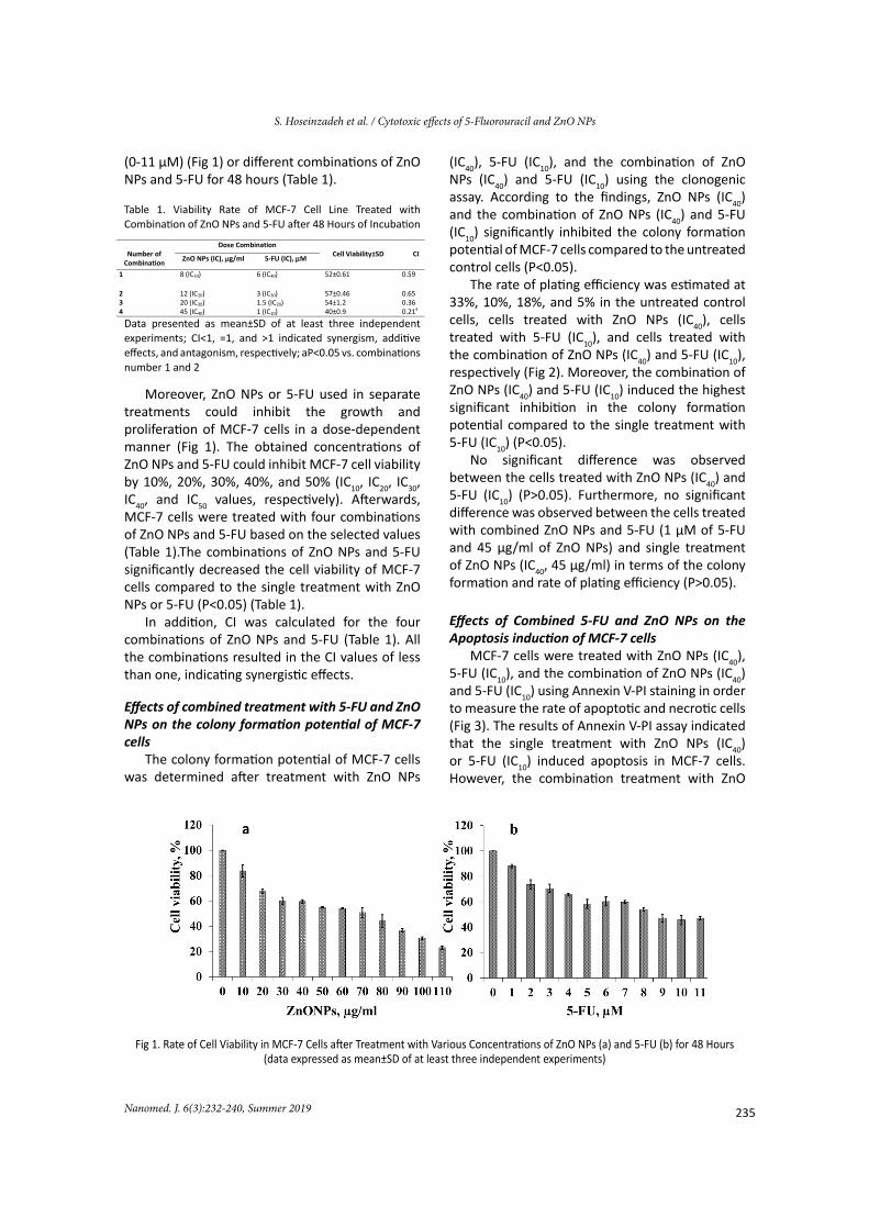
(0-11 µM) (Fig 1) or different combinations of ZnO NPs and 5-FU for 48 hours (Table 1).
Table 1. Viability Rate of MCF-7 Cell Line Treated with Combination of ZnO NPs and 5-FU after 48 Hours of Incubation
Data presented as mean±SD of at least three independent experiments; CI˂1, =1, and ˃1 indicated synergism, additive effects, and antagonism, respectively; aP<0.05 vs. combinations number 1 and 2
Moreover, ZnO NPs or 5-FU used in separate treatments could inhibit the growth and proliferation of MCF-7 cells in a dose-dependent manner (Fig 1). The obtained concentrations of ZnO NPs and 5-FU could inhibit MCF-7 cell viability by 10%, 20%, 30%, 40%, and 50% (IC10, IC20, IC30, IC40, and IC50 values, respectively). Afterwards, MCF-7 cells were treated with four combinations of ZnO NPs and 5-FU based on the selected values (Table 1).The combinations of ZnO NPs and 5-FU significantly decreased the cell viability of MCF-7 cells compared to the single treatment with ZnO NPs or 5-FU (P<0.05) (Table 1).
In addition, CI was calculated for the four combinations of ZnO NPs and 5-FU (Table 1). All the combinations resulted in the CI values of less than one, indicating synergistic effects.
Effects of combined treatment with 5-FU and ZnO NPs on the colony formation potential of MCF-7 cells
The colony formation potential of MCF-7 cells was determined after treatment with ZnO NPs
(IC40), 5-FU (IC10), and the combination of ZnO NPs (IC40) and 5-FU (IC10) using the clonogenic assay. According to the findings, ZnO NPs (IC40) and the combination of ZnO NPs (IC40) and 5-FU (IC10) significantly inhibited the colony formation potential of MCF-7 cells compared to the untreated control cells (P<0.05).
The rate of plating efficiency was estimated at 33%, 10%, 18%, and 5% in the untreated control cells, cells treated with ZnO NPs (IC40), cells treated with 5-FU (IC10), and cells treated with the combination of ZnO NPs (IC40) and 5-FU (IC10), respectively (Fig 2). Moreover, the combination of ZnO NPs (IC40) and 5-FU (IC10) induced the highest significant inhibition in the colony formation potential compared to the single treatment with 5-FU (IC10) (P<0.05).
No significant difference was observed between the cells treated with ZnO NPs (IC40) and 5-FU (IC10) (P>0.05). Furthermore, no significant difference was observed between the cells treated with combined ZnO NPs and 5-FU (1 µM of 5-FU and 45 µg/ml of ZnO NPs) and single treatment of ZnO NPs (IC40, 45 µg/ml) in terms of the colony formation and rate of plating efficiency (P>0.05).
Effects of Combined 5-FU and ZnO NPs on the Apoptosis induction of MCF-7 cells
MCF-7 cells were treated with ZnO NPs (IC40), 5-FU (IC10), and the combination of ZnO NPs (IC40) and 5-FU (IC10) using Annexin V-PI staining in order to measure the rate of apoptotic and necrotic cells (Fig 3). The results of Annexin V-PI assay indicated that the single treatment with ZnO NPs (IC40) or 5-FU (IC10) induced apoptosis in MCF-7 cells. However, the combination treatment with ZnO
7
Number of
Combination
Dose Combination Cell Viability±SD
CI ZnO NPs (IC), g/ml 5-FU (IC), M
1 8 (IC10) 6 (IC40) 52±0.61 0.59
2 12 (IC20) 3 (IC30) 57±0.46 0.65 3 20 (IC30) 1.5 (IC20) 54±1.2 0.36 4 45 (IC40) 1 (IC10) 40±0.9 0.21a
1
Fig 1. Rate of Cell Viability in MCF-7 Cells after Treatment with Various Concentrations of ZnO NPs (a) and 5-FU (b) for 48 Hours (data expressed as mean±SD of at least three independent experiments)

236
S. Hoseinzadeh et al. / Cytotoxic effects of 5-Fluorouracil and ZnO NPs
Nanomed. J. 6(3):232-240, Summer 2019
NPs (IC40) and 5-FU (1 µM) caused a significant elevation in the rate of apoptosis (79.53%) compared to the untreated control cells (P<0.05) (Fig 3).
Moreover, no significant difference was observed between the cells treated with combined ZnO NPs and 5-FU compared to those with the single treatment of ZnO NPs (IC40, 45 µg/ml) in terms of apoptosis (P>0.05).
Effects of single and combined treatment with 5-FU and ZnO NPs on apoptosis-related gene expression and MCF-7 cell invasion
Fig 4 shows the effects of single and combined treatment with 5-FU and ZnO NPs on p53 gene expression in MCF-7 cells. P53 gene expression significantly increased in the cells with the combined treatment (3.6-fold) compared to the untreated control cells (P<0.05).
2
Fig 2. Clonogenic Formation of MCF-7 Cells after Treatment with ZnO NPs (45 µg/ml), 5-FU (1 µM) or combination of ZnO NPs (45 µg/ml) and 5-FU (1 µM) Using Clonogenic Assay (Clonogenic assay was conducted 14 days after the treatment; A: a) untreated cells, b) treated cells with IC10 of 5-FU [1 µM], c) treated cells with IC40 of ZnO NPs [45 µg/ml], d) treated cells with IC10 of 5-FU [1 µM] and IC40 of ZnO NPs [45 µg/ml]; B; Rate of plating efficiency in MCF-7 cells; data expressed as mean±SD of at least three independent
experiments; aP<0.05 vs. control cells, bP<0.05 vs. 5-FU)
3
Fig 3. A: Annexin V-FITC/PI Analyzed by Flow Cytometry, B: Rate of Total Apoptosis Cells Treated with ZnO NPs (45 µg/ml), 5-FU (1 µM) or Combination of ZnO NPs (45 µg/ml) and 5-FU (1 µM) for 48 Hours (data expressed as mean±SD of at least three independent
experiments; aP<0.05 vs. control cells, bP<0.05 vs. 5-FU)

237Nanomed. J. 6(3):232-240, Summer 2019
S. Hoseinzadeh et al. / Cytotoxic effects of 5-Fluorouracil and ZnO NPs
Moreover, the expression of p53 was significantly higher in the MCF-7 cells with combined treatment compared to those with the single treatment of 5-FU (P<0.05). On the other hand, no significant difference was observed between the single treatment with ZnO NPs (IC40, 45 µg/ml) and combined ZnO NP and 5-FU treatment in terms of the p53 gene expression in the cells (P>0.05).
Western blot was used to measure the cellular Bcl-2 level in MCF-7 cells after 48 hours (Fig 5). The anti-apoptotic protein Bcl-2 decreased significantly following the combined treatment of the cells with ZnO NPs (IC40) and 5-FU (IC10) compared to the untreated control cells, as well as the cells treated with 5-FU only (P< 0.05). In addition, no significant difference was observed in the Bcl-2 levels between the cells with combined ZnO NP (IC40) and 5-FU treatment (IC10) and those with the single treatment of ZnO NPs (IC40, 45 µg/ml) (P>0.05). Fig 6 depicts the effects of ZnO NPs (IC40), 5-FU (IC10), and their combination on the invasion of MCF-7 cells.
Accordingly, the invasion of the MCF-7 cells that were with ZnO NPs (IC40), 5-FU (IC10), and their combination significantly reduced compared to the untreated cells by 21.37%, 61.2%, and 9.82%, respectively.
Fig 4. Changes in P53 Gene Expression Treated with ZnO NPs (45 µg/ml), 5-FU (1 µM) or Combination of ZnO NPs (45 µg/
ml) and 5-FU (1 µM) for 48 Hours (data expressed as mean±SD of at least three independent experiments; aP<0.05 vs. control
cells, bP<0.05 vs. 5-FU)
DISCUSSIONMultimodal drug combination has gained
pertinent consideration in anti-cancer therapeutic approaches [21]as well as the do’s and don’ts in drug combination studies, in terms of experimental design, data acquisition, data interpretation, and computerized simulation. The Chou-Talalay method for drug combination is based on the median-effect equation, derived from the mass-action law principle, which is the
unified theory that provides the common link between single entity and multiple entities, and first order and higher order dynamics. This general equation encompasses the Michaelis-Menten, Hill, Henderson-Hasselbalch, and Scatchard equations in biochemistry and biophysics. The resulting combination index (CI. The current research aimed to investigate the anti-cancer effects of 5-FU and ZnO NPs on MCF-7 cells as separate or combined treatment in an in-vitro setting. Our findings are consistent with the previous studies in this regard, including the reports on the synergistic, anti-cancer effects on MCF-7 cells. For instance, Sanad et al. reported the enhanced effectiveness of 5-FU in MCF-7 cells through the adherence of ZnO NPs to the 5-FU surface, while presenting no data on colony formation, Western blot, and cell invasiveness [22]. Furthermore, the mechanism of ZnO NP cytotoxicity was assumed to be exerted through cell endocytosis, leading to cytoplasmic aggregation [23].
ZnO NPs are inorganic compounds reported to inhibit the growth of MCF-7 cells by inducing apoptosis [18]. In this regard, Deng et al. used ZnO NPs in adjunction to ultraviolet radiation, demonstrating enhanced intra SMMC-7721 cell doxorubicin concentration, which in turn induced the inhibition of cancer cell growth [24]. In line with the latter, the cellular uptake of daunorubicin was reported to be facilitated by ZnO NPs in the setting of K562 and K562/A02 leukemia cancer cells [25]. Therefore, it could be inferred that the dosage of ZnO NPs exerts its anti-cancer effects by facilitating the cellular uptake of chemotherapy agents. On the other hand, ZnO NP dose could enhance cellular oxidative stress, destroy cell organelles (e.g., DNA, RNA, and endoplasmic reticulum), and induce apoptosis [18, 26].
5-FU is an anti-cancer agent used in the treatment of various cancer types owing to its antimetabolite properties by incorporating DNA (in the place of thymine) or RNA (in the place of uracil) and inducing cell apoptosis [27]; however, therapeutic resistance to 5-FU remains a challenge in the clinical setting. In the present study, the in-vitro effects of separate or molecule-free 5-FU/ZnO NPs were assessed in a free-molecule combination on MCF-7 cells using the clonogenic assay, Western blot, and invasion methods. In contrast to the previous findings regarding the surface insertion of ZnO NPs onto 5-FU molecules, the combination of 5-FU and ZnO NPs showed superior anti-cancer cell effectiveness compared
4

238
S. Hoseinzadeh et al. / Cytotoxic effects of 5-Fluorouracil and ZnO NPs
Nanomed. J. 6(3):232-240, Summer 2019
to the separate treatment with each of these agents. This is consistent with the previous study in this regard [22].
In the current research, the four titrations of 5-FU/ZnO NPs combination demonstrated the presumed synergistic effects based on specified CI value and MTT data (Table 1). In addition, the extent of synergism varied depending on the ratio of the 5-FU/ZnO NP concentrations used in the combination therapy. The observed reduction in cell proliferation was in line with the previous findings in this regard, indicating the facilitating role of drug delivery (e.g., nano-based drug delivery) in 5-FU effectiveness [28, 29].
The long-term cytotoxicity of 5-FU/ZnO NP combination number four (Table 1; ZnO NPs at IC40 and 5-FU at IC10) was also assessed using the clonogenic assay after 14 days. The latter was observed to inhibit the colony formation of MCF-7 cells more significantly compared to the single treatment with 5-FU or ZnONPs (Fig 2). As a result, cell growth inhibition was sustained by the expected synergistic effects of ZnONPs on 5-FU. Moreover, MCF-7 cell apoptosis was assessed using flow cytometry, and the obtained results indicated that 5-FU/ZnO NP combination number four induced the highest apoptotic ratio (Fig 3), confirming the presumed synergistic effects. In this regard, Deng et al. reported that ZnONPs could induce p53 gene expression, thereby leading to apoptosis in human malignancies [24]surface coating, and cosmetics, and recently, they have been explored in biologic and biomedical applications. Therefore, this study was undertaken to investigate the effect of ZnO NPs on cytotoxicity, apoptosis, and autophagy in human ovarian cancer cells (SKOV3.
Several studies have demonstrated that the combination of ZnONPs with chemotherapy agents induced apoptosis through the activation of caspase-3, caspase-8, and caspase-9 release from cytochrome c and down-regulation of Bcl-2 [9, 24]surface coating, and cosmetics, and recently, they have been explored in biologic and biomedical applications. Therefore, this study was undertaken to investigate the effect of ZnO NPs on cytotoxicity, apoptosis, and autophagy in human ovarian cancer cells (SKOV3, which is in line with the results of the present study. In the current research, p53 gene expression significantly enhanced based on the results of RT-PCR, which in turn led to MCF-7 cell death via mediating apoptosis using the 5-FU/ZnONP combination number four (ZnONPs at IC40
and 5-FU at IC10) (Fig 4). Evading apoptosis is a therapeutic issue associated with the production of nonfunctional p53 protein or up-regulation of the Bcl-2 family of anti-apoptotic proteins [30]. The tumor suppressor gene p53 stimulates a network of signals, which act via apoptotic pathways [31].
Fig 5. A: Western Blot Image of Bcl-2 and B-action after Treatment with Combination of ZnO NPs (45 µg/ml) and 5-FU (1 µM) and Single Treatment with ZnO NPs or 5-FU for 48 Hours; B: Densitometry Calculations for Western Blot Data
Using Image J (aP<0.05 vs. control cells, bP<0.05 vs. 5-FU)
Fig 6. A: Effects of ZnO NPs, 5-FU, and Their Combination on MCF-7 Cell Invasion for 48 Hours; B: Treated MCF-7 Cells for 48 Hours with ZnO NPs (45 µg/ml), 5-FU (1 µM) or Combination of ZnO NPs (45 µg/ml) and 5-FU (1 µM) (Invading cells were counted using inverted microscope bars; aP<0.05 vs. control
cells, bP<0.05 vs. 5-FU)
According to the findings of the current research, MCF-7 cell viability reduced based on the MTT assay (Fig 1), which is inconsistent with the previous findings in this regard, denoting the role of p53 protein in apoptosis induction [8, 32]. An in-vitro study in this regard demonstrated the elevation of p53 gene expression by inserting ZnO NPs onto the surface of 5-FU (ZnO NPs and 5-FU) in the same setting of MCF-7 cells [22]. P53 regulates the cell cycle by acting as a major anti-cancer barrier [32]. According to the literature, ZnO NPs could induce cell apoptosis, and the p53
5
6

239Nanomed. J. 6(3):232-240, Summer 2019
S. Hoseinzadeh et al. / Cytotoxic effects of 5-Fluorouracil and ZnO NPs
activity accompanied by reducing Bcl-2 proto-oncogene [33, 34]. The results of the present study indicated a significant reduction in the level of Bcl-2 expression in the MCF-7 cells that were treated with combination number four of 5-FU and ZnO NPs. Apoptosis increases in various cancer cells due to Bcl-2 inhibition [35], which is accompanied by cell growth inhibition; this is in congruence with our findings (Fig 5). Meanwhile, the combination of 5-FU and ZnO NPs was observed to inhibit the invasion of the MCF-7 cell line (Fig 6), thereby resulting in the inhibition of MCF-7 colony formation. Therefore, it could be concluded that the elevation of p53 gene expression, along with the reduction of Bcl-2 and cellular invasion, were resulted from the combination treatment with 5-FU and ZnO NPs.
CONCLUSIONAccording to the results of this in-vitro study,
the synergistic effects of ZnO NPs and 5-FU combination therapy on MCF-7 human breast cancer cells were exerted via Bcl-2 inhibition and the up-regulation of p53 expression, which also decreased cell invasion to a lesser extent.
ACKNOWLEDGEMENTS This article was extracted from the MSc thesis
conducted by Safoura Hoseinzadeh. Hereby, we extend our gratitude to the Clinical Biochemistry Research Center of Shahrekord University of Medical Sciences, Iran for assisting us in this research project.
REFERENCES1.Torre LA, Bray F, Siegel RL, Ferlay J, Lortet-tieulent J, Jemal
A. Global Cancer Statistics. 2012. CA Cancer J Clin. 2015; 65(2): 87–108.
2.Torre LA, Islami F, Siegel RL, Ward EM, Jemal A. Global Cancer in Women: Burden and Trends. Cancer Epidemiol. Biomarkers Prev. 2017; 26(4): 444–457.
3.Smith RA, Andrews KS, Brooks D, Fedew SA, Manassaram-Baptiste D, Saslow D. Brawley O.W, Wender R.C. A review of current American Cancer Society Guidelines and current issues in cancer screening. CA Cancer J Clin. 2018; 68(4): 297–316.
4.Neilson HK, Farris MS, Stone CR, Vaska MM, Brenner DR, Friedenreich CM. Moderate-vigorous recreational physical activity and breast cancer risk. stratified by menopause status. Menopause. 2017; 24(3): 322–344.
5.DeSantis CE, Lin CC, Mariotto AB, Siegel RL, Stein KD, Kramer JL, Alteri R, Robbins A.S, Jemal A. Cancer treatment and survivorship statistics. CA Cancer J Clin. 2014; 64(4): 252–271.
6.Pérez-Herrero E, Fernández-Medarde A. Advanced targeted therapies in cancer: Drug nanocarriers, the future of
chemotherapy. Eur J Pharm Biopharm. 2015; 93: 52–79. 7.Zhang RX, Lun Wong H, Xue HY, Eoh JY, Wu XY.
Nanomedicine of synergistic drug combinations for cancer therapy – Strategies and perspectives. J Control Release. 2016; 240: 489-503.
8.Akpinar B, Bracht E, Reijnders D, Safarikova B, Jelinkova I, Grandien A, Vaculova A, Zhivotovsky B, Olsson M. Oncotarget BS. 5-Fluorouracil-induced RNA stress engages a TRAIL-DISC-dependent apoptosis axis facilitated by p53. Oncotarget. 2015; 6(41): 43679-43681.
9.Bai D-P, Zhang XF, Zhang GL, Huang YF, Gurunathan S. Zinc oxide nanoparticles induce apoptosis and autophagy in human ovarian cancer cells. Int J Nanomedicine. 2017; 12: 6521–6535.
10.Malhotra SPK, Mandal TK. Biomedical applications of Zinc oxide nanomaterials in cancer treatment . A review Scirea J Chem. 2016; 1(2): 67–89.
11.Mishra PK, Mishra H, Ekielski A, Talegaonkar S, Vaidya B. Zinc oxide nanoparticles: a promising nanomaterial for biomedical applications. Drug Discov. 2017; 22(12): 1825–1834.
12.Fu YZ, Yan YY, He M, Xiao QH, Yao WF, Zhao L, Wu HZ, Yu ZJ, Zhou MY, Lv MT. Salinomycin induces selective cytotoxicity to MCF-7 mammosphere cells through targeting the Hedgehog signaling pathway. Oncol Rep. 2016; 35(2): 912–922.
13.Hernández-Vargas H, Ballestar E, Carmona-Saez P, von Kobbe C, Bañón-Rodríguez I, Esteller M, Moreno‐Bueno G, Palacios J. Transcriptional profiling of MCF7 breast cancer cells in response to 5-Fluorouracil: Relationship with cell cycle changes and apoptosis, and identification of novel targets of p53. Int J Cancer. 2006; 119(5): 1164–1175.
14.Senthilraja P, Kathiresan K. In vitro cytotoxicity MTT assay in Vero, HepG2 and MCF-7 cell lines study of Marine Yeast. J Appl Pharm Sci. 2015; 5(03): 80-84.
15.Bendale Y, Bendale V, Paul S. Evaluation of cytotoxic activity of platinum nanoparticles against normal and cancer cells and its anticancer potential through induction of apoptosis. Integr Med Res. 2017; 6: 141–148.
16.Pauzi AZM, Yeap SK, Abu N, Lim KL, Omar AR, Aziz SA, Chow ALT, Subramani T, Tan SG, Alitheen NB. Combination of cisplatin and bromelain exerts synergistic cytotoxic effects against breast cancer cell line MDA-MB-231 in vitro. Chin Med. 2016; 11(1): 46.
17.Roy R, Singh S, Chauhan L, Das M, Tripathi A, Dwivedi PD. Zinc oxide nanoparticles induce apoptosis by enhancement of autophagy via PI3K/Akt/mTOR inhibition. Toxicol Lett. 2014; 227(1): 29-40.
18.Wahab R, Siddiqui MA, Saquib Q, Dwivedi S, Ahmad J, Musarrat J, Al-Khedhairy AA, Shin HS. ZnO nanoparticles induced oxidative stress and apoptosis in HepG2 and MCF-7 cancer cells and their antibacterial activity. Colloids Surf B Biointerfaces. 2014; 117: 267–276.
19.Bradford MM. A rapid and sensitive method for the quantitation of microgram quantities of protein utilizing the principle of protein-dye binding. Anal Biochem. 1976; 72(1–2): 248–254.
20.Heidarian E, Keloushadi M, Ghatreh-Samani K, Jafari-Dehkordi E. Gallic acid inhibits invasion and reduces IL-6 gene expression, pSTAT3, pERK1/2, and pAKT cellular signaling proteins in human prostate cancer DU145 cells. Int J Cancer Manag. 2017; 10(11): 3-6.
21.Chou TC. Drug combination studies and their synergy

240
S. Hoseinzadeh et al. / Cytotoxic effects of 5-Fluorouracil and ZnO NPs
Nanomed. J. 6(3):232-240, Summer 2019
quantification using the chou-talalay method. Cancer Res. 2010; 70(2): 440–446.
22.Sanad F, Nabih S, Goda M.A. A lot of promise for ZnO-5FU nanoparticles cytotoxicity against breast cancer cell lines. J Nanomed Nanotechnol. 2018; 09(01): 1–8.
23.Panariti A, Miserocchi G, Rivolta I. The effect of nanoparticle uptake on cellular behavior: disrupting or enabling functions. Sci App. 2012; 5: 87–100.
24.Deng Y, Zhang H. The synergistic effect and mechanism of doxorubicin-ZnO nanocomplexes as a multimodal agent integrating diverse anticancer therapeutics. Int J Nanomedicine. 2013; 8: 1835–1841.
25.Guo D, Wu C, Jiang H, Li Q, Wang X, Chen B. Synergistic cytotoxic effect of different sized ZnO nanoparticles and daunorubicin against leukemia cancer cells under UV irradiation. J. Photochem. Photobiol B Biol. 2008; 93(3): 119–126.
26.Shoeb M, Singh BR, Khan JA, Khan W, Singh BN, Singh HB, Naqvi AH. ROS-dependent anticandidal activity of zinc oxide nanoparticles synthesized by using egg albumen as a biotemplate. Adv Nat Sci Nanosci Nanotechnol. 2013; 4(3): 1-11.
27.Cheung-Ong K, Giaever G, Nislow C. DNA-damaging agents in cancer chemotherap: serendipity and chemical biology. Chem Biol.2013; 20: 648-659.
28.Arias JL. Novel strategies to improve the anticancer action of 5-fluorouracil by using drug delivery systems. Molecules.
2008; 13(10): 2340–2369. 29.Chandran SP, Natarajan SB, Chandraseharan S, Shahimi
MSBM. Nano drug delivery strategy of 5-fluorouracil for the treatment of colorectal cancer. J Cancer Res Pract. 2017; 4(2): 45–48.
30.Leverson JD, Phillips DC, Mitten MJ, Boghaert ER, Diaz D, Tahir SK, Belmont LD, Nimmer P, Xiao Y, Ma X.M. Exploiting selective BCL-2 family inhibitors to dissect cell survival dependencies and define improved strategies for cancer therapy. Sci Transl Med. 2015; 7(279): 279ra40.
31.Wang X, Simpson ER, Brown KA. p53: Protection against tumor growth beyond effects on cell cycle and apoptosis. Cancer Res. 2015; 175(23): 5001–5007.
32.Mello SS, Attardi LD. Deciphering p53 signaling in tumor suppression. Curr Opin Cell Biol. 2018; 51: 65–72.
33.Ganeshpurkar A, Saluja AK. The pharmacological potential of rutin. Saudi Pharm J. 2017;25(2):149–164.
34.Mirza MB, Elkady AI, Al-Attar AM, Syed FQ, Mohammed FA, Hakeem KR. Induction of apoptosis and cell cycle arrest by ethyl acetate fraction of Phoenix dactylifera L. (Ajwa dates) in prostate cancer cells. J Ethnopharmacol. 2018; 218: 35–44.
35.Placzek WJ, Wei J, Kitada S, Zhai D, Reed JC, Pellecchia M. A survey of the anti-apoptotic Bcl-2 subfamily expression in cancer types provides a platform to predict the efficacy of Bcl-2 antagonists in cancer therapy. Cell Death Dis. 2010; 1(5):1-9.