effects of deep breathing exercise on …
TRANSCRIPT

EFFECTS OF DEEP BREATHING EXERCISE ON
CARDIOPULMONARY FUNCTION IN THE
ADOLESCENTS
Dissertation submitted to
THE TAMILNADU DR.M.G.R.MEDICAL UNIVERSITY
CHENNAI – 600 032
in partial fulfillment for the requirement for the award of the degree of
Doctor of Medicine in Physiology (Branch V)
M.D. (PHYSIOLOGY)
APRIL 2012
DEPARTMENT OF PHYSIOLOGY
TIRUNELVELI MEDICAL COLLEGE
TIRUNELVELI -11.

CERTIFICATE
This dissertation entitled “Effects of Deep Breathing Exercise on
Cardiopulmonary function in the adolescents” is submitted to the Tamil
Nadu Dr. M.G.R. Medical University, Chennai, in partial fulfillment of
regulations for the award of M.D. Degree in Physiology in the examinations to
be held during April 2012.
This dissertation is a record of fresh work done by the candidate
Dr. S. KAVITHA, during the course of the study (2009 – 2012).
This work was carried out by the candidate herself under my supervision.
Dean Dr. M. TAMILKODI, M.D.,
Tirunelveli Medical College, Professor of Physiology,
Tirunelveli – 627 011. Tirunelveli Medical College,
Tirunelveli – 627 011.

ACKNOWLEDGEMENT
I am deeply indebted to Dr. M. Tamilkodi, M.D., Professor of
Physiology, Tirunelveli Medical College, Tirunelveli for the able guidance,
inspiration, support and encouragement she rendered at every stage of this
study.
I express my sincere thanks to Dr. M. Manoharan, M.S., Dean,
Tirunelveli Medical College, Tirunelveli for granting me permission to do this
dissertation.
I am extremly thankful to Dr. N. Palaniappan, M.D., Vice Principal,
Tirunelveli Medical College, Tirunelveli for his encouragement during this
study.
I express my profound thanks to Dr. K. Krishnamoorthy, M.D.,
Associate Professor and Head of the Department, Department of Thoracic
Medicine, Tirunelveli Medical College Hospital, Tirunelveli for his guidance in
doing this study.
I express my heartful gratitude to Dr. S. Rajathi, M.D., Assistant
Professor and all the Staff members of Department of Physiology, Tirunelveli
Medical College, Tirunelveli for their valuable guidance in doing this study.
I express my profound thanks to my Post Graduate Colleagues,
Department of Physiology, Tirunelveli Medical College for their kind
co-operation in doing this study.
Last, but not the least, I gratefully acknowledge the student volunteers
who co-operated to submit themselves for the study.

CONTENTS
S. No TITLES Page No
1.
2.
3.
4.
5.
6.
7.
8.
Introduction
Aim and objectives of the study
Review of literature
a) Cardiac Function
- Functional Anatomy of the Cardiovascular
system.
- Cardiac Function tests
b) Pulmonary Function
- Functional Anatomy of Respiratory system
- Pulmonary Function Tests
c) Modern Life-Style causing stress conditions in
daily life
d) Effects of Stress on cardiovascular function
e) Effects of Stress on Respiratory function
f) Deep Breathing Exercise.
g) Effects of Deep Breathing Exercise on stress
Reduction
h) Effects of Deep Breathing exercise on
cardiovascular function
i) Effects of Deep Breathing exercise on
Respiratory function.
Materials and Methods
Results Analysis
Discussion
Summary and conclusion
Bibliography
1
2
3
14
18
25
30
32
35
36
38
39
41
42
49
60
64

EFFECTS OF DEEP BREATHING EXERCISE ON
CARDIOPULMONARY FUNCTION IN THE
ADOLESCENTS
ABSTRACT
Recently, deep breathing exercise(DBE) is used as a non- pharmacological
adjunct in the treatment of hypertension, anxiety and diabetes mellitus. The present
study was undertaken in 146 student volunteers, doing the first year M.B.B.S
course in Tirunelveli Medical College, Tirunelveli in the adolescent age group
(17-19 years). The aim was to study the effects of deep breathing exercise on the
cardiopulmonary function and to assess the anxiety level by pretest and post-test
technique in those individuals, who were trained for doing deep breathing exercise
for a period of 12 weeks. The cardiopulmonary parameters include pulse rate(/min)
, systolic and diastolic blood pressure (mm Hg) and FVC, FEV1, FEV1/ FVC, PEF
at the beginning prior to the DBE, and at the end of 4, 8, and 12 weeks; and
anxiety level in terms of Brief Patient Health Questionnaire (English) before and
after DBE. The lung parameters were carried out with a computerized
spirometer—Super Spiro. The various data was collected, tabulated, and
statistically analysed and all the parameters were found to be highly significant
(p<0.001). Results indicate that, regular practice of DBE increases parasympathetic

activity and lung compliance. To conclude, this study reveals the usefulness of
deep breathing exercise as non-pharmacological adjunct in reducing the anxiety
among the students and improving the efficacy of pulmonary and cardiovascular
functions.
KEY WORDS Deep Breathing Exercise, Cardiovascular and pulmonary
parameters, Anxiety level.

1
INTRODUCTION
The Modern Age is the age of stress and stress related diseases, are
posing a great challenge to the present society1,2 .The increased standard of
living has brought great comfort to mankind; still, the modern man suffers
more psychological stress than the physical stress. There is a remarkable
transition in the lifestyle pattern of majority of Indians. In today’s world,
most deaths are attributable to non-communicable diseases (35 million) and
just over half of these (17million) occur as a result of cardiovascular
diseases3. The younger generations in India are, up to 10 times, more prone to
cardiovascular diseases as compared to the western countries. Psychological
distress due to stress manifest in disrupted breathing, as in the tachypnoea
seen in anxiety disorders1.
The ancient Indian therapeutic traditions as well as modern
research have shown, that natural healthy respiration not only increases
longevity but also supports our overall wellbeing and self development; slow
deep breathing helps in the improvement of medical conditions like asthma,
hypertension, anxiety, insomnia etc…1.Deep slow breathing trials have been
published in various journals around the world4.Studies targeting younger age
group provide an estimate of the future magnitude of the problem and assist
in developing strategies for applying deep breathing exercise as a non-
pharmacological adjunct in the treatment of hypertension, stress related
asthma, anxiety, diabetes mellitus etc…Hence, the present study is
undertaken.


2
AIM AND OBJECTIVES
1) To assess the cardiovascular and pulmonary function in the selected
volunteers of medical students aged 17 to19 years - by the estimation
of Pulse rate, Blood pressure, FVC, FEV1, FEV1/FVC ratio and PEF.
2) To train the study group for performing deep breathing exercise,
which has to be done daily for a period of 12 weeks.
3) To repeat the tests for cardiopulmonary functions, mentioned above, at
the end of 4, 8 and 12 weeks in the students.
4) To assess the anxiety level in them by pre-test and post-test by
Brief Patient Anxiety Questionnaire (ENGLISH).
5) To record the values in the form of tabular column and
6) To analyze and compare the results obtained.


3
REVIEW OF LITERATURE
CARDIAC FUNCTION
FUNCTIONAL ANATOMY OF THE CARDIOVASCULAR SYSTEM
Introduction
“The heart is the beginning of life; the sun of the microcosm.... for it
is the heart by whose virtue and pulse the blood is moved, perfected made apt
to nourish, and is preserved from corruption and coagulation; it is the
household divinity which, discharging its function, nourishes, cherishes,
quickens the whole body, and is indeed the foundation of life, the source of
all action.” – William Harvey, 16285.
The primary role of circulatory system is the distribution of dissolved
gases and other molecules for nutrition, growth and repair. Secondary roles
have also evolved; (1) Fast chemical signaling to cells, by means of
circulating hormones or neurotransmitters,(2) Dissipating heat, by delivering
heat from the core to the surface of the body, and (3) Mediating inflammatory
and host defense responses, against invading micro- organisms.
The circulatory system of human integrates three basic functional
components: a pump (the heart), that circulates a liquid (the blood), through
a set of containers (the vessels). This integrated system, is able to adapt to

4
the changing circumstances of normal life. Demand of the circulation
fluctuates, widely between sleep and wakefulness, between rest and exercise,
with acceleration / deceleration, during changes in body position or
intrathoracic pressure, during digestion, and during emotional or thermal
stress. To meet these variable demands, the entire system requires
sophisticated and integrated regulation.
Structure and function of the heart
The heart acts as a dual pump that drives the blood in two serial
circuits- the systemic and pulmonary circulation. The atria are thin walled
structures that act as weak priming pumps for the ventricles, which provide
most of the energy to the circulation. The interatrial septum separates the two
atria. The right atrium receives de-oxygenated blood from Superior and
Inferior vena cavae and coronary sinus, and Right Ventricle circulates the
blood to the lungs, where it is oxygenated.[Pulmonary Circulation]. The left
atrium receives oxygenated blood from four pulmonary veins - two from
each of the left and right lungs and the left ventricle circulates blood to the
rest of the body [Systemic Circulation]. Flow of blood through the heart is
unidirectional, which is achieved by the appropriate arrangement of flap
valves.
Cardiac muscle fibers, which make the bulk of atria and ventricles, are
arranged in a Latticework and they are striated with typical myofibrils


5
containing actin and myosin filaments. The fibres branch and interdigitate,
but each is a complete unit, surrounded by the cell membrane that fuses with
one another forming intercalated disc. They fuse in such a way, that they
form permeable gap junctions that allow, almost totally, free diffusion of
ions. Thus, cardiac muscle acts as a functional syncytium - atrial and
ventricular - allows the atria to contract, a short time ahead of ventricular
contraction, which is important for effectiveness of cardiac pumping.
Conduction System of the Heart
The heart is made up of specialized excitatory and conduction
system of the heart, that controls cardiac contractions. The cardiac impulse
originates in the SA node and spreads to the atria like ripples of a pond; the
internodal pathways conduct the impulse to the AV node and, there is a delay
of 0.1 sec at the AV node. AV node continues with the Bundle of His; and it,
after penetrating through the atrioventricular septum for 5-15 mm, divides
into right and left bundle branches-- the left branch again into Lt Anterior
fascicle and Lt Posterior fascicle. Each of three bundles spreads down
towards the apex of the heart progressively dividing into smaller branches,
which spread the impulse to all parts of ventricular myocardium through
specialized purkinje fibres. Transmission of the impulse then occurs from
endocardium to epicardium.


6
Cardiac impulse
The membrane potential of pacemaker cells decays with time (the
Pacemaker Potential). Decay is due to multiple inward currents (Na+ currents
if, ib, ca2+ currents ica-T, ica-L, exchanger current iNa-ca) and a decaying, delayed
rectifier K+ permeability. The potential decay rate determines, the time taken
to reach threshold and fire an action potential, which initiates the next
heartbeat. Heart rate is thus controlled by the slope of the pacemaker
potential. The nodal action potential is small, sluggish, and generated solely
by L-type Ca2+ channels
6.
Action of autonomic nerves supplying the heart
Sympathetic nerve fibres release Nor-adrenaline7, which activates
cardiac β1 – adrenoreceptors . β1 –activation in the SA node accelerates the
decay of the pacemaker potential and thus increases heart rate (the
chronotropic effect) and in atrial and ventricular myocytes increases force
of contraction (the ionotropic effect). Reuptake of free Ca2+ by the SR
pumps is enhanced, so systole is shortened and relaxation speeded up (the
lusitropic effect). The effects of β1 activation are mediated through the
adenylate cyclase – cAMP – proteinkinase A pathway. Parasympathetic
fibres from the Vagi release acetylcholine, which activates muscarinic M2 –
receptor, which slows the rate of decay of the pacemaker potential and it also
hyperpolarize pacemaker cells. This produces a brisk fall in heart rate. The


7
parasympathetic action is mediated by inhibition of the adenylate cyclase –
cAMP – PKA chain.
The heart meets increased peripheral demand through increases in
heart rate and stroke volume. Stroke volume can be raised by (i) diastolic
stretch, which increases contractile force; (ii) catecholamine – mediated
increases in contractility; or (iii) a reduction of the arterial pressure opposing
ejection.
Frank-Starling Mechanism
Ventricular diastolic stretch is determined by the filling pressure,
namely central venous pressure (CVP) on the right and pulmonary venous
pressure on the left. Stretch increases the change in the sensitivity of the
myocyte contractile proteins to ca2+ leading to a stronger contraction and
increases stroke work (stroke volume x mean arterial pressure). This is called
the length tension relation / Frank starling mechanism /starling’s law of the
heart. The Frank starling mechanism equalizes right and left stroke volumes.
CVP and therefore stroke volume, is affected by blood volume, gravity,
orthostasis, sympathetic mediated peripheral venous tone, the skeletal muscle
pump and breathing. An increase in contractile force, which is not due to
increased stretch, is called increased contractility or positive ionotropism.
Increases in contractility shifts the starling curve (Stroke work vs. filling
pressure) upwards and to the left. The shift is graded in proportion to

8
sympathetic activity. The normal inotropes are the sympathetic
neurotransmitter, Noradrenaline and circulating adrenaline6.
Vascular system
When the blood is ejected from the heart, the compliant aorta expands
to accommodate the volume of blood, and its elastic recoil, sustains blood
pressure and flow following cessation of cardiac contraction. The windkessel
effect in the arteries, prevents the excessive rise in systolic blood pressure,
while sustaining diastolic blood pressure, thereby reducing cardiac after-load
and maintaining coronary perfusion.
Now, the blood flows from elastic arteries into muscular arteries,
before encountering the resistance vessels, namely small arteries and
arterioles. They act as control conduits through which blood reaches the
capillary bed, where there is exchange of nutrients, oxygen and waste
products of metabolism. The venules collect the blood from the capillaries
and they gradually coalesce into progressively large veins. The veins
function as conduits for transport of blood from the venules back to the
heart.
The pattern of blood flow is laminar in arteries and veins, turbulent
in the ventricles and bolus (single file) in capillaries. The pressure wave
propagates rapidly along the arterial tree, with distal systolic augmentation in


9
young subjects. Arterial stiffening raises cardiac work and oxygen demand.
Vascular resistance is located chiefly in the terminal arteries and arterioles,
across which the pressure falls from 80mmHg to 35 mmHg. Resistance
equals 8ηL/πr4, where η is blood viscosity, L is length and r is radius
(Poiseuille’s Law). Radius is controlled by vascular smooth muscle. Due to
the r4 effect, small increases in vascular tone greatly reduce blood flow, and if
widespread, increase the total peripheral resistance and mean arterial
pressure. The total cross sectional area of the capillary bed, is very large and
the velocity of blood flow is inversely related to the cross-sectional area at
any point along the vascular system6.
Coronary blood flow
The right and left coronary arteries arise from the aorta immediately
above the cusps of the aortic valve. The left coronary artery supplies, mainly
the left ventricle and septum and the right coronary artery mainly the right
ventricle. However, in 50% individuals, the predominant supply is by right
coronary artery. The coronary circulation is the shortest in the body and its
mean transit time is only 6-8s in a resting human.
The coronary circulation must deliver oxygen at a high basal rate, in
order to keep pace with cardiac demand. Even in a resting subject, the
myocardial oxygen consumption is very high 8ml O2 per min per 100g. This
is 20 times greater than in resting skeletal muscle. During exercise, the

10
cardiac work rate can increase over five-fold, so the coronary circulation
must increase the oxygen delivery correspondingly. The extra oxygen
required, at high work rates is supplied chiefly by increased blood flow rather
than increased extraction, since extraction is already high(65-75% of the
arterial oxygen whereas for the whole body it is 25% at rest) at basal outputs.
The energy source of the heart is 2/3rd to 3/4
th free fatty acid; the rest is
glucose and lactate .The high energy phosphate reserve is low, so increased
myocardial performance is tightly dependant on increased coronary oxygen
delivery7,8.
Coronary blood flow is under the influence of physical, neural and
metabolic factors, among which metabolic activity of the myocardium plays a
key role in determining the coronary blood flow (Shipley and Gregg, 1956).
Nerve Supply of the Cardiovascular System
The heart is innervated by the autonomic nervous system namely,
sympathetic and parasympathetic. The parasympathetic fibres originate in the
medulla oblongata - Dorsal Nucleus of Vagus. The vagal fibres are
distributed on epicardial fat pads near SA node, AV node and atria. The
sympathetic fibres arise from intermediolateral columns of upper 5 or 6
thoracic and lower one or two cervical segments of the spinal cord. They are
distributed to chambers of the heart as an epicardial plexus and they penetrate
the myocardium, accompanying the coronary vessels. Both divisions of

11
autonomic nervous system tonically influence the cardiac pacemaker as well
as the changes in heart rate and force of contraction.
Parasympathetic nervous system inhibits autorhythmicity as well as
contractility. In resting healthy conditions, vagal inhibitory activity
predominates and the heart rate is slow. It has short latency and duration of
action. Sympathetic nervous system enhances autorhythmicity as well as
contractility. It is slow to act and has longer duration of action. Adrenergic
stimulation associated with emotional stress, exercise, etc... causes the heart
rate and force of contraction to increase8. If both the divisions are blocked,
heart rate in adults is about 100 beats per minute (i.e.) the intrinsic heart rate.8
Heart rate and its regulation
It is defined as the number of heart beats per unit of time (expressed
per minute). The average resting heart rate is about 70 beats/minute in normal
adults. During stress and emotional excitement/muscular activity, it may
accelerate to rates well above 100.Heart rate is under the dual control
of:1)Regulatory mechanisms intrinsic to the heart and 2) Neural and
hormonal pathways that is extrinsic to the heart. The autonomic nervous
system is the principle means by which the heart rate is regulated
intrinsically. The extrinsic mechanisms are the baroreceptors reflex,
respiratory sinus arrhythmia, Bainbridge reflex, atrial receptors, atrial
natriuretic peptide, chemoreceptor reflex and ventricular receptor reflex8.

12
Blood Pressure
Arterial blood pressure is defined as the lateral pressure exerted by
the moving column of blood on the walls of arteries. Since the cardiac
pumping is pulsatile, the arterial pressure alternates between a systolic
pressure of 120 mmHg and a diastolic pressure of 80 mmHg.
In 1733, Rev.Stephen Hales measured arterial blood pressure for the
first time in his illustrious mare , by inserting a cannula into the carotid artery
connected to a manometer. The direct method was first employed in man in
1856, by Favre, a French physician. First successful estimations of arterial
pressure in human were performed in England by Mahomed in 1879, and
later the technique was perfected by Riva Rocci of Italy in 1896 and
korotkoff of Russia in 1905.
Determinants of arterial blood pressure
The determinants of arterial blood pressure are divided into a) Physical
factors and b) Physiological factors. The physical factors are blood volume
and elasticity of the arterial walls. The physiological factors are cardiac
output (which equals heart rate × stroke volume) and peripheral resistance.
Among these, physiological factors are important, and as cardiac output is the
product of stroke volume and heart rate, any factor that affects either of these
two parameters affect cardiac output. The stroke volume is affected by


13
preload, after load and myocardial contractility, and the heart rate is mainly
affected by the autonomic nervous system. The changes in cardiac output,
mainly affects systolic blood pressure and alteration in peripheral resistance
mainly affects diastolic blood pressure. Emotion as well as acute stress,
increases the cardiac output and peripheral resistance.
Regulation of Blood Pressure
The blood pressure regulatory mechanisms are divided into: Short-
term, Intermediate-term and Long-term processes. Short-term regulation
functions from moment-to-moment and minute-to-minute, mainly through
baroreceptors, that are present in carotid sinus and aortic arch.They are
stimulated by distension of their walls by the pressure inside. They act when
there is sudden increase or decrease in blood pressure and restore the blood
pressure to the normal levels.
The intermediate and long term regulation of blood pressure involves
Capillary fluid-shift mechanism and Renin-Angiotensin-Aldosterone
mechanisms.


14
CARDIAC FUNCTION TESTS
The history of the studies on cardiovascular function is a remarkable
story with origins in antiquity, centered initially on clinical observations and
palpation of the pulse. Until the 17th century, the clinical examination
consisted of palpation of the pulse; arterial pulse recordings were explained
by James Mackenzie in his “The Study of the Pulse” (1902)5, 9, 10.11
.
Arterial pulse
The blood forced into the aorta during systole not only moves the
blood in the vessels forward, but also sets up a pressure wave, that is
transmitted along the arteries to the periphery. The pressure wave expands
the arterial walls as it travels and expansion is palpable as the pulse. Arterial
pulse can be palpated from any superficial artery, e.g. radial, femoral, dorsalis
pedis and carotid etc... Most frequently, pulse is examined from radial
artery, because it is conveniently approached without exposing the body and
can be easily palpated as it is placed superficially against the bone.
The tracings of arterial pulse can be made by1) Manometric technique,
2) Dudgeon’s sphygomograph method- By this technique, we can record :
Arterial pulse, jugular venous pulse and heart rate.3) Electronic transducer
method.

15
Normally, in healthy individuals, heart rate equals the pulse rate
and hence, we take pulse rate as a measure of heart rate routinely.
Arterial Blood Pressure
The term, blood pressure, refers to the force created by the blood as it
passes against and attempts to stretch the walls of blood vessels. The pressure
in the aorta and in the brachial and other large arteries in a young adult
human rises to a peak value (systolic pressure) of about 120mm Hg during
each cardiac cycle and falls to a minimum (diastolic pressure) of about
70mmHg.The arterial pressure is conveniently written as systolic pressure
over diastolic pressure, for example 120/70 mm Hg.
Measurement of Blood Pressure
The blood pressure can be measured by a) directly by inserting an
arterial cannula into either carotid artery or femoral artery and connecting it
to a manometer – the procedure done commonly in animal experiments and
b) indirectly by using sphygmomanometer. Obviously, the direct method is
not suitable as a routine clinical procedure.
The mercury manometer is employed throughout the world to measure
the arterial blood pressure in human using sphygmomanometry. An
inflatable rubber sac within a cotton sleeve called a Riva- Rocci cuff is
wrapped around the upper arm in such a way that, it is located medially over

16
the brachial artery and the artery must be at the heart level. The cuff is
inflated sufficiently to obliterate the radial pulse and the cuff pressure is
measured by mercury manometer. Now, the diaphragm of the stethoscope is
placed over the brachial artery in the cubital fossa and no sound is heard, as
there is no blood flow. The cuff pressure is gradually lowered at the rate of 2-
4 mm Hg per second and at a point, when cuff pressure is just below systolic
pressure, the artery opens briefly and turbulent flow of blood causes a tapping
sound to appear (Korotkoff Sound)- and that pressure, we call it as Systolic
Blood Pressure. As cuff pressure is lowered further, the korotkoff’s sound
grow louder and undergoes a series of changes in quality and intensity and at
one point, the sound becomes muffled and then disappears. The pressure at
this point is noted as Diastolic Blood Pressure6,12.
Other Techniques
1. Chest x-ray-to determine the size and shape of the heart.
2. Electrocardiography- study of recording of electrical activity of
myocardial tissue during each cardiac cycle; can be detected by electrode
pairs on the body surface. It is used to assess the cardiac rhythm and
conduction and also provides information about the cardiac chamber size.
3. Echocardiography- Non- invasive Technique, that uses a Piezoelectric
crystal transducer, which emits ultrasound waves at a frequency of 2MHz
and is placed on the body surface. The reflected waves (echo) from the

17
various parts of the heart are detected by the transducer, which acts as a
receiver. The reflections are recorded on a photosensitive paper using an
oscilloscope. It provides specific and sensitive information about the
thickness of the walls of the chambers and septum, chamber diameters
and their changes, valve thickness, and volume and velocity of blood
flow through the valves( if combined with Doppler technique).
4. Plethysmography- Non-invasive technique for measurement of changes
in the volume of a limb and thereby, to measure the blood flow.
5. Recent tests
a) Computerized Tomography (CT)- Useful for imaging the cardiac
chambers, great vessels, pericardium, and mediastinal structures.
b) Magnetic Resonance Imaging (MRI)- Used to generate cross-
sectional images of the heart, lungs and mediastinal structure. It
provides better differentiation of soft tissue structures.
c) Single photon emission computed tomography (SPECT) and
d) Coronary angiography-to study the coronary vasculature are also
used.13
18
PULMONARY FUNCTION
FUNCTIONAL ANATOMY OF RESPIRATORY SYSTEM
Introduction
For millennia, people have regarded breathing as being synonymous
with life. Life begins and ends with breathing7. Respiration includes those
processes that contribute to the gaseous exchange- the uptake of oxygen and
elimination of carbon-di-oxide – between an organism and its environment.
Studies on chemistry of gases by Boyle, Henry, Avogadro and others laid
the theoretical foundation for the importance of oxygen and carbon-di-oxide
in life. Later work by Spallanzani, showed that mitochondrial respiration
(i.e., the oxidation of carbon containing compounds to form carbon-di-oxide)
is responsible for the oxygen consumption and carbon-di-oxide production .
This aspect of respiration is often called internal respiration or oxidative
phosphorylation at cellular level. The external respiration involves dual
processes of
1) Transporting oxygen from the atmosphere to the lungs.
2) Transporting carbon-di-oxide from the lungs to the atmosphere7.
Respiratory Tract
The respiratory tract is arbitrarily divided into the upper and lower
respiratory tract. The portion which lies above the cricoid cartilage is the

19
upper respiratory tract which includes nasal airways, posterior pharynx, the
glottis, vocal cords and the larynx. The portion which lies below the cricoid
cartilage is the lower respiratory tract which consists of trachea, two main
bronchi, lobar bronchi which in turn divide into segmental bronchi, smaller
and smaller bronchioles until reaching the alveoli.14,15,16
The right lung, located in the right hemi- thorax, is divided into three
lobes (upper, middle and lower)by two inter-lobar fissures(oblique and
horizontal),whereas the left lung, located in the left hemi- thorax, is divided
into two lobes (upper, including the lingual, and lower) by the oblique
fissure. Both the lungs are covered by a thin membrane called the visceral
pleura and are encased by another membrane called the parietal pleura,
which lines the interior of the thoracic cavity. The region of the lung,
supplied by a segmental bronchus is called a broncho-pulmonary segment
and is the functional anatomical unit of the lung.
Functions of the respiratory tract
The airways from the nose to the terminal bronchioles are the
conducting airways, which serve not only to move the air into those
regions of the lung that participates in gas exchange, but also is involved in
the regulation of air temperature and humidity; also handles the air-borne
particles and removes large part of the inhaled harmful foreign particles .
At the end of inspiration, the volume of air remaining in the conducting


20
airways is the anatomical dead space, which amounts to 150 ml in healthy
young males.
The aggregation of all airways arising from a single terminal
bronchiole i.e., the respiratory bronchioles, alveolar ducts and alveolar sacs,
along with their associated blood and lymphatic vessels is a terminal
respiratory unit, the fundamental unit of gas exchange. However, any
alveoli that are ventilated with air but not perfused with pulmonary
capillary blood, cannot participate in gas exchange; so also poorly ventilated
alveoli. Those alveoli are therefore, referred to as non-functioning alveoli.
Characteristics of thoracic wall and lungs
Elasticity is a property of matter, which causes it to return to its resting
shape, after having been distorted by some external force. Both the lungs
and thoracic wall exhibit elastic recoil. The interaction between the lungs
and thoracic cage determines lung volume. At equilibrium, the inward elastic
recoil of the lungs exactly balances the outward elastic recoil of the chest
wall. The elastic properties of the lung and its tendency to recoil can be
represented by plotting lung volume against the distending pressure. The
slope of this Pressure –Volume Curve is the Compliance of the lung,
which is defined as the volume change per unit of pressure change across the
lung.

21
Volumes and capacities of the lung
The lung volumes and capacities may either be static or dynamic,
depending on whether or not time factor has been taken into consideration.
A] Static Volumes and Capacities: These measurements are those,
where the time factor is not taken into consideration and they are
expressed in litres and include: 1)Tidal volume, inspiratory reserve
volume, expiratory reserve volume and residual volume. 2)
Inspiratory capacity, vital capacity, functional residual capacity and
total lung capacity.
B] Dynamic Volumes and Capacities: Indicate that they are time-
dependent and expressed in l/sec or l/min and include:1) Forced
Vital Capacity (FVC), 2) Timed vital capacity (FEV1),3) Peak
Expiratory Flow Rate(PEFR) 4)Maximum Voluntary Ventilation
(MVV) and 5) Maximum mid-expiratory flow rate (MMER).14
Mechanism of respiration
The muscles of respiration are important components of ventilation.
The process of respiration or gas exchange begins with the act of inspiration
and the most important muscle of inspiration is the diaphragm which
consists of thin dome- shaped sheet of muscles inserted into the lower ribs.
When it contracts, the abdominal contents are forced downwards and


22
forwards and the vertical dimension of chest cavity is increased and by lifting
the rib margins along with external intercostals muscles, the transverse
diameter of the thorax is increased. Expiration, during normal breathing,
is passive due to elastic recoil. But, it becomes active during exercise and
hyperventilation.
Airway resistance
It is the pressure between the alveoli and the mouth divided by flow
rate. By direct pressure measurements, it has been shown that the major site
of resistance is the medium sized bronchi, and the very small bronchioles
contribute relatively little to the resistance. Most of the pressure drop occurs
in the airways up to the 7th generation. Small airways (less than 2mm in
diameter) contribute only 20 percent of the airway resistance.
Work of breathing
The muscle effort to raise lung volume above Functional Residual
Capacity (FRC) during inspiration is a form of work. Part of this is elastic
work, used to stretch the tissues and surface lining of the lung and another is
frictional work, required to overcome airflow resistance in the airways. The
elastic work stored in stretched fibres on inspiration provides the energy
required to push air out in the subsequent passive exhalation. Normally, the

23
energy consumed by the process of breathing is<1ml/min of O2 consumption
for each 1litre/min of ventilation.
Control of respiration
The process of ventilation is remarkably regulated, so that arterial
Po2 and Pco2 are normally kept within limits in spite of widely differing
demands of the body as well as changes in the atmosphere. The respiratory
control system consists of three basic elements
1) Central control situated in the brain stem.
2) Sensors (chemo-receptors and other lung receptors) which gather
information about the status of body oxygen and carbon-di-oxide
and feed to the central control and
3) Efferent to the Respiratory muscles, which actually bring about the
process of ventilation.
The three form a co-ordinated loop and a feedback system is
developed to bring about a fine control of ventilation.
The dorsal respiratory group (DRG) and ventral respiratory group
(VRG) of neurons in the medulla generate the basic cyclical breathing
pattern. DRG is composed of cells in the Nucleus Tractus Solitarius located
in the dorsomedial region of the medulla. They receive afferent input through
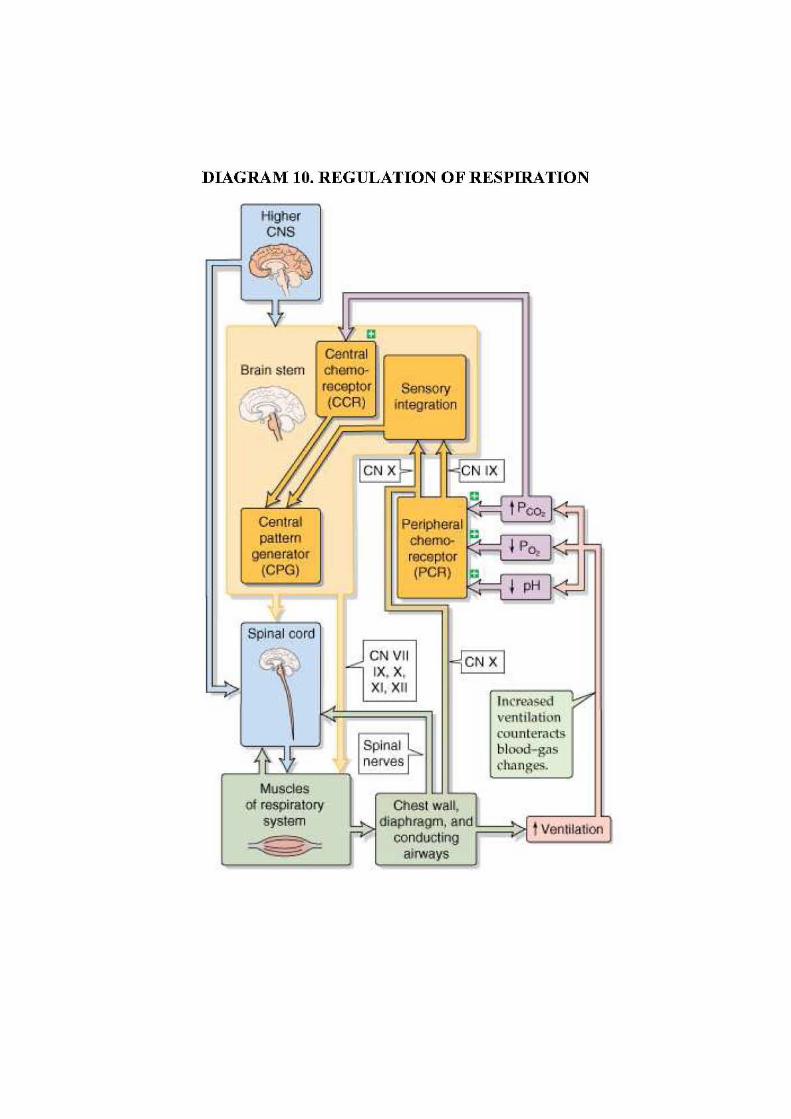

24
the 9th and 10
th cranial nerves, which carry impulses from airways and the
lungs.
The primary stimulus for breathing in healthy individuals is arterial
CO2, mediated through the central chemo-receptors through H+ formed
by the reaction between H2O and CO2 molecules. Peripheral chemo-
receptors, located mainly in the carotid bodies, respond to arterial O2.. The
primary stimulus to peripheral chemoreceptor in healthy individuals is
arterial hypoxemia, which is not clinically significant until PaO2 falls below
60mmHg.17

25
PULMONARY FUNCTION TESTS
History of PFT
Pulmonary function tests refer to a wide range of diagnostic procedures
to measure and evaluate lung function. Borelli (1681) is considered to be the
first physiologist, who established the quantity of air received by a single
inspiration. In 1700, Humphrey measured the residual volume by
hydrogen dilution technique using his mercurial air holding machine. and this
technique was improved by Darling, Cournand and Richards using O2
breathing to wash nitrogen out of the lungs. The collection of analysis of the
volume of exhaled N2 allowed the Functional Residual Capacity (FRC) to be
estimated and in the early 1950s, Fowler developed the single breath N2
wash out technique to assess FRC. Using simple spirometry combined with
FRC determinations allows Total Lung Capacity (TLC) and residual volume
to be calculated. Later, Body Plethysmography which was developed by
Comroe and Dubois is used to measure FRC and airway resistance. In
1845,Vieordt did a very exact determination of the volumetric parameters.
John Hutchinson was the first person, who started his work with
spirometer in 1844. He coined the term ‘vital capacity’, since he realized
that compromise of this crucial measurement was predictive of premature
morbidity and also, he defined the functional subdivisions of lung volume.
And after his discovery, there had been various modified forms of spirometer,

26
which was more simple to use. In 1854, Vintrich developed a modern
spirometer and confirmed that body height, weight and age determine the
vital capacity. Salter in 1866, added the kymograph to the spirometer, to
record time as well as the volume obtained. It was only one hundred years
later Hutchinson, Tiffeneau (1947) and colleagues timed the expired volume
and described the FEV1, which has now turned out to be a most widely used
parameter to measure mechanical properties of the lungs and medium sized
airways. In 1959, Wright B.M and Mckerrow C.B introduced the peak flow
meter and later in 1969, DuBois A.B and van de Woestijne K.P presented the
whole body plethysmograph on humans and in 1974, Campbell et al
presented a cheap and light development of a peak flow meter.
Since 1950s, however pulmonary physiologists took the advantage of
the opportunities provided by the growing fields of electronics, transducers
and computers, and since then, there has been tremendous progress in the
arena of pulmonary function testing. Single breath pulmonary function tests
are widely used in the modern computerized age. Digital spirometry
measures the mechanical function of lungs, chest wall and respiratory
muscles by assessing the total volume of air exhaled, from total lung capacity
to residual volume. It can graphically display the results and show predicted
values and their interpretations as required. The unit will have in memory
all the prediction tables for males and females across all age groups.There are

27
a few variables such as age, gender and body size which have an impact on
the lung function of one individual compared to another.
In 1831, Thomas Graham of Scotland described diffusion of gases
(Graham’s Law). The basis for the modern single-breath diffusing capacity
(DLco) test was described by August and Marie Krogh in 1911 and later
around 1950, Forrester revised the method and they developed it as a tool to
measure the gas exchange capacity of the lung. Measurement of blood gases
in the blood began since the early 1900s. In 1957, Sanz introduced the glass
electrode to measure pH of fluids potentiometrically, and in 1958,
Serveringhaus added an outer jacket containing a bicarbonate buffer to the
gas electrode. In 1956, Leland clark covered a platinum electrode with a
polypropylene membrane. These three electrodes (pH, pCO2 and pO2 ) were
the basic measurement device in blood gas analyzers, which was gradually
replaced by Miniature electrodes. In 1967, combined pH-clark-
Serveringhaus was developed for rapid blood gas analysis. The ear oximetry
was developed in 1974 and the pulse oximeter to monitor oxygen saturation
in 1980s. Modern microprocesses have allowed pulse oximeters that are
capable of measuring COHb in addition to oxygen saturation. To measure the
perfusion function of the lung, Swan-Ganz catheter was developed in 1970,
for measurement of pulmonary artery pressure and recently, Lung scan has
become the routine to determine pulmonary blood flow.18,19

28
Pulmonary function tests are useful 1) To assess physical fitness and
effects of physical training, 2) as an objective assessment of the functional
status of the respiratory system and indicate the nature and extent of
functional disturbances and, 3) serial measurements are useful in following
the course of the disease, evaluating therapy and determining prognosis.20
Recently Pulmonary function is assessed by the following tests:
I] Tests for ventilation
a. Spirometry: - For assessing Vital capacity (VC), mid-expiratory
flow rate (MEFR), peak expiratory flow rate (PEFR), minute
ventilation (MV), maximum voluntary ventilation (MVV),
b. Functional residual capacity is determined by single breath
nitrogen washout/Helium Dilution Method.
II] Tests for gas exchange function- analysis of blood gases using
miniature blood gas electrodes (give the value of pO2, pCO2 and pH)
and pulse oximeters with finger or ear probes for assessment of
oxygen saturation.
III] Tests for Perfusion function- Lung scan-to determine any blockage
in the blood flow from the heart into the lungs and determination of
pulmonary vascular pressure by catheterization.
IV] Plain X-ray chest – provides information on the lung fields
V] Computerized Axial Tomography (CT)

29
VI] Magnetic Resonance Imaging (MRI)
VII] Endoscopic Examination: Bronchoscopy- for direct visualization of
trachea and lower bronchial passages; Histopathological and
cytological examination of biopsy material.

30
MODERN LIFESTYLE CAUSING STRESS CONDITIONS IN DAILY
LIFE
In spite of the leaps and bounds in medical sciences, we are surrounded
by a very dangerous foe, the life-style disorders. As a result of modern
technology and automation, the need for physical activity is reduced and
thereby, we lead a sedentary life-style; it was defined as per Centre for
Disease Control and Prevention as, no leisure time physical activity or
activities done for less than 20 minutes or fewer than 3 times per week.21
Diseases, directly linked to modern life-style, like Diabetes, coronary artery
diseases, hypertension, obesity, eating disorders, mood disorders, mental
illness and psychosocial disorders are a great threat to human life and
constitute the major bulk of morbidity and mortality of the 21st century. Our
diet has changed, from high fibre, low fat to high fat, low fibre type and
processed food rich in salt and fat content.1
Stress also contributes to the formation of unhealthy life- style habits
like overeating, decreased physical activity, addiction to smoking, alcohol,
drugs etc. which lead to obesity, insulin resistance and atherogenic
dyslipidemia. Nearly, every man complains of life stress- Physical stress
from physical overactivity, as well as, mental, emotional and psychological
stress at the work place and in personal relationships with partners, family
and friends. Occupational stressors including long working hours (Theorell


31
T et al.1972)22,23
, shift work (Alfredsson L et al 1982)24, and high levels of
job responsibility (Kittel F et al.1980)25 may directly contribute to increased
health risks for cardiovascular diseases and respiratory diseases.
Multinational Time Budget Data Archive and the Australian Time Use
Survey suggest that women are now bearing a Dual Burden as both family
providers and family carers. Absence of reciprocal and joint emotion
management within family is a nagging stressor for women. For men,
workplace stress can have extreme consequences. In Japan, work stress
related suicide rate among men has risen over the last 15 years. According to
the Government’s Statistical Bureau, in Japan, the highest suicide rate occurs
in men from 35-44 years old, making it the 13th most common cause of death.
Sanjay Chugh, a leading Indian Psychologist, says that 70% to 90% of adults
visit primary care Physicians, for work stress- related problems.


32
EFFECTS OF STRESS ON CARDIOVASCULAR
FUNCTION
Among the many different type of stressors, mental and psychosocial
stressors, exert profound effects on the circulatory system. Psychosocial
stress has been demonstrated to increase oxidative stress, which disturbs the
endothelial integrity and induces endothelial injury and dysfunction. These
processes activate beta-1 adrenoreceptors that cause sympathoadrenal
activation, which, in turn, enhances atherosclerosis.26,27
Mental stress
increases the myocardial oxygen demand via sympatho-adrenal activation
and also involves a reduction in the myocardial oxygen supply, an eventually
fatal consequence.26 A possible biological explanation for long working hours
eliciting an acute myocardial infarction might be changes in the activity of
the autonomic nervous system. Work-induced tension that increases
sympathetic nervous activity, increases blood pressure. Blood pressure is
increased in both normotensives and hypertensive subjects. While working
the longer the working hours, the higher the daily mean blood pressure. In
addition, reduced activity of the parasympathetic nervous system
increases the risk of coronary heart disease. In particular, mental stress
may cause paradoxical constrictions, in patients with CAD/atherosclerosis,
especially, at points of stenosis- a response that correlates with the extent of
atherosclerosis(plaque) and with the endothelium- dependent response to an

33
infusion of acetylcholine (verification of endothelial dysfunction).26,29
An
early psychosomatic hypothesis put forth by Alexander(1939) stated that,
feelings of anxiety might lead to elevation of blood pressure, and when
prolonged, predisposes to the development of hypertension. A paper from the
Framingham Heart Study indicated that, anxiety levels were a significant
predictor of the subsequent incidence of hypertension among middle-aged
men (Markovitz et al 1993)30
Sympatho- adrenergic stimulation is achieved by a combination of
1. Nor-epinephrine released from the terminal neurons of
postganglionic fibres running from left or right stellate ganglion.
2. Epinephrine release from adrenal medulla.
Nor-epinephrine excites mainly alpha-receptors, but excites the beta
receptors to a lesser extent as well. Conversely, epinephrine excites both
types of receptors, approximately equally. Therefore, the types of receptors
in the organs determine the relative effects of nor-epinephrine and
epinephrine. Nor-epinephrine or epinephrine acts on cardiac beta-
adrenoreceptors, chiefly beta-1. As a consequence of sympathetic
stimulation, the heart rate, rate of conduction and the force of contraction of
the heart increases, while the refractory period is reduced30. Sustained stress
may lead to chronic sympathetic arousal and thereby produce
vasoconstriction and lead to hypertension.

34
In research conducted by Dr.David Anderson of the National Institute
of health, shallow breathing in animals was linked to elevated salt and higher
blood pressure. A similar process is suspected to be at play in human. When
people are under stress, they tend to take shallow breathes, which in turn
makes the blood more acidic and hence, the kidneys are less efficient in
removing sodium from the blood.
The studies by Julies et al.1976, have especially well-illuminated the
central autonomic involvement of both heart and blood vessels with clear
signs of a centrally reduced vagal tone to the heart in association with signs
of sympathetic activation.

35
EFFECTS OF STRESS ON RESPIRATORY FUNCTION
Whenever we are stressed, there is a shallow and fast breathing and
too much carbon dioxide is lost due to hyper- ventilation; constriction of
arteries and arterioles (since CO2 is a most potent vasodilator) and the
suppressed Bohr effect (less oxygen is released in tissues due to increased
affinity of oxygen to red blood cells caused by hypocapnia) leading to
reduced cells oxygen content. This means that the heart has to work hard to
attempt to get oxygen, when it is needed more in the body and brain.
Stressful situations, emotional arousal and psychological stress has been
shown to influence our respiratory system, by stimulation of sympathetic
nervous system, which produces adrenaline and this alters not only
respiratory system, but also heart, and other organs. It increases respiratory
rate and respiratory irregularity.
Another important effect of stress on the respiratory system is, Non-
allergic asthma which does not involve the immune system. It can be
triggered by stress, anxiety, sadness, smoke or a viral infection. It is also
associated with an elevated prevalence of anxiety and depressive disorders.
Asthma and these psychological states may mutually potentiate each other,
through direct psycho-physiological factors, non- adherence to medical
regimen etc. 32, 33,34

36
DEEP BREATHING EXERCISE
With our normal sedentary way of living, we use only about one
tenth of our lung capacity. Deep breathing is the act of breathing, done by
expanding one’s chest and thereby allowing the diaphragm to move down,
which becomes flattened (by creating a vacuum that draws the air) creating
more room for the lungs to expand rather than only through chest wall
expansion/movement of the rib cage. When we exhale, the diaphragm returns
to its dome shape, pushing air out of the body, which helps to remove waste
products, resulting from body’s metabolic processes, while providing oxygen
for energy. Deep breathing exercises increases the amount and percentage of
air, which enters into the gaseous exchange processes, the alveoli. This
slower, deeper breathing, combined with the rhythmical pumping of our
diaphragm and with the other muscles of respiration, helps to turn on our
parasympathetic nervous system - our "relaxation response." Such breathing
helps to harmonize our nervous system and reduce the amount of stress in
our lives. And this, of course, has a positive impact on our overall health.
Jerath and colleagues (2006) have reported that, slow deep breathing
increases frequency and duration of inhibitory neural impulses, by activating
stretch receptors of the lungs; as in Hering-Breuer reflex. Inhibitory impulses,
produced by slowly adapting – receptors in the lungs during inflation play a
role in controlling autonomic functions such as systemic vascular resistance
and heart rate. Inhibitory current synchronizes rhythmic cellular activity


37
between the cardiorespiratory centers and the central nervous system.
Inhibitory current regulates excitability of nervous tissue and is known to
elicit synchronization of neural elements, which typically is indicative of a
state of relaxation. Synchronization within the hypothalamus and the
brainstem is likely responsible for inducing the parasympathetic response
during breathing exercises.35

38
EFFECTS OF DEEP BREATHING EXERCISE ON STRESS
REDUCTION
The increasing stress of modern living makes us breathe more quickly
and less deeply. This does not allow enough oxygen to reach the organs and it
can cause hyperventilation that result in carbon-di-oxide wash out rather than
oxygen uptake. Modern technology and automation reduces our need for
physical activity. There is less need to breathe deeply, so we develop the
shallow breathing habit. Deep breathing exercises and stretching of muscles,
especially those primarily concerned with controlling inhaling and exhaling,
should be sought. Apart from its health giving benefits, it soothes the nerves
and quickly induces the state of peace and calm. Deep breathing
significantly decreases several negative mood traits, including tension-
anxiety in psychiatric inpatients. Relaxation training including deep breathing
has been found to be effective in treating stress and anxiety disorders 36 and is
therefore an alternative treatment for anxiety. Oxidative stress may contribute
to pathophysiology of many chronic diseases such as psychosocial stress. A
study using stress reduction from slow deep breathing exercise revealed
significantly lower levels of lactate and higher levels of superoxide
dismutase, glutathione and catalase compared with controls . In another
study, hemodynamic response to acute stress to healthy volunteers, revealed
significantly positive effect when slow deep breathing technique was
positively applied. 37

39
EFFECTS OF DEEP BREATHING EXERCISE ON
CARDIOVASCULAR FUNCTION
The cardiovascular system is controlled mainly by the autonomic
nervous system. Baroreceptors are branched and myelinated nerve endings,
that are present in the carotid sinus and aortic arch, which respond to changes
in blood pressure, whenever there is variation in blood pressure over a wide
range of 60-180 mm Hg. It sends impulses to the cardiac centre in the
brainstem via afferent fibres in the glossopharyngeal and vagus nerve .At the
normal blood pressure there is impulse discharge in these nerves at a low
frequency and is responsible for vagal tone.38 Baroreflex sensitivity can be
enhanced significantly by slow deep rhythmic breathing, by stimulating the
medullary cardio-inhibitory area and that leads to reduction in sympathetic
activity that inhibits the tonic discharge of the vasoconstrictor nerves and
excites the vagal innervations of the heart. As the sympathetic activity to the
heart is reduced, it leads to decrease in heart rate i.e decrease in work of the
heart , leading to fall in systolic blood pressure. As the vasoconstrictor fibres
supplying the resistance vessels i.e. the arterioles are inhibited, it leads to
widespread vasodilatation and hence fall in diastolic blood pressure.39 Thus,
slow deep breathing alters the heart rate, strength of cardiac contractions, and
diameter of blood vessels.

40
Deep breathing leads to a greater pressure differential in the lungs,
which leads to an increase in the circulation with increased oxygen level in
the blood, that can increase the efficient functioning of the heart.38

41
EFFECTS OF DEEP BREATHING EXERCISE ON
RESPIRATORY FUNCTION
During deep breathing exercise, there is increase in alveolar ventilation
that favours diffusion of oxygen and hence oxygenation of the tissues is also
greater. The shallower the breathing, the larger becomes the percentage of
dead air in each breath. Deep breathing has the effect of increasing both the
amount and percentage of air, which enters actively into the gaseous
exchange processes. Deep breathing exercises increase the elasticity of the
lungs and rib cage and also causes enhancement of respiratory muscle
efficiency. Stimulation of pulmonary stretch receptors by deep inspiration
causes reflex relaxation of smooth muscles of the larynx and
tracheobronchial tree and that modifies the airway caliber, thereby reducing
airway resistance.40 This creates an increased breathing capacity all day, not
just during the actual breathing period. This means all the above benefits also
occur all day and hence protect the individuals from respiratory illness.


42
MATERIALS AND METHODS
STUDY DESIGN : This is an interventional trial.
STUDY PLACE AND STUDY PERIOD : This study was conducted in the
Department of Physiology, Tirunelveli Medical College, Tirunelveli. The
study period extended from November 2010 to February 2011.
STUDY SUBJECTS : 146 student volunteers, doing the first year M.B.B.S
course in Tirunelveli Medical College, Tirunelveli in the adolescent age
group (17-19 years) were selected for the study.
INCLUSION CRITERIA : Healthy volunteers of both sexes in the age
group of 17-19 years.
EXCLUSION CRITERIA : Students suffering from any respiratory illness
like bronchial asthma, physically handicapped-any chest/spinal deformity.
MATERIALS USED FOR THE STUDY
• Proforma- to record the details of the subjects and to record the clinical
findings.
• Standardised mercury sphygmomanometer-to record the Blood
pressure.
• Portable weighing machine-to record the body weight in kilograms.
• Digital spirometer- to measure the lung function tests.

43
METHODOLOGY
The study was approved by the Ethical Committee of Tirunelveli
Medical College, Tirunelveli. After getting the informed written consent
from the individuals, the proforma for details such as age, sex, height,
weight, hours of active exercise, menstrual history (in case of women ) was
filled up. A thorough clinical examination of the study subjects was done.
The following parameters were measured in the selected group for
assessing the cardiovascular and pulmonary functions- Pulse rate (/min),
Blood pressure (mmHg), FEV1 (L), FVC (L), FEV1/FVC (%), PEF (L/min).
These tests were recorded in batches of 25 students daily for 6 days.
The anxiety level of the students was assessed by pretest technique,
with the help of Brief Patient Health Questionnaire (English), a version of
Patient Health Questionnaire (PHQ) Family of Measures41, 42, 43
. It was
designed by Drs.Robert L.Spitzer et al 1999.It consisted of 7 questions and
the anxiety level is calculated by assigning scores of 0,1,2, and 3 to the
response categories of ‘Not at all’, ‘Several days’, ‘more than half the day,
and ‘nearly every day’ respectively. Total score for the seven items ranges
from 0-21.Students were asked to answer the 7 questions in the format to the
best of their knowledge. The scores were calculated for each individual and
those with 1-5, 6-10, and 11-15 were said to have mild, moderate and severe
anxiety respectively.

44
The students were trained for performing deep breathing exercise, and
they were requested to do the same daily for a period of 12 weeks. They were
asked to sit upright comfortably with eyes closed, and they were instructed to
take a slow deep breathing (which includes inspiration and expiration), that
was continued for 15 minutes in the morning session, in the presence of
instructor from 11 AM to 11.15 AM, and they were asked to repeat the same
procedure for 15 minutes in the evening at their convenient time. Thus, the
procedure was done for 30 minutes/day, for a period of 12 weeks.
The various tests for assessing the cardiovascular and pulmonary
functions were carried by the procedures mentioned below and repeated at
the end of 4 weeks, 8 weeks and 12 weeks; Recording of the Pulse rate,
Blood Pressure and Spirometry was done in batches of 25 students daily, for
6 days-146 students totally and the anxiety level was analysed by post-test
technique at the end of 12 weeks. The above parameters were recorded in the
proforma and then tabulation was done separately. The results were then
compared, analyzed statistically to find out the significance of variations in
them, before and after deep breathing exercise.
1. Measurement of pulse rate
The arterial pulse rate, (which is an indirect indicator of the heart rate
in a healthy individual), was measured by the examination of the radial
artery. The radial pulse was best felt with the tips of the three middle fingers


45
with the forearm of the subject in semipronated position and the wrist slightly
flexed. The counting was not done, as soon as the fingers were first laid upon
the pulse, but only when it had resumed to normal rate; it was counted for
one minute and was expressed as pulse rate/minute.12
2. Measurement of Blood Pressure
The arterial blood pressure was measured by the non-invasive
technique using sphygmomanometer by auscultatory method (Russian
Physician, Korotkoff,1905). A Hawksley random–zero sphygmomanometer
(Gelman Hawksley, Northampton) was used for all, recording with a cuff
(Riva-Rocci cuff) of appropriate size (for adults-23×12.5 cm) as proposed by
Long et al. Blood Pressure was recorded by asking the student to sit
comfortably in a chair and the uninflated cuff was wrapped firmly around the
upper left arm (as recommended by Rose and Blacksmith44), 2.5-3cm above
the elbow and the chest piece of stethoscope was kept at the elbow medial to
the tendon of biceps, where pulsations of brachial artery was felt. Now, the
cuff was inflated rapidly until the pressure in it was well above the systolic
blood pressure i.e.30mm Hg above the level at which radial pulsation is no
longer felt (as measured by the palpatory method) and gradually the cuff
pressure was lowered by opening the valve till a clear, sharp, tapping sound
was heard with the stethoscope (corresponds to korotkoff’s phase 1) and the
pressure at this moment was taken as Systolic Blood Pressure. The pressure


46
in the cuff was progressively lowered (at 2-3mm Hg/sec) and at the same
time listening to the muffling/disappearance of sound, the pressure was noted
(corresponds to Korotkoff’s Phase4/5 respectively) and it was taken as
Diastolic Blood Pressure.
3. Measurement of
[a] Forced Vital Capacity
[b] Forced Expiratory Volume in first second
[c] FEV1/FVC ratio and
[d] Peak Expiratory Flow Rate.
were done using Computerised Spirometer- Super Spiro.
The Micro Medical Super Spiro (Micro Medical, Rochester, Kent,
England) consisting of a (1) microcomputer unit incorporating a 1/4s d A
screen with 64 colour display with touch screen,data entry keypad and all
associated circuitry, (2)digital volume transducer, (3) transducer holder and,
(4) mains adapter. The Micro Medical Digital Volume Transducer, which
measures expired air directly at B.T.P.S (Body Temperature and Pressure
with Saturated water vapour), thus avoiding inaccuracies of temperature
correction.45


47
Preparation of the student for spirometry: Preparation consists mainly
of instructions given to them in advance of the actual test session. To obtain
valid data, they were instructed to refrain from vigorous exercise immediately
before testing and to be relaxed and comfortable during the test session.
Demonstration of the procedure was given at the start of test session.
The test was done in the sitting position with the head slightly elevated.
The individual was instructed to take deep inspiration, until their lungs were
completely full, then seal their lips around the mouth piece and blow out as
hard and as fast as possible, until they cannot push any more air out (at
least for a minimum of 6 seconds). Then to breathe in fully, immediately after
the expiratory maneuver, thus completing the flow volume loop. Instruction
about maintaining a good seal on the mouth piece was given46.By this
maneuver, the flow-volume loop was displayed on the computer monitor
screen. The computer analyses the signal from the spirometer, then calculates
and displays the FVC,FEV1, FEV1/FVC, and PEF. The procedure was
repeated up to 3 times and best of the three was taken as the final reading.19
• Forced vital capacity (FVC)
Defined as the maximum volume of gas that can be expired, when the
patient exhales as forcefully and rapidly as possible, after a maximum
inspiration.

48
• Forced expiratory volume in one second (FEV1)
Defined as the volume of air which can be forcibly exhaled from the
lungs in the first second of a Forced expiratory maneuver.
• FEV1/FVC ratio- FEV1%
Indicates what percentage of the total FVC is expelled from the lungs
during the first second of forced exhalation. The normal values of FEV1%
are 80-85% in young adults.
• FVC and FEV1 are both expressed in litres. FVC, FEV1 and FEV1% are
reliable indices of ventilator capacity and indicator of pulmonary
ventilation.
• Peak Expiratory Flow Rate (PEFR/PEF)
Defined as the maximum rate of air flow achieved during the FVC
maneuver, beginning after full inspiration and starting and ending with
maximal expiration. It is expressed in L/min. Normal Range- 350-600 litres
per minute.46

PROFORMA
EFFECTS OF DEEP BREATHING EXERCISE ON
CARDIOPULMONARY FUNCTION IN ADOLESCENTS
No: AGE:
NAME : SEX:
ADDRESS: PARENT’S INCOME:
HEIGHT: cm WEIGHT: kg
PERSONAL HISTORY: Hours of active exercise
Menstrual history (in case of women)
O/E: GENERAL EXAMINATION-
SYSTEMIC EXAMINATION: CVS
RS
EXAMINATION OF CARDIOVASCULAR & RESPIRATORY SYSTEM
S. No Tests Done Initial
reading
End of 4
weeks
End of 8
weeks
End of 12
weeks
1. PR(/min)
2. BP(mm Hg)
3. FEV1(L)
4. FVC(L)
5. FEV1/FVC (%)
6. PEF(L/min)
INFORMED CONSENT:
I consent for this study having been informed about the procedure.

BRIEF PATIENT HEALTH QUESTIONNAIRE (ENGLISH)
Over the last 2 weeks, how
often have you been
bothered by the following
problems? (Use “√” to
indicate your answer)
Not at all
Several days
(less than half
the days)
More
than half
the day
Nearly
everyday
a. Feeling nervous, anxious
or on edge
b. Not being able to stop or
control worrying
c. worrying too much about
different thing
d. Trouble relaxing
e. Being so restless that it is
hard to sit still
f. Becoming easily annoyed
or irritable
g. Feeling afraid as if
something awful might
happen.


49
RESULTS ANALYSIS
The results obtained after the performance of the tests to assess the
cardiovascular and pulmonary functions in the study group were tabulated.
The findings were compared with reference to the age and the effects of the
deep breathing exercise at the intervals of 4, 8, and 12 weeks; the anxiety
level assessed by pre-test and post-test were also analysed and compared.
The statistical analysis of the results was also undertaken. All the
values obtained before and after performing Deep Breathing Exercise, were
expressed as mean ± S.D. The results were analyzed and interpreted by
repeated measures of ANOVA and the student’s paired t test was used to
compare parameters within the study group. The above statistical procedures
were performed with the help of statistical package namely PASW Statistics-
18 (Predictive and analysis software).

50
TABLLE.1
AGE & SEXWISE DISTRIBUTION OF THE STUDY GROUP
AGE (YEARS) MEN WOMEN TOTAL
17 23 46 69
18 30 32 62
19 4 11 15
TOTAL 57 89 146
Among the 146 students in the study group, 57 are men and 89 are
women.

51
TABLE.2
EFFECT OF DEEP BREATHING EXERCISE ON ANXIETY LEVEL.
NO
ANXIETY
MILD
ANXIETY
MODERATE
ANXIETY
SEVERE
ANXIETY
TOTAL
PRE
TEST
2 80 53 11 146
POST
TEST
24 112 9 1 146
The facts that the number of students with no anxiety level has
increased from 2 to 24 and the number of students with moderate and severe
anxiety level has been reduced markedly (after 12 weeks of DBE), show the
response due to deep breathing exercise among them. The same was analysed
statistically and the changes were found to be highly significant.
STATISTICAL ANALYSIS
Anxiety Level
P value Significance
Mean ± S.D
Initial Reading 5.5 ± 3.3
<0.001
Highly
Significance End of 12 Weeks 1.7 ± 1.2


52
TABLE.3
VARIATION IN PULSE RATE BEFORE AND AFTER DBE*
Age in
Years
No. of
students
Average pulse rate/min
Initial reading
At the end of
4 weeks 8 weeks 12 weeks
17 69 96.29 89.18 85.62 82.68
18 62 93.12 86.51 83.64 81.19
19 15 95 85.33 81.46 80.40
There is a gradual decrease in pulse rate/min following the deep
breathing exercise and it is found to be statistically significant (p<0.001).
STATISTICAL ANALYSIS
Pulse Rate (/min) F value
(ANOVA)
P value Significance
Mean ± S.D
Initial reading 94.8 ± 10.9
165.090 < 0.001
Highly
significant
End of 4 weeks 87.6 ± 11.3
End of 8 weeks 84.0 ± 9.6
End of 12 weeks 81.4 ± 8.9
*DBE – Deep Breathing Exercise


53
TABLE. 4
VARIATION IN SYSTOLIC BLOOD PRESSURE BEFORE AND
AFTRE DBE.
Age in
years
No. of
students
Average Systolic Blood pressure in mmHg
Initial reading
At the end of
4 weeks 8 weeks 12 weeks
17 69 120 112.92 109.33 108.20
18 62 119.29 114.32 113.16 109.64
19 15 125.19 118. 117.33 114
There is a gradual reduction in systolic blood pressure (mm Hg)
following the deep breathing exercise and statistically it is found to be highly
significant. (p<0.001).
STATISTICAL ANALYSIS
Systolic blood
pressure (mm Hg)
F value
(ANOVA)
P value Significance
Mean ± S.D
Initial reading 120.3 ± 13.6
67.882 < 0.001
Highly
significant
End of 4 weeks 113.3 ± 12.2
End of 8 weeks 112.5 ± 12.2
End of 12 weeks 110.1 ± 14.2

54
TABLE. 5
VARIATION IN DIASTOLIC BLOOD PRESSURE BEFORE AND
AFTER DBE
Age in
years
No. of
students
Average Diastolic blood pressure in mm Hg
Initial reading
At the end of
4 weeks 8 weeks 12 weeks
17 69 80.81 76.69 74.43 72.52
18 62 80.77 75.77 74.38 72.41
19 15 83.06 78.53 77.73 73.73
Following the deep breathing exercise, there is a gradual decline in
diastolic blood pressure (mm Hg), which, when analysed statistically is
highly significant. (p<0.001).
STATISTICAL ANALYSIS
Diastolic blood
pressure (mmHg)
F value
(ANOVA)
P value Significance
Mean ± S.D
Initial reading 81.2 ± 10.4
49.666 < 0.001
Highly
significant
End of 4 weeks 77.5 ± 12.5
End of 8 weeks 74.7 ± 7.8
End of 12 weeks 72.5 ± 7.8

55
TABLE.6
VARIATION IN FVC* (L) BEFORE AND AFTER DBE
Age in
years
No. of
students
Average FVC (L)
Initial reading At the end of
4 weeks 8 weeks 12 weeks
17 69 2.97 3.06 3.24 3.36
18 62 3.00 3.12 3.36 3.50
19 15 2.75 2.94 3.09 3.30
There is a gradual increase in FVC (L), following deep breathing
exercise and it is statistically significant.
STATISTICAL ANALYSIS
FVC (L) F value
(ANOVA)
P value Significance
Mean ± S.D
Initial reading 3.0 ± 0.7
61.169 < 0.001
Highly
significant
End of 4 weeks 3.1 ± 0.6
End of 8 weeks 3.3 ± 0.6
End of 12 weeks 3.4 ± 0.6
*FVC – Forced Vital Capacity

56
TABLE. 7
VARIATION IN FEV1* (L) BEFORE AND AFTER DBE
Age in
years
No. of
students
Average FEV1 (L)
Initial reading At the end of
4 weeks 8 weeks 12 weeks
17 69 2.58 2.78 3.02 3.25
18 62 2.62 2.84 3.10 3.38
19 15 2.34 2.59 2.83 3.12
There is a gradual rise in FEV1 (L) , following deep breathing exercise
and it is found to be hightly significant statistically.
STATISTICAL ANALYSIS
FEV1 (L) F value
(ANOVA)
P value Significance
Mean ± S.D
Initial reading 2.6 ± 0.7
143.439 < 0.001
Highly
significant
End of 4 weeks 2.8 ± 0.6
End of 8 weeks 3.0 ± 0.6
End of 12 weeks 3.3 ± 0.6
*FEV1 – Forced expiratory volume in one second.

57
TABLE. 8
VARIATION IN FEV1/FVC (%) BEFORE AND AFTER DBE
Age in
years
No. of
students
Average FEV1/FVC (%)
Initial reading
At the end of
4 weeks 8 weeks 12 weeks
17 69 88.37 91.22 93.72 95.48
18 62 87.60 89.49 93.37 94.58
19 15 85.53 88.40 91.06 94.26
Following deep breathing exercise, there is a gradual increase in
FEV1/FVC(%), and it is found to be statistically significant.
STATISTICAL ANALYSIS
FEV1/FVC
(%)
F value
(ANOVA)
P value Significance
MEAN ± S.D
Initial reading 87.8 ± 6.4
161.692 < 0.001
Highly
significant
End of 4 weeks 90.2 ± 6.7
End of 8 week 93.3 ± 4.2
End of 12 weeks 95.1 ± 3.3


58
TABLE. 9
VARIATION IN PEF* (L/min) BEFORE AND AFTER DBE
Age in
years
No. of
students
Average PEF (L/min)
Initial reading
At the end of
4 weeks 8 weeks 12 weeks
17 69 273.37 293.37 323.89 356.31
18 62 272.33 296.50 335.95 367.85
19 15 239.00 256.00 295.33 333.26
Following deep breathing exercise there is marked increase in
PEF (L/min), and it is found to be statistically significant.
STATISTICAL ANALYSIS
PEF (L/min) F value
(ANOVA)
P value Significance
Mean ± S.D
Initial reading 268.0 ± 87.3
199.868 < 0.001
Highly
significant
End of 4 weeks 201.0 ± 90.2
End of 8 weeks 323.5 ± 79.4
End of 12 weeks 357.6 ± 76.8
*PEF – Peak Expiratory Flow


59
TABLE.10
STATISTICAL ASSESSMENT OF EFFICACY OF DBE ON
CARDIOPULMONARY FUNCTION AND ANXIETY LEVEL
VARIABLES
BEFORE
(INITIAL
READING)
AFTER
(END OF 12
WEEKS)
DIFFER-
-ENCE t d.f P value
MEAN ±
S.D MEAN ± S.D
MEAN ±
S.D
PULSE RATE
(/MIN) 94.8± 10.9 81.4± 8.9 13.4± 8.9 17.302 145 <0.001
SYSTOLIC
BLOOD
PRESSURE (mm
Hg)
120.3± 13.6 110.1± 14.2 10.2± 11.8 10.386 145 <0.001
DIASTOLIC
BLOOD
PRESSURE (mm
Hg)
81.2± 10.4 72.5± 7.8 8.7± 7.8 13.486 145 <0.001
FEV1 (Litres) 2.6± 0.6 3.3±0.6 0.7± 0.5 17.344 145 <0.001
FVC (Litres) 3.0± 0.7 3.4±0.6 0.4±0.5 10.504 145 <0.001
FEV1/FVC (%) 87.8±6.4 95.1± 3.3 7.3± 5.1 17.430 145 <0.001
PEF (Litres/min) 268.0± 87.3 357.6±76.8 89.6± 53.4 19.910 145 <0.001
ANXIETY 5.5±3.3 1.7±1.2 3.8±2.9 16.132 145 <0.001


60
DISCUSSION
The present study involves the measurements of Pulse rate and Blood
pressure for the assessment of cardiovascular function and that of FVC,
FEV1, FEV1/FVC ratio and PEF for the assessment of respiratory function. It
is well known that, both cardiovascular and respiratory functions are
regulated mainly by autonomic nervous system, which is a part of central
nervous system and they are interlinked. Hypothalamus, that is considered
as the “ Head-ganglion” of autonomic nervous system plays the role of
neuroendocrine structure; structurally it has neuronal elements
predominantly, and functionally capable of secreting hormones like
Vasopressin, CRF and other releasing factors that control the hypophysis
cerebri, which acts as the “Mastergland of Endocrine Orchestra”.
Modern lifestyle mostly sedentary in type, that consists of physical
inactivity, overeating, exposure to physical and emotional stress acts to cause
anxiety in young individuals. Anxiety, when continued as stress, can cause
physiological changes in our body—in the form of sympathetic stimulation
with reduction in parasympathetic activity; which in turn, causes alterations
in the cardiovascular and respiratory functions. Chronic stress is found to
cause stress-related diseases.

61
In the present study, anxiety level in the student volunteers at the
beginning of the study shows predominant levels—Out of 146 students
studied, only 2 were of no anxiety level, whereas all others had varying
anxiety levels. At the end of the study i.e., 12 weeks after Deep breathing
exercise, the anxiety level in all of them was re-assessed and it was found
that, there was significant level of reduction in anxiety. This correlates with
the earlier report by Colleen M. Johnson et al 2009.47 Our brains are only 2%
of our body weight, yet they consume 20% of the oxygen we inhale. The
shallow breathing associated with stress and negative emotions, reduces the
oxygen level of our blood. When there is a slow deep breathing, our body
gets enough oxygen and that breaks the stress cycle. Slow and deep
breathing itself has a calming effect on the mind and helps an individual to
de-stress 48. This calming effect may also exert profound physiological
effects on pulmonary, cardiovascular, and mental functions of the brain.
In our study, there is a significant decrease in the pulse rate from the
mean value of 94.8 to 81.4/min at the end of 12 weeks after the practice of
deep breathing exercise. This indicates that, the practice of deep breathing
exercise improves vagal activity49. The systolic and diastolic blood pressure
also shows significant change, before and after deep breathing exercise; The
mean SBP has reduced from 120.3 to 110.1 mm of Hg and the mean DBP has
reduced from 81.2 to 72.5 mm of Hg. Diastolic blood pressure depends on

62
peripheral resistance and lung inflation has been shown to decrease systemic
vascular resistance. This response is initiated by pulmonary stretch receptors,
which bring about withdrawal of sympathetic tone in skeletal muscle blood
vessels, leading to widespread vasodilatation, thus bringing up decrease in
peripheral resistance, which leads to decrease in diastolic blood
pressure 40. The slow deep breathing enhances the baroreflex sensitivity,
leading to improvement of vagal tone and reduced sympathetic discharge,
thereby decreasing systolic blood pressure.50 Our present study correlates
well with the study of Grossman A & Grossman E in 200351. Deep and slow
breathing found to improve small vessels blood flow and thereby decreases
the peripheral resistance, leading to reduction in blood pressure and pulse
rate reduction.47 A large study of 21,565 subjects practicing deep breathing
for 30 seconds induced significant blood pressure decreases in
normotensives, untreated and treated hypertensives (Mori et al.,2005)37
Thus, the reduction in pulse rate and blood pressure found in our study,
indicates a shift in the balancing components of autonomic nervous
system towards the parasympathetic activity which was reported by Anand
BK et al in 1991.52 This modulation of autonomic nervous system activity
might have been brought about through the conditioning effect of deep
breathing exercise on autonomic functions and mediated through the limbic
system and higher areas of central nervous system as reported by
Selvamurthy et al.52 The better response seen in the present study, can be

63
attributed to more elastic blood vessels and also better reflex activity, which
is seen in the younger age group.
Pulmonary function is a valuable tool for the general health
assessment. The mean values of FEV1, FVC, FEV1/FVC, and PEF were
assessed before and after deep breathing exercise. All the parameters were
found to be increased significantly indicating the improvement in efficiency
of pulmonary function. The reason could be that, deep breathing exercise
causes an increase in the elasticity of lungs and the rib cage; there is
increased oxygenation of blood, as alveolar ventilation increases without
changing minute ventilation 39. The findings correlate with earlier reports.
Richa Ghay Thaman et al in 2010 have reported that medical students who
had sedentary life-style had lower pulmonary function; physical training and
exercise improved the lung function parameters in BSF trainees.21
K
Upadhyay Dhungal et al40 in 2008 have reported increase in PEFR after slow,
deep breathing exercise in young individuals, which also correlates with our
finding.


64
SUMMARY AND CONCLUSION
146 student volunteers, doing the first year M.B.B.S course in Tirunelveli
Medical College, Tirunelveli in the adolescent age group (17-19 years) were
selected for the study. During this study, the measurement of pulse rate/min, blood
pressure (mm Hg) and estimation of FEV1,FVC,FEV1/FVC,PEF were done before
and after Deep Breathing Exercise at the intervals of 4 weeks,8weeks and 12
weeks.
The present study indicates that, the anxiety level among the medical
students is significantly reduced after deep breathing exercise for 12 weeks—by
the findings in Pre-Test and Post-Test with specific questionnaire.
Anxiety, as stress when mild, acts beneficially to improve our activities, by
bringing about physiological changes through Psycho-Hypothalamo-Hypophyseal-
Adrenal axis as explained by Hans Selye (1907-1982). Stress, when prolonged,
interferes with various body regulatory mechanisms resulting in stress-related
diseases like hypertension, diabetes mellitus and bronchial asthma. Therefore, to
reduce anxiety among youngsters, the deep breathing exercise is found to be very
helpful.
The study also reveals the significant reduction in pulse rate (which directly
correlates with heart rate) and blood pressure after 12 weeks duration of deep

65
breathing exercise; this effect is due to increasing parasympathetic activity,
which is accompanied by reduction in sympathetic stimulation, which in turn is
the result of reduction in anxiety level in the students. Sedentary life-Style,
prevalent among modern student population from marked physical inactivity (as a
result of lack of exercise, usage of TV and Computers at leisure and working
hours) together with anxiety or stress while driving vehicles, facing tests causes
rapid, shallow breathing; This causes reduction in the vital parameters of
pulmonary function, as evidenced by the various studies.
In this study, we find the measurement of Forced Vital Capacity,
Forced Expiratory Volume in the first second, Ratio of FEV1/FVC and Peak
Expiratory Flow, done for assessing the efficacy of pulmonary ventilation are
significantly raised after 12 weeks of deep breathing exercise, when compared to
those values recorded at the beginning of the study. The changes are due to slow
deep breathing exercise, which results in better enlargement of thoracic cavity
improving the contraction of external intercostals muscles and diaphragm, namely
respiratory efficiency, which is sustained during the day. This deep breathing effort
increases the alveolar ventilation, thereby facilitating diffusion and transport of
O2 and CO2 across the alveoli. Better oxygenation of the blood, that reaches the
brain helps in improving the nervous activity, that reduces the anxiety and its
related effects on various systems of the body.

66
To conclude, this study reveals the usefulness of deep breathing exercise as
non-pharmacological adjunct in reducing the anxiety among the students and
improving the efficacy of pulmonary and cardiovascular functions. When pursued
further, the simple procedure can also help in preventing and postponing the
cardiovascular disorders such as hypertension, myocardial ischemia etc.


i
BIBLIOGRAPHY
[1] G. Sharma, L.K. Sharma & S. Sood: Synergistic Approach of Applied
Physiology & Yoga to Combat Lifestyle Diseases. The Internet
Journal of Alternative Medicine. 2009 Volume 7 Number1
[2] Anand BK. Yoga and medical sciences. Ind J Physiol Pharmacol, 1991
Apr; 35(2):84-87.
[3] Park’s Textbook of Preventive and Social Medicine 20th ed. Pg.314
[4] Breathing control lowers Blood Pressure- Journal of human
hypertension. April 15, 2001, Vol.15 No.4, Pg 263-269.
[5] Hurst’s THE HEART 12th ed.Vol.2.
[6] An introduction to Cardiovascular Physiology J R Levick 4th edition.
[7] Walter F. Boron & Emile L. Boulpaep MEDICAL PHYSIOLOGY
2nd ed.
[8] Berne & Levy. Principles of Physiology.6th ed .Pg 353-356.
[9] Harvey W. Anatomical Studies on the Motion of the heart and Blood.
Leake CD, transl. Springfield, 1L:Charles C Thomas;1970.
[10] Acierno LJ, The History of Cardiology. London: Parthenon; 1994.
[11] Willius FA, Dry TJ, A History of the Heart and the Circulation.
Philadelphia; Saunders; 1948.
[12] G K Pal Pravati Pal Textbook of Practical Physiology 3rd ed.
[13] Davidson’s Principle &Practice of Medicine.21st ed.
[14] Textbook of Pulmonary Medicine D.Behera; Volume.1; pg 46-61.

ii
[15] Proctor DF. The upper airways. I. Nasal physiology and defense of the
lungs. Am Rev Respir Dis 1977;115:97.
[16] Proctor DF. The upper airways. II. The larynx and trachea. Am Rev
Respir Dis 1977;115:315.
[17] Robert L.Wilkins, James K.Stoller, Robert M. Kacmarek. Egan’s
Fundamentals of Respiratory Care 9th ed.
[18] Grippi MA, Metzeger LF,Sack AV, Fishman AP, Pulmonary Function
Testing. Fishman AP,Elias JA, Fishman JA ,Grippi MA, Kaiser
LR,Senior RM, Editors: Fishman’s Pulmonary Diseases and
Disorders 3rd International ed. Mc Graw-Hill; Health
Professional Division;1998;pp533-7.
[19] Manual of Pulmonary Function Testing Gregg L.Ruppel 9th edition.
[20] Manual of practical physiology-A.K.Jain.
[21] Richa Ghay Thaman, Anterpreet Arora and Rachna Bachhel. Effect of
Physical Training ON Pulmonary Function Tests in Border
Security Force Trainees of India. J Life Sci,2(1):11-15 (2010).
[22] Theorell T, Rahe R. Behaviour and life satisfactions characteristics of
Swedish subjects with myocardial infarction. J Chronic Dis
1972; 25: 139-47.
[23] Theorell T, Lind E. Systolic blood pressure, serum cholesterol and
smoking in relation to sociocultural factors and myocardial
infarctions. Journal of Psychosomatic Research 1973; 17: 327-
32.

iii
[24] Alfredsson L,et al. Myocardial work and psychosocial work
environment: an analysis of the male Swedish working force.
Soc Sci Med 1982;16: 463-7.
[25] Kittel F et al. Coronary heart disease and job stress in two cohorts of
bank clerks. Psychother Psychosom 1980;34: 110-23.
[26] HamidReza Roohafza MD, Isfahan University of Medical Sciences:
STRESS AND HEART. ARYA Journal, 2006, 2(2):60-61.
[27] Kaplan JR, Adams MR, Clarkson TB,et al. Psychosomatic factors, Sex
differences and atherosclerosis. Lessons from animal models.
Psychosomatic Medicine,1996;58:598-611.
[28] Joho S, Asanoi H, Takagawa J, et al. cardiac sympathetic denervation
modulates the sympatho-excitatory response to acute myocardial
ischemia. Journal Of the American College of
Cardiology,2002;39:436-442.
[29] Black PH, Garbutt LD. Stress, inflammation and cardiovascular
disease. Journal of Psychosomatic Research,2002;52:1-23.
[30] Markovitz JH, Matthews KA, Kannel WB, et al. Psychological
predictors of hypertension in the Framingham study: Is there
tension in hypertension? JAMA 1993;270: 2439-2443.
[31] Lionel H Opie. Control of Circulation. In: The Heart:Physiology, from
cell to circulation: 3rd ed. New York: Lippincort-Raven
Publishers; 1998,pp. 17-23.
[32] Psychological aspects of asthma. Lehrer, Paul; Feldman, Jonathan;
Giardino, Nicholas; Song, Hye-Sue; Schmaling, Karen

iv
[33] Niloofar Afari, Karen B. Schmaling, Scott Barnhart and Dedra
Buchwald Journal of Consulting and Clinical Psychology, Vol
70(3), Jun 2002, 691-711. Psychiatric Comorbidity and
Functional Status in Adult Patients with Asthma
[34] Thomas Ritz, PhD and Andrew Steptoe, PhD . Psychosomatic
Medicine 62:808-815 (2000). Emotion and Pulmonary Function
in Asthma: Reactivity in the Field and Relationship With
Laboratory Induction of Emotion
[35] Jerath R, Edry JW, Barnes VA, Jerath V. (2006). Physiology of long
pranayamic breathing:neural respiratory elements may provide a
mechanism that explains how slow deepbreathing shifts the
autonomic nervous system. Med Hypotheses. 67(3), 566-5
[36] Larzelere, M. & Jones G. (2008). Stress and health. Journal of Primary
Care,35(4),pg. 839-56. Ahmed SM, Lemkau JP. Psychosocial
influences on health. In: Rakel RE, ed. Textbook of Family
Medicine. 7th ed. Philadelphia, Pa: Saunders Elsevier; 2007:
chap 4.
[37] Ravinder Jerath, & Vernon A. Barnes. Augmentation of Mind-body
Therapy and Role of Deep Slow Breathing. Journal of
Complementary and Integrative Medicine. 2009; Vol. 6 :Iss. 1,
Article 31.
[38] Sarada Subrahmanyam, K.Madhavankutty Textbook of Human
Physiology.6th ed.
[39] Circulation; Journal of the American Heart Association.Vol.112
Nov.23 Dec.6 2005.

v
[40] K Upadhyay Dhungel, V Malhotra, D Sarkar and R Prajapati Effect of
alternate nostril breathing exercise on cardiorespiratory
functions. Nepal Med Coll J 2008;10(1):25-27.
[41] Spitzer RL, Kroenke K, Williams JBW, Lowe B.A brief measure for
assessing generalized anxiety disorder; the GAD-7. Arch Intern
Med 2006; 166: 1092-1097.
[42] Kroenke K,Spitzer RL, Williams JBW, Monahan PO, Lowe B. Anxiety
disorders in primary care: prevalence, impairement,comorbidity,
and detection. Ann Intern Med 2007;146:317-325.
[43] Kroenke K,Spitzer RL, Williams JBW, Lowe B. The Patient Health
Questionnaire somatic, anxiety, and depressive symptom scales:
a systematic review. Gen Hosp Psychiatry 2010
[44] Rose GA Blackburn H. Cardiovascular survey methods Geneva WHO
1968.
[45] Micro Medical Super Spiro Operating Manual, August 2005.
[46] Pulmonary function testing. http://jan.ucc.nau.edu. Pg 1-10.
[47] Collen M. Johnson, Heidi A. Larson, Steven R. Conn, Lincoln A.
Estes, and Amanda B. Ghibellini. The Impact of Relaxation
Techniques on Third Grade Students’Self –Perceived Levels of
Test Anxiety. 2009 March 19-23.
[48] N.K.Subbulakshmi, S.K.saxena, Urmimala,and Urban J.A.D.’Souza.
Immediate effect of ‘Nadi-Shodhana Pranayama’ on some
selected parameters of cardiovascular, pulmonary, and higher
functions of brain. Thai Journal Of Physiological Sciences
Volume 18 No.2 August, 2005 Pg 10-16.

vi
[49] Pal GK, Velkumary S, Madanmohan. Effect of short-term practice of
breathing exercises on autonomic functions in normal human
volunteers. Indian J Med Res. 2004 Aug;120(2):115-21.
[50] Bhargava R, Gogate MG, Mascarenhas JF. Autonomic responses to
breath holding and its variations following pranayama. Indian J
Physiol Pharmacol 1988; 42 : 257-64.
[51] Grossman A, Grossman E. Treatment of hypertension with device-
guided breathing exercise. 2003 Oct;142(10):677-9, 718.
[52] Indla Devasena , Pandurang Narhare Effect of yoga on heart rate and
blood pressure and its clinical significance Int J Biol Med Res.
2011; 2(3): 750-753.


Init
ial
End
of
4 w
eeks
End
of
8 w
eeks
End
of
12 w
eeks
Init
ial
End
of
4 w
eeks
End
of
8 w
eeks
End
of
12 w
eeks
Init
ial
End
of
4 w
eeks
End
of
8 w
eeks
End
of
12 w
eeks
Init
ial
End
of
4 w
eeks
End
of
8 w
eeks
End
of
12 w
eeks
Init
ial
End
of
4 w
eeks
End
of
8 w
eeks
End
of
12 w
eeks
Init
ial
End
of
4 w
eeks
End
of
8 w
eeks
End
of
12 w
eeks
Init
ial
End
of
4 w
eeks
End
of
8 w
eeks
End
of
12 w
eeks
1 18 F 19 90 100 90 86 110 110 110 110 72 70 68 68 2.30 2.34 2.34 3.28 2.34 2.36 2.38 3.4 98 99 98 97 260 271 278 292 11 2
2 17 F 21.3 108 88 86 86 128 120 120 120 86 84 80 72 2.64 2.70 2.65 2.83 2.88 2.89 3.00 3.02 92 93 92 94 210 223 230 375 4 1
3 17 F 22 98 92 88 88 150 140 130 120 90 80 80 80 2.44 2.42 2.50 2.59 2.7 2.68 2.70 2.92 90 90 93 89 224 235 240 406 1 0
4 17 F 22.6 110 110 90 88 120 100 100 100 70 76 70 70 2.66 2.72 2.84 3.24 2.8 2.81 2.9 3.26 95 97 98 99 260 283 290 307 9 0
5 18 M 26.2 70 70 70 68 130 120 120 120 84 70 70 70 3.4 3.1 3.12 3.22 3.8 3.14 3.3 3.4 89 92 95 95 280 319 335 391 10 2
6 17 F 17.7 100 96 90 88 120 120 114 110 60 70 70 70 2.63 3.01 3.40 3.68 3.01 3.32 3.50 3.88 87 91 97 97 340 358 360 379 8 3
7 19 F 18.4 112 100 92 90 120 120 120 110 74 80 80 70 1.97 2.21 2.46 2.93 2.80 2.97 3.05 3.31 70 74 81 87 180 193 210 275 8 1
8 19 F 22.2 88 66 68 66 130 120 120 120 86 80 80 80 2.62 2.08 2.74 3.10 3.03 2.30 3.00 3.30 86 90 91 94 150 163 180 225 6 1
9 18 F 22 98 92 78 78 126 120 110 110 90 80 80 80 2.40 2.75 2.8 3.54 2.68 3.00 3.00 3.60 89 92 93 95 280 290 310 382 6 0
10 18 M 25.2 90 78 70 70 158 150 150 148 100 86 84 84 2.68 3.37 3.48 3.69 3.2 3.87 3.6 3.81 83 87 96 97 360 388 420 442 1 0
11 17 M 18.1 102 86 72 72 110 108 110 110 80 70 70 80 3.01 3.25 3.21 3.90 3.32 3.45 3.45 4.00 87 94 93 98 310 323 350 381 9 1
12 19 M 20.7 86 84 84 84 120 110 110 110 84 80 80 80 2.63 3.59 3.71 4.37 3.01 3.80 3.08 4.40 87 94 98 99 290 320 362 405 2 0
13 19 F 25.1 89 72 72 72 140 120 120 120 88 76 74 70 2.21 2.46 2.50 3.01 2.97 3.05 2.71 3.14 74 81 92 98 300 329 360 391 5 2
14 19 F 21.2 110 96 90 88 130 120 120 110 86 78 74 70 2.45 2.63 2.74 3.10 3.02 3.01 3.00 3.28 81 87 91 93 190 230 310 365 10 2
15 19 F 21.8 94 88 88 86 120 110 110 110 80 78 78 80 1.83 1.97 2.21 2.56 2.06 2.83 3.00 3.15 70 70 74 81 140 123 234 270 7 3
16 18 M 19.5 110 104 90 90 120 120 120 120 80 70 70 80 1.44 1.83 2.61 2.84 2.05 2.52 2.99 3.10 70 73 87 91 160 124 252 298 6 1
17 17 F 23.2 90 88 82 82 120 110 110 120 70 68 68 80 2.08 2.41 3.2 3.72 2.30 2.58 3.04 3.81 90 93 94 97 210 247 318 364 6 1
18 18 F 21.7 80 78 78 76 126 120 120 110 76 70 70 60 2.40 2.44 2.63 3.01 2.58 2.52 2.68 3.14 90 97 98 98 182 206 290 303 6 2
MASTER CHART
BLOOD PRESSURE (mm Hg)PR(/min)
Sl. N
o
Age Sex BMI
SYSTOLIC DIASTOLIC PEF (L/min)
LUNG FUNCTION TESTS ANXIETY
SCORE
Pre
test
Pos
t T
est
FEV1(L) FVC(L) FEV1/FVC(L)

Init
ial
End
of
4 w
eeks
End
of
8 w
eeks
End
of
12 w
eeks
Init
ial
End
of
4 w
eeks
End
of
8 w
eeks
End
of
12 w
eeks
Init
ial
End
of
4 w
eeks
End
of
8 w
eeks
End
of
12 w
eeks
Init
ial
End
of
4 w
eeks
End
of
8 w
eeks
End
of
12 w
eeks
Init
ial
End
of
4 w
eeks
End
of
8 w
eeks
End
of
12 w
eeks
Init
ial
End
of
4 w
eeks
End
of
8 w
eeks
End
of
12 w
eeks
Init
ial
End
of
4 w
eeks
End
of
8 w
eeks
End
of
12 w
eeks
BLOOD PRESSURE (mm Hg)PR(/min)
Sl. N
o
Age Sex BMI
SYSTOLIC DIASTOLIC PEF (L/min)
LUNG FUNCTION TESTS ANXIETY
SCORE
Pre
test
Pos
t T
est
FEV1(L) FVC(L) FEV1/FVC(L)
19 18 F 19.5 90 86 86 86 110 110 110 106 70 70 70 66 1.97 2.41 2.63 3.20 2.80 3.29 3.01 3.41 70 73 87 92 125 174 283 321 6 1
20 17 M 19.2 90 84 82 104 120 120 120 130 80 86 80 80 2.44 3.18 3.16 3.78 3.11 3.80 3.51 4.06 78 84 90 93 164 243 264 310 3 1
21 18 F 19.2 100 80 78 96 100 90 90 90 80 60 60 70 1.86 2.12 2.58 2.67 2.18 2.34 2.69 2.75 85 91 96 97 192 202 253 308 7 3
22 17 M 20.8 84 82 80 76 110 100 100 100 68 68 60 60 2.42 2.83 3.25 3.74 2.98 3.41 3.15 3.08 81 83 93 95 210 227 264 311 10 2
23 18 M 22.6 68 68 66 96 140 124 120 140 84 84 80 80 2.18 3.90 3.91 3.96 2.60 4.00 4.01 4.02 84 98 98 99 349 360 398 420 4 1
24 17 F 27.5 98 86 86 80 130 110 110 110 90 80 80 86 1.92 2.09 2.30 3.39 2.14 2.30 2.37 2.45 90 91 97 98 230 252 260 311 2 1
25 17 M 22.2 100 90 88 86 150 140 140 140 96 94 90 90 3.70 3.78 4.67 4.63 2.15 4.06 4.75 4.07 89 93 97 98 291 440 445 331 2 0
26 19 F 24 104 90 86 86 110 100 100 100 70 60 60 60 2.11 1.99 2.39 2.04 2.19 2.07 2.45 2.44 96 96 98 98 230 254 280 293 5 2
27 18 F 15.4 104 102 100 90 100 100 100 90 74 70 68 60 1.61 1.24 1.92 2.84 2.05 2.87 2.29 2.70 79 43 83 84 89 122 212 236 7 2
28 17 F 22.9 112 110 100 92 110 106 100 100 80 80 80 70 2.06 2.24 2.52 2.48 2.38 2.38 2.63 2.54 87 94 97 98 155 256 307 345 11 3
29 17 F 17.2 110 116 98 84 130 116 110 110 80 70 70 70 2.74 2.73 2.39 3.50 2.74 2.75 2.45 3.60 99 99 97 97 303 311 329 341 4 1
30 18 F 17.5 100 96 90 86 106 110 100 100 70 70 70 70 2.18 2.34 2.85 2.81 2.60 2.80 3.34 3.15 84 84 86 89 269 280 310 337 2 0
31 17 F 20.3 100 90 84 84 100 100 96 100 70 60 60 60 1.82 1.76 2.53 3.65 2.19 2.10 2.98 2.15 83 84 92 94 154 161 231 248 4 2
32 18 F 22.3 100 90 90 86 112 110 110 110 70 60 60 60 2.72 2.42 2.57 2.68 2.96 2.88 2.65 2.74 92 84 97 98 160 221 245 281 2 0
33 17 F 15.8 110 104 90 86 90 100 100 90 70 66 60 60 2.39 2.67 3.35 3.52 2.45 2.75 3.66 3.62 98 91 97 97 190 225 256 273 5 2
34 17 M 20.1 96 86 84 84 110 110 110 110 70 70 70 70 2.97 3.4 3.45 3.43 3.23 3.58 3.76 3.05 92 95 97 97 298 328 256 381 3 1
35 18 F 20.6 110 100 90 86 110 110 110 100 60 60 60 60 1.89 1.94 3.26 2.46 2.01 2.15 2.39 2.52 94 90 96 98 149 338 317 325 9 2
36 18 M 22.9 74 72 70 68 120 110 110 110 90 80 70 60 3.25 3.24 3.84 3.98 3.51 3.64 3.09 4.09 93 89 95 97 264 319 364 401 0 0
37 17 M 22.9 92 90 90 80 120 98 90 90 70 68 70 66 2.95 2.68 3.99 3.55 3.44 3.10 3.45 3.81 86 86 91 93 249 307 368 384 2 0

Init
ial
End
of
4 w
eeks
End
of
8 w
eeks
End
of
12 w
eeks
Init
ial
End
of
4 w
eeks
End
of
8 w
eeks
End
of
12 w
eeks
Init
ial
End
of
4 w
eeks
End
of
8 w
eeks
End
of
12 w
eeks
Init
ial
End
of
4 w
eeks
End
of
8 w
eeks
End
of
12 w
eeks
Init
ial
End
of
4 w
eeks
End
of
8 w
eeks
End
of
12 w
eeks
Init
ial
End
of
4 w
eeks
End
of
8 w
eeks
End
of
12 w
eeks
Init
ial
End
of
4 w
eeks
End
of
8 w
eeks
End
of
12 w
eeks
BLOOD PRESSURE (mm Hg)PR(/min)
Sl. N
o
Age Sex BMI
SYSTOLIC DIASTOLIC PEF (L/min)
LUNG FUNCTION TESTS ANXIETY
SCORE
Pre
test
Pos
t T
est
FEV1(L) FVC(L) FEV1/FVC(L)
38 18 M 26.8 100 86 84 82 160 150 140 130 98 80 80 80 2.82 3.35 4.12 4.52 3.28 3.81 4.34 4.98 84 88 91 91 256 328 354 382 3 1
39 17 M 16.2 92 80 80 78 120 100 100 100 76 70 70 70 3.14 3.34 3.50 3.67 3.88 3.72 3.73 3.82 81 90 93 96 418 387 445 462 5 2
40 17 F 20.06 100 100 98 80 116 110 110 100 84 80 80 70 2.59 2.49 2.90 3.29 2.91 2.88 3.09 3.53 89 86 91 93 299 213 326 335 5 1
41 18 M 25 80 76 76 72 110 110 100 96 70 70 70 70 2.20 2.24 2.57 2.71 2.50 2.62 2.76 2.96 88 85 92 94 241 314 381 401 7 2
42 18 F 19.6 90 88 86 86 118 120 116 106 90 80 80 70 3.15 3.27 3.52 3.74 3.51 3.42 3.57 3.8 90 96 98 98 291 370 382 411 3 3
43 18 F 21.1 100 90 90 72 110 100 100 100 80 70 70 70 2.22 2.11 2.71 2.09 2.63 2.73 2.19 3.2 84 77 89 91 148 134 290 310 6 2
44 18 M 16.7 110 104 100 90 110 106 100 100 80 70 70 66 1.90 1.93 2.27 2.54 2.09 2.01 2.39 2.59 91 96 97 98 161 177 262 283 6 5
45 17 F 18.4 86 86 82 72 110 100 100 100 76 70 70 60 2.77 3.00 3.37 3.62 3.61 3.24 3.52 3.8 77 93 95 95 175 205 282 310 10 2
46 17 F 20.6 100 90 90 80 110 100 100 100 80 80 70 70 2.16 2.63 3.59 3.87 3.06 2.93 3.71 3.99 90 90 95 97 380 347 390 404 3 5
47 17 F 24.9 100 98 96 86 116 110 110 110 76 76 76 70 2.47 2.48 3.00 3.37 2.69 2.84 3.22 3.52 92 87 93 95 334 229 371 392 5 2
48 18 F 23.1 92 88 88 86 110 106 106 100 84 72 70 70 2.02 1.94 2.32 2.60 2.28 2.71 2.53 2.18 89 88 92 93 155 161 227 262 5 2
49 17 F 21.6 90 80 80 80 108 106 100 100 80 70 70 70 2.45 2.64 2.94 3.15 3.19 3.24 3.26 3.62 77 81 84 87 162 274 321 361 9 3
50 18 M 22.2 68 60 60 58 130 120 120 120 72 70 70 70 3.71 3.7 2.17 4.30 4.19 4.0 4.36 4.53 89 93 92 93 434 415 462 474 3 1
51 17 M 18.2 80 66 66 76 120 120 120 120 90 78 78 70 3.20 3.17 3.44 3.54 3.43 3.32 3.49 3.59 93 95 98 98 304 289 354 372 7 0
52 18 M 21.5 106 104 100 80 140 120 120 120 110 80 80 80 3.78 3.76 4.00 4.19 4.15 4.13 4.24 4.31 91 91 93 95 443 439 461 483 4 2
53 17 F 29.1 96 84 84 80 120 110 110 120 90 80 70 70 2.42 2.52 2.74 2.83 2.52 2.57 2.79 2.88 96 98 98 98 211 274 301 325 7 1
54 17 F 19.1 78 78 78 80 100 96 90 90 70 70 70 60 2.38 1.98 2.69 2.97 2.69 2.32 3.16 3.23 88 85 89 92 294 154 321 362 1 0
55 18 F 24.6 90 80 80 80 110 110 110 110 70 80 80 70 2.19 2.17 3.00 3.12 2.31 2.27 3.15 3.2 95 96 96 97 167 253 284 312 4 0
56 18 F 19.7 106 90 90 90 120 110 110 110 70 70 70 70 2.19 2.17 2.27 3.34 2.27 2.36 3.39 3.4 96 92 97 98 228 179 264 285 2 0

Init
ial
End
of
4 w
eeks
End
of
8 w
eeks
End
of
12 w
eeks
Init
ial
End
of
4 w
eeks
End
of
8 w
eeks
End
of
12 w
eeks
Init
ial
End
of
4 w
eeks
End
of
8 w
eeks
End
of
12 w
eeks
Init
ial
End
of
4 w
eeks
End
of
8 w
eeks
End
of
12 w
eeks
Init
ial
End
of
4 w
eeks
End
of
8 w
eeks
End
of
12 w
eeks
Init
ial
End
of
4 w
eeks
End
of
8 w
eeks
End
of
12 w
eeks
Init
ial
End
of
4 w
eeks
End
of
8 w
eeks
End
of
12 w
eeks
BLOOD PRESSURE (mm Hg)PR(/min)
Sl. N
o
Age Sex BMI
SYSTOLIC DIASTOLIC PEF (L/min)
LUNG FUNCTION TESTS ANXIETY
SCORE
Pre
test
Pos
t T
est
FEV1(L) FVC(L) FEV1/FVC(L)
57 17 F 21.1 100 86 84 84 130 124 120 120 76 74 70 70 1.64 1.90 2.28 3.11 1.96 2.24 2.64 3.35 84 85 87 93 252 174 214 241 8 1
58 18 F 27.5 82 72 74 84 120 100 106 100 80 70 70 70 1.97 2.32 2.85 3.21 2.80 2.73 3.32 3.52 70 84 86 91 117 148 192 224 10 2
59 17 F 27.5 96 70 70 70 150 130 120 130 120 94 80 80 3.09 3.30 3.57 3.65 3.44 3.53 3.72 3.8 90 93 95 96 262 284 314 334 5 3
60 17 F 22.6 110 100 84 84 130 120 120 120 80 80 80 76 2.32 2.59 3.27 3.34 2.53 2.71 3.39 3.45 92 95 97 97 169 115 215 276 2 0
61 17 F 21.9 110 98 74 82 110 100 100 120 80 70 70 80 2.45 2.90 3.95 4.02 2.68 3.12 4.00 1.2 91 93 95 96 179 211 265 282 5 2
62 18 F 21.6 90 84 80 80 100 110 100 110 90 80 70 70 1.98 2.34 2.61 2.84 2.23 2.58 2.8 3.04 89 91 93 93 136 190 245 261 7 2
63 17 M 15.5 80 76 74 70 126 100 100 110 80 70 70 70 2.52 2.66 2.71 2.82 2.78 2.9 2.9 3 91 92 93 94 334 365 380 410 2 1
64 18 M 20.1 80 76 76 70 100 100 100 96 70 70 68 70 3.84 4.09 4.12 4.31 4.43 4.6 4.6 4.72 87 89 90 91 490 518 484 528 4 2
65 18 M 17.7 100 98 88 78 130 120 110 100 80 80 76 70 2.92 3.10 3.12 3.24 3.15 3.30 3.32 3.4 93 94 94 95 407 410 390 435 4 1
66 17 M 18.7 106 106 100 90 120 110 100 100 86 80 70 70 2.23 4.43 2.54 2.62 2.64 2.77 2.85 2.84 84 88 89 92 148 170 282 290 3 2
67 18 M 26.2 86 86 84 84 110 110 110 100 80 80 80 70 3.63 3.66 3.54 3.92 4.30 4.31 4.4 4.32 84 85 87 91 408 420 434 445 4 2
68 17 F 19.8 100 100 90 90 100 100 100 100 74 70 70 70 2.24 2.37 2.44 2.52 2.36 2.44 2.52 2.6 95 97 97 97 202 236 252 274 4 0
69 18 M 18.1 96 96 90 80 130 130 130 120 80 80 80 76 3.39 3.41 3.52 3.62 3.71 3.66 3.7 3.76 91 93 95 96 332 360 484 492 4 1
70 19 F 18 100 100 90 90 128 120 120 110 70 70 70 66 3.55 2.52 2.55 3.01 1.61 2.6 2.6 3.07 96 97 98 98 149 159 170 210 6 2
71 18 F 22.9 86 86 86 84 110 110 110 110 86 80 80 80 2.18 2.34 2.52 2.66 2.43 2.52 2.65 3.82 90 93 94 94 249 266 190 310 4 3
72 18 M 25.9 88 78 76 70 130 120 120 120 80 80 80 80 2.74 3.03 3.2 3.32 3.15 3.35 3.51 3.56 87 90 91 93 416 433 460 474 7 5
73 17 F 26 116 100 100 98 130 130 130 130 90 90 90 90 2.79 2.92 3.11 3.24 3.23 3.29 3.4 3.54 86 89 91 92 388 391 408 420 4 1
74 17 M 22.2 86 84 84 68 140 140 140 130 100 90 90 90 4.11 4.19 4.22 4.34 4.74 4.78 4.79 4.78 87 88 89 91 464 492 497 510 11 3
75 17 F 22.5 100 100 90 78 124 120 120 120 70 120 70 70 2.45 2.52 2.64 2.17 2.99 3.00 3.05 3.00 82 84 87 90 262 280 310 353 2 1

Init
ial
End
of
4 w
eeks
End
of
8 w
eeks
End
of
12 w
eeks
Init
ial
End
of
4 w
eeks
End
of
8 w
eeks
End
of
12 w
eeks
Init
ial
End
of
4 w
eeks
End
of
8 w
eeks
End
of
12 w
eeks
Init
ial
End
of
4 w
eeks
End
of
8 w
eeks
End
of
12 w
eeks
Init
ial
End
of
4 w
eeks
End
of
8 w
eeks
End
of
12 w
eeks
Init
ial
End
of
4 w
eeks
End
of
8 w
eeks
End
of
12 w
eeks
Init
ial
End
of
4 w
eeks
End
of
8 w
eeks
End
of
12 w
eeks
BLOOD PRESSURE (mm Hg)PR(/min)
Sl. N
o
Age Sex BMI
SYSTOLIC DIASTOLIC PEF (L/min)
LUNG FUNCTION TESTS ANXIETY
SCORE
Pre
test
Pos
t T
est
FEV1(L) FVC(L) FEV1/FVC(L)
76 17 F 18.7 100 96 90 86 120 110 110 106 80 76 70 70 1.81 2.01 2.35 2.55 2.16 2.32 2.55 2.58 84 87 92 99 208 230 252 255 6 2
77 18 M 16.9 84 80 80 78 100 100 100 100 70 70 66 60 3.24 3.32 3.45 3.67 3.85 3.90 3.97 3.99 84 85 87 92 214 234 252 254 4 2
78 17 F 24.2 84 76 76 72 120 110 110 110 80 80 78 70 2.03 2.12 2.34 2.47 2.28 2.35 2.54 2.59 89 90 92 93 252 283 293 311 0 0
79 17 M 20.8 84 70 70 70 120 110 110 96 70 60 60 60 4.03 4.11 4.23 4.47 4.21 4.3 4.38 4.5 96 96 97 99 504 530 564 610 4 2
80 18 M 24.2 92 72 72 72 150 150 150 150 110 100 100 100 3.02 3.33 3.57 3.68 3.61 3.94 4.11 3.99 84 85 87 92 308 340 165 497 5 1
81 19 M 19.8 96 90 86 84 140 130 130 130 110 90 90 90 2.62 2.73 2.84 3.00 3.30 3.30 3.35 3.44 79 83 85 87 248 214 308 221 9 2
82 18 M 22.9 88 88 84 84 126 120 120 120 84 180 80 80 2.98 3.01 3.21 3.31 3.19 3.22 4.41 3.42 93 93 95 97 382 404 410 432 4 1
83 19 F 20.5 88 88 84 84 100 100 100 90 70 70 70 70 2.10 2.22 2.37 2.45 2.56 2.64 2.71 3.55 82 84 87 96 206 236 264 365 6 2
84 17 F 20.9 88 84 84 80 100 100 90 90 80 80 64 60 3.09 3.1 3.11 3.22 3.48 3.45 3.4 3.5 89 90 91 92 274 290 310 324 13 3
85 18 F 17.3 96 86 86 70 100 90 90 90 70 70 70 70 2.10 2.13 2.22 2.41 2.31 2.39 2.45 2.54 91 89 92 95 181 213 280 204 7 2
86 18 F 21.5 90 86 86 84 104 1014 100 100 64 68 66 60 2.43 2.68 2.75 2.84 3.05 3.24 3.2 3.15 80 83 86 90 223 260 305 320 4 1
87 17 F 22.2 100 92 84 80 120 120 120 100 90 80 80 80 2.00 2.21 2.32 2.44 2.37 2.62 2.60 2.67 84 86 89 91 253 257 290 312 3 1
88 18 M 23.1 90 88 78 70 130 130 126 110 88 80 80 70 3.05 3.40 3.57 3.58 4.03 4.20 4.14 4.13 76 81 86 97 214 272 331 471 12 3
89 18 F 21.9 90 90 90 68 110 110 110 100 74 70 64 64 2.20 2.33 2.44 2.57 2.69 2.59 2.60 2.71 83 90 94 95 242 257 289 310 3 1
90 18 M 20.3 86 86 84 64 120 110 110 110 78 70 70 68 3.04 3.15 3.28 3.35 3.44 3.49 3.49 3.54 88 90 94 95 229 360 392 436 4 2
91 17 F 21.5 100 100 100 84 120 120 120 100 70 70 70 68 2.22 2.35 2.45 2.57 2.64 2.56 2.60 2.70 87 92 94 95 289 290 310 342 4 1
92 18 M 16.5 80 80 80 78 130 130 130 130 80 70 70 70 3.04 3.34 3.38 3.65 3.36 3.67 3.63 3.86 90 91 93 95 240 464 472 483 3 1
93 18 F 18 102 100 98 90 120 110 110 100 70 70 70 70 2.31 2.34 2.47 2.59 2.47 2.50 2.56 2.68 94 94 96 97 187 201 278 290 2 0
94 18 M 21.5 80 78 70 70 150 130 130 120 90 90 80 76 3.37 3.54 3.67 3.68 4.16 4.00 4.00 4.02 81 89 92 92 337 370 401 439 18 3

Init
ial
End
of
4 w
eeks
End
of
8 w
eeks
End
of
12 w
eeks
Init
ial
End
of
4 w
eeks
End
of
8 w
eeks
End
of
12 w
eeks
Init
ial
End
of
4 w
eeks
End
of
8 w
eeks
End
of
12 w
eeks
Init
ial
End
of
4 w
eeks
End
of
8 w
eeks
End
of
12 w
eeks
Init
ial
End
of
4 w
eeks
End
of
8 w
eeks
End
of
12 w
eeks
Init
ial
End
of
4 w
eeks
End
of
8 w
eeks
End
of
12 w
eeks
Init
ial
End
of
4 w
eeks
End
of
8 w
eeks
End
of
12 w
eeks
BLOOD PRESSURE (mm Hg)PR(/min)
Sl. N
o
Age Sex BMI
SYSTOLIC DIASTOLIC PEF (L/min)
LUNG FUNCTION TESTS ANXIETY
SCORE
Pre
test
Pos
t T
est
FEV1(L) FVC(L) FEV1/FVC(L)
95 18 M 22.8 94 94 92 88 150 130 130 120 88 80 80 80 3.54 3.71 3.89 3.97 3.90 4.03 4.13 4.18 91 92 94 95 284 310 340 478 4 1
96 17 F 26.2 116 100 78 90 124 120 120 120 86 80 80 80 2.16 2.21 2.32 2.47 2.59 2.64 2.67 2.73 83 84 97 90 313 240 260 305 4 2
97 18 F 21.1 104 96 94 88 110 110 110 100 70 70 70 70 2.62 2.73 2.79 2.87 2.80 2.87 2.94 2.96 94 95 95 97 256 264 279 285 1 0
98 19 F 20.3 100 80 78 78 110 110 110 110 80 80 80 60 2.49 2.64 2.70 2.77 2.77 2.87 2.90 2.92 90 92 93 95 233 249 305 323 6 1
99 19 F 21.8 78 66 66 70 130 120 120 100 70 80 80 70 2.67 2.74 2.94 3.15 2.90 2.95 3.10 3.25 92 93 95 97 237 390 421 452 6 2
100 17 F 24.8 96 90 88 90 130 120 120 120 90 90 90 90 2.51 2.69 2.84 3.05 2.86 2.90 3.01 3.19 90 93 94 96 280 210 256 264 7 2
101 17 F 22.9 88 86 86 86 120 100 100 100 90 70 70 70 2.59 2.84 2.92 3.23 3.05 3.28 3.29 3.54 85 87 89 91 293 310 324 346 6 1
102 17 M 23.1 110 86 84 84 116 110 110 110 88 80 80 70 3.16 3.35 3.54 3.68 3.77 3.67 3.80 3.86 85 91 93 95 415 430 454 467 6 2
103 17 F 16.2 110 102 98 90 120 106 106 100 70 70 70 60 1.82 1.85 2.32 2.41 1.87 1.90 2.36 2.44 97 97 98 99 289 310 334 374 7 1
104 18 M 19.0 104 90 88 106 126 106 106 120 90 70 60 80 3.10 3.18 3.29 3.34 3.52 3.49 3.35 3.54 88 91 93 94 376 412 421 439 5 4
105 17 F 19.3 90 88 86 86 100 106 106 90 68 60 60 60 2.37 2.44 2.52 2.65 2.78 2.85 2.89 2.90 85 86 87 91 249 265 291 308 3 2
106 17 F 21.6 96 86 80 80 100 100 100 90 76 70 70 70 2.55 2.63 2.75 2.98 2.86 2.94 3.00 3.13 89 89 92 95 272 295 331 365 9 2
107 17 M 20.8 82 80 70 70 130 126 120 108 90 86 80 76 3.19 3.34 3.41 3.54 3.48 3.60 3.60 3.68 92 93 95 96 380 394 409 426 3 1
108 19 M 18.8 100 90 78 78 120 110 110 110 78 70 70 70 2.89 3.03 3.12 3.24 3.22 3.30 3.38 3.44 90 92 92 94 353 361 390 410 5 3
109 18 M 18.6 82 70 70 70 120 120 120 110 80 80 80 70 3.15 3.18 3.24 3.41 3.54 3.49 3.51 3.61 89 91 92 94 485 490 511 532 4 1
110 17 F 22.8 88 70 70 66 114 100 100 90 80 80 70 62 1.83 2.24 2.32 2.41 1.98 2.41 2.43 2.50 92 93 95 96 289 310 342 364 1 0
111 17 F 19.1 104 100 100 98 110 110 110 100 90 80 80 80 2.54 2.63 2.75 2.87 2.66 2.74 2.84 2.94 95 96 97 98 334 374 392 410 11 2
112 17 F 22.0 100 104 100 96 120 126 120 110 90 100 90 80 2.53 2.90 3.37 3.56 2.79 3.12 3.49 3.6 91 93 97 98 353 374 391 417 5 1
113 17 M 21.7 90 82 82 80 140 130 130 120 84 98 80 70 2.85 2.90 3.37 3.42 3.17 3.12 3.49 3.50 90 93 97 98 244 289 310 370 2 0

Init
ial
End
of
4 w
eeks
End
of
8 w
eeks
End
of
12 w
eeks
Init
ial
End
of
4 w
eeks
End
of
8 w
eeks
End
of
12 w
eeks
Init
ial
End
of
4 w
eeks
End
of
8 w
eeks
End
of
12 w
eeks
Init
ial
End
of
4 w
eeks
End
of
8 w
eeks
End
of
12 w
eeks
Init
ial
End
of
4 w
eeks
End
of
8 w
eeks
End
of
12 w
eeks
Init
ial
End
of
4 w
eeks
End
of
8 w
eeks
End
of
12 w
eeks
Init
ial
End
of
4 w
eeks
End
of
8 w
eeks
End
of
12 w
eeks
BLOOD PRESSURE (mm Hg)PR(/min)
Sl. N
o
Age Sex BMI
SYSTOLIC DIASTOLIC PEF (L/min)
LUNG FUNCTION TESTS ANXIETY
SCORE
Pre
test
Pos
t T
est
FEV1(L) FVC(L) FEV1/FVC(L)
114 18 F 25.1 90 82 82 82 110 100 110 190 70 68 70 70 2.45 2.56 3.32 3.45 3.02 2.91 3.64 3.70 81 88 91 93 239 252 335 364 8 1
115 18 M 21.9 110 100 96 94 140 130 130 130 90 80 80 70 3.75 3.94 3.95 4.25 4.24 4.08 4.00 4.34 88 97 98 98 374 382 384 421 8 3
116 18 F 21.5 100 100 96 96 130 116 110 100 80 86 86 80 2.42 2.65 2.85 3.12 2.80 2.88 3.08 3.27 86 92 93 95 262 281 290 324 12 2
117 18 F 19.0 84 80 80 80 100 90 100 100 80 78 76 68 2.42 2.50 3.07 3.37 2.70 2.72 3.19 3.47 90 92 96 97 377 360 372 391 9 1
118 17 F 27.5 100 96 94 94 120 120 110 100 90 80 80 80 2.52 3.90 3.27 3.49 2.74 3.12 3.39 3.56 92 93 97 98 325 340 364 382 4 2
119 18 F 19.5 98 88 84 72 110 120 110 100 80 80 78 76 2.00 2.70 3.27 3.32 2.15 2.92 3.39 3.42 93 93 96 97 142 161 241 284 2 1
120 18 F 26.2 110 100 98 90 120 130 130 130 100 90 90 80 1.82 2.33 3.25 3.42 2.41 2.74 3.60 3.68 76 85 91 93 243 252 280 290 4 2
121 17 M 25.8 88 78 76 76 134 126 126 120 90 84 80 80 3.45 3.90 4.10 4.24 3.80 4.12 4.18 4.37 91 93 96 97 427 440 445 484 10 2
122 17 F 19.9 110 100 90 78 100 110 110 100 70 80 80 70 2.10 2.60 2.00 2.87 2.18 2.69 2.15 2.96 96 97 93 97 236 268 214 290 6 1
123 18 F 31.3 110 92 90 86 120 110 110 110 90 80 80 80 2.33 3.03 3.10 3.24 2.74 3.30 3.32 3.42 85 92 93 95 196 215 256 275 3 1
124 19 F 24.6 110 110 100 90 130 120 120 110 90 80 80 80 2.13 2.78 3.25 3.41 2.19 2.83 3.30 3.45 97 98 98 99 240 259 292 310 3 1
125 19 M 21.9 70 60 60 60 150 140 140 140 110 106 100 90 2.90 3.33 3.95 4.24 3.12 3.51 4.00 4.3 93 95 98 99 299 310 344 384 8 3
126 18 M 17.6 76 68 68 68 116 104 110 110 80 70 70 70 3.37 3.78 4.25 2.34 3.49 3.83 4.30 4.41 97 98 98 98 373 394 411 432 1 0
127 17 M 19.4 84 80 80 70 130 120 120 100 90 80 80 70 4.14 4.90 4.78 4.87 4.49 5.12 4.83 4.91 92 93 98 99 281 294 310 344 4 1
128 17 M 18.0 108 100 98 82 130 120 120 100 90 80 80 80 3.24 3.27 3.45 3.57 3.47 3.39 3.50 3.6 93 97 97 99 392 420 445 462 12 5
129 17 M 21.3 82 76 76 76 100 110 100 100 70 70 70 70 2.93 3.23 3.37 3.59 3.20 3.41 3.49 3.63 92 95 97 99 359 362 374 396 16 2
130 18 F 17.3 110 104 100 110 106 90 90 90 70 70 70 60 2.25 2.62 3.10 3.32 2.51 2.81 3.18 3.40 90 92 96 98 272 285 310 347 2 2
131 17 M 21.8 84 76 76 80 120 110 110 110 80 70 70 70 4.20 4.33 4.47 4.59 5.13 4.74 4.49 4.70 82 85 93 98 386 410 401 434 9 3
132 17 F 30.9 110 104 100 78 120 120 120 120 90 84 80 80 2.65 2.73 3.23 3.44 3.00 2.99 3.41 3.54 88 91 95 98 191 215 244 295 6 2

Init
ial
End
of
4 w
eeks
End
of
8 w
eeks
End
of
12 w
eeks
Init
ial
End
of
4 w
eeks
End
of
8 w
eeks
End
of
12 w
eeks
Init
ial
End
of
4 w
eeks
End
of
8 w
eeks
End
of
12 w
eeks
Init
ial
End
of
4 w
eeks
End
of
8 w
eeks
End
of
12 w
eeks
Init
ial
End
of
4 w
eeks
End
of
8 w
eeks
End
of
12 w
eeks
Init
ial
End
of
4 w
eeks
End
of
8 w
eeks
End
of
12 w
eeks
Init
ial
End
of
4 w
eeks
End
of
8 w
eeks
End
of
12 w
eeks
BLOOD PRESSURE (mm Hg)PR(/min)
Sl. N
o
Age Sex BMI
SYSTOLIC DIASTOLIC PEF (L/min)
LUNG FUNCTION TESTS ANXIETY
SCORE
Pre
test
Pos
t T
est
FEV1(L) FVC(L) FEV1/FVC(L)
133 18 M 17.7 100 68 70 68 116 120 120 120 70 80 80 80 2.66 2.82 3.00 3.35 3.15 3.10 3.15 3.49 84 90 95 96 209 247 265 295 3 1
134 17 F 23.1 102 100 100 72 120 108 100 100 70 68 68 70 2.78 2.80 2.88 3.05 2.83 2.89 2.93 3.10 98 97 97 98 272 294 312 345 5 2
135 18 F 21.2 100 90 80 80 106 110 106 100 74 80 74 80 2.56 2.80 2.23 3.42 2.86 3.02 3.41 3.50 90 93 95 98 224 249 312 392 6 3
136 18 M 23.2 80 78 78 78 130 130 130 130 100 90 90 80 3.64 3.82 3.94 4.14 4.02 4.11 4.23 4.35 91 93 93 95 350 365 370 - 17 6
137 17 M 21.5 100 100 98 90 140 140 140 130 98 70 70 70 2.59 2.98 3.00 3.22 2.78 3.12 3.10 3.30 93 96 97 98 404 410 410 - 1 1
138 18 F 21.9 96 90 88 88 110 110 110 110 78 80 78 76 2.28 2.51 2.55 2.74 4.49 2.56 2.60 2.77 92 98 98 99 304 354 360 370 4 3
139 17 F 32.1 100 82 72 84 138 130 130 120 78 80 78 76 2.35 2.50 2.55 2.72 2.92 2.90 2.92 3.00 80 86 87 90 168 190 210 330 0 0
140 17 F 21.0 100 88 82 84 120 100 96 90 80 76 76 70 2.20 2.32 2.41 2.54 2.45 2.44 2.52 2.58 90 95 96 96 264 280 282 325 5 3
141 17 F 21.5 90 72 70 78 130 120 110 110 90 88 80 80 2.15 2.98 3.11 3.25 2.88 3.12 3.24 3.30 92 96 96 98 404 412 420 432 11 4
142 18 F 21.6 100 90 88 88 120 112 110 110 80 86 80 78 2.51 2.53 2.64 2.77 2.62 2.60 2.70 2.78 96 97 98 99 245 230 232 250 7 6
143 18 M 18.5 100 84 82 80 110 110 110 110 90 80 80 80 2.24 3.33 3.42 3.57 3.69 3.51 3.05 3.61 88 95 98 99 267 480 482 510 4 3
144 17 M 24.6 80 78 78 78 120 110 110 110 78 70 70 70 2.92 3.01 3.41 3.57 4.27 3.33 3.60 3.68 68 90 95 97 233 248 250 294 6 4
145 17 M 20.8 90 80 82 80 120 100 100 100 80 70 70 70 2.12 2.33 2.45 2.73 2.32 2.43 2.50 2.80 91 96 98 98 163 210 215 260 2 2
146 17 F 20.8 70 72 70 80 110 100 100 110 80 80 80 70 2.57 2.62 2.66 3.02 3.24 3.00 3.01 3.29 79 87 88 92 214 220 232 352 5 1