effects of the local mechanical environment on vertebrate tissue
TRANSCRIPT

Mechanobiology, a science that relates mechanical loadinghistory to every level of tissue formation from geneticexpression and tissue differentiation to tissue architecture andmechanical properties, has shown great promise in furtheringour understanding of tissue development, ontologicaladaptation and repair. Its roots date back to the work of Wolff(1892), who suggested that the daily mechanical environmentwill influence the gross morphology of skeletal tissuearchitecture. Recent models of the local mechanical
environment within skeletal tissues suggest that thisphenomenon plays a crucial role in creating, maintaining orrepairing the skeleton (Beaupre et al., 2000; Carter andBeaupre, 2001; Carter, 1987; Carter et al., 1991; Cullinane etal., 1999, 2002; van der Meulen et al., 1993, 1995; van derMeulen and Huiskes, 2001; Whalen, 1993). Mechanicalstability has likewise been shown to influencerevascularization within a healing defect (Claes et al., 2002),a necessary stage in the formation of new bone tissues.
2459The Journal of Experimental Biology 206, 2459-2471© 2003 The Company of Biologists Ltddoi:10.1242/jeb.00453
The local mechanical environment is a crucial factor indetermining cell and tissue differentiation duringvertebrate skeletal development and repair. Unlike thebasic response of bone to mechanical load, as describedin Wolff’s law, the mechanobiological relationshipbetween the local mechanical environment and tissuedifferentiation influences everything from tissue type andmolecular architecture to the formation of complex joints.This study tests the hypothesis that precisely controlledmechanical loading can regulate gene expression, tissuedifferentiation and tissue architecture in the adultskeleton and that precise manipulation of the defect’slocal mechanical environment can initiate a limitedrecapitulation of joint tissue development. We generatedtissue type predictions using finite element models (FEMs)interpreted by published mechanobiological fate maps oftissue differentiation. The experiment included a custom-designed external fixator capable of introducing dailybending, shear or a combination of bending and shearload regimens to induce precisely controlled mechanicalconditions within healing femoral defects. Tissue typesand ratios were characterized using histomorphometricsand molecular markers. Tissue molecular architecturewas quantified using polarized light and Fouriertransforms, while immunological staining and in situ
hybridization were used to characterize gene expression.The finite element models predicted the differentiationof cartilage within the defects and that substantialfibrous tissues would develop along the extremeexcursion peripheries in the bending group. The threeexperimentally induced loading regimens producedcontiguous cartilage bands across all experimental defects,inhibiting bony healing. Histomorphometric analysis ofthe ratios of cartilage to bone in the experimental groupswere not significantly different from those for theknee joint, and Fourier transform analysis determinedsignificantly different collagen fibril angle specializationswithin superficial, intermediate and deep layers of allexperimental cartilages (P<0.0001), approximating thosefor articular cartilage. All stimulations resulted in theexpression of collagen type II, while the bendingstimulation also resulted in the expression of the joint-determining gene GDF-5. These findings indicate that thelocal mechanical environment is an important regulatorof gene expression, tissue differentiation and tissuearchitecture.
Key words: local mechanical environment, mechanical loading,mechanobiology, tissue differentiation, tissue architecture, geneexpression, finite element model, skeleton, cartilage, bone.
Summary
Introduction
Effects of the local mechanical environment on vertebrate tissue differentiationduring repair: does repair recapitulate development?
Dennis M. Cullinane1,3,*, Kristy T. Salisbury1,3, Yaser Alkhiary2, Solomon Eisenberg3,Louis Gerstenfeld1 and Thomas A. Einhorn1
1Orthopaedic Research Laboratory, Department of Orthopaedic Surgery, Boston University Medical Center, 715Albany Street, Housman-205, Boston, MA 02118-2526, USA, 2Department of Restorative Sciences and Biomaterials,Boston University School of Dental Medicine, Boston, MA 02118, USA and 3Department of Biomedical Engineering,
Boston University, Boston, MA 02115, USA*Author for correspondence (e-mail: [email protected])
Accepted 10 April 2003

2460
However, aside from simple changes in tissue architecture,this mechanosensitivity of the skeleton is also likely to includea direct influence on gene expression, tissue moleculararchitecture and tissue type during the processes ofdevelopment and healing (Carter et al., 1998a,b; Claes andHeigele, 1999; Claes et al., 2002; Cullinane et al., 1999, 2002;Hartman and Tabin, 2001; Elder et al., 2001; Gardner et al.,2000; Loboa et al., 2001; Smith-Adaline et al., 2002; Waanderset al., 1998). In fact, several studies have made direct parallelsbetween joint development and fracture repair based on thisrelationship (Cullinane et al., 2002; Ferguson et al., 1999).Thus, not only does the mechanical environment initiate tissueformation and resorption due to exercise and disuse, forexample, but it can also regulate the very type of tissue thatwill form during development or healing.
Appropriate mechanical stimulation is essential in directingcomplex tissue differentiation and architecture during jointdevelopment (Carter et al., 1998b; Eckstein et al., 2002;Heegaard et al., 1999; Sarin and Carter, 2000; Smith et al.,1992; van der Meulen and Carter, 1995), and the mechanicalproperties of the resulting tissues can be correlated to theapplied load (Grodzinsky et al., 2000). Thus, cartilage, andspecifically articular cartilage, demonstrates direct dependenceon the mechanical environment for normal development andmaintenance (Beaupre et al., 2000; Grodzinsky et al., 2000;Loboa-Polefka et al., 2002). Evidence of this relationshipcan be found in studies of joint immobilization in whichthe absence of mechanical loading significantly alterstissue developmental pathways even in developmentallypredetermined joint tissues (de Rooji et al., 2001; Hall, 1972;Smith et al., 1992). In this way, the mechanical environmentcan specifically foster cartilage formation instead of bone orfibrous tissues, and it can regulate the architecture of thosetissues down to their molecular configuration (Cullinane et al.,2002).
The model
This study was designed to empirically test themechanobiological paradigm as it applies to gene expression,tissue differentiation and tissue architecture in a healingskeletal defect. The goal of this experimental design was tomimic the local mechanical environment during early jointdevelopment (post-segmentation) using a custom-designedexternal fixation device capable of inducing bending and shearloads within a healing bone defect. Finite element models(FEMs) generate estimates of stress and strain distributionswithin the defects that are then used to predict tissue type anddistribution based on a mechanobiologically derived tissuedifferentiation fate map (MFM) based on Carter et al. (1988).
Materials and methodsFinite element models
Finite element models (FEMs) were used to predictdevelopment of specific tissue types and their spatialdistribution within the defect in response to each mechanical
treatment. Predictions of tissue compositions required arepresentative three-dimensional reconstruction of the defectbased on applied input loads and tissue material properties(Fig.·1). The FEMs generated estimates of experimentallyimposed stress and strain distributions within the modeleddefect. These models were based on a bone defect FEM byCarter et al. (1988), using ideal geometric tubes to representthe femur and a mid-tube segment to represent the defect.
The cortical bone of the femoral diaphysis served as a rigidboundary because the bone is several orders of magnitudestiffer than the materials within the early healing defect. Thedefect is represented by a middle segment of the tube withdifferent mechanical properties from the cortical bone portionof the tube and the medullary canal. Values for the mechanicalproperties of the defect tissues were taken from the literaturefor an equivalent early stage of maturation (Gardner et al.,2000). It was expected that the early callus would berepresentative of a fluid to semi-solid phase material withhydrostatic forces dominating. The bending and shear models
D. M. Cullinane and others
Fig.·1. A finite element model of the defect. Cortical bone isrepresented as an ideal tube of appropriate thickness, while the defectis represented as a mid-segment of the tube. The mechanicalproperties of the defect tissues are taken from the literature for callusmechanics, while the cortical bone is modeled as an incompressiblesolid. The model incorporates geometry, mechanical properties andload characteristics and generates stress and strain distribution fieldsthat are used to create tissue differentiation predictions.
2461Does repair recapitulate joint tissue development?
were used to (1) estimate local mechanical loading conditionsand, using that information, to (2) predict the patterns of tissuedifferentiation within the defect.
The FEM is comprised of a number of nodes and brickelements. We estimated the brick elements to be 0.05·mm inmagnitude. The FE analysis was performed using I-DEASsoftware (Schroff Development Corp., Mission, KS, USA).Solids including bone and condensed cell masses were meshedinto elements using mesh generation software. Stress and straindistributions were estimated by the FEMs, and tissue typeswere assigned based on a mechanobiologically derived tissuedifferentiation fate map (MFM) based on Carter et al. (1988,1998a,b). Hydrostatic stress and maximum principal tensilestrain were calculated for the different mechanical actions,and spatial tissue predictions were assigned based uponquantitative, as well as relative, stress and strain levelsaccording to Giori et al. (1993). The tissue types we predictedbased on our mechanical stimulations included cartilage (underrelatively high hydrostatic compressive stress), fibrocartilage(under relatively high hydrostatic stress and high tensilestrain), bone (under relatively low hydrostatic stress and lowhydrostatic strain) and fibrous tissue (under relatively lowhydrostatic stress but high tensile strain).
External fixation
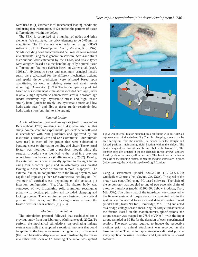
A total of twelve Sprague–Dawley rats (Rattus norvegicusBerkkenhaut 1769) weighing 421±34·g were used in thisstudy. Animal care and experimental protocols were followedin accordance with NIH guidelines and approved by ourinstitution’s Animal Care and Use Committee. Four animalswere used in each of the groups that were subjected tobending, shear or alternating bending and shear. The externalfixator was modified from a previous model, while thesurgical procedure was identical to that used in a previousreport from our laboratory (Cullinane et al., 2002). Briefly,the external fixator was surgically applied to the right femurusing four bicortical pins, and an osteotomy was createdleaving a 2·mm defect within the femoral diaphysis. Theexternal fixator, in conjunction with the linkage system, wascapable of imposing either 12° symmetrical bending or 10%symmetrical cortical shear, depending on the actuator pininsertion configuration (Fig.·2A). The fixator body wascomposed of two articulating solid aluminum rectangularprisms with cortical pin holes and included clamping andlocking screws. The clamping screws fastened the corticalpins into the fixator, and the locking screws arrested thefixator pivot or shear actions (Fig.·2B).
Mechanical stimulation
The stimulation protocol followed that established for aprevious study from our laboratory (Cullinane et al., 2002). Toperform the mechanical stimulations, an oscillating linkagesystem was built that supplied a rotational moment that couldbe applied to the fixators as an oscillating vertical displacement(Fig.·3). The vertical displacement was translated by the fixatorinto either 10% shear or 12° bending. The action was applied
using a servomotor (model #2602-010, QCI-23-5-E-01;Quicksilver Controls Inc., Covina, CA, USA). The speed of themotor was controlled using PC-based software. The shaft ofthe servomotor was coupled to one of two eccentric shafts ofa torque transducer (model #1102-50; Lebow Products, Troy,MI, USA). The other shaft of the transducer was connected tothe linkage system. A torque sensor incorporated within thesystem was connected to an external data acquisition board(model #100; InstruNet Inc., Cambridge, MA, USA) and actedas a bridge voltage sensor, measuring the torque transduced tothe fixator. Based on the manufacturer’s specifications, thetorque sensor was mapped to 278.6·mV Nm–1, with the inputtorque sampled at 60·Hz for the duration of each experimentalsession. The peak torque required to induce the respectivemotions prior to animal attachment was recorded as thebaseline value. The loading apparatus was calibrated prior toevery application using InstruNet and Quicksilver PC-basedsoftware.
Fig.·2. An external fixator mounted on a rat femur with an AutoCadrepresentation of the device. (A) The pin clamping screws can beseen facing out from the animal. The device is in the straight andlocked position, maintaining rigid fixation within the defect. Thehealed surgical incision site can be seen below the fixator. (B) Thebicortex pins are situated in the pin channels (green arrows) and arefixed by clamp screws (yellow arrows). The black arrow indicatesthe axis of the bending fixator. When the locking screws are in place(white arrows), the device is capable of rigid fixation.

2462
There were three mechanical stimulation protocols executedduring this study: (1) bending at 12°, (2) 10% shear and (3)alternating 12° bending and 10% shear (percentage of cortexdiameter). All mechanical stimulations were symmetrical tothe alignment of the cortices and cyclical for the 15-minstimulation period. Starting at post-operative day three andcontinuing for six weeks, the mechanical stimulations wereinduced for six consecutive days, with one day of rest eachweek. The fixator on each animal was attached to the linkagesystem that instituted the respective bending and shear actionsinitiated by the motor. The results from these three treatmentgroups were compared with those of previous controlspecimens.
During each mechanical session, the treatment animals wereanesthetized, the fixators were attached to the linkage, thelocking screws were removed, and cyclic stimulations wereapplied for 15·min at a frequency of 1·Hz. A dedicatedcomputer coordinated the application of the mechanicaltreatment and data acquisition during calibration and treatment.The locking screws were replaced upon completion of eachsession. Once recovered from the anesthesia, the animals werereturned to the housing room and allowed to ambulate freelyin their cages.
Moment analysis
A torque sensor incorporated within the system wasconnected to an external data acquisition board (model #100;InstruNet Inc.) and acted as a bridge voltage sensor, measuringthe torque transduced to the fixator and the resistance to theapplied torque. Based on the manufacturer’s specifications, thetorque sensor was mapped to 278.6·mV Nm–1, with the inputtorque sampled at 60·Hz for the duration of each experimental
session. The loading apparatus was calibrated prior to everyapplication using InstruNet and Quicksilver PC-basedsoftware. The peak torque required to induce the respectivemotions prior to animal attachment was recorded as thebaseline value. The torque sensor then constantly monitoredthe torsional resistance to the motor via the skeletal defectwithin the animal. These resistance data were captured for eachanimal during every daily stimulation period, the data werethen averaged among all the animals for every day ofstimulation, and the result was a mean daily moment resistancefor every day of the stimulation protocol. The mean relativemoment resistance was charted for the entire 35-daystimulation period.
Histology
Animals were euthanized at the termination of the study andthe femora were excised. Standard histological methods wereemployed to generate serial 5·µm sagittal sections for standardhistology and histomorphometry (Cullinane et al., 2002). Thesections were mounted on glass slides, and even-numberedslides were stained using Safranin-O (cartilage) and FastGreen (bone), while odd-numbered slides were stained withAlcian Blue and counter-stained with eosin (proteoglycans).The 5·µm decalcified histological specimens were examinedunder a light microscope using 1.25× to 40× objectives. Dark-field images were obtained through the use of a polarizingfilter, which highlighted collagen fibrils for quantification oftheir orientation and conformity within the extracellularmatrix.
Histomorphometrics
Tissue type composition
Tissue type area composition was quantified usingImagePro® software (Atlanta, GA, USA). We quantified thepercentage of bone and cartilage for each of the treatmentgroups and the control group, as well as rat knee and lumbarintervertebral joints. The entire defect and joint were quantifiedfor tissue percentage within a standardized area of interest,including 2.5·mm in both directions proximal and distal to thedefect or joint center. Tissue type ratios were generated foreach treatment group and the controls, as well as actual nativerat joints. Comparisons were made to identify similarities intissue composition ratios between the treatment groups and thenative rat joints.
Collagen architecture quantification
In order to characterize the molecular organization of thenewly formed cartilage tissues, collagen fibril orientationand angular agreement were quantified using polarizing lightmicroscopy and histomorphometric analyses using Matlab®
and ImagePro®. Fast Fourier transforms were performed ondigitized images of polarized light micrographs, and thepreferred collagen fibril orientation was determined by themost intense region in their power spectra (Fig.·4). Thisprocedure was performed as previously described by Cullinaneet al. (2002). Polarized light micrographs were taken from
D. M. Cullinane and others
Fig.·3. The linkage system connecting the motor and torque sensor tothe fixator, which is inserted by pins into the rat femur. As the wheel(bottom right) rotates, the horizontal actuator arm (bottom) drives thevertical accuator arm (bottom left), which is attached to one side ofthe fixator (hidden by the plate). As the fixator bends on its axis ordisplaces in shear, the defect is stimulated. The rat is lying in a slinghammock with its tail protruding to the left of the image.

2463Does repair recapitulate joint tissue development?
predetermined superficial, intermediate and deep regions of theexperimentally derived cartilage tissues in order to highlightcollagen fibrils. These images were then incorporated into aMatlab Fourier transform analysis to determine mean collagenfibril orientation and fibrillar agreement (Cullinane et al.,2002).
Molecular analyses
Molecular analyses of the expression of specific genes orproteins was carried out by in situ hybridization andimmunostaining in order to confirm tissue types and toidentify expression of the growth and differentiating factor 5(GDF-5), respectively. In situ hybridization was carried outfor collagen type II using a commercially available probe forRNA labeling. Linearized plasmids containing this gene werepurchased from Pharmigen Corp. (San Diego, CA, USA).Single stranded 35S-labeled cRNA probes were generated byin vitro transcription (Pharmigen Corp.). Linearized plasmidscontaining each of the selected genes for analysis weretranscribed using [35S]uridine triphosphate ([35S]UTP; NENLife Science Products, Inc., Boston, MA, USA) and T7 RNApolymerase, then digested with DNase, phenol extracted andethanol precipitated. The labeling efficiency for the cRNAproducts was determined by scintillation counting andadjusted to a concentration of 3×10⁵·c.p.m.·µl–1 of probe foreach in situ assay.
Tissue procurement
Tissue samples were fixed overnight in freshly prepared 4%paraformaldehyde at 0°C, followed by decalcification in 14%EDTA for up to eight weeks. Decalcified samples wereparaffin embedded.
Tissue preparation and sectioning
Fixed and decalcified tissues were dehydrated in gradedethanol up to 100%, transferred to xylenes, then embedded inparaffin. 5·µm-thin paraffin sections were placed on poly L-lysine-coated slides, dried overnight and used immediately orstored at 4°C.
Probe preparation
Sense and antisense 35S-labeled cRNA probes were usedfor hybridization. Vectors were appropriately linearized andincubated with either T7 or SP6 RNA polymerase in thepresence of [35S]UTP, unlabeled nucleotides, 10·mmol·l–1
dithiothreitol (DTT) and RNasin RNase inhibitor (Promega,Madison, WI, USA). Labeled cRNA probes wereseparated from free nucleotides using a Mini Quick SpinRNA column (Roche Molecular Biochemicals, Indianapolis,IN, USA).
Prehybridization
Slides were deparaffinized in xylenes followed byrehydration in graded ethanol solutions, rinsed in 0.85% NaC1(5·min) and 1× phosphate-buffered saline (PBS; 5·min).Sections were treated with proteinase K (20·µg·ml–1) for8·min at 37°C. Slides were dipped successively in 1× PBS(5·min), 4% paraformaldehyde (5·min), acetylated in 0.25%acetic anhydride. GDF-5 expression was examined byimmunohistochemistry. For these studies, an antibody to GDF-5 was obtained from Santa Cruz Biotechnology, Inc. (SantaCruz, CA, USA). Briefly, histochemical staining was carriedout using antigen retrieved at 199°F for 10·min in 10·mmol·l–1
sodium citrate. The anti-GDF-5 antibody (0.5·µg·ml–1)was applied to the sections, followed by a biotinylated
0
5
10
0 50 100 150 2002
4
6
8
Angle (deg.)
Sum
of p
ower
spe
ctru
m
JtF 3815.tifanalysis 2
The ratio of elliptical axes is:3.9416
The orientation (deg.) is:153.9382
Fig.·4. An exampleFourier transform ofcollagen fibrilarchitecture. The panelsrepresent the stagesof Fourier analysisincluding a thresholdedimage, ellipse andfrequency distribution.The transform identifiesthe predominant patternangle within an image(in this case, fibrillarorientation). Thepredominant angle,identified by highestfrequency, is relative tothe original capturedimage’s orientation, butin the final analysis theangle is relative to apresumed defect midline,perpendicular to the bonelong axis.
2464 D. M. Cullinane and others
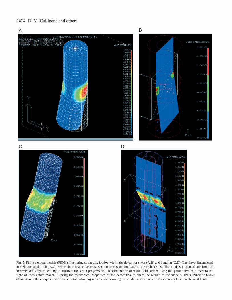
Fig.·5. Finite element models (FEMs) illustrating strain distribution within the defect for shear (A,B) and bending (C,D). The three-dimensionalmodels are to the left (A,C), while their respective cross-section representations are to the right (B,D). The models presented are from anintermediate stage of loading to illustrate the strain progression. The distribution of strain is illustrated using the quantitative color bars to theright of each active model. Altering the mechanical properties of the defect tissues alters the results of the models. The number of brickelements and the composition of the structure also play a role in determining the model’s effectiveness in estimating local mechanical loads.

2465Does repair recapitulate joint tissue development?
secondary antibody and horseradish peroxidase (HRP)-conjugated streptavidine complex, and visualized with DABchromogen.
Statistical analysis
Data are presented as means ±S.D. All histomorphometricresults, including collagen preferred fiber angle and fiber angleconformity, were compared between the control and treatmentgroups using analysis of variance (ANOVA) and Tukey’s post-hoctest at an α level of 0.05, with P values of <0.05 interpretedas significant. All sample sizes for the specific groups weredetermined by power statistics calculations: based on a
coefficient of variation of 25% in the data and accepting α andβ errors of 5.0%.
ResultsFinite element models
The results of the FEMs indicated unique distributions ofstress and strain between the bending and shear groups (Fig.·5).The distribution of tensile strain within the bending defectpeaked at the defect periphery and subsided linearly in thedirection of the defect center. In the bending model, thecompressive stresses acted in opposite response to the tensilestrain, peaking almost simultaneously but in the vicinity of theopposite cortex. The peak strain levels corresponded with themagnitude of displacement of the cortices on the tensile sideduring bending excursion. The distribution of compressivestress spanned the entire bending defect, with diminishingvalues approaching the defect center.
Peak strain levels in the bending group reached 7.87×10⁻⁶,while peak strain in the shear groups reached only 1.95×10⁻¹¹,with a more narrow range of strain distribution in theproximal–distal direction. According to the MFM based onCarter et al. (1988), fibrous tissues would form in the bendinggroup within the estimated range of 7.87×10⁻⁶ to 6.07×10⁻⁶, withcartilage forming in the range from 5.84×10⁻⁶ to 3.37×10⁻⁶ andbone within the range from 2.70×10⁻⁶ to 2.20×10⁻⁶. The sheargroup tissue differentiation ranges for strain include 1.95×10⁻¹¹
to 1.56×10⁻¹¹for fibrous tissue, 1.47×10⁻¹¹to 8.79×10⁻¹²forcartilage and 7.82×10⁻¹² to 2.93×10⁻¹² for bone.
The stress and strain distributions were then incorporatedinto graphic models of expected tissue differentiation foreach of the mechanical stimulations (Fig.·6). The graphicmodel predictions were based on the stress and strainresults from the FEMs, interpreted by the MFM. The areasof higher compressive stress were predicted to encouragecartilage differentiation whereas the areas of extreme
Bone Cartilage Fibrous
A B Fig.·6. Graphic stress- and strain-based tissue prediction diagramscreated from finite element results for (A) 12° bending and (B) shear.The areas in green represent putative cartilage, the areas in redrepresent fibrous tissues, and the blue areas represent bone. Thebending model predicts two opposing bone elements but does notpredict that the cartilage element will completely segment betweenthe proximal and distal halves. The shear model predicts cartilagesegmentation between the halves.
50 10 15 20 25 30 35–5
0
5
10
15
20
25
30
35
40
Indu
ced
torq
ue (
norm
aliz
ed)
Days of stimulation
Torisonal resistance over experimental period
Bending onlyShear onlyCombination
Fig.·7. Moment resistance data for bending, shear andcombination loading. The bending (dotted line), shear (solid line)and combination (dashed line) data correlate highs and lows verywell, except for an initial lag in the combination group. Tissueappearance seems constrained by the timing of physiologicalprocesses and mechanobiological principles, while thearchitecture and maintenance of tissues seems to be controlledprimarily by the mechanical environment. Note how the timingof the peaks and troughs correlates well among the threetreatment regimens, while the magnitude of the momentresistance varies by treatment.

2466
tensile strain were predicted to promote the differentiation offibrous tissue. The areas within the high compressive stressregion but that are shielded by previous cartilage formation arepredicted to foster bone. These areas of subchondral bone werepredicted to form arch-like structures, peaking at the neutralaxis of bending.
Moment analysis
Fig.·7 details the results of the moment analysis for thethree mechanical stimulation regimens for the entire 35-daystimulation period, normalized to the stimulation devicewithout an animal attached. ANOVA found a significantdifference among the groups (P<0.001, N=4), with the bendinggroup being significantly different from both the shear andcombination groups. The bending group experienced thegreatest moment resistance, followed by the shear andcombination groups. After initial fluctuations, the three groupsappeared to cycle together, with some temporal offset initiallyin the combination group, and with a magnitudinal differencein the bending group. The bending and combination groupsexperienced an initial peak at approximately 8–10·daysfollowing the onset of stimulation. This peak was followed bya day 15–17 mutual low for all three groups. A subsequentmutual peak at days 23–25 then followed for all three groups,followed by a mutual low at days 30–32. The day 10 peakcoincides with the maturation and peak of the cartilaginousstage of callus healing.
Radiology
The weekly radiographs illustrated the onset of bonybridging across the defects in the control specimens, while thetreatment defects each demonstrated defect translucency andcomplete non-union in all specimens (Fig.·8). Areas of reduceddensity represent cartilage or fibrous tissue, while high-densityareas represent mineralized tissues such as bone. A distinctivearch-shaped structure spanning the defect cortices can be seenin several of the bending group specimens.
General histology
The shear treatment was preceded by an experimental testto determine an appropriate shear magnitude. This test, usingtwo shear magnitudes, demonstrated two completely differenttissue outcomes. One group experienced 10% shear magnitudewhile the other experienced 25% shear. The 10% magnitudeshear group developed a cartilage band across the entire defect,while the 25% shear defect developed only fibrous tissueacross the defect (Fig.·9).
The mechanical treatment groups all demonstrated thepresence of a cartilage band spanning the entire defect, whilethe control specimens demonstrated bony bridging of thedefect (Fig.·10). The cartilage tissues stained red while thebone and fibrous tissues stained blue-green. The bendingspecimens acquired an arched appearance to their cartilage andthe underlying subchondral bone arch on at least one side ofthe defect, while the shear and combination groups showedparallel and evenly distributed cartilage bands.
D. M. Cullinane and others
Fig.·8. Radiographs of (A) control, (B) bending and (C) shearspecimens with the external fixators attached. In every case, themechanically stimulated defects resulted in non-union. The cartilagetissues in the experimental treatment defects are represented bytranslucencies in the gaps between the segments. All experimentaltreatments were for a 35-day (six-week) duration. The controlspecimen example is from a four-week control specimen,demonstrating the rapid bony bridging occurring in the controls.

2467Does repair recapitulate joint tissue development?
Histomorphometrics
Tissue type composition
We found that the ratio of cartilage to bone in ourexperimental tissues was very similar to that in articular
cartilage, especially in comparison with control endochondralhealing callus (Table·1). The mean control ratio of bone tocartilage was 94:6. In the experimental results, the meanbending group ratio of bone to cartilage was 78:22 and the
shear group was 80:20. The meancombination group ratio was 82:18.None of the treatment groups weresignificantly different from the kneejoint but all were significantly differentfrom the control (P<0.05, N=4 pergroup). Thus, we found consistentratios of cartilage to bone in ourexperimental tissues, and thesemirrored the knee joint mean of roughly80:20. The intervertebral joint wasunique in that its cartilage to bone ratiowas significantly different from allothers (P<0.00062), approximating50:50.
Collagen architecture quantification
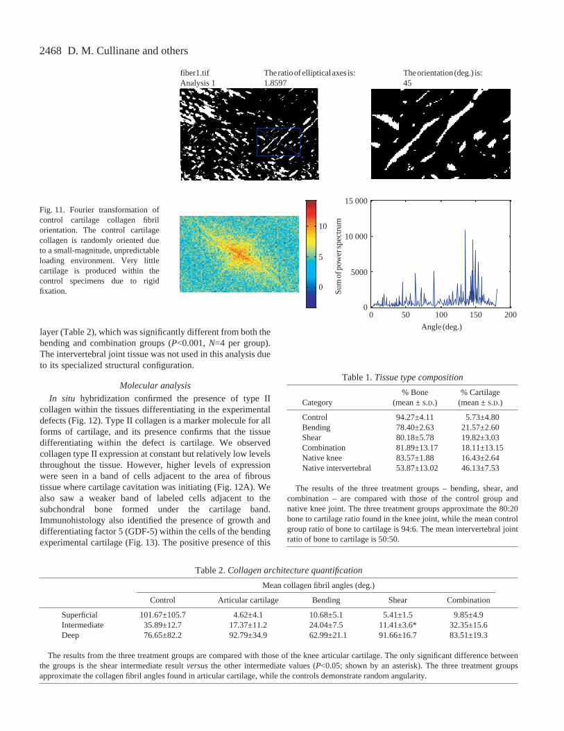
The experimentally generatedcartilage tissues demonstrated visuallydistinct zones of collagen fibrilorganization with specialized fiberorientations in each zone (Fig.·11).Obvious were the superficial and deepzones, with a less obvious transitionalintermediate zone. Mean collagen fibrilangles were not significantly differentamong the different treatment groupsfor each of the layers, with theexception of the shear intermediate
Fig.·10. The histological results from (A) control, (B) bending, (C) bending and shear, and (D)shear stimulations. Note that the control exhibits very little cartilage, while the treatmentgroups all present cartilage bands (shown in red) spanning the defect. Note also the archednature of the cartilage band in the bending specimen, a further mechanobiological response tothe bending action.
Fig.·9. An illustrative example of the mechanobiological paradigm’s predictive value. Here, the magnitude of strain graphically dictates thedifferentiation of cartilage (shown in red) versusfibrous tissue (shown in blue) within the mechanically stimulated defects. The defect shown inA underwent 10% cortex diameter shear, whereas the defect shown in B underwent 25% shear. Thus, a threshold exists between these shearmagnitudes that determines cartilage versusfibrous tissue outcomes.
2468
layer (Table·2), which was significantly different from both thebending and combination groups (P<0.001, N=4 per group).The intervertebral joint tissue was not used in this analysis dueto its specialized structural configuration.
Molecular analysis
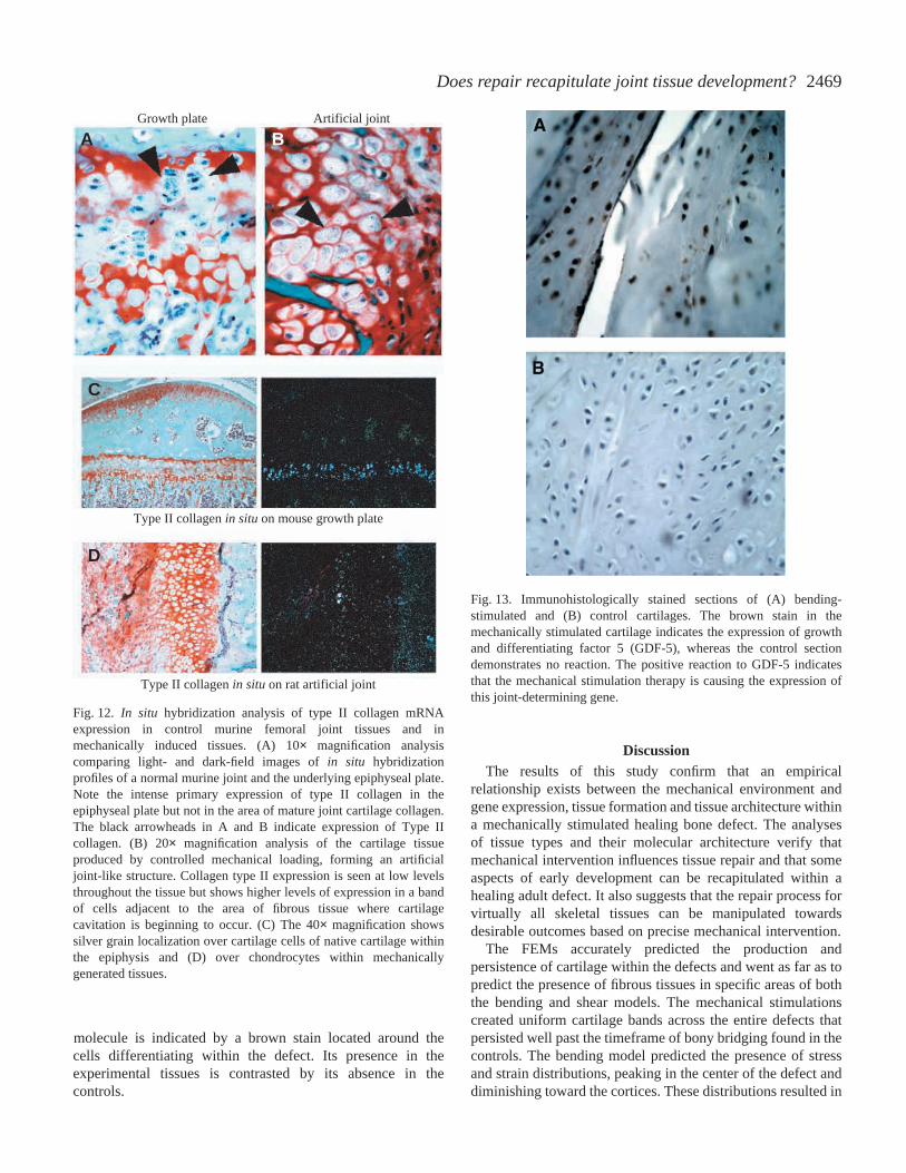
In situ hybridization confirmed the presence of type IIcollagen within the tissues differentiating in the experimentaldefects (Fig.·12). Type II collagen is a marker molecule for allforms of cartilage, and its presence confirms that the tissuedifferentiating within the defect is cartilage. We observedcollagen type II expression at constant but relatively low levelsthroughout the tissue. However, higher levels of expressionwere seen in a band of cells adjacent to the area of fibroustissue where cartilage cavitation was initiating (Fig.·12A). Wealso saw a weaker band of labeled cells adjacent to thesubchondral bone formed under the cartilage band.Immunohistology also identified the presence of growth anddifferentiating factor 5 (GDF-5) within the cells of the bendingexperimental cartilage (Fig.·13). The positive presence of this
D. M. Cullinane and others
0
5
10
0 50 100 150 2000
5000
10 000
15 000
Angle (deg.)S
um o
f pow
er s
pect
rum
fiber1.tifAnalysis 1
The ratio of elliptical axes is:1.8597
The orientation (deg.) is:45
Fig.·11. Fourier transformation ofcontrol cartilage collagen fibrilorientation. The control cartilagecollagen is randomly oriented dueto a small-magnitude, unpredictableloading environment. Very littlecartilage is produced within thecontrol specimens due to rigidfixation.
Table 2.Collagen architecture quantification
Mean collagen fibril angles (deg.)
Control Articular cartilage Bending Shear Combination
Superficial 101.67±105.7 4.62±4.1 10.68±5.1 5.41±1.5 9.85±4.9Intermediate 35.89±12.7 17.37±11.2 24.04±7.5 11.41±3.6* 32.35±15.6Deep 76.65±82.2 92.79±34.9 62.99±21.1 91.66±16.7 83.51±19.3
The results from the three treatment groups are compared with those of the knee articular cartilage. The only significant difference betweenthe groups is the shear intermediate result versusthe other intermediate values (P<0.05; shown by an asterisk). The three treatment groupsapproximate the collagen fibril angles found in articular cartilage, while the controls demonstrate random angularity.
Table 1.Tissue type composition
% Bone % Cartilage Category (mean ±S.D.) (mean ±S.D.)
Control 94.27±4.11 5.73±4.80Bending 78.40±2.63 21.57±2.60Shear 80.18±5.78 19.82±3.03Combination 81.89±13.17 18.11±13.15Native knee 83.57±1.88 16.43±2.64Native intervertebral 53.87±13.02 46.13±7.53
The results of the three treatment groups – bending, shear, andcombination – are compared with those of the control group andnative knee joint. The three treatment groups approximate the 80:20bone to cartilage ratio found in the knee joint, while the mean controlgroup ratio of bone to cartilage is 94:6. The mean intervertebral jointratio of bone to cartilage is 50:50.
2469Does repair recapitulate joint tissue development?
molecule is indicated by a brown stain located around thecells differentiating within the defect. Its presence in theexperimental tissues is contrasted by its absence in thecontrols.
DiscussionThe results of this study confirm that an empirical
relationship exists between the mechanical environment andgene expression, tissue formation and tissue architecture withina mechanically stimulated healing bone defect. The analysesof tissue types and their molecular architecture verify thatmechanical intervention influences tissue repair and that someaspects of early development can be recapitulated within ahealing adult defect. It also suggests that the repair process forvirtually all skeletal tissues can be manipulated towardsdesirable outcomes based on precise mechanical intervention.
The FEMs accurately predicted the production andpersistence of cartilage within the defects and went as far as topredict the presence of fibrous tissues in specific areas of boththe bending and shear models. The mechanical stimulationscreated uniform cartilage bands across the entire defects thatpersisted well past the timeframe of bony bridging found in thecontrols. The bending model predicted the presence of stressand strain distributions, peaking in the center of the defect anddiminishing toward the cortices. These distributions resulted in
Growth plate
AArtifici al joint
B
Type II collagen in situ on mouse growth plate
C
Type II collagen in situ on rat artifici al joint
D
Fig.·12. In situ hybridization analysis of type II collagen mRNAexpression in control murine femoral joint tissues and inmechanically induced tissues. (A) 10× magnification analysiscomparing light- and dark-field images of in situ hybridizationprofiles of a normal murine joint and the underlying epiphyseal plate.Note the intense primary expression of type II collagen in theepiphyseal plate but not in the area of mature joint cartilage collagen.The black arrowheads in A and B indicate expression of Type IIcollagen. (B) 20× magnification analysis of the cartilage tissueproduced by controlled mechanical loading, forming an artificialjoint-like structure. Collagen type II expression is seen at low levelsthroughout the tissue but shows higher levels of expression in a bandof cells adjacent to the area of fibrous tissue where cartilagecavitation is beginning to occur. (C) The 40× magnification showssilver grain localization over cartilage cells of native cartilage withinthe epiphysis and (D) over chondrocytes within mechanicallygenerated tissues.
Fig.·13. Immunohistologically stained sections of (A) bending-stimulated and (B) control cartilages. The brown stain in themechanically stimulated cartilage indicates the expression of growthand differentiating factor 5 (GDF-5), whereas the control sectiondemonstrates no reaction. The positive reaction to GDF-5 indicatesthat the mechanical stimulation therapy is causing the expression ofthis joint-determining gene.

2470
an arch-like bony structure forming across the medullary canalin the bending specimens. The results of the shear pilot studyare particularly interesting because they underline theextremely divergent tissue types one can expect based solelyon differences in the magnitude of the local mechanicalenvironments. These results in particular serve as a potentexample of the predictive power of the mechanobiologicalparadigm.
The moment resistance data is interesting because it appearsto coincide with tissue developmental timing events such asthe onset of cartilage formation (15–20·days post-surgery) andperhaps even the formation of ligament-like connective tissueson the periphery of the defect. Tissue segmentation events(chiefly cartilage) probably resulted in the steep declines inmoment resistance following the day 10 and day 25 peaks,while connective tissue formations such as ligament-liketissues probably resulted in increases in moment resistance.The magnitude of moment resistance is probably related to themechanics of bending versus shear and, specifically, theextreme tension and compression generated via bending. Thisinformation goes far in explaining the significant difference inthe bending group data. The individual highs and lows areprobably related to the tissue types being formed withinthe defects, and thus are time-dependent as well asmechanobiologically dependent. Thus, the appearance of atissue is constrained by time and physiological processes aswell as by mechanobiological principles, while the architectureand maintenance of a tissue is related more to the mechanicalenvironment.
The histomorphometric results verify that the ratio ofcartilage to bone within the experimentally treated defects isin large part controlled by mechanics. The collagen fibrilswithin the experimentally derived tissues demonstrateorganized patterns that resemble those found in articularcartilage. Finally, specific mechanical stimuli can trigger theexpression of the genes encoding collagen type II (cartilageformation) and GDF-5 (bone and joint formation). Theseresults further suggest that induced mechanical stimulationduring the process of bone defect repair can cause arecapitulation of developmental events from joint formation. Itis interesting to note that during stable fracture repair, GDF-5expression appears in a tightly defined window during theendochondral phase of fracture and disappears as soon as bonereplacement is initiated (Cho et al., 2002). Such results suggestthat in the absence of continued mechanical intervention, theexpression of this gene is downregulated and would furthersuggest that it plays an important role either in the maintenanceof cartilage or in the retardation of further endochondralmaturation. It may reflect the possibility that theexperimentally induced activity of this gene is a vestigialattribute of the mechanisms of original joint formation.
Our molecular results are encouraging because theydemonstrate two principal findings: first, the presence ofcollagen type II confirms that our experimentally derivedtissues are true cartilage and, second, a gene associated withthe in uterodevelopment of joints (GDF-5) is upregulated as
a result of the bending stimulation (Storm and Kingsley, 1999).The comparison between the in situ reactions in normalpostnatal long bones and those obtained from the mechanicallyinduced cartilage was very informative. High levels ofcartilage mRNA expression were not observed in fullydifferentiated joint tissues but were observed with veryintensely labeled areas of cartilage formation within theepiphyseal growth plate. Similarly, in the areas ofmechanically induced cartilage formation we observedcollagen type II expression at low levels throughout the tissue.However, higher levels of expression were seen in a band ofcells adjacent to the area of fibrous tissue where cartilagecavitation was initiating. We also saw a weaker band of labeledcells adjacent to the subchondral bone that formed under thecartilage band. These results suggest that the mechanicalenvironment has a direct and quantifiable effect on geneexpression and tissue differentiation within healing bonedefects.
The origin of the cells that populate the defect following thesurgical procedure and during the duration of the experimentis an intriguing question. Very little is known about the preciseorigin of the cells invading the callus during the many stagesof defect repair (Denker et al., 2001; Hunziker et al., 2001;O’Driscoll and Fitzsimmons, 2001). The hematoma probablyarises from vascular cells that invade the defect and fill thegap, while cells that form the cartilage tissue originate fromthe periosteum (Denker et al., 2001; Hunziker et al., 2001;O’Driscoll and Fitzsimmons, 2001). In bone formation, cellsmust invade from the marrow as they do during initialendochondral bone formation. The origin of the cells withinthe defect may be academic since it seems that the localmechanical environment can regulate and direct theirmaturation trajectory to mature cells.
Finally, the experimentally generated tissues and theirmolecular architecture took on joint-like characteristics inseveral aspects of our analyses. This is an intuitive outcome inour estimation, as the mechanical interventions were designedto mimic the actions of a developing joint. These resultsemphasize, on numerous levels, the importance of themechanical environment in tissue differentiation during bothdevelopment and repair. It should also be noted that cases ofmechanically unstable fractures will likewise demonstrate thepresence of cartilage within a healing bone defect, but thelocation, amount and architecture of that cartilage differsmarkedly from our precisely mechanically generatedcartilages. A classic pseudoarthrosis or ‘false joint’ is typicallya random conglomeration of fibrotic tissue, cartilage and bone.This configuration is directly related to the random instabilityof the local mechanical environment and its variantmagnitudes.
The outcomes of this study confirm that mechanobiologicalprinciples can accurately predict gene expression, tissuedifferentiation and tissue architecture based on manipulationsof the local mechanical environment during healing. Theresults further emphasize the important role the localmechanical environment plays in the everyday development
D. M. Cullinane and others

2471Does repair recapitulate joint tissue development?
and repair of the vertebrate body. Further studies need to beconducted in order to determine the precise relationshipsbetween the physical environment and gene expression, tissuedevelopment and tissue repair.
The authors would like to acknowledge students GeraldoSanabria, Nichole Cottetta, Allison Weiner, CharleneHumphrey, Justin Voigt, Sarah Brennan, Susan Christensenand Lisa Foley and technicians Jennifer Fitch and Alfie Tsaywho contributed to this work. Funding for the project and aportion of Dr Cullinane’s salary were provided by theArthritis Foundation; Kristy Salisbury is funded by a graduatefellowship from the Whitaker Foundation.
ReferencesAthanasiou, K. A., Rossenwasser, M. P., Buckwalter, J. A., Malini, T. I.
and Mow, V. C. (1991). Interspecies comparison of in situ intrinsicmechanical properties of distal femoral cartilage. J. Orthop. Res.9, 330-340.
Beaupre, G. S., Stevens, S. S. and Carter, D. R. (2000). Mechanobiology inthe development, maintenance, and degeneration of articular cartilage. J.Rehabil. Res. Dev.37, 145-151.
Carter, D. R. (1987). Mechanical loading history and skeletal biology. J.Biomech. 20, 1095-1109.
Carter, D. R., Blenman, P. R. and Beaupre, G. S. (1988). Correlationsbetween mechanical stress history and tissue differentiation in initialfracture healing. J. Orthop. Res.6, 736-748.
Carter, D. R., Wong, M. and Orr, T. E. (1991). Musculoskeletal ontogeny,phyolgeny, and functional adaptation. J. Biomech.24, 3-16.
Carter, D. R., Beaupre, G. S., Giori, N. J. and Helms, J. A. (1998a).Mechanobiology of Skeletal regeneration. Clin. Orthop.355, S41-S55.
Carter, D. R., van der Meulen, M. C. H. and Beaupre, G. S. (1998b).Mechanobiologic regulation of osteogenesis and athrogenesis. In SkeletalGrowth and Development: Clinical Issues and Basic Science Advances(ed.J. A. Buckwalter, M. G. Ehrlich, L. J. Sandell and S. B. Trippel), pp. 99-130. Rosemont, IL: American Academy of Orthopedic Surgery.
Carter, D. R. and Beaupre, G. S. (2001). Skeletal tissue histomorphometryand mechanics. In Skeletal Function and Form(ed. D. R. Carter and G. S.Beaupre), pp. 31-52. Cambridge: Cambridge University Press.
Cho, T.-J., Gerstenfeld, L. C. and Einhorn, T. A. (2002). Differentialtemporal superfamily during murine fracture expression of members of theTGF-beta healing. J. Bone Min. Res. 17, 513-520.
Claes, L., Eckert-Hubner, K. and Augat, P. (2002). The effect of mechanicalstability on local vascularization and tissue differentiation in callus healing.J. Orthop. Res. 20, 1099-1105.
Claes, L. E. and Heigele, C. A. (1999). Magnitudes of local stress and strainalong bony surfaces predict the course and type of fracture healing. J.Biomech. 32, 255-266.
Cullinane, D. M., Inoue, N., Meffert, R., Tis, J., Rafiee, B. and Chao, E.Y. S. (1999). Regulating bone at the cellular level: a little evidence forWolff’s Law. Am. Zool. 39, 96.
Cullinane, D. M., Fredrick, A., Eisenberg, S. R., Pacicca, D., Elman, M.V., Lee, C., Salisbury, K. Gerstenfeld, L. C. and Einhorn, T. A. (2002).Induction of articular-like cartilage and a joint-like structure by controlledmotion in an experimental mid-femoral defect. J. Orthop. Res. 20, 579-586.
Denker, A. E., Van Rheeden, R., Watson, M. and Sandell, L. J. (2001).Bone marrow cell distribution during early fracture healing. 47th Ann. Meet.Orthop. Res. Soc., San Francisco.
de Rooji, P. P., Siebrecht, M. A., Tagil, M. and Aspenberg, P. (2001). Thefate of mechanically induced cartilage in an unloaded environment. J.Biomech. 34, 961-966.
Eckstein, F., Faber, S., Muhlbauer, R., Hohe, J., Englmeier, K. H., Reiser,
M. and Putz, R. (2002). Functional adaptation of human joints tomechanical stimuli. Osteoarthr. Cart.10, 44-50.
Elder, S. H., Goldstein, S. A., Kimura, J. H., Soslowsky, L. J. andSpengler, D. M. (2001). Chondrocyte differentiation is modulated byfrequency and duration of cyclic loading. Ann. Biomed. Eng. 29, 476-482.
Ferguson, C., Alpern, E., Miclau, T. and Helms, J. A. (1999). Doesadult fracture recapitulate embryonic skeletal formation? Mech. Dev. 87,57-66.
Gardner, T. N., Stoll, T., Marks, L., Mishra, S. and Tate, M. K. (2000).The influence of mechanical stimulus on the pattern of tissue differentiationin a long bone frature – a FEM study. J. Biomech. 33, 415-425.
Giori, N. J., Beaupre, G. S. and Carter, D. R. (1993). Cellular shape andpressure may mediate mechanical control of tissue composition in tendons.J. Orthop. Res. 11, 581-591.
Grodzinsky, A. J., Leveston, M. E., Jin, M. and Frank, E. H. (2000).Cartilage tissue remodeling in response to mechanical forces. Annu. Rev.Biomed. Eng. 2, 691-713.
Hall, B. K. (1972). Immobilization and cartilage transformation into bone inthe embryonic chick. Anat. Rev. 173, 391-403.
Hartmann, C. and Tabin, C. J. (2001). Wnt-14 plays a pivotal role ininducing synovial joint formation in the developing appendicular skeleton.Cell 104, 341-351.
Heegaard, J. H., Beaupre, G. S. and Carter, D. R. (1999). Mechanicallymodulated cartilage growth may regulate joint surface morphogenesis. J.Orthop. Res. 17, 509-517.
Hunziker, E. B., Driesang, I. M. K. and Morris, E. A. (2001). Experimentalmodels of cartilage repair: chondrogenesis in cartilage repair is induced bymembers of the transforming growth factor-beta superfamily. Clin. Orthop.Rel. Res. 391 (suppl.), S171-S181.
Loboa, E. G., Beaupre, G. S. and Carter, D. R. (2001). Mechanobiology ofinitial pseudoarthrosis formation with oblique fractures. J. Orthop. Res. 19,1067-1072.
Loboa-Polefka, E. G., Wren, T. A. L. and Carter, D. R. (2002).Mechanobiology of soft tissue regeneration. 48th Annual Meeting Orthop.Res. Soc., Dallas.
O’Driscoll, S. W. and Fitzsimmons, J. S. (2001). Experimental models ofcartilage repair: the role of periosteum in cartilage repair. Clin. Orthop. Rel.Res. 391 (suppl.), S190-S207.
Sarin, V. K. and Carter, D. R. (2000). Mechanobiology and joint conformityregulate endochondral ossification of sesamoids. J. Orthop. Res.18, 706-712.
Smith, R. L., Thomas, K. D., Schurman, D. J., Carter, D. R., Wong, M.and van der Meulen, M. (1992). Rabbit knee immobilization: Boneremodeling precedes cartilage degredation. J. Orthop. Res. 10, 88-95.
Smith-Adaline, E. A., Ignelzi, M. A., Volkman, S. K., Slade, J. M. andGoldstein, S. A. (2002). Chondrogenesis is influenced by mechanicalenvironment during fracture healing concommitant with altered expressionof BMP-4, VEGF, and IHH. 48th Annual Meeting Orthop. Res. Soc.,Dallas.
Storm, E. E. and Kingsley, D. M. (1999). GDF-5 coordinates bone and jointformation during digit development. Dev. Biol. 209, 11-27.
van der Meulen, M. C., Beaupre, G. S. and Carter, D. R. (1993).Mechanobiologic influences in long bone cross-sectional growth. Bone14,635-642.
van der Meulen, M. C., Morey-Holton, C. R. and Carter, D. R. (1995).Hindlimb suspension diminishes femoral cross-sectional growth in the rat.J. Orthop. Res.13, 700-707.
van der Meulen, M. C. H. and Carter, D. R. (1995). Developmentalmechanics determine long bone geometry. J. Theor. Biol. 172, 323-327.
van der Meulen, M. C. H. and Huiskes, R. (2001). Why mechanobiology?J. Biomech.35, 401-414.
Waanders, N. A., Richards, M., Steen, H., Kuhn, J. L., Goldstein, S. A.and Goulet, J. A. (1998). Evaluation of the mechanical environment duringdistraction osteogenesis. Clin. Orthop.349, 225-234.
Whalen, S. (1993). Musculoskeletal adaptation to mechanical forces on Earthand in space. Physiologist36, S127-S130.
Wolff, J. (1892). Das Gaesetz der Transformation der Knochen(ed. AHirchwald). Berlin.