energy expenditure during ambulation in dysvascular … · energy expenditure during ambulation in...
TRANSCRIPT
Department ofVeterans Affairs
Journal of Rehabilitation Research andDevelopment Vol . 32 No . 2, May 1995Pages 111-119
Energy expenditure during ambulation in dysvascular and
traumatic below-knee amputees : A comparison of five
prosthetic feet
Leslie Torburn, MS, PT; Christopher M. Powers, MS, PT; Robert Guiterrez, MD ; Jacquelin Perry, MD
Rancho Los Amigos Medical Center, Pathokinesiology Laboratory, Downey, CA 90242
Abstract—Recent advancement in prosthetic technology hasled to the development of dynamic elastic response feet (DER),which are reported to store and release energy to facilitate gait.To date, there has been no objective evidence to suggest energyconservation while using these foot designs . The purpose of thisstudy was to compare the energy expenditure of five commer-cially available prosthetic feet (SACH and four DER feet) inboth the traumatic and dysvascular populations during levelwalking . Seventeen male subjects with below-knee amputation(nine traumatic and seven dysvascular) were tested for energyexpenditure (Douglas Bag technique) during a 20-min walkwhile wearing each of the prosthetic feet . The DER prostheticfoot designs were not shown to reduce the energy cost (ml 02/kg-m) or rate of energy expenditure (ml 0 2/kg-min) compared to
the SACH foot . Overall, the traumatic amputees had a similaroxygen consumption per meter traveled compared to the dys-vascular amputees ; however, the rate of energy consumption wasmuch higher in the traumatic group . This increased rate was afunction of the greater walking velocity employed by the trau-matic subjects, made possible by their better physical fitness.
Key words : amputees, energy cost, prosthetics.
Address all correspondence and requests for reprints to : Jacquelin Perry, MD,Rancho Los Amigos Medical Center, Pathokinesiology Laboratory, Bldg 304,7601 East Imperial Hwy, Downey, CA 90242.This study was supported by Department of Veterans Affairs contract #V600P-4176-89 .
INTRODUCTION
Advancement in prosthetic technology during the lastdecade has led to the development of new prosthetic footdesigns which have been termed Dynamic ElasticResponse or DER feet . Compared to the traditional solidconfiguration of the SACH foot, the DER feet are reportedto store and release energy in a manner that facilitates am-bulation (1,2) . The internal keel of these feet is designedto elastically deform under load bearing (thus storing en-ergy) and then release when the amputee advances overthe foot (3) . This supposedly reproduces the energyabsorption and generation characteristics of the normalfoot and ankle in addition to providing better mobility.Theoretically, this would reduce the high energy cost ofambulation associated with this population.
Despite the reported energy storing and releasingproperties of the DER feet, previous studies have failed toshow statistically significant reductions in energy expen-diture (i .e ., oxygen consumption) when compared to theconventional SACH foot (4-7) . To date, there has beenonly one report as to how the energy expenditure of thetwo primary populations of persons with below-knee (B K)amputation, due to trauma or vascular dysfunction, com-pares while walking with different prosthetic feet (7). Inaddition, biomechanical analysis during level walking hasdemonstrated that the SACH and DER feet have knee andhip power curves of comparable magnitude, implying thatthe metabolic energy consumption should be similar (8).
111
112
Journal of Rehabilitation Research and Development Vol . 32 No . 2 1995
These studies have been limited in sample size and havebeen inconsistent regarding the amputee population underinvestigation.
Compared to traumatic amputees, the dysvascularpopulation tends to be less physically active, with a greaterincidence of health-related problems (9) . Thus, the trau-matic amputee is more likely able to effectively compen-sate for the biomechanical limitations imposed by using aprosthesis than is the dysvascular amputee.
Studies have demonstrated that the energy expendi-ture per minute (02 rate) when walking at a self-selectedcomfortable speed, is equal between the traumatic am-putees, dysvascular amputees, and subjects without am-putation (10,11) . Differences in walking velocity betweenthese groups was responsible for the consistent 02 rate.The energy cost (ml O2/kg-m) for dysvascular amputees,however, has been reported to be greater when comparedto traumatic amputees, with both groups having a greaterenergy cost than non-amputees because of a decrease inambulation efficiency (11).
Little evidence exists that the energy "stored" in theDER feet is actually utilized to spare metabolic energyexpenditure for either the dysvascular or traumatic am-putee . The purpose of this study was to compare the ef-fects of five different commercially available prostheticfeet (SACH and four DER feet) on the energy expenditureof walking in both the traumatic and dysvascular popula-tions . This information could assist in providing a basisfor prosthetic foot prescription for a patient with BK am-putation (BKA), and would be beneficial in determiningwhether any of the DER feet provide the capacity for en-ergy conservation.
METHODS
SubjectsSeventeen males with BKA, 10 traumatic and 7 dys-
vascular, participated in this study . The etiology of the dys-vascular group was related to vascular disease secondaryto diabetes . Subjects were recruited from the Long BeachDepartment of Veterans Affairs Medical Center (LBVA)STAMP program and from Rancho Los Amigos MedicalCenter Prosthetic Service.
Criteria for participation included independent com-munity ambulation without use of an assistive device, anda history of compliance . All subjects consented to partic-ipate following explanation of the testing procedures and
review of the informed consent form (approved by theLBVA Subcommittee for Human Studies) . Followingcompletion of the study, the subjects were able to chooseone of the five feet tested to retain on a permanent basis.
Prosthetic ManagementFive prosthetic feet were tested in random order for
each subject : SACH,' Carbon Copy 11, 2 Seattle Lite,'Quantum,' and Flex-Foot .' To ensure the fitting of appro-priate foot components and keel, each manufacturer wasprovided with each subject's age, weight, height, con-tralateral shoe size, activity level, amputation level, andlength of residual limb . The selection of the SACH footheel wedge was based on the weight of the subject ac-cording to the guidelines of the developer. The appropri-ateness of each foot component/keel selection was thenconfirmed or modified at the time of prosthetic fitting.
The fit of each prosthesis was clinically optimizedand reviewed by a team of three certified prosthetists.Alignment of the first foot followed established prostheticprinciples . The Vertical Fabrication Jig' was used for sub-sequent alignments when more than the interchange of afoot-bolt was required.
InstrumentationEach subject was fitted with three precordial electro-
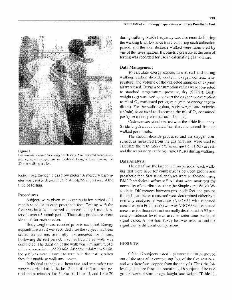
cardiograph electrodes to monitor heart rate, a compres-sion closing switch taped to the bottom of one shoe torecord stride frequency (converted to cadence), and a har-ness with a telemetry system (Figure 1) . A nose clip wasplaced on the subject to prevent nasal breathing . The har-ness was equipped with a mouthpiece attached to a T-seg-ment with two one-way valves which allowed inspirationof ambient air and expiration into the collection bags (mod-ified Douglas bags). A multiported system allowed at-tachment of multiple collection bags . A thermistor placedin the T -segment detected difference in temperature be-tween the ambient air and expired air, allowing recordingof respiration rate.
A level, 60 .5 m outdoor track was used for the walk-ing trials . Each meter of the track was marked for moni-toring the distance traveled.
Respiration rate, heart rate, and stride frequency wererecorded via telemetry on a strip chart recorder .' Gas ana-lyzers were used to determine the carbon dioxide and oxy-gen content of the collected sample of expired air .' Thetemperature of the sampled air was monitored by a ther-mistor placed in the sample flow line . The volume of thecollected expired air was measured by evacuating the col-
113
TORi3URN et aL Energy Expenditure with Five Prosthetic Feet
Figure 1.Instrumentation used for energy cost testing . A multiported harness sys-tem collected expired air in modified Douglas bags during the20-min walking session.
lection bag through a gas flow meter.' A mercury barom-eter was used to determine the atmospheric pressure at thetime of testing.
ProceduresSubjects were given an accommodation period of 1
month to adjust to each prosthetic foot . Testing with thefive prosthetic feet occurred at approximately 1-month in-tervals over a 5-month period . The testing procedures wereidentical for each session.
Body weight was recorded prior to each trial . Energyexpenditure at rest was recorded after the subject had beenseated for 30 min and fully instrumented for 5 min.Following the rest period, a self-selected free walk wascompleted . The duration of the walk was a minimum of 5min and a maximum of 20 min . After the minimum 5 min,the subjects were allowed to terminate the testing whenthey felt unable to walk any longer.
Individual gas samples, heart rate, and respiration ratewere recorded during the last 2 min of the 5 min rest pe-riod and at minutes 4 to 5, 9 to 10, 14 to 15, and 19 to 20
during walking. Stride frequency was also recorded duringthe walking trial . Distance traveled during each collectionperiod, and the total distance walked were monitored byone of the investigators . Barometric pressure at the time oftesting was recorded for use in calculating gas volumes.
Data ManagementTo calculate energy expenditure at rest and during
walking, carbon dioxide content, oxygen content, tem-
perature, and volume of the collected samples of expiredair were used . Oxygen consumption values were convertedto standard temperature, pressure, dry (STPD) . Bodyweight (kg) was used to convert the oxygen consumptionto ml of 02 consumed per kg-min (rate of energy expen-diture) . For the walking data, body weight and velocity(m/min) were used to determine the ml of 02 consumedper kg-m (energy cost per unit distance).
Cadence was calculated as twice the stride frequency.Stride length was calculated from the cadence and distancewalked per minute.
The carbon dioxide produced and the oxygen con-sumed, as measured from the gas analyses, were used tocalculate the respiratory exchange quotient (RQ) at rest,and the respiratory exchange ratio (RER) during walking.
Data AnalysisThe data from the last collection period of each walk-
ing trial were used for comparisons between groups andprosthetic feet . Statistical analyses were performed usingBMDP statistical software . 1" All data were analyzed fornormality of distribution using the Shapiro and Wilk's W-statistic . Differences between prosthetic feet and groupsfor each parameter measured were determined either by atwo-way analysis of variance (ANOVA) with repeatedmeasures, or a Friedman's two-way ANOVA with repeatedmeasures for those data not normally distributed . A 95 per-cent confidence level was used to determine statisticalsignificance. A post-hoc Tukey test was used to find thesignificantly different comparisons.
RESULTS
Of the 17 subjects tested, I (a traumatic BKA) movedout of the area after completing four of the five sessions,and was therefore dropped from the analysis . Thus, the fol-lowing data are from the remaining 16 subjects . The twogroups were of similar age, height, and weight (Table 1) .
114
Journal of Rehabilitation Research and Development Vol . 32 No . 2 1995
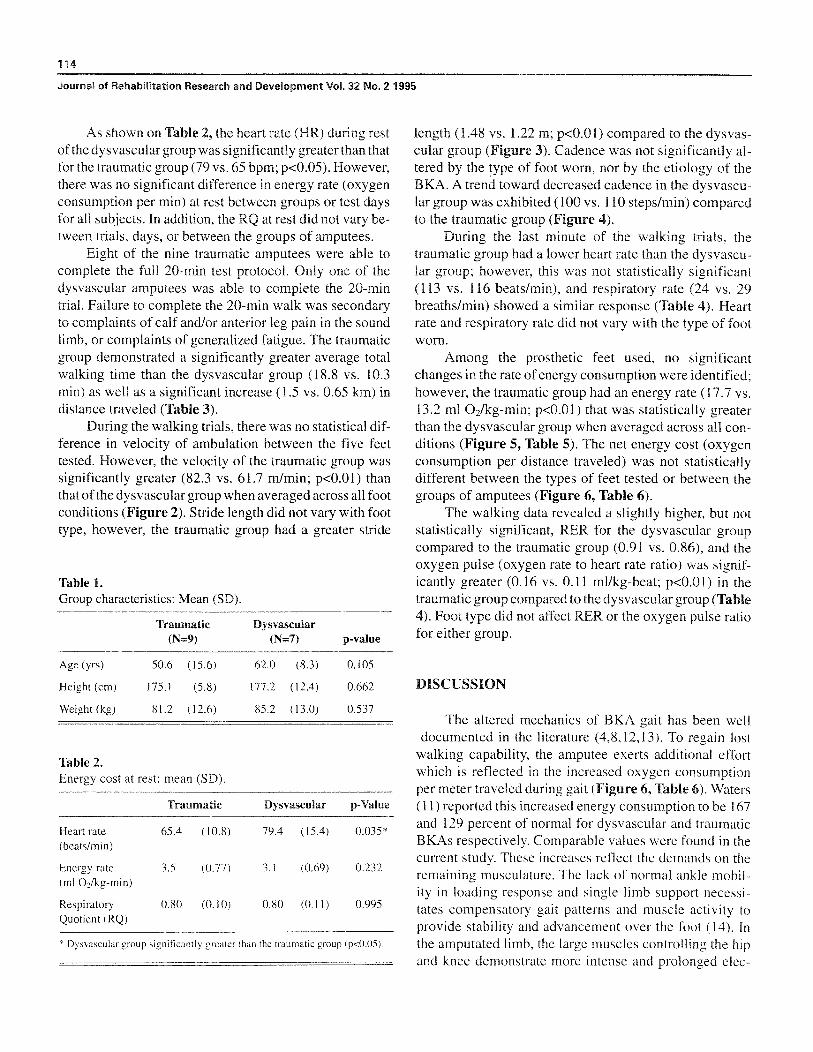
As shown on Table 2, the heart rate (HR) during restof the dysvascular group was significantly greater than thatfor the traumatic group (79 vs . 65 bpm ; p<0 .05) . However,there was no significant difference in energy rate (oxygenconsumption per min) at rest between groups or test daysfor all subjects . In addition, the RQ at rest did not vary be-tween trials, days, or between the groups of amputees.
Eight of the nine traumatic amputees were able tocomplete the full 20-min test protocol . Only one of thedysvascular amputees was able to complete the 20-mintrial . Failure to complete the 20-min walk was secondaryto complaints of calf and/or anterior leg pain in the soundlimb, or complaints of generalized fatigue . The traumaticgroup demonstrated a significantly greater average totalwalking time than the dysvascular group (18 .8 vs. 10.3min) as well as a significant increase (1 .5 vs . 0 .65 km) indistance traveled (Table 3).
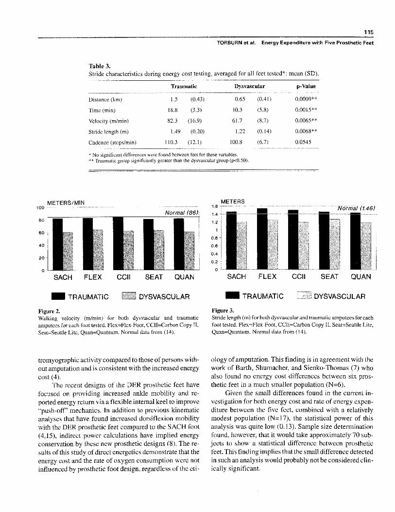
During the walking trials, there was no statistical dif-ference in velocity of ambulation between the five feettested . However, the velocity of the traumatic group wassignificantly greater (82 .3 vs . 61 .7 mlmin; p<0 .01) thanthat of the dysvascular group when averaged across all footconditions (Figure 2). Stride length did not vary with foottype, however, the traumatic group had a greater stride
Table I.Group characteristics : Mean (SD).
Traumatic dysvascular(N=9) (N=7) p-value
Age (yrs) 50 .6 (15 .6) 62.0 (8 .3) 0 .105
Height (cm) 175 .1 (5 .8) 177 .2 (12 .4) 0 .662
Weight (kg) 81 .2 (12 .6) 85 .2 (13 .0) 0 .537
Table 2.Energy cost at rest : mean (SD).
Traumatic Dysvascular p-Value
Heart rate 65 .4 (10 .8) 79 .4 (15 .4) 0 .035*(beats/min)
Energy rate 3 .5 (0 .77) 3 .1 (0 .69) 0 .232(ml 0 2 /kg-min)
Respiratory 0 .80 (0.10) 0 .80 (0 .11) 0 .995Quotient (RQ)
x Dysvascular group significantly greater than the traumatic group (p<0 .05) .
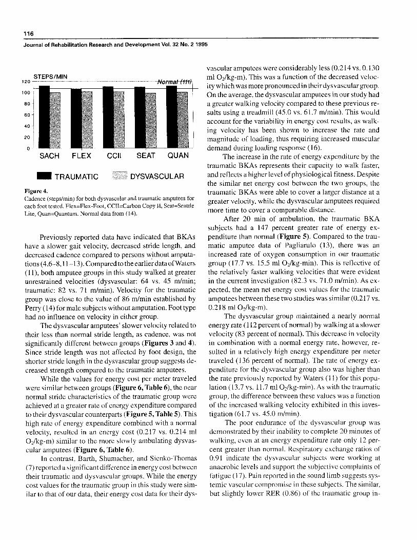
length (1 .48 vs . 1 .22 m ; p<0 .01) compared to the dysvas-cular group (Figure 3) . Cadence was not significantly al-tered by the type of foot worn, nor by the etiology of theBKA. A trend toward decreased cadence in the dysvascu-lar group was exhibited (100 vs. 110 steps/min) comparedto the traumatic group (Figure 4).
During the last minute of the walking trials, thetraumatic group had a lower heart rate than the dysvascu-lar group ; however, this was not statistically significant(113 vs . 116 beats/min), and respiratory rate (24 vs . 29breaths/min) showed a similar response (Table 4) . Heartrate and respiratory rate did not vary with the type of footworn .
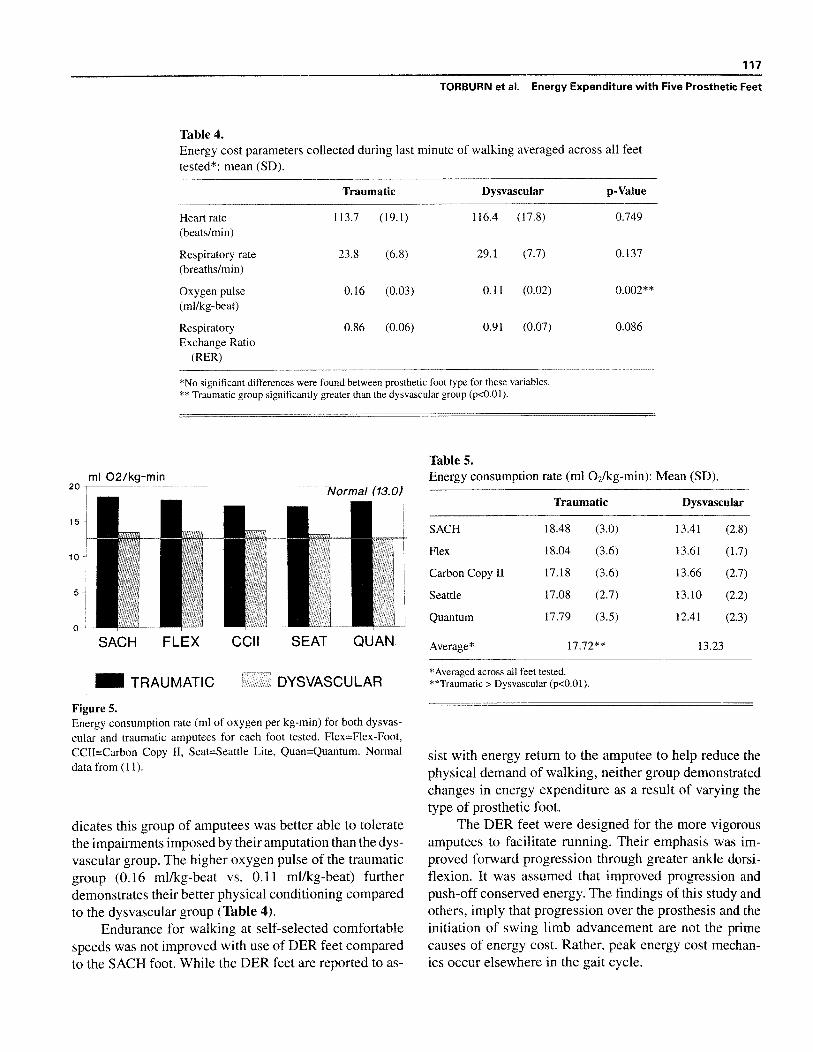
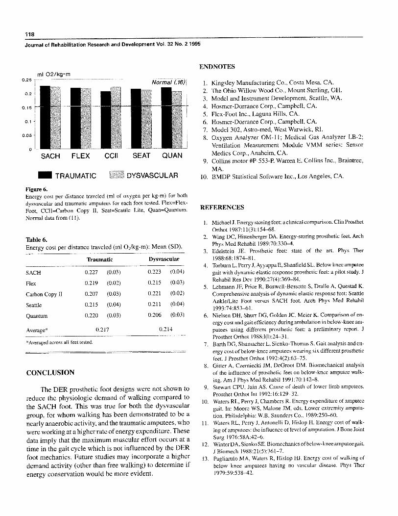
Among the prosthetic feet used, no significantchanges in the rate of energy consumption were identified;however, the traumatic group had an energy rate (17 .7 vs.13 .2 ml Oz/kg-min ; p<0.01) that was statistically greaterthan the dysvascular group when averaged across all con-ditions (Figure 5, Table 5) . The net energy cost (oxygenconsumption per distance traveled) was not statisticallydifferent between the types of feet tested or between thegroups of amputees (Figure 6, Table 6).
The walking data revealed a slightly higher, but notstatistically significant, RER for the dysvascular groupcompared to the traumatic group (0.91 vs . 0 .86), and theoxygen pulse (oxygen rate to heart rate ratio) was signif-icantly greater (0 .16 vs . 0 .11 ml/kg-beat; p<0.01) in thetraumatic group compared to the dysvascular group (Table4). Foot type did not affect RER or the oxygen pulse ratiofor either group.
DISCUSSION
The altered mechanics of BKA gait has been welldocumented in the literature (4,8,12,13) . To regain lost
walking capability, the amputee exerts additional effortwhich is reflected in the increased oxygen consumptionper meter traveled during gait (Figure 6, Table 6) . Waters(11) reported this increased energy consumption to be 167and 129 percent of normal for dysvascular and traumaticBKAs respectively. Comparable values were found in thecurrent study. These increases reflect the demands on theremaining musculature . The lack of normal ankle mobil-ity in loading response and single limb support necessi-tates compensatory gait patterns and muscle activity toprovide stability and advancement over the foot (14) . Inthe amputated limb, the large muscles controlling the hipand knee demonstrate more intense and prolonged elec .-
115
TORBURN et al . Energy Expenditure with Five Prosthetic Feet
Table 3.Stride characteristics during energy cost testing, averaged for all feet tested* : mean (SD).
Traumatic Dysvascular p-Value
Distance (km) 1 .5 (0 .43) 0.65 (0 .41) 0 .0009**
Time (min) 18 .8 (3 .3) 10.3 (5 .8) 0DV!5**
Velocity (m/min) 82 .3 (10 .9) 61 .7 (8 .7) 0 .0065**
Stride length (m) 1 .49 (0 .20) 1 .22 (0 .14) 0 .0060**
Cadence (steps/min) 110 .3 (12 .1) 100.8 (6 .7) 0 .0545
* No significant differences were found between feet for these variables.~~Tmomauc group significantly greater than the dysvascular group (p<0 .50).
METERS/MIN
oSACH FLEX CCU SEAT QUAN
FLEX CCU SEAT QUAN
—El TRAUMATIC ~~~~~~TRAUMAT!C DYSVASCULAR
Figure 2.Walking velocity (mlmin) for both dysvascular and traumaticamputees for each foot tested . pq ux=Bo^-povt.CCD=CarhvuCopy D.Smt=8oauloLite .Vuuu=Auuomm . Normal data from (14) .
Figure 3.Stride length (m) for both dysvascular and traumatic amputees for eachfoot Flex=Flex-Foot, CCD=CurbouCopy D .Scat=Seaule Lite,Quan=Quantum . Normal data from (14).
tromyographic activity compared to those of personsamputation and is consistent with the increased energy
cost (4).The recent designs of the DER prosthetic feet have
focused on providing increased ankle mobility and re-ported energy return via a flexible internal keel to improve"push-off" mechanics. In addition to previous kinematicanalyses that have found increased dorsiflexion mobilitywith the DER prosthetic feet compared to the SACH foot(4,15), indirect power calculations have implied energyconservation by these new prosthetic designs (8) . The re-sults of this study of direct energetics demonstrate that theenergy cost and the rate of oxygen consumption were notinfluenced by prosthetic foot design, regardless of the eti-
ology amputation . This finding is in agreement with thework of Barth, Shumacher, and Sienko-Thomas (7) whoalso found no energy cost differences between six pros-thetic feet in a much smaller population (N=6).
Given the small differences found in the current in-vestigation focbodbuuecgy cost and rate of energy expen-diture between the five feet, combined with a relativelymodest population (N=17) . the statistical power of thisanalysis was quite low (0.13). Sample size determinationfound, however, that it would take approximately 70 sub-jects to show a statistical difference between prostheticfeet . This finding implies that the small difference detectedin such an analysis would probably not be considered clin-ically miguifiouut.
116
Journal of Rehabilitation Research and Development Vol . 32 No . 2 1995
Figure 4.Cadence (steps/min) for both dysvascular and traumatic amputees foreach foot tested . Flex=Flex-Foot, CCII=Carbon Copy II, Seat=SeattleLite, Quan=Quantum . Normal data from (14).
Previously reported data have indicated that BKAshave a slower gait velocity, decreased stride length, anddecreased cadence compared to persons without amputa-tions (4,6—8,11—13) . Compared to the earlier data of Waters(11), both amputee groups in this study walked at greaterunrestrained velocities (dysvascular : 64 vs . 45 m/min;traumatic: 82 vs. 71 m/min) . Velocity for the traumaticgroup was close to the value of 86 m/min established byPerry (14) for male subjects without amputation . Foot typehad no influence on velocity in either group.
The dysvascular amputees' slower velocity related totheir less than normal stride length, as cadence, was notsignificantly different between groups (Figures 3 and 4).Since stride length was not affected by foot design, theshorter stride length in the dysvascular group suggests de-creased strength compared to the traumatic amputees.
While the values for energy cost per meter traveledwere similar between groups (Figure 6, Table 6), the nearnormal stride characteristics of the traumatic group wereachieved at a greater rate of energy expenditure comparedto their dysvascular counterparts (Figure 5, Table 5) . Thishigh rate of energy expenditure combined with a normalvelocity, resulted in an energy cost (0 .217 vs . 0 .214 ml02/kg-m) similar to the more slowly ambulating dysvas-cular amputees (Figure 6, Table 6).
In contrast, Barth, Shumacher, and Sienko-Thomas(7) reported a significant difference in energy cost betweentheir traumatic and dysvascular groups . While the energycost values for the traumatic group in this study were sim-ilar to that of our data, their energy cost data for their dys-
vascular amputees were considerably less (0 .214 vs . 0 .130ml 02/kg-m). This was a function of the decreased veloc-ity which was more pronounced in their dysvascular group.On the average, the dysvascular amputees in our study hada greater walking velocity compared to these previous re-sults using a treadmill (45 .0 vs . 61 .7 mlmin) . This wouldaccount for the variability in energy cost results, as walk-ing velocity has been shown to increase the rate andmagnitude of loading, thus requiring increased musculardemand during loading response (16).
The increase in the rate of energy expenditure by thetraumatic BKAs represents their capacity to walk faster,and reflects a higher level of physiological fitness . Despitethe similar net energy cost between the two groups, thetraumatic BKAs were able to cover a larger distance at agreater velocity, while the dysvascular amputees requiredmore time to cover a comparable distance.
After 20 min of ambulation, the traumatic BKAsubjects had a 147 percent greater rate of energy ex-penditure than normal (Figure 5) . Compared to the trau-matic amputee data of Pagliarulo (13), there was anincreased rate of oxygen consumption in our traumaticgroup (17 .7 vs . 15 .5 ml 0 2/kg-min). This is reflective ofthe relatively faster walking velocities that were evidentin the current investigation (82 .3 vs . 71 .0 m/min) . As ex-pected, the mean net energy cost values for the traumaticamputees between these two studies was similar (0 .217 vs.0.218 ml 0 2/kg-m).
The dysvascular group maintained a nearly normalenergy rate (112 percent of normal) by walking at a slowervelocity (83 percent of normal) . This decrease in velocityin combination with a normal energy rate, however, re-sulted in a relatively high energy expenditure per metertraveled (136 percent of normal) . The rate of energy ex-penditure for the dysvascular group also was higher thanthe rate previously reported by Waters (11) for this popu-lation (13 .7 vs . 11 .7 ml 0 2/kg-min) . As with the traumaticgroup, the difference between these values was a functionof the increased walking velocity exhibited in this inves-tigation (61 .7 vs . 45 .0 m/min).
The poor endurance of the dysvascular group wasdemonstrated by their inability to complete 20 minutes ofwalking, even at an energy expenditure rate only 12 per-cent greater than normal . Respiratory exchange ratios of0.91 indicate the dysvascular subjects were working atanaerobic levels and support the subjective complaints offatigue (17). Pain reported in the sound limb suggests sys-temic vascular compromise in these subjects . The similar,but slightly lower RER (0.86) of the traumatic group in-
SACH FLEX
= TRAUMATIC
SEAT QUAN
DYSVASCULAR
CCII
117
TORBURN et al . Energy Expenditure with Five Prosthetic Feet
Table 4.Energy cost parameters collected during last minute of walking averaged across all feettested* : mean (SD).
Traumatic Dysvascular p-Value
Heart rate 113 .7 (19.1) 116 .4 (17 .8) 0 .749(beats/min)
Respiratory rate 23 .8 (6.8) 29 .1 (7 .7) 0 .137(breaths/min)
Oxygen pulse 0.16 (0.03) 0 .11 (0 .02) 0 .002**(ml/kg-beat)
Respiratory 0.86 (0.06) 0 .91 (0 .07) 0 .086Exchange Ratio
(RER)
*No significant differences were found between prosthetic foot type for these variables.** Traumatic group significantly greater than the dysvascular group (p<0 .01).
ml 02/kg-min20
Norma! (13.0
SACH FLEX
CCII
SEAT QUAN
= TRAUMATIC
DYSVASCULAR
Figure 5.Energy consumption rate (ml of oxygen per kg-min) for both dysvas-cular and traumatic amputees for each foot tested . Flex=Flex-Foot,CCII=Carbon Copy II, Seat=Seattle Lite, Quan=Quantum . Normaldata from (11).
dicates this group of amputees was better able to toleratethe impairments imposed by their amputation than the dys-vascular group . The higher oxygen pulse of the traumaticgroup (0 .16 ml/kg-beat vs . 0.11 ml/kg-beat) furtherdemonstrates their better physical conditioning comparedto the dysvascular group (Table 4).
Endurance for walking at self-selected comfortablespeeds was not improved with use of DER feet comparedto the SACH foot . While the DER feet are reported to as-
Table 5.Energy consumption rate (ml O 2/kg-min) : Mean (SD).
Traumatic Dysvascular
SACH 18 .48 (3 .0) 13 .41 (2 .8)
Flex 18 .04 (3 .6) 13 .61 (1 .7)
Carbon Copy II 17 .18 (3 .6) 13 .66 (2 .7)
Seattle 17 .08 (2.7) 13 .10 (2 .2)
Quantum 17 .79 (3 .5) 12.41 (2.3)
Average* 17 .72** 13 .23
*Averaged across all feet tested.**Traumatic > Dysvascular (p<0.01).
list with energy return to the amputee to help reduce thephysical demand of walking, neither group demonstratedchanges in energy expenditure as a result of varying thetype of prosthetic foot.
The DER feet were designed for the more vigorousamputees to facilitate running . Their emphasis was im-proved forward progression through greater ankle dorsi-flexion. It was assumed that improved progression andpush-off conserved energy. The findings of this study andothers, imply that progression over the prosthesis and theinitiation of swing limb advancement are not the primecauses of energy cost . Rather, peak energy cost mechan-ics occur elsewhere in the gait cycle.
118
Journal of Rehabilitation Research and Development Vol. 32 No . 2 1995
ENDNOTESml 02/kg-m
= TRAUMATIC
DYSVASCULAR
Figure 6.Energy cost per distance traveled (ml of oxygen per kg-m) for bothdysvascular and traumatic amputees for each foot tested. Flex=Flex-Foot, CCII=Carbon Copy II, Seat=Seattle Lite, Quan=Quantum.Normal data from (11).
Table 6.Energy cost per distance traveled (ml 02/kg-m): Mean (SD).
Traumatic Dysvascular
SACH 0 .227 (0.03) 0 .223 (0 .04)
Flex 0 .219 (0.02) 0 .215 (0 .03)
Carbon Copy II 0 .207 (0 .03) 0 .221 (0.02)
Seattle 0 .215 (0 .04) 0.211 (0.04)
Quantum 0 .220 (0 .03) 0.206 (0.03)
Average* 0 .217 0.214
*Averaged across all feet tested.
CONCLUSION
The DER prosthetic foot designs were not shown to
reduce the physiologic demand of walking compared to
the SACH foot. This was true for both the dysvascular
group, for whom walking has been demonstrated to be a
nearly anaerobic activity, and the traumatic amputees, who
were working at a higher rate ofenergy expenditure . These
data imply that the maximum muscular effort occurs at a
time in the gait cycle which is not influenced by the DER
foot mechanics . Future studies may incorporate a higher
demand activity (other than free walking) to determine if
energy conservation would be more evident .
1. Kingsley Manufacturing Co ., Costa Mesa, CA.
2. The Ohio Willow Wood Co ., Mount Sterling, OH.
3. Model and Instrument Development, Seattle, WA.
4. Hosmer-Dorrance Corp., Campbell, CA.
5. Flex-Foot Inc ., Laguna Hills, CA.
6. Hosmer-Dorrance Corp ., Campbell, CA.
7. Model 302, Astro-med, West Warwick, RI.
8. Oxygen Analyzer OM-11 ; Medical Gas Analyzer LB-2;Ventilation Measurement Module VMM series : Sensor
Medics Corp., Anaheim, CA.
9. Collins motor #P-553-P, Warren E . Collins Inc ., Braintree,
MA.10. BMDP Statistical Software Inc ., Los Angeles, CA.
REFERENCES
1. Michael J . Energy storing feet: a clinical comparison . Clin ProsthetOrthot 1987 :11(3) :154-68.
2. Wing DC, Hittenberger DA . Energy-storing prosthetic feet. ArchPhys Med Rehabil 1989 :70 :330-4.
3. Edelstein JE . Prosthetic feet : state of the art . Phys Ther1988 :68 :1874-81.
4. Torburn L, Perry J, Ayyappa E, Shanfield SL . Below knee amputeegait with dynamic elastic response prosthetic feet : a pilot study. JRehabil Res Dev 1990 :27(4) :369-84.
5. Lehmann JF, Price R, Boswell-Bessette S, Dralle A, Questad K.Comprehensive analysis of dynamic elastic response feet : SeattleAnkle/Lite Foot versus SACH foot . Arch Phys Med Rehabil1993 :74 :853-61.
6. Nielsen DH, Shur. DG, Golden JC, Meier K. Comparison of en-ergy cost and gait efficiency during ambulation in below-knee am-putees using different prosthetic feet : a preliminary report. JProsthet Orthot 19881(1) :24-31.
7. Barth DG, Shumacher L, Sienko-Thomas S . Gait analysis and en-ergy cost of below-knee amputees wearing six different prostheticfeet. J Prosthet Orthot 1992:4(2) :63-75.
8. Gitter A, Czerniecki JM, DeGroot DM . Biomechanical analysisof the influence of prosthetic feet on below-knee amputee walk-ing . Am J Phys Med Rehabil 1991 :70 :142-8.
9. Stewart CPU, Jain AS . Cause of death of lower limb amputees.Prosthet Orthot Int 1992 :16 :129-32.
10. Waters RL, Perry J, Chambers R. Energy expenditure of amputeegait . In : Moore WS, Malone JM, eds . Lower extremity amputa-tion . Philadelphia: W .B . Saunders Co ., 1989 :250-60.
11. Waters RL, Perry J, Antonelli D, Hislop H . Energy cost of walk-ing of amputees: the influence of level of amputation . J Bone JointSurg 1976 :58A :42-6.
12. Winter DA, Sienko SE. Biomechanics of below-knee amputee gait.J Biomech 1988 :21(5) :361-7.
13. Pagliarulo MA, Waters R, Hislop DJ . Energy cost of walking ofbelow knee amputees having no vascular disease . Phys Ther1979 :59 :538-42.
SACH FLEX CCII SEAT QUAN

119
TORBURN et al .
Energy Expenditure with Five Prosthetic Feet
14 .
Perry
J .
Gait
analysis,
normal
and
pathological
function . 16 . Skinner SR . The relationship of gait velocity to the rate of lower
Thorofare, NJ : Charles B . Slack, 1992 . extremity
loading
and
unloading .
Trans
Ortho
Res
Soc
15 . Wagner J, Sienko S, Supan T, Barth D . Motion analysis of SACH 1980 :5 :273.
vs . Flex-Foot in moderately active below-knee amputees . Clin 17 . Astrand PO, Rodahl, K. Textbook of work physiology. 2nd ed.
Prosthet Orthot 1987 :11(1) :55—62 . New York: McGraw-Hill Book Co ., 1986 :513 .