evidence on how a conserved glycine in the hinge region of
TRANSCRIPT
Evidence on How a Conserved Glycine in the Hinge Region ofHapR Regulates Its DNA Binding AbilityLESSONS FROM A NATURAL VARIANT*□S
Received for publication, December 4, 2010, and in revised form, March 7, 2011 Published, JBC Papers in Press, March 7, 2011, DOI 10.1074/jbc.M110.209346
Mitesh Dongre1, Naorem Santa Singh1,2, Chetna Dureja, Nagesh Peddada2, Ashish K. Solanki2, Ashish3,and Saumya Raychaudhuri4
From the Institute of Microbial Technology, Chandigarh, Council of Scientific and Industrial Research, Sector 39A,Chandigarh 160036, India
HapR has been recognized as a quorum-sensing master regu-lator in Vibrio cholerae. Because it controls a plethora of dispa-rate cellular events, the absence of a functional HapR affects thephysiology of V. cholerae to a great extent. In the current study,we pursued an understanding of an observation of a natural pro-tease-deficient non-O1, non-O139 variantV. cholerae strainV2.Intriguingly, a nonfunctional HapR (henceforth designated asHapRV2) harboring a substitution of glycine to aspartate at posi-tion 39 of theN-terminal hinge region has been identified. An invitro gel shift assay clearly suggested the inability of HapRV2 tointeract with various cognate promoters. Reinstatement of gly-cine at position 39 restores DNA binding ability of HapRV2(HapRV2G), thereby rescuing the protease-negative phenotypeof this strain. The elution profile of HapRV2 and HapRV2G pro-teins in size-exclusion chromatography and their circular di-chroism spectra did not reflect any significant differences toexplain the functional discrepancies between the two proteins.To gain insight into the structure-function relationship of thesetwoproteins, we acquired small/wide angle x-ray scattering datafrom samples of the native andG39Dmutant. AlthoughGuinieranalysis and indirect Fourier transformation of scattering indi-cated only a slight difference in the shape parameters, structurereconstruction using dummy amino acids concluded thatalthough HapR adopts a “Y” shape similar to its crystal struc-ture, the G39D mutation in hinge drastically altered the DNAbinding domains by bringing them in close proximity. Thisaltered spatial orientation of the helix-turn-helix domains inthis natural variant provides the first structural evidence on thefunctional role of the hinge region in quorum sensing-relatedDNA-binding regulatory proteins of Vibrio spp.
Studies on the quorum-sensing signal network of Vibriocholerae have produced a rich harvest of data where the peri-odic appearance and performance of two regulatory proteins,namely LuxO and HapR, determine the fate of a plethora ofdisparate cellular events (1). Of these, HapR has been given thestatus of a master regulator because it controls a wide range ofdiverse physiological activities, thus exerting a profound influ-ence on the survival and pathogenic potential of this bacterium.Collectively, it represses biofilm development and the produc-tion of primary virulence factors (2) while it stimulates theproduction of HA/protease (3), promotes chitin-induced com-petence (4), increases resistance to protozoan grazing (5),enhances the survival against oxidative stress (6), and controlsthe expression of the gene encoding Hcp (7). In a recent effort,Zhu and co-workers have elegantly characterized additionalnovel direct targets ofHapR and illustrated two distinct bindingmotifs (motif 1 andmotif 2) in all target promoters (8). Becauseit modulates a multitude of diverse cellular parameters, theabsence of a functional HapR affects the physiology of V. chol-erae to a great extent. Being a master regulatory protein of aquorum-sensing circuit, a great deal of work has therefore beendedicated to understanding the various structural and func-tional aspects of HapR. Although previous analysis has identi-fied certain residues contributing to the DNA binding activityofHapR (9), the role of residues in the hinge region has not beenevaluated in this context.While unraveling the necessary causeof a protease-negative phenotype of a non-O1, non-O139 strainofV. cholerae, we discovered a variantHapRharboring a glycineto aspartate substitution in the hinge region. Herein, our struc-ture-function results underscore the significance of a hingeregion glycine moiety at position 39 in mediating HapR inter-action with its cognate promoters.
MATERIALS AND METHODS
Bacterial Strains andMedia—The bacterial strains and plas-mids used in this study are listed in supplemental Table 1. V.cholerae strains were derived from a non-O1, non-O139 strainV2, serogroup O37. Strains were maintained at �70 °C inLuria-Bertani (LB) medium containing 20% glycerol. Esche-richia coli BL21 (DE3) (Novagen) was used for the overexpres-sion of proteins. All strains were propagated at 37 °C in liquidwith agitation or on solid (1.5% agar) in Luria broth unlessmen-tioned otherwise. For the protease assay, V. cholerae strainswere grown with aeration at 37 °C in tryptic soya broth without
* This work was supported by the grants from the Council of Scientific andIndustrial Research India, Department of Biotechnology Grant BT/PR6918/BRB/10/454/2005 and Supra Institutional Project SIP-10/IMTECH/CSIR. Useof the National Synchrotron Light Source, Brookhaven National Labora-tory, was supported by the U.S. Department of Energy, Office of Science,Office of Basic Energy Sciences, under Contract DE-AC02-98CH10886.
□S The on-line version of this article (available at http://www.jbc.org) containssupplemental Tables 1 and 2 and Figs. 1– 4.
1 Both authors contributed equally to this work.2 Supported by Council of Scientific and Industrial Research and Department
of Biotechnology, India, research fellowships.3 To whom correspondence may be addressed. Tel.: 91-172-6665256; Fax:
91-172-2690585; E-mail: [email protected] To whom correspondence may be addressed. Tel.: 91-172-6665256; Fax:
91-172-2690585; E-mail: [email protected] or [email protected].
THE JOURNAL OF BIOLOGICAL CHEMISTRY VOL. 286, NO. 17, pp. 15043–15049, April 29, 2011© 2011 by The American Society for Biochemistry and Molecular Biology, Inc. Printed in the U.S.A.
APRIL 29, 2011 • VOLUME 286 • NUMBER 17 JOURNAL OF BIOLOGICAL CHEMISTRY 15043
by guest on February 15, 2018http://w
ww
.jbc.org/D
ownloaded from

dextrose (TSB-D).5When appropriate, the growthmediumwassupplemented with ampicillin (100 �g ml�1) or chloramphen-icol (17 �g ml�1). All antibiotics were purchased from Sigma-Aldrich and GEHealthcare. Media ingredients were purchasedfrom Himedia and Difco. To disrupt the chromosomal copy ofluxO, conjugationwas conducted between recipientV. choleraestrainV2 and donorE. coli SM10:� pir transformedwith pSVM(supplemental Table 1). luxO mutants of V2 were screened bystreaking onto thiosulphate-citrate-bile salt-sucrose platescontaining ampicillin (100 �g ml�1). Disruption was furtherconfirmed by Southern hybridization. The resulting strain wasdesignatedasV2-SVM(supplementalTable1).TodisrupthapR inV2, a similar strategy was adopted where conjugation was carriedout between recipient V2 and donor E. coli SM10: � pir harboringpCD (supplemental Table 1). hapRmutants of V2 were screenedby streakingontoTCBSplates containing chloramphenicol (17�gml�1). Disruption was further confirmed by Southern hybridiza-tion. The recombinant strain was named V2S.Protease Assay—Protease activity was measured using an
azocasein assay as described earlier (10). Briefly, wild-type andrecombinant derivatives ofV. cholerae strain V2 (supplementalTable 1) were grown in TSB-D containing chloramphenicol (17�g ml�1) and ampicillin (100 �g ml�1) accordingly, with agita-tion to stationary phase at 37 °C. 100 �l of stationary phaseculture supernatant was incubated with 100 �l of azocasein (5mg ml�1 in 100 mM Tris, pH 8.0) for 1 h at 37 °C. The reactionwas stopped by the addition of 400 �l of 10% tricholoroaceticacid. After centrifugation, supernatant was transferred to 700�l of 525mMNaOH, and theAwas determined at 442 nm. Oneazocasein unit was defined as the amount of enzyme producingan increase of 0.01 A unit/h.Site-specific Mutagenesis—A D39G mutation in the hapRV2
ORF was generated on plasmid pSV2 (supplemental Table 1)using a Gene Tailor mutagenesis kit from Invitrogen accordingto the manufacturer’s guidelines. The primers are listed in sup-plemental Table 2. Positive clones were checked by sequencing.One such clone designated as pSV2G was further transformedinto V2S, and the recombinant strain was designated asV2S-RV2G.Protein Purification and Electrophoretic Gel Mobility Shift
Assay with Promoter Regions of aphA, hapA, and vc0900—HapRV2 (Asp39) and HapRV2G (D39G) proteins were purifiedby Ni2�-nitrilotriacetic acid chromatography. The wild-typegene and aspartate variants of HapRwere cloned into theNdeI-BamHI site of the pET15b vector (Novagen) to generate anN-terminal His6-HapR fusion protein. All clones were con-firmed by sequencing and transformed into E. coli BL21 (DE3).After induction with 0.4 mM isopropyl 1-thio-�-D-galactopyra-noside, HapR proteins were purified through Qiagen Ni2�-ni-trilotriacetic acid columns. All proteins were dialyzed overnightin a solution of buffer A containing 10 mM Tris, pH 7.9, 100 mM
KCl, 0.1 mM EDTA, 0.1 mM DTT, 5% glycerol. Gel mobility shiftassay was done essentially as described earlier (9). Briefly, threefragments of 399, 665, and 467 bp corresponding to promoter
regions of aphA, hapA, and vc0900, respectively, were amplifiedwithprimer pairs as listed in supplementalTable 2.The fragmentswere gel-purified and end-labeled with [�-32P]dATP using T4polynucleotide kinase (New England Biolabs). The binding reac-tion was carried out with 4 ng of labeled fragment in 10 mM Tris-HCl, pH 7.9, 1mMEDTA, 1mMDTT, 60mMKCl, 10% glycerol, 5�g of BSA, and 1 �g of poly(dI�dC) in a 20-�l reaction volume for20 min at 26 °C. The reaction mixture was applied to a 5% poly-acrylamide gel and subjected to electrophoresis in 1� Tris-ace-tate-EDTA, pH 8.5, at 4 °C. The gel was dried and autoradio-graphed to examine the shift of the band.Circular Dichroism Measurement—HapRV2 and HapRV2G
were examined by circular dichroism using a Jasco J-810 spec-tropolarimeter. Measurements in the far ultraviolet region(250–190 nm) were performed on protein solutions (0.2mg/ml) employing a cell with path length of 0.1 cmat 25 °C.Themean residue ellipticity, [�], was calculated using a mean resi-due molecular mass of each protein. Each spectrum reported isan average of 10 scans.Molecular Weight Determination—HapRV2 and HapRV2G
were dialyzed overnight in buffer A. All proteins were subjectedto molecular sieve chromatography using a Bio-Sil SEC 125analytical column (300� 7.8mm) (Bio-Rad) and “Biologic Duoflow” chromatography system (Bio-Rad). Elution volumes weredetermined by monitoring the absorbance at both 230 and 280nm. All mutant proteins were eluted at the same volume asdetermined for wild-type HapR.Source of Protein Samples for SAXS Experiments—The two
proteins were purified to homogeneity from the AKTAexplorer FPLC system using an S200 column and concentratedusingMillipore membrane concentrators with 10 kDa. UV-vis-ible absorption results suggested that the concentrations ofproteins HapRV2 and HapRV2G were about 2.8 and 2.5 mg/ml,respectively. To estimate the beam intensity at zero angles, henegg white lysozyme purchased from ACROS Organics (MorrisPlains, NJ) was dissolved, dialyzed, and purified by gel filtration in40mM sodium acetate buffer, pH 3.8, containing 150mMNaCl.Synchrotron SAXS/WAXS Data Acquisition and Processing—
The SAXS data were collected at beam line X9 at the NationalSynchrotron Light Source (Brookhaven National Laboratory).Two charge-coupled detectors simultaneously collected data atsmall (SAXS) and wider (WAXS) angles. The wavelength of thebeam was 0.873 Å, and the ratio of the detector distance fromsample to the diameter of charge-coupled detector was 20.8. 45�l of HapRV2 and HapRV2G samples and their matched bufferwere exposed for 90 s in a quartz flow cell at 15 °C with a flowrate of 30 �l/min. SAXS on lysozyme concentration series wasalso collected under identical conditions/set-up. The imagesrecorded on two charge-coupled detectors from protein solu-tions were circularly averaged, buffer subtracted, and scaled toobtain relative scattering intensity (I) as a function of momen-tum transfer vector, Q (Q � [4�sin�]/�), where � is the beamwavelength and � is the scattering angle. The SAXS andWAXSintensity profiles were scaled and merged using the Q databetween 0.12 and 0.2 Å�1. All SAXS experiments were carriedout in duplicate. No protein appeared to have suffered degra-dation during exposure to x-rays as characterized by themigra-tion pattern in SDS-PAGE.
5 The abbreviations used are: TSB-D, tryptic soya broth without dextrose; PDB,Protein Data Bank; SAXS, small angle x-ray scattering; WAXS, wide anglex-ray scattering.
HapR Hinge Region Glycine and DNA Binding Ability
15044 JOURNAL OF BIOLOGICAL CHEMISTRY VOLUME 286 • NUMBER 17 • APRIL 29, 2011
by guest on February 15, 2018http://w
ww
.jbc.org/D
ownloaded from
SAXS Data Analysis—Guinier approximation was employedto estimate the RG of the scattering particle. According to thisapproximation, for a monodisperse sample of globular protein,a plot of ln (I (Q)) versus Q2, where Q � RG � 1.3, should belinear and fits into the following equation,
ln�I�Q�� � ln�I0� �RG2/3� Q2 (Eq. 1)
where I0 is defined as the intensity of scattering at zero angles, isdirectly proportional to the product of molar concentrationand molecular mass of the scattering sample, and can beapproximated by extrapolating SAXS data to Q 0 (12). RG isdefined as the root mean square of all elemental volumes fromthe center of mass of the particle, weighted by their scattering
densities and is characteristic of the overall shape of the mole-cule. For this study, Guinier analysis was performed using thePrimus software package (13). Using GNOM45 software (14),indirect Fourier transformation of the scattering data over themeasuredQ range computed a pairwise distribution function ofinteratomic vectors, P(r) (Equation 2).
P�r� � �1/ 2��� I�Q�Q r sin�Q r�dQ (Eq. 2)
P(r) is a histogram of the frequency of vector lengths connect-ing small volume elements within the entire volume of the scat-tering particle. During indirect Fourier transformation, P(r)
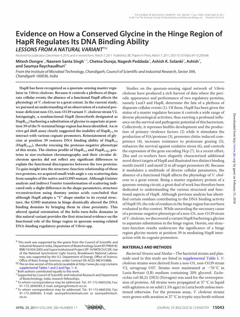
FIGURE 1. Protease activity. A, wild-type and recombinant derivatives of V. cholerae strain V2 analyzed for hemagglutinin protease (Hap) production in thecell-free culture supernatants. The indicated strains were grown in TSB-D for 12 h at 37 °C (200 rpm). Protease activity was assayed through digestion ofazocasein in triplicate. Enzyme activities are the mean of three independent cultures. S.D. are indicated with error bars. B, conserved glycine hinge region ofvarious TetR family quorum-sensing master regulator proteins of Vibrio spp. The underlined conserved glycine 39 is substituted by aspartate in V. cholerae V2.The asterisks indicate the conserved residues in different HapR homologs. C, ribbon representation of the crystal structure of HapR dimer (PDB ID code 2PBX)highlighting the position of the conserved linker region (blue) and the Phe55 residues (red stick) important for DNA binding function. D, in silico docking-basedmodels of complexes of DNA duplex-HapR dimer, VC0900 (motif 1, dark yellow space filled representation), VCA0865 (phapA, motif 2, cyan cpk), and VC2647(paphA, motif 2, black space filled representation).
HapR Hinge Region Glycine and DNA Binding Ability
APRIL 29, 2011 • VOLUME 286 • NUMBER 17 JOURNAL OF BIOLOGICAL CHEMISTRY 15045
by guest on February 15, 2018http://w
ww
.jbc.org/D
ownloaded from
was considered to be zero for vector lengths equal to 0 andDmax. The analysis also provided RG and I0 from the secondmoment and the start of P(r), respectively. Kratky analysis ofthe HapRV2 and HapRV2G molecules was carried out by inter-preting the shape of the I (Q)Q2 versusQ plot.Ab Initio Structure Restoration—Employing dummy residues
and constraints provided within the SAXS profile, the three-dimensional shape of the two proteins was restored using theDAMMINIQ program (15). Ten models were generated with-out any predefined shape or symmetry bias. An average modelthat best represented all of the individual solutions was gen-erated using the DAMAVER suite of programs (16). ForHapRV2G, SUPCOMB software was used to superimpose theinertial axes of our SAXS data-basedmodel and its known crys-tal structure (Protein Data Bank ID code 2PBX) in an auto-mated manner (17).DNA-HapR Docking Calculations—For DNA-protein dock-
ing, we used the GRAMM program freely available fromProf. Vasker’s website. We used parameters for high resolu-tion docking with an increase in repulsion factor to 50. 100low energy models were written out for each run. For inter-action analysis, the model of the complex with lowest energyinteracting with the helix-turn-helix binding region wasselected (and presented). For DNA duplex, we used pro-moter regions of VC0900 (motif 1), VCA0865 (motif 2), andVC2647 (motif 2).Graphical Analysis and Representations—Open Source
PyMOL 0.99rc6 was used for graphical analysis and figuregeneration.
RESULTS AND DISCUSSION
Identification of a Nonfunctional HapRV2 That Is Unable toStimulate Protease Production in V. cholerae—The aim of thepresent study was to investigate the molecular mechanismleading to the protease-negative phenotype ofV. cholerae strain
V2 (Fig. 1A). In the light of current knowledge, this phenotypicbehavior could be explained in the following manners: (i) exis-tence of a nonfunctional HapR (18), (ii) a problem in proteasesecretory machinery, or (iii) repression of hapR due to consti-tutively active LuxO (11). To begin with, a chromosomal copyof luxOwas disrupted, and resultant strainV2-SVM(V2luxO)was found to remain protease-negative, thus ruling out any pos-sibility of a constitutively active LuxO-mediated suppression ofprotease production (Fig. 1A). As the protease-negative pheno-type could also be due to mutations occurring in HapR (6), thecorrectness of gene encoding hapR was examined in strain V2.Together, sequence analysis and ClustalW alignment ofHapRV2 (GenBank accession number DQ379712) identified asingle point mutation (GGT to GAT) that converts a glycine 39to aspartate (G39D) (supplemental Fig. 1). This glycine alongwith other residues is also conserved in other HapR homologs(Fig. 1B and supplemental Fig. 1). There are a total of nine �helices where the first three helices of each HapR monomerform the putative DNA binding domain. The HTH motif liesbetween helices �2 and �3. The remaining six �-helices (�4 to�9) are located in the large C-terminal domain. Interestingly,the conserved glycine-rich hinge region (G34IGRGG39) thatlinks the �1 and �2 helices has been highlighted in the crystalstructure resolved for HapR dimer (PDB ID code 2PBX) (Fig.1C). We carried out rigid-body docking calculations to gaininsight into the putative manner in which DNA duplexesbelonging to motif 1 and motif 2 may bind to HapR dimer(Fig. 1D and supplemental Fig. 3). Interestingly, in the lowenergy models of all complexes, the Phe55 of HapR waswithin the interaction zone to the oligonucleotides. Earlier,the role of this residue of HapR in its DNA binding ability hasbeen proven experimentally (9). The docking results indicatethat both motif 1 and motif 2 duplexes may prefer to interactgrossly in the same manner, but differences do remain in the
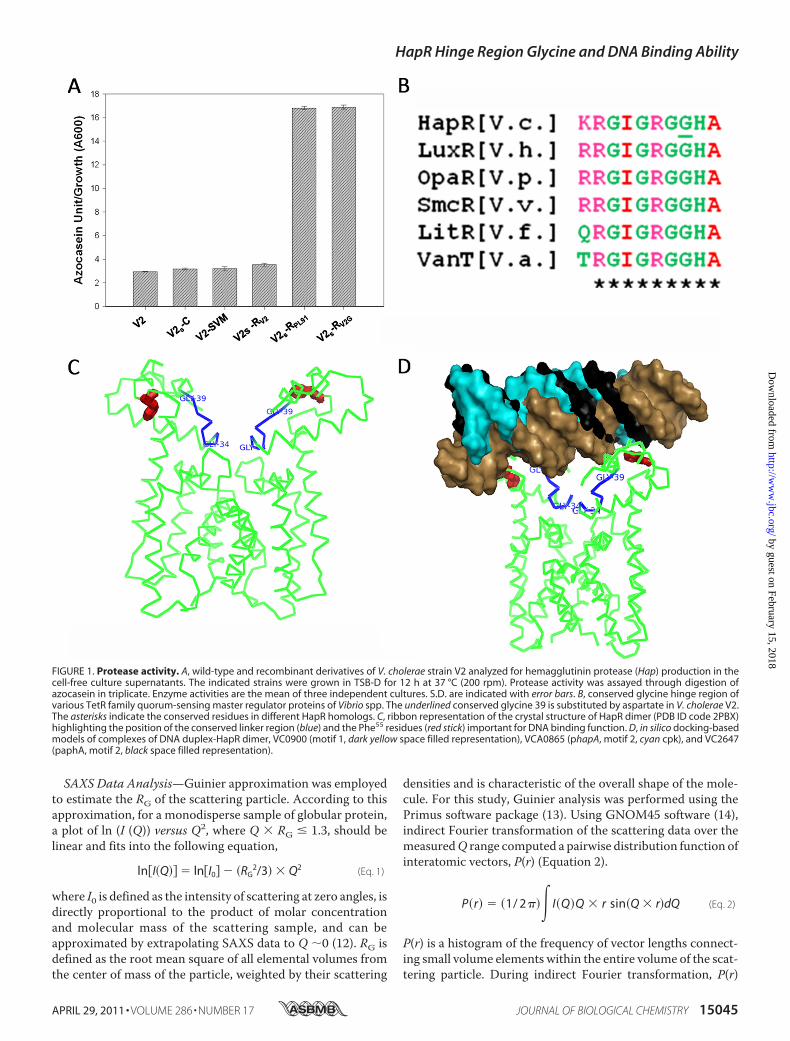
FIGURE 2. Gel shift experiments. Electrophoretic mobility shift assay of purified HapRV2 and HapRV2G was carried out with 32P-labeled promoter regions ofvc0900, hapA, and aphA. Open and filled wedges represent HapRV2 and HapRV2G proteins, respectively.
HapR Hinge Region Glycine and DNA Binding Ability
15046 JOURNAL OF BIOLOGICAL CHEMISTRY VOLUME 286 • NUMBER 17 • APRIL 29, 2011
by guest on February 15, 2018http://w
ww
.jbc.org/D
ownloaded from

angle and rotated position while interacting with HapR. Thenucleotides that play the contact points with the HapRdimer, as seen in the low energy structural models, aredepicted in supplemental Fig. 4. Possibly, this might be thereason for differential binding efficacy of the HapR to differ-ent motifs which eventually leads to a varied response levelin eliciting function.To determine its functionality, recombinant construct of
hapRV2 (pSV2) was transformed into V2S (hapR disruptedstrain of V2) to generate V2S-RV2 (supplemental Table 1). Inaddition, V2S was also transformed with empty vector (V2S-C)and hapRPL91 (V2S-RPL91) where latter served as a positive con-trol. Protease activity was measured in the cell-free culturesupernatants of V2S-C, V2S-RPL91, and V2S-RV2. In contrast toV2S-RPL91, the remaining strains turned out to be protease-negative (Fig. 1A), thus raising the possibility of V2 being ahapRmutant. These data also suggested that there is no defect
in the protease secretory pathway in this strain. UnlikeHapRV2,glycine 39 is conserved in HapRPL91 and other functional HapR(supplemental Fig. 1). To further confirm, glycine was restoredat position 39 by site-directed mutagenesis, and the recombi-nant construct hapRV2G was able to rescue the protease-nega-tive phenotype of strain V2.Evaluate DNA Binding Ability of Variant HapRV2—HapR
justifies its role as amaster regulator by interactingwith a rangeof cognate promoter sequences. To assess the DNA bindingability of HapRV2, a gel shift assay was employed with hapA,aphA, and vc0900 promoter regions. It should be noted thatthe promoter region of vc0900 contains a motif 1 binding sitewhereas the promoter regions of hapA and aphA containa motif 2 binding site. Unlike its functional counterpartHapRV2G, HapRV2 fails to bind to any of these promoterregions, thus indicating a compromise in binding ability ofHapRV2 (Fig. 2).
FIGURE 3. Circular dichroism spectra for wild-type (HapRV2) and mutant (HapRV2G). Far-UV CD spectra of proteins were obtained between wavelengths250 and 190 nm. Mean residual ellipticity (MRE) was calculated and plotted against the wavelength.
HapR Hinge Region Glycine and DNA Binding Ability
APRIL 29, 2011 • VOLUME 286 • NUMBER 17 JOURNAL OF BIOLOGICAL CHEMISTRY 15047
by guest on February 15, 2018http://w
ww
.jbc.org/D
ownloaded from

MolecularWeight and Secondary Structure Determination ofHapRV2 and HapRV2G—Being a prominent member of TetRfamily transcriptional regulators, it has been suggested thatHapR acts as a dimer (9, 10). There remains a possibility thatsubstitution of glycinewith aspartate at position 39might affectthe dimer stability and/or its overall structure which eventuallymight be perturbing the function of this regulatory protein. Todelve further into the structural aspects, we estimated themolecular weight of these proteins by size-exclusion chroma-tography (supplemental Fig. 2) and carried out circular dichro-ism analysis of HapRV2 and HapRV2G (Fig. 3). Interestingly, nosignificant difference was observed in the gel filtration elutionprofiles of the purified His-tagged HapRV2 and HapRV2G pro-teins and their corresponding circular dichroism spectra. Even,K2D analysis of the CD data (in the range of 250–190 nm)suggested that only a minor loss of � helical content (�4%)occurred as a result of the G39D mutation. Overall, this lowresolution information helped in ruling out a grossly misfoldedshape of theHapRV2 protein, but lacked in explaining the loss ofDNA binding function.Structure Reconstruction from SWAXS Data—To aid us in
visualizing the structure-function role, the crystal structure ofHapR has been resolved in an unliganded form, which illus-trates a dimeric, two-domain molecule having an N-terminalDNA binding domain and C-terminal dimerization domain
(PDB ID code 2PBX) (3). In both chains of dimer, the conservedG34IGRGG39 forms an unstructured link between the helix 1and helix 2. Although themolecular docking basedmodel of theHapR-DNA complex illustrates the significance of Phe55 in theDNA binding domain in mediating the interaction (9), it doesnot elucidate any role of the glycine hinge region in enabling theDNA binding ability of HapR. Analysis of the crystal structurerevealed that to affect the turn structure in the hinge region,two of the four glycines (Gly34 and Gly36) occupied the D-sideof the Ramachandran map with �, values 99, 148, and 68,�12, respectively. On the other hand, the residues Gly38 andGly39 adopted backbone torsion angles �162, 146, and �136,175, respectively. We could possibly reason that to affect theturn structure with some flexibility, nature used and retainedthe only coding residues capable of adopting conformation onthe right side of Ramachandran map, but still the answer to thequestion that how the naturally occurring mutation in HapRV2resulted in a protease-negative strain remained elusive.To gain an insight into the global architecture of HapR as
a function of mutation, small/wide angle x-ray scattering(SWAXS) experiments were carried out. The measuredSWAXS intensity data from the samples of HapRV2 andHapRV2G and their Guinier region are presented in Fig. 4A.Presuming the globular nature of the scattering species, theslope of the linear fit of Guinier analysis over the Q range of
FIGURE 4. SWAXS of HapRV2 and HapRV2G. A, SWAXS intensity profiles are shown as a function of Q (HapRV2G, purple; HapRV2, green). Inset depicts the linearregion of the Guinier approximation. The computed P(r) curves from the SWAXS data are plotted in the right panel. B, averaged models of HapRV2G (left) andHapRV2 (right) reconstructed within shape constraints offered in the measured scattering data are presented. The model of HapRV2G was overlaid on the crystalstructure solved for the same protein (PDB ID code 2PBX). C, two orthogonal views of the manual superimposition of the dummy atom models restored forHapRV2G and HapRV2 highlight similarities and local differences in the global structure of the two proteins. D, schematic representation highlights the distortionof the hinge region upon G39D mutation which grossly alters the spatial disposition of the DNA binding domains.
HapR Hinge Region Glycine and DNA Binding Ability
15048 JOURNAL OF BIOLOGICAL CHEMISTRY VOLUME 286 • NUMBER 17 • APRIL 29, 2011
by guest on February 15, 2018http://w
ww
.jbc.org/D
ownloaded from

0.004–0.057 Å�1 and 0.004–0.054 Å�1 for HapRV2 andHapRV2G provided anRGof 23.4� 0.2 and 24.3� 0.1Å, respec-tively. Importantly, the ability to fit a linear equation to the lowangle region confirmed a complete lack of aggregation in thesamples. The P(r) analysis over a wider Q range (0.004–1.0 Å)provided a more complete estimation of the structural param-eters specific to the predominant shape preferred by these pro-teins in solution (Fig. 4B). The P(r) calculated for functional(HapRV2G) and its inactive variant (HapRV2) showed a singlepeak profile with aDmax of 78 and 82 Å, and an RG of 23.8� 0.3and 24.7 � 0.6 Å, respectively. Indirect Fourier transformationsuggested that the I0 values for the samples of HapRV2 andHapRV2G were 410 and 350 units, respectively. Based on anestimated I0/c value of 10 units for lysozyme and considering amass of 46 kDa (as both these proteins form a dimer stabilizedby an intermolecular disulfide bond), we estimated the concen-tration of theHapRV2 andHapRV2G proteins to be2.6 and 2.3mg/ml, respectively. The only differences lie in the proteins athigher Q or smaller dimensions in real space. To gain visualinsight into how Gly to Asp mutation is reorienting the DNAbinding domains, wemodeled the global structure of HapR andits inactive mutant. An average of 10 individual solutions of themodel restored for HapR confirmed that it adopts a “Y”-shapedglobular structure which agrees well with the crystal structuresolved for the same protein (PDB ID code 2PBX) (Fig. 4B).Superimposition of inertial axes of ourmodel and crystal struc-ture concluded that the two binding domains of HapR are in anopen geometry suitable for binding to DNA. Surprisingly, com-parison of a similar model calculated for HapRV2 with themodel generated for HapR brought forth a dramatic loss of theopen architecture of DNA binding domains (Fig. 4B). Overlayof the core models computed for HapRV2G and HapRV2 clearlydemonstrated how there is a large scale redistribution of scat-tering shape only on one end of the dimeric molecule, verylikely the DNA-binding portion (Fig. 4C). Taking together ourmolecular and structural biology results, we present here firststructural evidence on the critical contribution of a hinge gly-cine residue in regulating theDNAbinding ability ofHapR (Fig.4D). Similarity in secondary structural content but difference inglobal structure suggests that the mutation-driven shapechange is localized in the hinge region which may occur in theprotein without significant change in its global energy.Accumulated evidences underpin the significance of hinge
region as a multifunctional domain of various DNA-bindingproteins (19–21). In a recent effort, Hopfner and co-workershave elegantly resolved the architecture of a hinge domain ofeukaryotic SMC (structural maintenance of chromosomes)
proteins and showed how the hinge domain fold is conservedfrom prokaryotes to eukaryotes. Their data also illustrate theevolution of the hinge domain within eukaryotic SMC proteinsto serve specific functions. The glycine hinge region of HapR isalso conserved in other TetR family quorum sensing regulatoryproteins (supplemental Fig. 2). It would be interesting to exam-ine how the conserved residues in the glycine hinge region ofHapR and HapR homologs help them to discern their targetgenes. Additional studies are planned to address this issue.
Acknowledgments—We thank Drs. Giesla Storz and Dhruba Chatto-raj for providing pKK177–3R1 and pDS132 plasmids, respectively,andDr. Jun Zhu for primer sequences of various promoter regions.Wealso thankRicha Singh for critical comments on the preparation of themanuscript.
REFERENCES1. Lenz, D. H., Mok, K. C., Lilley, B. N., Kulkarni, R. V., Wingreen, N. S., and
Bassler, B. L. (2004) Cell 118, 69–822. Zhu, J., Miller, M. B., Vance, R. E., Dziejman, M., Bassler, B. L., and Me-
kalanos, J. J. (2002) Proc. Natl. Acad. Sci. U.S.A. 99, 3129–31343. Silva, A. J., and Benitez, J. A. (2004) J. Bacteriol. 186, 6374–63824. Meibom, K. L., Blokesch, M., Dolganov, N. A., Wu, C. Y., and Schoolnik,
G. K. (2005) Science 310, 1824–18275. Matz, C., McDougald, D., Moreno, A. M., Yung, P. Y., Yildiz, F. H., and
Kjelleberg, S. (2005) Proc. Natl. Acad. Sci. U.S.A. 102, 16819–168246. Joelsson, A., Kan, B., and Zhu, J. (2007) Appl. Environ. Microbiol. 73,
3742–37467. Ishikawa, T., Rompikuntal, P. K., Lindmark, B., Milton, D. L., and Wai,
S. N. (2009) PLoS One 4, e67348. Tsou, A. M., Cai, T., Liu, Z., Zhu, J., and Kulkarni, R. V. (2009) Nucleic
Acids Res. 37, 2747–27569. De Silva, R. S., Kovacikova, G., Lin, W., Taylor, R. K., Skorupski, K., and
Kull, F. J. (2007) J. Bacteriol. 189, 5683–569110. Pompeani, A. J., Irgon, J. J., Berger,M. F., Bulyk,M. L.,Wingreen,N. S., and
Bassler, B. L. (2008)Mol. Microbiol. 70, 76–8811. Raychaudhuri, S., Jain, V., and Dongre, M. (2006) Gene 369, 126–13312. Glatter, O., and Kratky, O. (1982) Small Angle X-ray Scattering, Academic
Press, New York13. Konarev, P. V., Volkov, V. V., Sokolova, A. V., Koch,M. H. J., and Svergun,
D. I. (2003) J. Appl. Crystallogr. 36, 1277–128214. Svergun, D. I. (1992) J. Appl. Crystallogr. 25, 495–50315. Svergun, D. I. (1999) Biophys J. 76, 2879–288616. Volkov, V. V., and Svergun, D. I. (2003) J. Appl. Crystallogr. 25, 860–86417. Kozin, M. B., and Svergun, D. I. (2001) J. Appl. Crystallogr. 34, 33–4118. Joelsson, A., Liu, Z., and Zhu, J. (2006) Infect. Immun. 74, 1141–114719. Little, J. W., and Hill, S. A. (1985) Proc. Natl. Acad. Sci. U.S.A. 82,
2301–230520. Haelens, A., Tanner, T., Denayer, S., Callewaert, L., and Claessens, F.
(2007) Cancer Res. 67, 4514–452321. Griese, J. J., Witte, G., and Hopfner, K. P. (2010) Nucleic Acids Res. 38,
3454–3465
HapR Hinge Region Glycine and DNA Binding Ability
APRIL 29, 2011 • VOLUME 286 • NUMBER 17 JOURNAL OF BIOLOGICAL CHEMISTRY 15049
by guest on February 15, 2018http://w
ww
.jbc.org/D
ownloaded from

Solanki, Ashish and Saumya RaychaudhuriMitesh Dongre, Naorem Santa Singh, Chetna Dureja, Nagesh Peddada, Ashish K.
DNA Binding Ability: LESSONS FROM A NATURAL VARIANTEvidence on How a Conserved Glycine in the Hinge Region of HapR Regulates Its
doi: 10.1074/jbc.M110.209346 originally published online March 7, 20112011, 286:15043-15049.J. Biol. Chem.
10.1074/jbc.M110.209346Access the most updated version of this article at doi:
Alerts:
When a correction for this article is posted•
When this article is cited•
to choose from all of JBC's e-mail alertsClick here
Supplemental material:
http://www.jbc.org/content/suppl/2011/03/07/M110.209346.DC1
http://www.jbc.org/content/286/17/15043.full.html#ref-list-1
This article cites 20 references, 9 of which can be accessed free at
by guest on February 15, 2018http://w
ww
.jbc.org/D
ownloaded from