experiment -1 1. aim 3. apparatus / equipment hp
TRANSCRIPT

1
EXPERIMENT -1
1. AIM -: Preparation and study of the Micro Structure of pure metals like Iron, Copper
and Aluminum.
2. Objectives: a) To Learn the preparation of specimen for microscopic observation.
b) To understand what Microscopy is, and how it can be used to observe
Microstructure of metals,
3. APPARATUS / EQUIPMENT –
3.1 Abrasive Cut-Off Wheel Machine
3 HP Motor, 1440 rpm
Manual model .
10” Diameter wheel
Fully enclosed
Cutting Capacity 50mm Diameter &
75mm
Height X 200mm Length.
This Equipment is used to cut the sample pieces form
the component of all kind of materials. These sample pieces
can be used microscopic observation.
3.2 Specimen Mounting Press
Manual Operation
1” Mould Capacity
Digital Temperature/ Time control unit
Pressure gauge for reading pressure
Alarm indicator for cycle completion.
This Equipment is used to mount specimen in bakelite.
Particularly is useful in hot mounting process.
3.3 Belt Linishing Machine
This Equipment is used for rough Grinding of the Specimen.
3.4 Polishing Machine -
Variable speeds 100 rpm - 800rpm
High torque PMDC motor with D. C. Drive.
Water In and Drain
Corrosion resistant fiber reinforced cover.
8” Disc
This Equipment is used to polish the specimen after
rough grinding with the help of polishing clothe. It also

2
useful for rough grinding with grinding disc.
3.5 Metallurgical Microscope.
Binocular model
Eye pieces: 10X, 15X & 2 OX
Magnification: 10X - 500X.
This Microscope is used for microscopic observation of specimen with the adjustable magnification.
CONSUMABLES/ RAW MATERIAL -:
Rod or Sample pieces of Pure Iron, Copper and Aluminum, Abrasive Polishing paper
80grit/ 100 grit. Polishing powder
4. THEORY -:
4.1 Metallography:
Metallography is the study of metals by optical and electron microscopes. Structures, which
are coarse enough to be discernible by the naked eye or under low magnifications, are termed
macrostructures. Useful information can often be gained by examination with the naked eye of the
surface of metal objects or polished and etched sections. Those, which require high magnification
to be visible, are termed microstructures. Microscopes are required for the examination of the
microstructure of the metals. Optical microscopes are used for resolutions down to roughly the
wavelength of light (about half a micron) and electron microscopes are used for detail below this
level, down to atomic resolution. The most commonly used microscope is the conventional light
microscope. In principle, optical microscopes may be used to look through specimens ('in
transmission') as well as at them ('in reflection'). Many materials, however, do not transmit light
and so we are restricted to looking at the surface of the specimens with an optical microscope.
Electron microscope can be used in the transmission e.g. Transmission Electron Microscope (TEM)
and to look at the surfaces e.g. Scanning Electron Microscope (SEM) Microscopy can give
information concerning a material's composition, previous treatment and properties. Particular
features of interest are
(1) Grain size (II) phases present (III) Chemical homogeneity (IV) distribution of phases (V)
Elongated structures formed by plastic deformation
4.2 Optical Microscopy:
With optical microscopy, the light microscope is used to study the microstructure; optical
illumination systems are its basic elements. For materials that are opaque to visible light (all metals,
many ceramics and polymers), only the surface is subject to observation, and the light microscope

3
must be used in a reflective mode. Contrasts in the image produced result from differences in
reflectivity of the various regions of the microstructure. Careful and meticulous surface preparations
are necessary to reveal the important details of the microstructure. The specimen surface must first be
ground and polished to a smooth and mirror like finish. This is accomplished by using successively
finer abrasive papers and powders.
The microstructure is revealed by a surface treatment using an appropriate chemical reagent
in a procedure termed etching. The etching reagents depend on the material used and after etching the
specimen must be washed with alcohol and ether to remove the grease. The atoms at the grain
boundaries are chemically more active, and consequently dissolve more readily than those within the
grains forming small grooves. These grooves become discernible when viewed under a microscope
because they reflect light at an angle different from that of the grains themselves. When the
microstructure of a two-phase alloy is to be examined, an etchant is chosen that produces a different
texture for each phase so that the different phases may be distinguished from each other.
5. PROCEDURE:
The preparation of metallic or other materials for microscopic examination and micro
structural characterization is in principal very simple. There are four basic processes that you will
need to become familiar with: sample cutting and sectioning, metallographic mounting, Surface
grinding and surface polishing.
-----------------------------------------------------------------------------------------------------------------------
5.1 Sample Cutting and Sectioning
5.1. J. Sectioning Sectioning means removal of convenient size specimen from large sample with minimal
damage to microstructure with the help of abrasive cut off machine. Abrasive cutting wheel/saw is
attached to cutting machine and for work piece holding proper vice is provided on machine. The
primary concern in this process is to minimize the heating of the sample due to the cutting. For this
reason, the cut-off saws that is equipped with either water-cooling systems.
5.2 Mounting
If sample is large enough (about 25 mm square or larger) than do not need to mount it, as it
will be able to control the sample during polishing without a mount. For smaller samples there are
two basic mounting techniques used in this laboratory. Mounting facilitates handling during
preparation and handling. It also avoids damage to polishing wheels during polishing. The most
common uses a thermosetting plastic compound (Bakelite) to encapsulate the specimen known as
hot-mounting process, and the second uses a room temperature curing epoxy known as cold-
mounting process. The Bakelite mounting is by far the most common and easiest. The room
temperature curing epoxy mount should only be used for samples that are extremely sensitive to
heat.
The Bakelite process uses a sample mounting press that applies a pressure to the
Bakelite/sample system during the cure to remove voids and gaps and to fully fill the sample spaces.
Bakelite comes in a variety of colors, which can be combined to produce easy sample identification.

4
5.2.2 The procedure for Hot mounting process.
The following process is used to encapsulate your specimen in Bakelite:
1) Place sample face down on the small piston inside the press, and lower the piston into the
cylinder by opening slightly the valve on the front of the press.
2) Approximately three tablespoons of Bakelite is poured over the sample, and the top of the
press gently screwed into place. DO NOT TIGHTEN THE TOP OF THE PRESS. It is only
necessary to engage all of the screw threads; you do not have to tightly secure the top.
3) The cylindrical heater is plugged in and turned on (the red light should turn on). Place the
heater around the mold. The heater is thermostatically controlled and will heat the mold to
about 135-150 °C. Close the valve, and pump up the cylinder using the hand lever. As the
Bakelite heats, it will begin to flow to fill the void spaces, and the pressure will drop.
Maintain the constant.
4) When the pressure stops rapidly dropping, the whole mold has reached I50°C. Begin timing
for 5-7 minutes to fully cure the Bakelite. Maintain the pressure during the entire heating and
cooling cycle.
5) At the end of the heating cycle, remove the heater and place the cooling collar on the mold
for an additional 6-8 minutes.
6) Crack the valve to release the pressure, and unscrew the top of the mold. When the mold top
is fully unscrewed (it may not come out due to adhesion with the Bakelite), close the valve
and slowly pump up the cylinder to push the sample fully out of the press. Mark the sample
on the back.
7) Clean any residual Bakelite off of the mold surfaces.
5.2.2 The procedure for Cold Mounting process.
The following process is used to room temperature curing epoxy process:
1) Apply mold release agent to mold. Place specimen in the mold.
2) Mix epoxy powder and bonding liquid in 1:2 ration in a cup.
3) Pour into mold, wait for 10 minutes.
4) Eject the mold.
5.3 Sample Surface Polishing
The goal of the surface polishing is to end up with a planar cross section of sample free from
scratches or disturbed metal introduced by the cutting and sectioning. This process is a step-wise
process that can be broken into three loosely separate parts: grinding, coarse polishing, and final
polishing.
5.3.1. Grinding
The first step in preparing your sample is to ensure that you have a flat surface to begin with.
A water-cooled abrasive grinder is available to form a flat initial surface from which to begin. After
getting a flat sample on the belt grinder, WASH sample thoroughly. The hand lapping station has
four graded abrasive papers to produce a sequentially finer surface finish. Be sure the water is turned
on and flowing uniformly over the abrasives. Start with the coarsest grit (240) and, using a firm and

5
uniform pressure, slowly move the specimen forward and back across the abrasive. This will produce
parallel scratches of uniform size. Continue this step until the entire surface of your sample is flat
and contains only scratches of the size of 240 grit abrasive. When the sample is flat and the only
scratches remaining are those due to the 240 grit abrasive, WASH your sample and your hands
thoroughly, and move to the 320 grit abrasive. Repeat this procedure for the 400 grit and the 600 grit
abrasive, checking after each step to be sure that only those scratches remain that are due to the
smallest grit.
5.3.2 Rough Grin dins
Before proceeding to the first polishing wheel (leftmost wheel), wash sample with water.
1) First, apply a small amount of water to the wheel, turn on the motor, and gently clean off the
wheel with your fingers.
2) Apply a small amount of abrasive slurry to the wheel. This wheel uses an A1203 abrasive in
a water suspension. The abrasive particles are 5 micrometers in diameter.
3) Carefully place your sample on the wheel while gripping it tightly. Slowly move the sample
in a circular motion against the rotation of the wheel. Use a moderate and even pressure. It is
important to ensure that you keep the sample flat on the wheel so that the final surface will be
completely planar.
4) After several minutes on the wheel, hold the sample in one place for a moment. This will
provide lots of parallel scratches that you can use to determine if you have removed the
damage from the grinding steps.
5) Examine the sample under the microscope to determine if all the scratches are the same size.
6) Repeat steps 1-5 on the middle polishing wheel. This wheel uses a 0.3 micrometer A1203
abrasive in a water suspension.
5.3.3. Final Polishing
1) Repeat steps 1-5 above on the right polishing wheel. This wheel uses a 0.05 micrometer
A1203 abrasive in a water suspension. At this point, the sample will be very smooth to the
eye and even the oils and dirt on your fingers will scratch it with larger scratches than the
abrasive. DO NOT TOUCH THE SAMPLE SURFACE FROM THIS POINT ON.
2) The last step in the process is to etch the sample to bring out the microstructure.
3) Use a cotton swab and a petri dish for the etching. Gently swab the surface of your sample
with the etchant. Roughly spreading the etchant will scratch your surface. Let the etchant
stand for15 seconds or so and rinse the sample with water to stop the etching, and rinse again
with methanol. Rinse the swab with water and throw into tlie trash bin.
4) Examine specimen under the microscope. You may require several etching steps to bring out
the microstructure
5) If the sample is over-etched, repeat the final polishing step and re-etch for a shorter time.
Samples to be examined at high magnification generally require shorter etching times than
those to be viewed at lower magnifications.
* After last polishing stage the sample looks mirror like.

6
5.4 Etching
Grains cannot be seen without etching. Cracks, pores and defects are observed without etching. Etchant reacts
with atoms and dissolves them. Atoms at grain boundaries dissolve quickly.
Dissolved grain boundaries appear dark.
Steps:
1) Apply enchant to polished surface for some time
2) Rinse with distilled water
5.5. Metallographic Observation
Observe microstructure. Place specimen on metallograph and adjust magnification, focus and position s adjust
micro High magnification - to study phases and Low magnification -to study grain size.
5.5. / Microphotography
In this laboratory, you will report the microstructures of prepared samples in specific
formats. You will be expected to sketch the microstructure that you see under the microscope by
hand. In sketching the microstructure there are several things to keep in mind. First, the
magnification that you use depends upon the scale of the microstructure you are looking for. It is
IMPORTANT to know in advance of the lab class what the expected microstructure for your
samples are and at what scale they should appear, hi sketching the microstructure, you should
indicate only the important features of the structure that you observe-don't make a photographic
reproduction of the microstructure. Simple sketches show that you know what the important
structures are and have identified them in the cross section.
An example of what is considered to be a good laboratory report sketch of the microstructures is
included in the appendix. Your sketch MUST INCLUDE:
1) The sample name and composition,
2) The metallurgical history of the sample,
3) A simple sketch of the important microstructure indicating
a) The magnification used (i.e. “MAG= 100 X”),
b) Important phases and features noted,
4) Etchant used.
6. OBSERVATION:

7
7. PRECAUTIONS:
There are several general instructions to keep in mind during this part of the process.
1) CLEANLINESS!!! Keep the room and the work areas clean, especially the polishing area. Each
step in the polishing process uses a finer grade of abrasive, so good results require that both your
sample and your hands are free from abrasive at each stage before proceeding to the next stage of
the process. Turn the power off and cover the wheels when you are through. Clean up any spilled
water and wipe up the countertop.
2) Throw away the leftover etchants into the sink with a liberal amount of water. Swabs should be
rinsed and thrown into the trash bin IMMEDIATELY AFTER USE. Wash your containers with
water, rinse in methanol, and place them on the shelf above the sink.
3) Use goggles and gloves while handling chemicals. Contact the T.A. or the Instructor in the case
of an acid spill, or if new etchants are needed. Be extra careful when using hydrofluoric acids.
4) Do not eat or drink anything while working in the lab. Wash your hands thoroughly with soap
before leaving the lab.
8. TROBLESHOOTING:
TROUBLE SHOOTING GUIDE for Grinding and polishing
Symptom Cause Action Specimen has fine matte like finish
This layer of particles on the surface
Clean surface swab 110% cotton swab and alcohol.
Clean specimen by flushing specimen and polishing pad with water for last 10-15 seconds of the polishing operation.
Excessive relief in surface finish
Over polishing Repeat step prior to final polish and shorten final polishing time
Scratches in specimen
Improper selection of polishing and
Contaminated pad
Removal of secondary phases
Review application guideline chart for polishing pads or select a softer pad
Replace polishing pad
Alternate between polishing and etching
Etching of specimen Alumina abrasive pH too high
Use lower pH alt

8
9. QUESTIONS:
1. What is Microscopy?
2. Why it is necessary to mount the specimen before grinding and polishing?
3. Which different etching agents used for specimen preparations?
4. What is the principal of metallurgical Microscope?
5. How is the microstructure of pure Iron?
6. How is the microstructure of pure Copper?
7. How is the microstructure of pure Aluminum?
8. How is the microstructure of low carbon steel?
9. How is the microstructure of medium carbon steel?
10. What is the purpose of Etching?

9
EXPERIMENT-2
1. Aim-: Preparation and study of the Micro Structure of pure metals Mild Steel, Low
Carbon steel and High Carbon Steel.
2. Objectives: a.) To Learn the preparation of specimen for microscopic observation.
b) To understand what Microscopy is, and how it can be used to observe
Microstructure of metals.
3. APPARATUS / EQUIPMENT
3.1 Abrasive Cat-Off Wheel Machine,
3 HP Motor, 1440 rpm
Manual model
10” Diameter-wheel
Fully enclosed
Cutting Capacity 50mm Diameter & 75mm
Height X 200mm Length.
This Equipment is used to cut the sample pieces form the component of all kind of materials.
These sample pieces can be used microscopic observation.
3.2 Specimen Mounting Press
• Manual Operation
• 1” Mould Capacity
• Digital Temperature/ Time control unit
• Pressure gauge for reading pressure
• Alarm indicator for cycle completion.

10
This Equipment is used to mount specimen in bakelite. Particularly is useful in hot mounting
process.
3.3 Belt Linishing Machine.
This Equipment is used for rough Grinding of the Specimen 3.4 Polishing Machine
Machine.
Variable speeds 100rpm - 800rpm
High torque PMDC motor with D. C. Drive.
Water In and Drain
Corrosion resistant fiber reinforced cover.
8” Disc
This Equipment is used to polish the specimen after rough grinding with the help of polishing clothe. It
also useful for rough grinding with grinding disc.
3.5 Metallurgical Microscope.
Binocular model
Eyepieces: 10X, 15X&20X
Magnification: 10X-500X.
This Microscope is used for microscopic observation of specimen with the adjustable
magnification.

11
CONSUMABLES/ RAW MATERIAL -:
Rod or Sample pieces of Pure Iron, Copper and Aluminum, Abrasive Polishing paper 80grit/
100 grit. Polishing powder
4. THEORY -:
Mild Steel, Low Carbon steel and High Carbon Steel are types of ferrous materials and
are most important to the engineering application because of their wide range of properties and
verity of applications. Theoretically, steels are the alloys of iron and carbon in which the
carbon content is between 0.008 to 2.0 per cent. The structures and properties can be discussed
with the help of Fe-C equilibrium diagram.
4.1 Iron -Iron Carbide Equilibrium Diagram
Fig 5 Fe-Fe3C Phase Diagram
Figure 5 shows the equilibrium diagram for combinations of carbon in a solid solution
of iron. The diagram shows iron and carbons combined to form Fe-Fe3C at the 6.67%C end of
the diagram. The left side of the diagram is pure iron combined with carbon, resulting in steel
alloys. Three significant regions can be made relative to the steel portion of the diagram.
They are the

12
eutectoid E, the hypoeutectoid A, and the hypereutectoid B. The right side of the pure iron line
is carbon in combination with various forms of iron called alpha iron (ferrite), gamma iron
(austenite), and delta iron. The black dots mark clickable sections of the diagram.
Allotropic changes take place when there is a change in crystal lattice structure. From 2802°-
2552°F the delta iron has a body-centered cubic lattice structure. At 2552°F, the lattice changes
from a body-centered cubic to a face-centered cubic lattice type. At 1400°F, the curve shows a
plateau but this does not signify an allotropic change. It is called the Curie temperature, where the
metal changes its magnetic properties.
Two very important phase changes take place at 0.83%C and at 4.3% C.
At 0.83%C, the transformation is eutectoid, called pearlite.
gamma (austenite) —> alpha + Fe3C (cementite)
At 4.3% C and 2066°F, the transformation is eutectic, called ledeburite.
L(liquid) --> gamma (austenite) + + Fe3C (cementite)
Definitions
Eutectoid: A eutectoid system occurs when a single-phase solid transforms directly to a two-
phase solid.
Hypereutectoid: Hypereutectoid systems exist below the eutectoid temperature.
Hypoeutectoid: Hypoeutectoid systems exist above the eutectoid temperature.
Ferrite: Body-centered cubic iron or an iron alloy based on this structure.
Austenite: Face-centered cubic iron or an iron alloy based on this structure.
Delta iron: The body-centered cubic phase which results when austenite is no longer the most
Stable form of iron. Exists between 2802 and 2552 degrees F. has BCC lattice
structure and is magnetic.
Body-centered: A structure in which every atom is surrounded by eight adjacent atoms, whether
the atom is located at a corner or at the center of a unit cell.
Face-centered: A structure in which there is an atom at the corner of each unit cell and one in
the center of each face, but no atom in the center of the cube.
Pearlite: A lamellar mixture of ferrite and carbide formed by decomposing austenite of
Eutectoid composition.
Cementite: The second phase formed when carbon is in excess of the solubility limit.
Ledeburite: Eutectic of cast iron. It exists when the carbon content is greater than 2 percent. It
contains 4.3 percent carbon in combination with iron.

13
4.2 Classification of Steel : The steels are classified by various methods and each method is
based on a definite criteria as follows,
i) Amount of carbon
a) Low carbon steels (0.008 - 0.3%C)
b) Medium carbon steels (0.30 - 0.60%C)
c) High carbon steels ( 0.60 - 2.00%C)
ii) Amount of alloying elements and carbon
iii) Amount of deoxidation
iv) Method of manufacture v) Form and use.
5. PROCEDURE:
The preparation of metallic or other materials for microscopic examination and micro
structural characterization is in principal very simple. There are four basic processes that you will
need to become familiar with: sample cutting and sectioning, metallographic mounting, Surface
grinding and surface polishing.
5.1 Sample Cutting and Sectioning 5.1.1. Sectioning
Sectioning means removal of convenient size specimen from large sample with minimal damage to
microstructure with the help of abrasive cut off machine. Abrasive cutting wheel/saw is attached to
cutting machine and for work piece holding proper vice is provided on machine. The primary
concern in this process is to minimize the heating of the sample due to the cutting. For this reason,
the cut-off saws that is equipped with either water-cooling systems.
5.2 Mounting
If sample is large enough (about 25 mm square or larger) than do not need to mount it, as it
will be able to control the sample during polishing without a mount. For smaller samples there are
two basic mounting techniques used in this laboratory. Mounting facilitates handling during
preparation and handling. It also avoids damage to polishing wheels during polishing. The most
common uses a thermosetting plastic compound (Bakelite) to encapsulate the specimen known as
hot-mounting process, and the second uses a room temperature curing epoxy known as cold-
mounting process. The Bakelite mounting is by far the most common and easiest. The room
temperature curing epoxy mount should only be used for samples that are extremely sensitive to
heat.
The Bakelite process uses a sample mounting press that applies a pressure to the
Bakelite/sample system during the cure to remove voids and gaps and to fully fill the sample
spaces. Bakelite comes in a variety of colors, which can be combined to produce easy sample
identification.
5.2.2 The procedure for Hot mounting process.
The following process is used to encapsulate your specimen in Bakelite:
1) Place sample face down on the small piston inside the press, and lower the piston into the
cylinder by opening slightly the valve on the front of the press.
2) Approximately three tablespoons of Bakelite is poured over the sample, and the top of the
press gently screwed into place. DO NOT TIGHTEN THE TOP OF THE PRESS. It is only
necessary to engage all of the screw threads; you do not have to tightly secure the top.
3) The cylindrical heater is plugged in and turned on (the red light should turn on). Place the

14
heater around the mold. The heater is thermostatically controlled and will heat the mold to
about 135-150 °C. Close the valve, and pump up the cylinder using the hand lever. As the
Bakelite heats, it will begin to flow to fill the void spaces, and the pressure will drop.
Maintain the constant.
4) When the pressure stops rapidly dropping, the whole mold has reached 150°C. Begin
timing for 5-7 minutes to fully cure the Bakelite. Maintain the pressure during the entire
heating and cooling cycle.
5) At the end of the heating cycle, remove the heater and place the cooling collar on the
mold for an additional 6-8 minutes.
6) Crack the valve to release the pressure, and unscrew the top of the mold. When the mold
top is fully unscrewed (it may not come out due to adhesion with the Bakelite), close the
valve and slowly pump up the cylinder to push the sample fully out of the press. Mark the
sample on the back.
7) Clean any residual Bakelite off of the mold surfaces.
5.2.2 The procedure for Cold Mounting process.
The following process is used to room temperature curing epoxy process:
1) Apply mold release agent to mold. Place specimen in the mold.
2) Mix epoxy powder and bonding liquid in 1:2 ration in a cup.
3) Pour into mo Id... wait for 10 minutes.
4) Eject the mold.
5.3 Sample Surface Polishing
The goal of the surface polishing is to end up with a planar cross section of sample free from
scratches or disturbed metal introduced by the cutting and sectioning. This process is a step-wise
process that can be broken into three loosely separate parts: grinding, coarse polishing, and final
polishing.
5.3.1. Grinding
The first step in preparing your sample is to ensure that you have a flat surface to begin with. A
water-cooled abrasive grinder is available to form a flat initial surface from which to begin. After
getting a flat sample on the belt grinder, WASH sample thoroughly. The hand lapping station has
four graded abrasive papers to produce a sequentially finer surface finish. Be sure the water is
turned on and flowing uniformly over the abrasives. Start with the coarsest grit (240) and, using
a firm and uniform pressure, slowly move the specimen forward and back across the abrasive.
This will produce parallel scratches of uniform size. Continue this step until the entire surface of
your sample is flat and contains only scratches of the size of 240 grit abrasive. When the sample
is flat and the only scratches remaining are those due to the 240 grit abrasive, WASH your
sample and your hands thoroughly, and move to the 320 grit abrasive. Repeat this procedure for
the 400 grit and the 600 grit abrasive, checking after each step to be sure that only those
scratches remain that are due to the smallest grit.

15
5.3.2 Rough Grinding
Before proceeding to the first polishing wheel (leftmost wheel), wash sample with water.
1) First, apply a small amount of water to the wheel, turn on the motor, and gently clean off
the wheel with your fingers.
2) Apply a small amount of abrasive slurry to the wheel. This wheel uses an AI203 abrasive
in a water suspension. The abrasive particles are 5 micrometers in diameter.
3) Carefully place your sample on the wheel while gripping it tightly. Slowly move the
sample in a circular motion against the rotation of the wheel. Use a moderate and even
pressure. It is important to ensure that you keep the sample flat on the wheel so that the
final surface will be completely planar.
4) After several minutes on the wheel, hold the sample in one place for a moment. This will
provide lots of parallel scratches that you can use to determine if you have removed the
damage from the grinding steps.
5) Examine the sample under the microscope to determine if all the scratches are the same
size.
6) Repeat steps 1-5 on the middle polishing wheel. This wheel uses a 0.3 micrometer A1203
abrasive in a water suspension.
5.3.3. Final Polishing
1) Repeat steps 1-5 above on the right polishing wheel. This wheel uses a 0.05 micrometer
A1203 abrasive in a water suspension. At this point, the sample will be very smooth to the
eye and even the oils and dirt on your fingers will scratch it with larger scratches than the
abrasive. DO NOT TOUCH THE SAMPLE SURFACE FROM THIS POINT ON.
2) The last step in the process is to etch the sample to bring out the microstructure.
3) Use a cotton swab and a petri dish for the etching. Gently swab the surface of your
sample with the etchant. Roughly spreading the etchant will scratch your surface. Let the
etchant stand for 15 seconds or so and rinse the sample with water to stop the etching,
and rinse again with methanol. Rinse the swab with water and throw into the trash bin.
4) Examine specimen under the microscope. You may require several etching steps to bring
out the microstructure.
5) If the sample is over-etched, repeat the final polishing step and re-etch for a shorter time.
Samples to be examined at high magnification generally require shorter etching times
than those to be viewed at lower magnifications.
* After last polishing stage the sample looks mirror like.
5.4 Etching
Grains cannot be seen without etching. Cracks, pores and defects are observed without etching.
Etchant reacts with atoms and dissolves them. Atoms at grain boundaries dissolve quickly.
Dissolved grain boundaries appear dark.
Steps:
1) Apply enchant to polished surface for some time

16
2) Rinse with distilled water
Enchants:
1) 50/50 HCI: equal parts hydrochloric acid (HCI) mixed with water.
2) Alcoholic Ferric Chloride: 5 grams FeC13; 2 ml concentrated HCI acid; 95 ml methyl
alcohol.
3) Aqueous Ferric Chloride: 10 grams FeC13; 20 ml concentrated HCI acid; 80 ml water.
4) Ammonia/Hydrogen peroxide: 1 part strong ammonia; 1 part hydrogen peroxide; 2 parts
water; FRESHLY MADE.
5) Mixed acids: 95 ml water; 1.5 ml concentrated HCI acid; 2.5 ml concentrated nitric
(HN03) acid; 0.5 ml hydrofluoric (HF) acid.
6) 2% Nital: 2 ml concentrated HN03; 98 ml methyl alcohol.
5.5. Metallographic Observation
Observe microstructure, Place specimen on metallograph and adjust magnification, focus and
position s adjust micro High magnification - to study phases and Low magnification -to study
grain size.
5.5.1 Microphotography
In this laboratory, you will report the microstructures of prepared samples in specific formats.
You will be expected to sketch the microstructure that you see under the microscope by hand. In
sketching the microstructure there are several things to keep in mind. First, the magnification
that you use depends upon the scale of the microstructure you are looking for. It is
IMPORTANT to know in advance of the lab class what the expected microstructure for your
samples are and at what scale they should appear. In sketching the microstructure, you should
indicate only the important features of the structure that you observe-don't make a photographic
reproduction of the microstructure. Simple sketches show that you know what the important
structures are and have identified them in the cross section.
An example of what is considered to be a good laboratory report sketch of the microstructures is included in
the appendix. Your sketch MUST INCLUDE:
1) The sample name and composition,
2) The metallurgical history of the sample,
3) A simple sketch of the important microstructure indicating
a) The magnification used (i.e. “MAG= 100 X”),
b) Important phases and features noted,
4) Etchant used.
6. OBSERVATION:

17
7.PRECAUTIONS:
There are several general instructions to keep in mind during this part of the process.
1) CLEANLINESS!!! Keep the room and the work areas clean, especially the polishing
area. Each step in the polishing process uses a finer grade of abrasive, so good results
require that both your sample and your hands are free from abrasive at each stage before
proceeding to the next stage of the process. Turn the power off and cover the wheels
when you are through. Clean up any spilled water and wipe up the countertop.
2) Throw away the leftover etchants into the sink with a liberal amount of water. Swabs
should be rinsed and thrown into the trash bin IMMEDIATELY AFTER USE. Wash
your containers with water, rinse in methanol, and place them on the shelf above the
sink.
3) Use goggles and gloves while handling chemicals. Contact the T.A. or the Instructor in
the case of an acid spill, or if new etchants are needed. Be extra careful when using
hydrofluoric acids.
4) Do not eat or drink anything while working in the lab. Wash your hands thoroughly with
soap before leaving the lab.
Symptom Cause Action Specimen has fine matte like finish
This layer of particles on the surface
Clean surface swab 110% cotton swab and alcohol.
Clean specimen by flushing specimen and polishing pad with water for last 10-15 seconds of the polishing operation.
Excessive relief in surface finish
Over polishing Repeat step prior to final polish and shorten final polishing time
Scratches in specimen
Improper selection of polishing and
Contaminated pad
Removal of secondary phases
Review application guideline chart for polishing pads or select a softer pad
Replace polishing pad
Alternate between polishing and etching
Etching of specimen Alumina abrasive pH too high
Use lower pH alt

18
7. QUESTIONS:
i) Why alloying elements are added to steels?
ii) How negative effects of sulphur in steels will be neutralized?
iii) What is the composition of stain less steel?
iv) What are the important characteristics of Tool steels?
v) What is the composition of H.S.S.?
vi) What makes High Carbon high chromium steel suitable for making dies?
vii) Show the heat treatment cycles, on Time-temperature diagrams for different types of
steels?
viii) Compare the properties of alloy steels with and without heat treatment?
19
EXPERIMENT - 3
1. AIM: Study of the Microstructures of Cast Iron.
2. Objectives: a.) To understand what Microscopy is, and how it can be used to
observe Microstructure of metals like cast iron.
b) To conduct typical engineering microscopic observation and be able to recognize the
microstructure of typical type of cast iron metal.
c) To learn the differences in microstructure of different type of cast irons.
3. APPERATUS: Metallurgical Microscope, Standard Specimens of Cast Iron materials.
3.1 Metallurgical Microscope.
Binocular model
• Eye pieces: 10X,15X & 20X
• Magnification: 10X-500X.
This Microscope is used
for -microscopic observation of specimen with the adjustable
magnification.
Fig. 1
4. THEORY:
4.1 Cast Iron
Cast irons typically contain 2-4 wt% of carbon with a high silicon concentrations and a greater
concentration of impurities than steels. The carbon equivalent (CE) of a cast iron helps to
distinguish the grey irons which cool into a microstructure containing graphite and the white
irons where the carbon is present mainly as cementite. The carbon equivalent is defined as:
St4-P
3%)(
PSiCwtCE
The term cast iron, like the term steel, identifies a large family of ferrous alloys. Cast irons are
multi component ferrous alloys. They contain major (iron, carbon, silicon), minor (<0.01%), and
often alloying (>0.01%) elements. Cast iron has higher carbon and silicon contents than steel.
Because of the higher carbon content, the structure of cast iron, as opposed to that of steel,
exhibits a rich carbon phase. Depending primarily on composition, cooling rate and melt
treatment, cast iron can solidify according to the thermodynamically metastable Fe-Fe3C system
or the stable Fc- Gr system. When the metastable path is followed, the rich carbon phase in the
eutectic is the iron carbide; when the stable solidification path is followed, the rich carbon phase
is graphite. Referring only to the binary Fe-Fe3C or Fe-Gr system, cast iron can be defined as an
iron-carbon alloy with more than 2% C. Important notice is that silicon and other alloying
elements may considerably change the maximum solubility of carbon in austenite (g). Therefore,
in exceptional cases, alloys with less than 2% C can solidify with a eutectic structure and
therefore still belong to the family of cast iron.

20
4.2 Types of Cast Iron
On the basis of Microstructure cast irons are classified as follows.
4.2.1 White Cast iron
Composition of the iron is appropriate or the cooling rate of the metal is sufficiently rapid
during solidification, the metal will solidify with the C combined with iron as iron carbide. This
compound, also called cementite, is hard and brittle and dominates the microstructure of white
iron. Thus, white iron is hard and brittle and has a white crystalline fracture because it is
essentially free of graphite.
White iron has a high compressive strength and excellent wear resistance, and it retains its
hardness for limited periods even up to a red heat. It can be produced in selected areas of a
casting—such as. on the periphery of a cam—by causing localized rapid solidification of the
iron. White iron at the surface of a casting is called chill. It is produced by making that portion of
the mold—where the white iron is desired—of-a material that can extract heat very rapidly, such
as iron or graphite. White iron does not have the easy castabiiity of other irons because its
solidification temperature is generally higher, and it solidifies with C in its combined form as
iron carbide. Application includes rollers of rolling mills, Dies of metal extrusion and where high
wear resistance is necessary.
Fig. 2 Microstructure of white cast iron Fig. 3. Microstructure of Gray cast iron
4.2.1 Gray Cast Iron When the composition of the molten iron and its cooling rate are appropriate, the C in the iron
separates during solidification and forms separate graphite flakes that are interconnected within
each eutectic cell. The graphite grows edgewise into the liquid and forms the characteristic flake
shape. When gray iron is broken, most of the fracture occurs along the graphite, thereby
Cast Iron
White Gray Nodular Malleable Alloy Cast iron Cast iron Cast iron Cast iron Cast iron

21
accounting for the characteristic gray color of the fractured surface. Because the large majority of
the iron castings produced are of gray iron, the generic term, cast iron, is often improperly used
to mean gray iron specifically.
The properties of gray iron are influenced by the size, amount and distribution of the graphite
flakes, and by the relative hardness of the matrix metal around the graphite. These factors are
controlled mainly by the C and Si contents of the metal and the cooling rate of the casting.
Slower cooling and higher C and Si contents tend to produce more and larger graphite flakes, a
softer matrix structure and lower strength. The flake graphite provides gray iron with unique
properties such as excellent machinability at hardness levels that produce superior wear-resisting
characteristics, the ability to resist galling and excellent vibration damping.
The amount of graphite present, as well as its size and distribution, are important to the
properties of the iron. Whenever possible, it is preferable to specify the desired properties rather
than the factors that influence them.4.2.2
Nodular cast Iron (Ductile iron, S. G Iron)
Ductile iron also referred to as nodular iron or spheroidal graphite iron, was patented in 1948.
After a decade of intensive development work in the 1950s, ductile iron had a phenomenal nine-
fold increase in use as an engineering material during the 1960s, and the rapid increase in
commercial application continues today. An unusual combination of properties is obtained in
ductile iron because the graphite occurs as spheroids rather than as individual flakes as in gray
iron. This mode of solidification is obtained by adding a very small, but specific, amount of Mg to
molten iron of a proper composition. The base iron is severely restricted in the allowable contents
of certain minor elements that can interfere with the graphite spheroid formation. The added Mg
reacts with the sulfur and oxygen in the molten iron and changes the way the graphite is formed.
Control procedures have been developed to make the processing of ductile iron dependable. The
high C and Si content of ductile iron provide the casting process advantages, but the graphite
spheroids have only a nominal influence on the mechanical properties of the metal. Ductile iron,
like malleable iron, exhibits a linear stress-strain relation, a considerable range of yield strengths
and, as its name implies, ductility. Castings are made in a wide range of sizes with sections that
can be either very thin or very thick.
The different grades are produced by controlling the matrix structure around the graphite either as-
cast or by subsequent heat treatment. Only minor compositional differences exist among the
regular grades, and these adjustments are made to promote the desired matrix microstructures.
Alloy additions may be made to ductile iron to assist in controlling the matrix structure as-cast or
to provide response to heat treatment. Special analysis ductile irons and high-alloy ductile irons
provide unusual properties for special applications.
4.2.3 Malleable Cast Iron
This type of iron is characterized by having the majority of its C content occur in the
microstructure as irregularly shaped nodules of graphite. This form of graphite is called temper
carbon because it is formed in the solid state during heat treatment. The iron is cast as a white iron
of a suitable chemical composition. After the castings are removed from the mold, they are given an
extended heat treatment starting at a temperature above 1650°F (900°C). This causes the iron
carbide to dissociate and the free carbon precipitates in the solid iron as graphite. The rapid
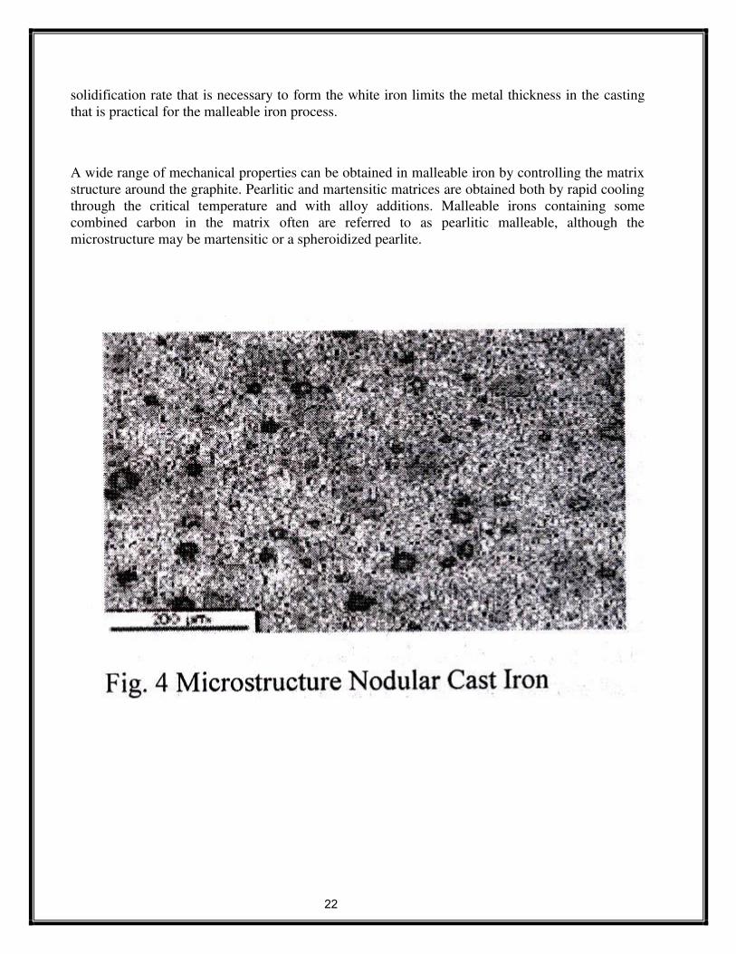
22
solidification rate that is necessary to form the white iron limits the metal thickness in the casting
that is practical for the malleable iron process.
A wide range of mechanical properties can be obtained in malleable iron by controlling the matrix
structure around the graphite. Pearlitic and martensitic matrices are obtained both by rapid cooling
through the critical temperature and with alloy additions. Malleable irons containing some
combined carbon in the matrix often are referred to as pearlitic malleable, although the
microstructure may be martensitic or a spheroidized pearlite.

23

24
4. PROCEDURE:
In this laboratory, you will report the microstructures of prepared samples in specific formats. You
will be expected to sketch the microstructure that you see under the microscope by hand. In sketching
the microstructure there are several things to keep in mind. First, the magnification that you use
depends upon the scale of the microstructure you are looking for. It is IMPORTANT to know in
advance of the lab class what the expected microstructure for your samples are and at what scale they
should appear. In sketching the microstructure, you should indicate only the important features of the
structure that you observe-don't make a photographic reproduction of the' microstructure. Simple
sketches show that you know what the important structures are and have identified them in the cross
section.
An example of what is considered to be a good laboratory report sketch of the microstructures is
included in the appendix. Your sketch MUST INCLUDE:

25
1) The sample name and composition,
2) The metallurgical history of the sample,
3) A simple sketch of the important microstructure indicating
a) The magnification used (i.e. “MAG= 100 X”),
b) Important phases and features noted,
4) Etchant used.
5. OBSERVATIONS:
6. PRECAUTIONS:
There are several general instructions to keep in mind during this part of the process.
6.1 CLEANLINESS!!! Keep the room and the work areas.
6.2 Don't touch etched and polished surface of the specimen.
6.3 Don't touch the lances of eyepiece with dirty hand.
6.4 Use clean clothes only to clean the lenses of eyepieces.
6.5 Handle the microscope with gently.
6.6 Return the standard specimen to the Lab Technician after observation.
6.7 Switch off the microscope after the observation.

26
7, Review Questions:
i) What are the important alloys of Copper & Zinc?
ii) What is composition of Muntz metal?
iii) What is the composition of Cartridge Brass?
iv) What is a Bronze?
v) What is composition of Gunmetal?
vi) What are the important applications of Gunmetal?
vii) What is a Bell metal? .
viii) What is melting point of Tin?
ix) What is use of Babbit metals? Explain why?
x) What is the microstructure of Tin based Babbit?

27
EXPERIMENT-4
1. AIM: Study of the Microstructures of Non Ferrous Metals.
2. Objectives: a.) To understand what Microscopy is, and how it can be used to observe
Microstructure of non-ferrous metals.
b) To conduct typical engineering microscopic observation and be able to recognize the
microstructure of typical type of copper, aluminum and its alloys.
c) To learn the differences in microstructure of different type of cast irons.
3. APPERATUS: Metallurgical Microscope, Specimens of Non Ferrous Metals.
3.1 Metallurgical Microscope.
Binocular model
• Eye pieces: 10X, 15X & 20X
• Magnification: 10X - 500X. This Microscope is used for
microscopic observation of specimen with the adjustable
magnification.
•
4. THEORY:
4.1 COPPER AND COPPER ALLOYS
4.1.1 COPPER -: The properties of copper are high electrical and thermal conductivity, good.
Corrosion resistance, machinability, strength and ease of fabrication.
Copper properties:-
Its melting pt is 1083C
Specific gravity is 8.9.
Has high electrical and thermal conductivity.
Its good ductility and malleability r due to its FCC structure.
Cu containing 0.3% As is called as ARSENICAL Cu
Cu containing 0.6% Al is called as FREE CUTTING Cu.
4.1.2 COPPER ALLOYS. There are two types of Cu alloys popularly known:- BRASS AND BRONZE.
BRASS:- Cu + Zn = Brass.
• Brass are again divided as alpha brass and alpha + beta brass.
// TYPES OF ALPHA BRASS //

28
1) CAP BRASS:-
Consists of 2-5% Zn'
Very very ductile alloy.
Zn is used as deoxidiser for deoxidation of Cu.
Used for cap of detonation..

29
2) GUILDING METAL:-
Zn-(5-15%).
Has color of gold.
Uses-artificial jewellary, condenser tubes, coils, needles, etc.
3) CARTRIDGE BRASS:-
Known as (70-30 ) brass
Has max ductility and malleability of all brasses.
Applications=radiator fins, lamp fixtures, rivets, springs, etc.
4) ADMIRALITY BRASS: -
1% Sn is added to cartridge brass to improve corrosion resistance.
Uses in condenser tubes and heat exchangers.
Containing 22%Zn,2%Al,0.04%Sn is used in marine applications.
//(ALPHA +BETA) BRASS//: -
Contains up to (32-40)%Zn.
Hard, strong and less corrosion resistance compared to alpha brasses.
1) MUNTZ METAL:-
Contains 40%Zn.
Alloy becomes single phase at about 700C
Also called as (60-40)brass.
Uses in utensils, shafts, nuts, bolts, condenser tubes, etc.
2) NAVAL BRASS:-
ADDING l%Sn to muntz metal inc corrosion resistance to marine environment
Also called as TOBIN BRONZE.
3) LEADED BRASS: -.
Lead added in small amount (l-3)%to improve machinability.
Appears as globules in microstructure.
// BRONZE // (Cu-Sn alloy)
1) Al BRONZE: -
An alloy of Cu and Ni.
Max solubility of Cu in Ni is 9.4%
Has good ductility, strength, toughness, corrosion resistance and fatigue resistance.
Called as IMITATION GOLD.
Applications “-components of valves, pump castings, park plug body.
2) TIN BRONZE:-
ALLOY OF Cu nd Sn.
Tin has affinity towards oxyzen. the tin oxide formed reduces ductility nd
malleability. Zn or P are added to deoxidize these.
COINAGE BRONZE Contains 5%Sn and l%Zn.
It is a ductile metal, having better formability and strength..
Application=manufacturing of coins.

30
GUN METEL
Contains 10%Sn and 2% Zn.
Known as (10-2) bronze.

31
1) PHOSPHOROUS BRONZE:-
Alloy of Cu and Zn with P.
Phosphorous is a strong deoxidizer and helps in inc fluidity.
Phosphorous content is max upto 1% depending on application,
(2.5-8)% Zn,(0.1 -0.35)%P known as WROUGHT PHOSPHOROUS BRONZE.
(5-13)% Zn and (0.3-l)%P is known as CAST PHOSPHOROUS BRONZE.
2) MONEL METAL:-
ALLOY OF Cu and Ni.
Cupronickel metal alloy consists of 15-30%Ni.
Cu and Ni are completely soluble into each other in both solid in liquid solution.
Application—blades of turbine, coins, bullet envelops.
ALUMINIUM
Properties:
Light in wt.
Castability and formability arc better.
Corrosion resistance of Al is excellent.
Powerful deoxidiser.
Carries more electricity than Cu.
Costlier metal in the family of light metal alloys.
Has good electrical and thermal conductivity.
A1 ALLOYS
1) Al-Si-Cu: -
LM2,LM6, LM8, LM13.
Used for production of castings, due to their excellent fluidity nd casting characteristics.
Higher Si content better are the mechanical properties.
2) Al-Mg (MAGNALIUM ALLOY):-
LM5NDLM10.
Used in marine environments. Due to its high strength nd resistant to corrosion.
Has good surface finish.
3) Al-Cu (DURALIUM): -
LM11 (CU-4.5%)
Susceptible to hot tearing. Applications are casted components used in aircrafts.
4) Y ALLOY (HIGH STRENGTH Al ALLOY):-
LM14.
CONTAINS 4%Cu, 2% Ni, and 1.5%Mg.
Application=heavy-duty petrol engine, piston block, etc.
5) RR350 (HINDUMINIUM):-
Contains 5%Cu, 1.5%Ni with small amounts of Mn, Ti, Sb, Co, Zr.
Used in aero engines and other continuous elevated temp service applications upto 300°C.

32
4. PROCEDURE:
In this laboratory, you will report the microstructures of prepared samples in specific formats.
You will be expected to sketch the microstructure that you see under the microscope by hand. In
sketching the microstructure there are several things to keep in mind. First, the magnification that you
use depends upon the scale of the microstructure you are looking for. It is IMPORTANT to know in
advance of the lab class what the expected microstructure for your samples are and at what scale they
should appeal'. In sketching the microstructure, you should indicate only the important features of the
structure that you observe-don't make a photographic reproduction of the microstructure. Simple
sketches show that you know what the important structures are and have identified them in the cross
section.
An example of what is considered to be a good laboratory report sketch of the microstructures is
included in the appendix. Your sketch MUST INCLUDE:
1) The sample name and composition,
2) The metallurgical history of the sample,
3) A simple sketch of the important microstructure indicating
a) The magnification used (i.e. “MAG= 100 X”),
b) Important phases and features noted,
4) Etchant used.
5. OBSERVATION:
6. PRECAUTIONS:
There are several general instructions to keep in mind during this part of the process.
6.8 CLEANLINESS!!! Keep the room and the work areas.
6.9 Don't touch etched and polished surface of the specimen.
6.10 Don't touch the lances of eyepiece with dirty hand.
6.11 Use clean clothes only to clean the lenses of eyepieces.
6.12 Handle the microscope with gently.
6.13 Return the standard specimen to the Lab Technician after observation.
6.14 Switch off the microscope after the observation.

33
7, Review Questions:
i) What are the important alloys of Copper & Zinc?
ii) What is composition of Muntz metal?
iii) What is the composition of Cartridge Brass?
iv) What is a Bronze?
v) What is composition of Gunmetal?
vi) What are the important applications of Gunmetal?
vii) What is a Bell metal? .
viii) What is melting point of Tin?
ix) What is use of Babbit metals? Explain why?
x) What is the microstructure of Tin based Babbit?

34
EXPERIMENT – 5
1. AIM: Study of the Microstructures of Heat Treated Steels.
2 OBJECTIVE.
a. To observe microstructure of heat traded steels.
b. To study the changes in macrostructure due to heat Treatment.
c. To study the one of the heat treatment process
3. APPERATUS: 1) Specimens of High Carbon Steel subjected
II) Metallurgical Microscope.
Binocular model
Eye pieces: 10X, 15X & 20X
Magnification: 10X-500X.
This Microscope is used for microscopic observation of Specimen with the
adjustable magnification.
4. THEORY:
4.1 Heat Treatment is the controlled heating and cooling of metals to alter their physical and
mechanical properties without changing the product shape. Heat treatment is sometimes done
inadvertently due to manufacturing processes that either heat or cool the metal such as
welding or forming.
Heat Treatment is often associated with increasing the strength of material, but it can also be
used to alter certain manufacturability objectives such as improve machining, improve
formability, and restore ductility after a cold working operation. Thus it is a very enabling
manufacturing process that can not only help other manufacturing process, but can also improve
product performance by increasing strength or other desirable characteristics.
Steels are particularly suitable for heat treatment, since they respond well to heat treatment and
the commercial use of steels exceeds that of any other material. Steels are heat treated for one of
the following reasons: Softening, Hardening, Material Modification
4.2 Heat Treatment Processes:
4.2.1 Annealing
The process consists of heating the steel to above A3 temperature for hypo-eutectoid steels and
above Al temperature for hypereutectoid steels by 30-50°C, holding at this temperature for a
definite period and slow cooling to below Al or to room temperature in the furnace. Due to slow
cooling, eutectoid phase transformation occurs very nearly in accordance with conditions
represented by Fe-C phase diagram.
Purpose of Annealing -:
To relive the internal stresses induced due to cold working, welding.
To reduce hardness and to increase ductility.
To refine the grain size.

35
To make the material homogeneous in respect of chemical composition.
To increase machinability
To increase the uniformity of phase distribution and to make the material isotropic in
respect to mechanical properties.
Types of Annealing -:
i.Full Annealing iv. Box annealing
ii.Spheroidized Annealing v. Isothermal Annealing
iii.Bright Annealing
4.2.2. Normalizing
The process consists of heating the steels above the upper critical temperature (A3) for
hypoeutectoid steels, and above Acm for hypoeutectoid steels by 30 to 50°C, holding long
enough at this temperature for homogeneous austenitization and cooling to room temperature in
air. Due to air cooling which is slightly fast as compared to furnace cooling employed to
annealing, normalized components show slightly different structure and properties than annealed
components. After normalizing, microstructure shows more peralite than observed in annealed
components.
Purpose -:
This process is used to eliminate the cementite network which formed due to slow
cooling in the temperature range from Acm to al.
To relieve internal stresses.
To make the material homogeneous in respect of chemical composition.
To increase machinability
To increase the uniformity of phase distribution and to make the material isotropic in
respect to mechanical properties.
4.2.3 Hardening:
The hardening process consists of heating the steel to above A3 temperature for hypoeutectoid
steels and above Al temperature.
Draw hardening cycle.
4.2.4 Tempering: Tempering is fallowed to reduce the effect of retained austenite in which the hardened steel is
reheated below the lower critical temperature and cooled slowly.
4.3 Time — Temperature — Transformation Diagram.
Draw the Time — Temperature — Transformation Diagram.
5 PROCEDURE:
In this laboratory, you will report the microstructures of prepared samples in specific formats.
You will be expected to sketch the microstructure that you see under the microscope by hand, in
sketching the microstructure there are several things to keep in mind. First, the magnification that

36
you use depends upon the scale of the microstructure you are looking for. It is IMPORTANT to
know in advance of the lab class what the expected microstructure for your samples are and at
what scale they should appear. In sketching the microstructure, you should indicate only the
important features of the structure that you observe-don't make a photographic reproduction of
the microstructure. Simple sketches show that you know what the important structures are and
have identified them in the cross section.
An example of what is considered to be a good laboratory report sketch of the microstructures is
included in the appendix. Your sketch MUST INCLUDE:
1) The sample name and composition,
2) The metallurgical history of the sample,
3) A simple sketch of the important microstructure indicating
a) The magnification used (i.e. “MAG= 100 X”),
b) Important phases and features noted,
4) Etchant used.
6. OBSERVATION:
7. Review Questions:
i) What is the Annealing temperature range of Hypo eutectoid steels?
ii) What is the hardening temperature range of Hyper eutectoid steels?
iii) Why hardened steel specimens are subjected to tempering?
iv) What is the normalizing temperature range of Hyper eutectoid steels?
v) How the soaking time in furnace is decided? Mention the times required for 1
cm thickness, 5 cm thickness, 10 cm thickness etc.
vi) Explain the properties of Hypo eutectoid, eutectoid, Hyper eutectoid steels,
before and after heat treatments?
vii) Slow Time Temperature diagram for different types of plain carbon steels?

37
EXPERIMENT - 6
1. AIM: To find out the Hardness of various treated and untreated steels.
2. OBJECTIVES:
1. To understand what hardness is, and how it can be used to indicate some properties
of materials.
2. To conduct typical engineering hardness tests and be able to recognize commonly used hardness
scales and numbers.
3. To learn the advantages and limitations of the common hardness test methods.
3. EQUIPMENTS*
Rockwell Standard Hardness Tester
Maximum Test height - 295mm
Depth of the throat - 150mm
Diamond Dial Indentor- 120 deg cone angle
Maximum load - ] 50 Kgf
Rockwell Calibration Test Blocks ( C scale), Hardness Test Specimens of various metal.
4. THEORY:
A commonly accepted engineering definition of hardness is the resistance to indentation.
Resistance to indentation is a function of the mechanical properties of the material, primarily
its elastic limit and to a lesser extent, its work-hardening tendency, and the modulus of
elasticity. For a given composition with a known history it is possible to relate the elastic
limit (for practical purposes, the yield strength) to the tensile strength, ductility, and
toughness. Hence, the hardness tests can provide information from which many important
mechanical properties can be derived. Since the hardness test can be conducted easily and
quickly, they are very popular and are used to control processing and for inspection and
acceptance of materials and components.
The common hardness tests rely upon the slow application of a fixed load to an indenter,
which is forced into the smooth surface of the specimen. Upon removal of the load either the
area or the depth of penetration is measured as an indication of resistance to the load. Three
types of tests are discussed below,

38
4.1 Rockwell Tests:
The Rockwell tests depend upon the measurement of the differential depth of a permanent
deformation caused by the application and removal of differential loads. Various penetrator
and load combinations are used to adapt different Rockwell tests to materials of varying
hardness and thickness. The penetrators include a cone-shaped diamond, known as a Brale,
and hard steel balls from 1/16-inch to 1/2 -inch in diameter.
4.1.1 Standard Rockwell Test:
The Standard Rockwell tests use a light load of 10 Kg to seat the penetrator firmly in the
surface of the specimen. This load is known as the minor load. After the application of the
minor load, the depth gauge is zeroed and a larger load, known as major load, is applied and
then removed. While the minor load still acts, the depth of permanent penetration is
measured. The depth gauge, which measures the penetration, is calibrated to read in hardness
numbers directly rather than in inches. Major loads for Standard Rockwell tests are 60, 100
or 150 Kg. The diamond penetrator is marked as “C-Brale”.
4.1.2. Superficial Rockwell Test:
Superficial Rockwell tests are used for measuring the hardness of thin specimens and
specimens which have only a thin hardened surface layer) known as a case) on a soft base
(known as a core). The ball penetrators available for superficial testing are the same as for
standard testing. The diamond brale is marked as “N-Brale”. The loads for superficial testing
are much lower than for standard testing, being 3 Kg for the minor load and 15, 30 or 45 Kg
for the major load.
The wide range of combinations of penetrators and loads permit application of Rockwell Test
to an equally wide range of materials of varying hardness. The diamond penetrator makes it
practical to test the hardest steels and the large balls permit testing of soft metals and even
plastics. Generally the Rockwell test is considered to be nondestructive because the light
loads and small penetrators produce very small impressions. However, because of the small
impressions several readings should be taken to obtain a representative result. Furthermore,
the smaller the impression, the greater is the care necessary in preparing the surface. Apart
from any special effort required for surface preparation, the Rockwell test is easier and more
quickly performed than the Brinell test.

39
5. BRINELL TEST:
The Brinell test relies on mechanical or hydraulic loads as large as 3000 Kg. acting through a
10 mm hard steel or carbide ball. In order to compensate for variations in the response of
materials to the application of the load, the time for which the load is applied is specified. For
hard materials such as steel, a 30-second loading period is adequate. Softer metals and alloys
such as brass or aluminum require about 60 seconds. After the load is removed, the diameter
of the impression made by the ball is measured in millimeters. The Brinell hardness number,
abbreviated as BHN, is the quotient of the load, P (kg), divided by the area, A, of the
impression:
2/))(( 2/122 pDdDD
PBHN
Where D is the diameter of the ball penetrator (mm) and d is the diameter of the impression
(mm). In practice, the BHN is read directly from a table listing different values of d for
various values of load, P.
The Brinell test makes a large impression on the surface of the piece tested. Unless such a
large impression can be tolerated, and often it cannot, the test is destructive. However, the
large impression is advantageous because it gives a more representative result than would a
smaller impression, which would be more sensitive to local soft or hard inhomogeneties. The
size of the impression also renders the test less sensitive to the presence of rough surface
finish and mill scale than is the case when tests are used which rely on small indentations.
6. PROCEDURE:
For carrying out test the following procedure should be adopted very carefully, any
negligence may lead damage to the Indentor.
1. Adjust the weights on plunger (3) or dashpot according to the Rockwell scale required as
shown in chart on machine by load selection disc (13).
2. Keep the lever (9) at position 'A'.
3. Place specimen securely on testing table.
4. Turn the hand wheel (10) clockwise so that specimen will push the Indentor and show a
reading on dial gauge (6) as small pointer at set (red spot) and long pointer close to 'O' of
outer scale (i.e. 'B' 30 inner scale).
5. Turn the lever (9) from position A to B slowly so that the total load is brought into action
without any jerks.
6. The long pointer of dial gauge retches a steady position when indentations complete. Then
take- back the lever (9) to 'A' position slowly (sudden return of lever (9) from 'B' to 'A' may
show erratic readings). The weights (4) are thereby lifted off, only the initial load remaining
active.
7. Read the figure against the long pointer. This is the direct reading of the Rockwell Hardness
of specimen. Use black or red scale as per the selection of Rockwell scale. Black scale foe

40
“C” scale and red for 'B' scale.
8. Turn back the hand wheel (10) and remove the specimen piece. Carry on the same procedure
for further tests.
9. The first hardness value so obtained may not be correct. All standards recommended
neglecting first two readings to ensure that specimen. The Indentor and the anvil are seating
correctly, further reading will be correct.
Standard Calibration.
1. Understand thoroughly the operation of each machine, and check its operation before
proceeding.
2. Check the calibration of the Rockwell Machines with Standard Calibration Test Blocks
for the scale selected.
3. Using the appropriate scale
(a) Check the hardness of each test specimen on a Rockwell Test Machine.
(b) Tabulate the results.
(c) Convert all readings to either RB or Re values.
4. Using the hardness conversion chart, find the Tensile Strength of the steel samples.
7. OBSERVATION:
The following is a sample hardness data as presented in a laboratory report. Use the same
format in your report. Material Rockwell Hardness
Scale, Major Load,
Type of Penetrator
Rockwell Hardness
Number
Tensile Strength
Example 1 (steel)
Example 2 (steel)
Example 3 (steel)

41
EXPERIMENT -7
JOMINY END QUENCH TEST
1. AIM: To determine the Hardenability of a given steel.
2. Objectives: a) To understand the concept of Hardeanability.
b) To study methods of determination of Hardeanability.
3. APPARATUS:
Jominy test apparatus, furnace, Rockwell hardness tester and a grinder.
JOMINEY END QUENCH TEST ROCKWELL HARDNESS TESTER
APPARATUS

42
4. THEORY :
Jominy end quench test is used to determine Hardenability of steels. The process of increasing
the hardness of steel is known as Hardening. Specific specimen with standard dimensions, used
for the test is given in fig. 8.1. The hardness of hardened bar is measured along its length.
4.1 Hardenability
The depth up to which steel can be hardened is defined as Hardenability. A steel having high
hardness need not have high Hardenability. Hardenability may be defined as susceptibility to
hardening by quenching. A material that has high Hardenability is said to he hardened more
uniformly throughout the section than one that has lower Hardenability.
M.A. Gross man devised a method to decide Hardenability,
4.1.1 Critical diameter:
The size of the bar in which the zone of 50% martensite occurs at center is taken as critical
diameter. This is a measure of Hardenability of steel for a particular quenching medium
employed.
4.1.2. Severity of Quench: The severity of quench is indicated by heat transfer equivalent
H = 17k
f- Heat transfer factor of quenching medium and the turbulance of the bath.
k = Thermal conductivity of bar material.
The most rapid cooling is possible with severity of quench as infinity.
4.1.3 Ideal Critical Diameter :
The Hardenability of steel can be expressed as the diameter of bar that will form a
structure composed of 50% martensite at the center when quenched with H = infinity. This
diameter is defined as ideal critical diameter.
5. Description of Apparatus:
Jominy end quench apparatus is shown in fig. 8.2.
The apparatus consists of a cylindrical drum. At the top of the drum provision is made for
fixing the test specimen. A pipe line is connected for water flow, which can be controlled by
means of a stop cock.

43
6. PROCEDURE:
a. Out of the given steel bar. the standard sample is to be prepared as per the
dimensions, shown in the fig. 8.1
b. The austenitising temperature and time for the given steel is to be determined
depending on its chemical composition.
c. The furnace is setup on the required temperature and sample is kept in the furnace.
d. The sample is to be kept in the furnace for a predetermined time (based on chemical
composition of steel) then it is taken out of the furnace and is kept fixed in the test
apparatus.
e. The water flow is directed onto the bottom end of the sample. The water flow is
adjusted such that it obtains shape of umbrella over bottom of sample.
f. The quenching is to be continued for approximately 15 minutes.
g. A flat near about 0.4 mm deep is grounded on the specimen.
h. The hardness of the sample can be determined at various points starting from the
quenched end and the results are tabulated,
i. The graph is plotted with hardness values versus distance from quenched end. From
the results and graph plotted the depth of hardening of the given steel sample can be
determined.
The Hardenability of the specimen is found by observing the structure under the
microscope. As detailed above the diameter at which the percentage of martensite is
50” indicates Hardenability of material. More this diameter, high will be the
hardenability. Now the important factor is the relationship between size are diameter
of a steel bar quenched in an ideal quenching medium which has the same cooing rate
at it center as a given position along the surface of a Jominy bar. This information is
furnished in fig.
8.4. Its importance is associated with the fact that if position on the Jommy bar where
the structure is' 50% martensite is known then the curve shown in fig. 8.4 makes it
possible to determine ideal critical diameter.
7. OBSERVATION TABLE
S.No Distance from quenched end Hardness
8. RESULTS:

44
9. CONCLUSIONS:
10 PRECAUTIONS:
1. The specimen is to be handled carefully while transferring from furnace to test
apparatus.
2. Proper water flow (at high pressure) over the bottom end of specimen is to be ensured.
10. REVIEW QUESTIONS:
i) What is the difference between Hardness & Hardenability?
ii) What is severity of quench?
iii) What is critical diameter?
iv) What is the ideal critical diameter?
v) What is the quenching medium employed in the test?
vi) What are the important precautions to be observed in the test?
vii) Why a flat is to be ground on the test specimen?
viii) What is the equipment used to measure the hardness of specimen in the
experiment?