expression of the fragile histidine triad gene in laryngeal … · 2014-01-08 · expression of the...
TRANSCRIPT

UHOD Number: 4 Volume: 20 Year: 2010 229
Expression of the Fragile Histidine Triad Gene in Laryngeal Carcinoma
Ediz TUTAR1, Erkan KARATAS2, Tekin BAGLAM2
1 GaziantepUniversity, Faculty of Medicine, Department of Pathology 2 GaziantepUniversity, Faculty of Medicine, Department of Otolaryngology– Head and Neck Surgery, Gaziantep, TURKEY
ABSTRACT
The aim of this study was to investigate Fragile Histidine Triad Gene (FHIT) expression in laryngeal squamous cell carcino-ma. The paraffin embedded tissue blocks of 64 laryngeal squamous cell carcinoma specimens were enrolled in the study.FHIT expression was detected by an immunohistochemical method. Rabbit polyclonal antibody was used for immunohis-tochemical study. FHIT expression was low in 40.6% of patients and high in 59.6% of patients. Results were compared withclinicopathological variables. There was a significant correlation between high FHIT expression and lymph node metastasis(p<0.05). There was no significant correlation between FHIT expression and age, histological grade, perineural invasion andvascular invasion. In conclusion, FHIT may be accepted to play a role in laryngeal squamous cell carcinoma tumorigenesisas a tumor suppressor gene.
Keywords: Fragile Histidine Triad Protein, Laryngeal neoplasms, Metastasis
ÖZET
Larinks Karsinomlar›nda Fragil Histidine Triad Gen Ekspresyonu
Bu çal›flman›n amac›, skuamoz hücreli larinks karsinomlar›nda Fragile Histidine Triad Gene ( FHIT ) gen ekspresyonunu arafl-t›rmakt›r. Bu çal›flmaya 64 skuamoz hücreli larinks karsinomu spesmenine ait parafine gömülmüfl doku bloklar› dahil edilmifl-tir. FHIT ekspresyonu immunhistokimyasal yöntem ile çal›fl›lm›flt›r. ‹mmunhistokimyasal çal›flmada tavflan poliklonal antikorukullan›lm›flt›r. FHIT ekspresyonu hastalar›n %40.6’s›nda düflük ve hastalar›n %59.6’unda yüksektir. Sonuçlar, klinikopatolojikde¤iflkenlerle karfl›laflt›r›lm›flt›r. Yüksek FHIT ekspresyonu ile lenf nodu metastaz› aras›nda anlaml› bir iliflki mevcuttur (p<0.05). FHIT ekspresyonu ile yafl, histolojik derece, perinöral invazyon vasküler invazyon aras›nda anlaml› bir iliflki mevcut de-¤ildir. Sonuç olarak FHIT ‘in skuamoz hücreli larinks karsinomu tümör oluflumunda, tümör supresör gen olarak rol oynad›¤›kabul edilebilir.
Anahtar Kelimeler: Fragile Histidine Triad Protein, Larinks neoplazmlar›, Metastaz
ULUSLARARASı HEMATOLOJI-ONKOLOJI DERGISI ARTICLE International Journal of Hematology and Oncology

INTRODUCTIONThe establishment of prognostic factors is indispen-sable in oncology. These factors are useful to pre-dict the response to therapy and to plan new treat-ment and prevention strategies. As for other can-cers of the body, many studies have been done onlaryngeal cancer to determine the prognostic signi-ficance of many clinical and pathological factors.1,2
Genetic alterations are crucial in the cancer disease.Understanding the genetic aspects of the cancerwill improve not only its treatment, but also helppredict the prognosis of the disease. This is true forthe head and neck cancers including the laryngealsquamous cell carcinoma (LSCC) which is the mostcommon cancer of the larynx.
The fragile histidine triad (FHIT) gene is located atchromosome 3p14.2. FHIT encompasses FRA3B,the most active common fragile site in the humangenome.3,4 It had been proposed as a tumor suppres-sor gene encoding a 1.1 kb cDNA.4 Siprashvilli etal revealed that tumorigenesis of cancer cells is in-hibited by replacement of the FHIT gene, and soFHIT may be accepted as a tumour suppresor gene.5
The reduced expression of FHIT has been identifi-ed in a number of carcinoma such as lung , breast ,cervix , nasopharynx, prostate carcinomas and ma-lignant mesothelioma.6-11 Tobacco smoke is a riskfactor of the larynx carcinoma.12 Soma et al showedthat the FHIT gene of esophageal mucosal cells wasinactivated by exposure to nicotine.13 Loss of FHITprotein expression has also been reported in someprecancerous lesion of the oral cavity.14
In this study we examined the immunohistochemi-cal expression of FHIT protein in laryngeal squ-amous cell carcinomas and compared the relations-hip between FHIT expression and conventional cli-nicopathologic prognostic parameters.
MATERIALS AND METHODS 64 patients with squamous cell carcinoma of thelarynx were randomly selected from pathology filesof our hospital. All the patients included in ourstudy had undergone total or partial laryngectomydepending on the extent of the lesion and the neckdissection. All of them had single primary tumorand none had undergone treatment prior to the sur-gery. T1 to T4 cases were included in this study.
The most representative parafin embedded block oftissue was chosen for each case and 5 µm sectionswere cut for immunohistochemistry. The FHIT an-tibodies (rabbit polyclonal antibody, Neomarkers,USA, dilution 1: 150) were used.
The tissue sections were deparaffinized in xyleneand rehydrated through graded concentrations of al-cohol. The sections were boiled in a microwaveoven for 20 min., in citrate buffer solution (10mmol/L, pH= 6.0). Endogenous peroxidase activitywas blocked by exposing sections to a 0.3% soluti-on of hydrogen peroxidase in phosphate- bufferedsaline (PBS) for 10 minutes at room temperature.
Sections were incubated with the primary antibodyfor 40 min at room temperature. Biotinylated goatanti-polyvalent and streptavidin peroxidase (LabVision, USA ) were applied for 10 minutes. Betwe-en incubations, the sections were washed in PBS(ph 7.00) for 3 min. Sites of peroxidase activity we-re visualized with 3,3-diaminobenzidine (DABplus, Lab Vision). The sections were counterstainedwith hematoxylin and mounted. Normal breast tis-sue was used as positive control. Negative controlswere incubated with Tris-buffered saline instead ofprimary antibody.
Evaluation of immunohistochemistry was done byone pathologist who was neither aware of originaldiagnosis nor of clinical data. Immunohistochemi-cal staining was graded, as reported previously byOtero- Garcia et al.15 The intensity of the staining inthe neoplastic cells was graded as 0= no significantstaining, 1= moderate staining, 2= intense staining.The extent of staining was graded as follows : 0= ≤10%; 1= 11-49%; 2= ≥ 50%. Scores of intensityand extent of staning were added for each tumor. Atotal score of 2 or less was considered as low FHITexpression. If the total score was greater than 2 itwas considered to be high expression of FHIT.
The chi-square test was used for statistical analysis.Data were analyzed using SPSS for Windows 10,0.Fisher’s exact test was used on 2x2 tables when anexpected frequency of a cell was less than 5. Alldifferences were considered significant if p< 0.05.
RESULTSThe main clinicopathologic features of our series ofpatients are summarized in Table 1. We analyzed
230 UHOD Number: 4 Volume: 20 Year: 2010

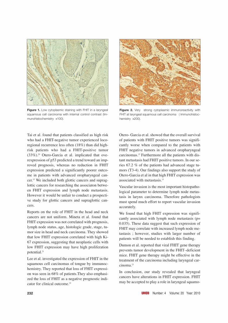
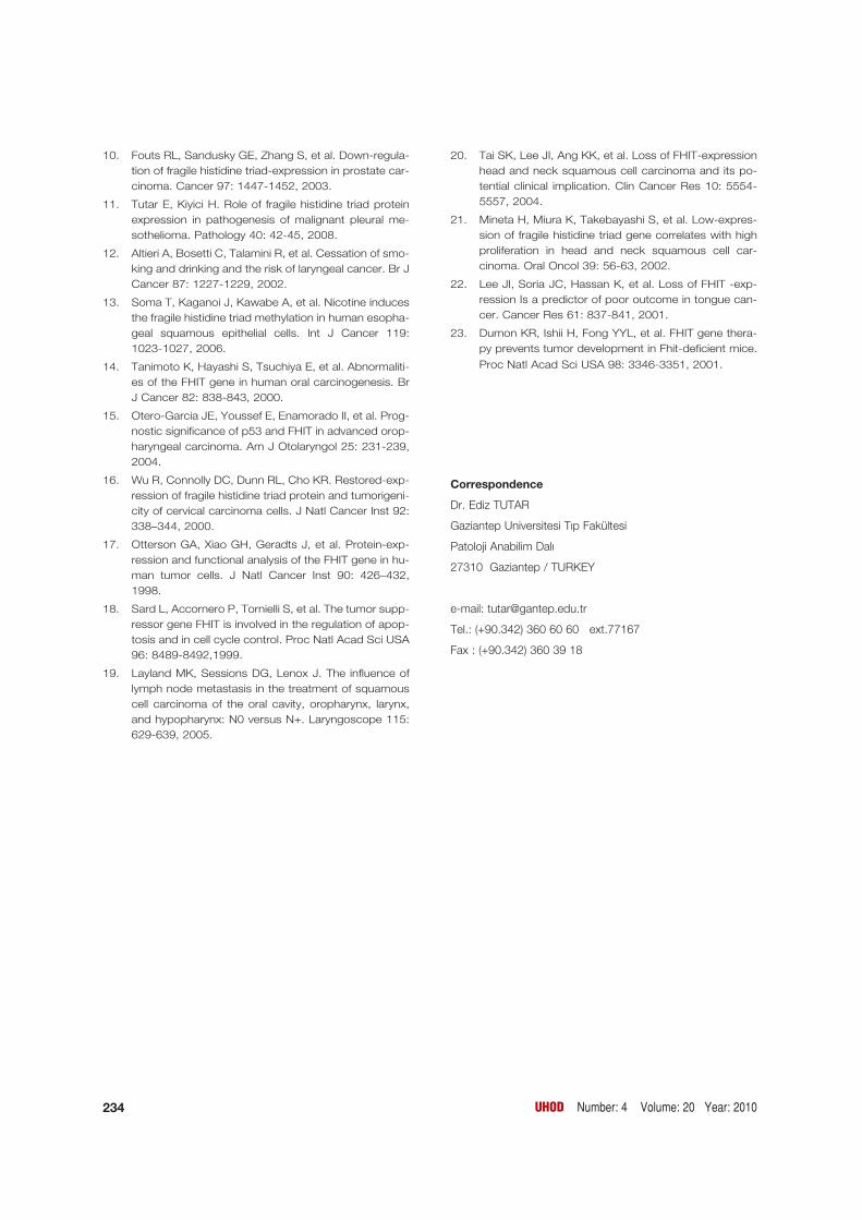
parafin embedded tissue blocks of 64 patients withsquamous cell carcinoma of the larynx. The medi-an age of the patients was 56.6 (±10.98). Sixty ofthe patients (93.8%) were male and 4(6.2%) werefemale. FHIT expression was low in 40.6% of pati-ents and high in 59.6% of patients (Figure 1, 2).
In our statistical analysis FHIT expression was se-parately compared with age, T stage, histologicalgrade, vascular and perineural invasion, lymph no-de metastasis (Table 2).
High FHIT expression was significantly associatedwith lymph node metastasis (p= 0.035).
FHIT expression was not statistically correlatedwith histological grade (p= 0.953), perineural inva-sion (p= 0.331), and vascular invasion (p= 0.663).There was no correlation between FHIT expressionand age (p= 0.358).
DISCUSSION This study was designed to evaluate abnormalFHIT expression as a prognostic marker in patientswith laryngeal carcinoma. FHIT expression waslow in 40.6% of our patients and high in 59.6% ofour patients. High FHIT expression was signifi-cantly associated with lymph node metastasis.
The FHIT gene is a candidate tumor suppressor ge-ne, although its precise mechanism of action rema-ins unclear. Restoration of this gene expression intumor cells has yielded conflicting results. Theseconflicting data suggest that the mechanisms invol-ved with the function of the FHIT gene may be dif-ferent from those of other classic tumor suppressorgenes such as p53 and Rb1.16,17 Sard et al. reportedthat the FHIT gene is involved in the regulation ofthe cell cycle and that its tumor suppressor activityis derived from its proapoptotic activity.18 However,Otterson et al. evaluated the function of the FHITgene and did not discover any regulation of the cellcycle or function with respect to induction of apop-tosis.17 These varied results may reflect that FHITfunctions in a tissue-specific fashion or at a particu-lar point in the multistage process of carcinogenesis.
T stage and lymph node metastasis negatively af-fects disease-specific survival in the laryngeal car-cinoma. Patients older than 65 years of age haveshort survival rates.19 In our study, it was found thatthere was no statistically significant relationship
between age of the patient, T stage of the tumor andFHIT expression. In a previous study of 103 pati-ents with cervical carcinoma, it was found that re-duced FHIT expression was significantly associ-ated with lymph node metastasis.8
Low levels of FHIT expression has been found in ahigh percentage of high-grade squamous epithelialneoplasia in many studies.6,8,15,21 A few studies hasbeen showed that FHIT expression was associatedwith locoregional recurrence paradoxically in headand neck squamous cell carcinoma (HNSCC).15,20
UHOD Number: 4 Volume: 20 Year: 2010 231
Table 1. Clinicopathologic data of 64 patients with laryngeal squamous cell carcinoma
Characteristics Patients (%)
Patients 64
Total no 56.64 (32-76)
Median age (years) [range] 60 (93.8%)
Male
Female 4 (6.2%)
Histological grade
Well differentiated 26 (40.6%)
Moderately differentiated 21 (32.8%)
Poorly differentiated 17 (26.6%)
T stage
T1 5 (7.8%)
T2 16 (25.0%)
T3 24 (37.5%)
T4 19 (29.7%)
Lymph node metastasis
Absent 38 (59.4%)
Present 26 (40.6%)
Vascular invasion
Absent 45 (70.3%)
Present 19 (29.7%)
Perineural invasion
Absent 52 (81.3%)
Present 12 (18.7%)
Tai et al. found that patients classified as high riskwho had a FHIT-negative tumor experienced loco-regional recurrence less often (18%) than did high-risk patients who had a FHIT-positive tumor(33%).20 Otero-Garcia et al. implicated that ove-rexpression of p53 predicted a trend toward an imp-roved prognosis, whereas no reduction in FHITexpression predicted a significantly poorer outco-me in patients with advanced oropharyngeal can-cer.15 We included both glottic cancers and suprag-lottic cancers for researching the associaton betwe-en FHIT expression and lymph node metastasis.However it would be unfair to conduct a prospecti-ve study for glottic cancers and supraglottic can-cers.
Reports on the role of FHIT in the head and neckcancers are not uniform. Mineta et al. found thatFHIT expression was not correlated with prognosis,lymph node status, age, histologic grade, stage, tu-mor size in head and neck carcinoma. They showedthat low FHIT expression correlated with high Ki-67 expression, suggesting that neoplastic cells withlow FHIT expression may have high proliferationpotential.21
Lee et al. investigated the expression of FHIT in thesquamous cell carcinomas of tongue by immunoc-hemistry. They reported that loss of FHIT expressi-on was seen in 68% of patients.They also emphasi-zed the loss of FHIT as a negative prognostic indi-cator for clinical outcome.22
Otero- Garcia et al. showed that the overall survivalof patients with FHIT positive tumors was signifi-cantly worse when compared to the patients withFHIT negative tumors in advanced oropharyngealcarcinomas.15 Furthermore all the patients with dis-tant metastasis had FHIT positive tumors. In our se-ries 67.2 % of the patients had advanced stage tu-mors (T3-4). Our findings also support the study ofOtero-Garcia et al in that high FHIT expression wasassociated with metastasis.15
Vascular invasion is the most important histopatho-logical parameter to determine lymph node metas-tasis in larynx carcinoma. Therefore pathologistsmust spend much effort to report vascular invasionaccurately.
We found that high FHIT expression was signifi-cantly associated with lymph node metastasis (p=0.035). These data suggest that such expression ofFHIT may correlate with increased lymph node me-tastasis ; however, studies with larger number ofpatients will be needed to establish this finding.
Dumon et al. reported that viral FHIT gene therapyprevents tumor development in the FHIT–deficientmice. FHIT gene therapy might be effective in thetreatment of the carcinoma including laryngeal car-cinoma.23
In conclusion, our study revealed that laryngealcancers have alterations in FHIT expression. FHITmay be accepted to play a role in laryngeal squamo-
232 UHOD Number: 4 Volume: 20 Year: 2010
Figure 1. Low cytoplasmic staining with FHIT in a laryngealsquamous cell carcinoma with internal control contrast (Im-munohistochemistry x100).
Figure 2. Very strong cytoplasmic immunoreactivity withFHIT at laryngeal squamous cell carcinoma ( Immunohistoc-hemistry x200).

us cell carcinoma. However, the exact molecularmechanism of FHIT function is unclear and rema-ins to be elucidated. Understanding the functionalstatus of the FHIT proteins may lead to develop-ment of new therapeutic options in the future.
REFERENCES
1. Vermund H, Krajci P, Eide TJ, Winther F. Laryngec-tomy whole organ serial sections--histological para-meters correlated with recurrence rate. Acta Oncol 43:98-107, 2004.
2. Yilmaz T, Hosal AS, Gediko¤lu G, et al. Prognostic sig-nificance of vascular and perineural invasion in cancerof the larynx. Am J Otolaryngol 19: 83-88, 1998.
3. Pekarsky Y, Zanesi N, Palamarchuk A, et al. FHIT:from gene discovery to cancer treatment and preven-tion. Lancet Oncol 3: 748-754, 2002.
4. Ohta M, Inoue H, Cottecelli MG, et al. The FHIT gene,spanning the chromosome 3p14.2 fragile site and re-nal carcinoma associated t(3;8) breakpoint, is abnor-mal in digestive tract cancers. Cell 84: 587-597, 1996.
5. Siprashvili Z, Sozzi G, Barnes LD, et al . Replacementof Fhit in cancer cells suppresses tumorigenicity. ProcNatl Acad Sci USA 94: 13771-13776, 1997.
6. Mascaux C, Martin B, Verdebout JM, et al. Fragile his-tidine triad protein-expression in nonsmall cell lungcancer and correlation with Ki-67 and with p53. EurRespir J 21: 753-758, 2003.
7. Guler G, Uner A, Guler N, et al. The fragile genes FHITand WWOX are inactivated coordinately in invasivebreast carcinoma. Cancer 100: 1605-1614, 2004.
8. Huang LW, Chao SL, Chen TJ. Reduced FHIT-expres-sion in cervical carcinoma: correlation with tumorprogression and poor prognosis. Gynecol Oncol 90:331-337 2003.
9. Deng YF, Tian F, Lu YD, et al. Mutation and abnormalexpression of the fragile histidine triad gene in nasop-haryngeal carcinoma. Laryngoscope 111: 1589-1592,2001.
UHOD Number: 4 Volume: 20 Year: 2010 233
Table 2. Correlation between FHIT-expression and other histopathologic parameters
Parameter Low FHIT –expression High FHIT –expression p value(n= 26) (n= 38)
Age
≤ 50 12 (18.8%) 12 (18.8%) p= 0.358 (NS)
> 50 14 (21.9%) 26 (40.6%)
Histological grade
Well differentiated 10 (15.6%) 16 (25%)
Moderately differentiated 9 (14.1%) 12 (18.8%) p=0.953 (NS)
Poorly differentiated 7 (10.9%) 10 (15.6%)
T stage
T1-2 9 (14.1%) 12 (18.8%) p= 1 (NS)
T3-4 17 (26.6%) 26 (40.6%)
Lymph node metastasis
Absent 20 (31.3%) 18 (28.1%) p= 0.035
Present 6 (9.4%) 20 (31.3%)
Vascular invasion
Absent 17 (26.6%) 28 (43.8%) p=0.663 (NS)
Present 9 (14.1%) 10 (15.6%)
Perineural invasion
Absent 23 (35.9%) 29 (45.3%) p=0.331 (NS)
Present 3 (4.7%) 9 (14.1%)
NS: nonsignificant
10. Fouts RL, Sandusky GE, Zhang S, et al. Down-regula-tion of fragile histidine triad-expression in prostate car-cinoma. Cancer 97: 1447-1452, 2003.
11. Tutar E, Kiyici H. Role of fragile histidine triad proteinexpression in pathogenesis of malignant pleural me-sothelioma. Pathology 40: 42-45, 2008.
12. Altieri A, Bosetti C, Talamini R, et al. Cessation of smo-king and drinking and the risk of laryngeal cancer. Br JCancer 87: 1227-1229, 2002.
13. Soma T, Kaganoi J, Kawabe A, et al. Nicotine inducesthe fragile histidine triad methylation in human esopha-geal squamous epithelial cells. Int J Cancer 119:1023-1027, 2006.
14. Tanimoto K, Hayashi S, Tsuchiya E, et al. Abnormaliti-es of the FHIT gene in human oral carcinogenesis. BrJ Cancer 82: 838-843, 2000.
15. Otero-Garcia JE, Youssef E, Enamorado II, et al. Prog-nostic significance of p53 and FHIT in advanced orop-haryngeal carcinoma. Am J Otolaryngol 25: 231-239,2004.
16. Wu R, Connolly DC, Dunn RL, Cho KR. Restored-exp-ression of fragile histidine triad protein and tumorigeni-city of cervical carcinoma cells. J Natl Cancer Inst 92:338–344, 2000.
17. Otterson GA, Xiao GH, Geradts J, et al. Protein-exp-ression and functional analysis of the FHIT gene in hu-man tumor cells. J Natl Cancer Inst 90: 426–432,1998.
18. Sard L, Accornero P, Tornielli S, et al. The tumor supp-ressor gene FHIT is involved in the regulation of apop-tosis and in cell cycle control. Proc Natl Acad Sci USA96: 8489-8492,1999.
19. Layland MK, Sessions DG, Lenox J. The influence oflymph node metastasis in the treatment of squamouscell carcinoma of the oral cavity, oropharynx, larynx,and hypopharynx: N0 versus N+. Laryngoscope 115:629-639, 2005.
20. Tai SK, Lee JI, Ang KK, et al. Loss of FHIT-expressionhead and neck squamous cell carcinoma and its po-tential clinical implication. Clin Cancer Res 10: 5554-5557, 2004.
21. Mineta H, Miura K, Takebayashi S, et al. Low-expres-sion of fragile histidine triad gene correlates with highproliferation in head and neck squamous cell car-cinoma. Oral Oncol 39: 56-63, 2002.
22. Lee JI, Soria JC, Hassan K, et al. Loss of FHIT -exp-ression Is a predictor of poor outcome in tongue can-cer. Cancer Res 61: 837-841, 2001.
23. Dumon KR, Ishii H, Fong YYL, et al. FHIT gene thera-py prevents tumor development in Fhit-deficient mice.
Proc Natl Acad Sci USA 98: 3346-3351, 2001.
Correspondence
Dr. Ediz TUTAR
Gaziantep Universitesi T›p Fakültesi
Patoloji Anabilim Dal›
27310 Gaziantep / TURKEY
e-mail: [email protected]
Tel.: (+90.342) 360 60 60 ext.77167
Fax : (+90.342) 360 39 18
234 UHOD Number: 4 Volume: 20 Year: 2010