familial hydronephrosis unlinked to the hla complex
TRANSCRIPT

Familial Hydronephrosis Unlinked to theHLA Complex
Alena Santava,1* Anna Utıkalova,2 Adela Bartova,3 Jirı Drabek,3 Jirı Santavy,1 and Jirı Scheinar2
1Department of Medical Genetics and Fetal Medicine, Palacky University, Olomouc, Czech Republic2Department of Urology, Palacky University, Olomouc, Czech Republic3Department of Immunology, Palacky University, Olomouc, Czech Republic
Clinical findings, management, and possiblelinkage of congenital hydronephrosiscaused by ureteropelvic junction stenosis tothe HLA complex were studied in four fami-lies. These families provide evidence of pos-sible autosomal dominant inheritance. HLAclass I antigen studies in all four and class II(HLA-DR) in three families were performed.These studies failed to show close linkage tothe chromosome 6 markers in two familiesbut there was consistent inheritance in theother two. Although formal linkage calcula-tions are not presented, it is apparent thatin some families HLA haplotyping is not use-ful in predicting prevence of renal obstruc-tion. Am. J. Med. Genet. 70:118–120, 1997.© 1997 Wiley-Liss, Inc.
KEY WORDS: congenital hydronephrosis;automosomal dominant in-heritance; HLA system
INTRODUCTION
Congenital hereditary hydronephrosis (HN), causedby obstruction of the ureteropelvic junction region, usu-ally occurs sporadically. However, familial occurrenceboth in subsequent generations or among siblings hasbeen documented repeatedly. Possible genetic mecha-nisms involve multifactorial or autosomal dominant in-heritance with reduced penetrance. Possible heteroge-neity of the disorder may be another factor. HLA stud-ies in some families with HN (performed because ofplanned renal transplantation) indicated identicalHLA haplotypes among affected relatives, with pos-sible linkage of HN to the HLA-gene complex on 6p[Paramo et al., 1991; Izquierdo et al., 1992].
This study addresses familial HN and possible link-age to the HLA complex. Ultrasound studies of the kid-
neys were done on all available first-degree relatives ofindex cases to obtain an early diagnosis (relevant forclinical prognosis) and to establish genetic status. Thiswas especially motivated because of non-specific andlate occurrence of symptoms of hydronephrosis-relatedrenal impairment, which (if noticed too late) requiresnephrectomy rather than corrective surgical interven-tion, such as pyeloplasty.
METHODS
Four families with possible autosomal dominant in-heritance are described. In all families clinical geneticexaminations, ultrasound kidney examinations, and,in one family, prenatal diagnosis, were performed.HLA class I antigens were determined using NIHmethods (microlymphocytotoxic tests). Finally, 15HLA-A, 34 HLA-B, and 8 HLA-C antigens were typed.The other 18 antigens of the HLA class II (HLA-DR)were typed using PCR allelic-specific primers (DYNALDR ‘‘low resolution’’ SSP kit [Miller et al., 1988; Olerupet al., 1992]). The details of the families are given in thefollowing family reports.
Family I
The propositus II-2 (Fig. 1), born in 1969, was admit-ted at the age of 14 months because of high fever andsevere illness due to acute pyelonephritis and septice-mia. The IVP showed HN grade IV. Nephrectomy of theleft kidney was indicated. During surgery, a dilatedsuppurative pelvicocalyceal system and multiple ab-scesses in the renal parenchyma were found. She wasfollowed until adolescence. The right kidney appearedhypertrophic and descended by 2.5 vertebrae. The fa-ther, I-1, born in 1951, developed macroscopic hematu-ria at the age of 14 after a sudden fall on his back. TheIVP showed HN grade IV, and nephrectomy of the leftkidney was performed. There were no other cases ofhereditary renal and/or ureteral malformations in thisfamily. The proposita married at the age of 20. She hadtwo first-trimester spontaneous abortions and duringher third pregnancy we performed ultrasonographicexamination of the fetal kidneys. In the 18th week, di-lation of the pelvicocalyceal collecting system of theright kidney of the fetus was evident. This finding re-mained unchanged during the later pregnancy. A girl
*Correspondence to: A. Santava, M.D., Department of MedicalGenetics and Fetal Medicine, Palacky University, I.P.Pavlova 6,775 20 Olomouc, Czech Republic.
Received 8 February 1996; Accepted 9 August 1996
American Journal of Medical Genetics 70:118–120 (1997)
© 1997 Wiley-Liss, Inc.
(III-I) weighing 3,400 g, 49 cm long, was born at term.Postnatal IVP confirmed HN grade IV. Pyeloplasty ofthe right kidney was performed four weeks after birthwith an uneventful recovery.
The HLA typing is presented in the pedigree (Fig. 1).In this family, both father and daughter with unilat-eral congenital HN share a haplotype not shared by theprenatally diagnosed affected granddaughter (III-1).
Family 2
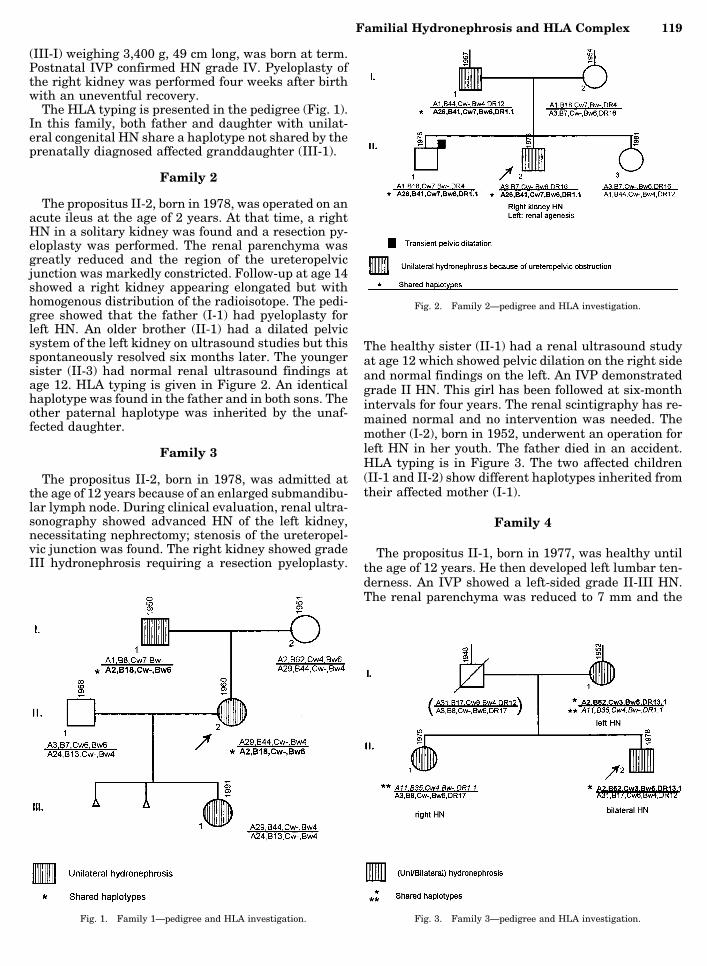
The propositus II-2, born in 1978, was operated on anacute ileus at the age of 2 years. At that time, a rightHN in a solitary kidney was found and a resection py-eloplasty was performed. The renal parenchyma wasgreatly reduced and the region of the ureteropelvicjunction was markedly constricted. Follow-up at age 14showed a right kidney appearing elongated but withhomogenous distribution of the radioisotope. The pedi-gree showed that the father (I-1) had pyeloplasty forleft HN. An older brother (II-1) had a dilated pelvicsystem of the left kidney on ultrasound studies but thisspontaneously resolved six months later. The youngersister (II-3) had normal renal ultrasound findings atage 12. HLA typing is given in Figure 2. An identicalhaplotype was found in the father and in both sons. Theother paternal haplotype was inherited by the unaf-fected daughter.
Family 3
The propositus II-2, born in 1978, was admitted atthe age of 12 years because of an enlarged submandibu-lar lymph node. During clinical evaluation, renal ultra-sonography showed advanced HN of the left kidney,necessitating nephrectomy; stenosis of the ureteropel-vic junction was found. The right kidney showed gradeIII hydronephrosis requiring a resection pyeloplasty.
The healthy sister (II-1) had a renal ultrasound studyat age 12 which showed pelvic dilation on the right sideand normal findings on the left. An IVP demonstratedgrade II HN. This girl has been followed at six-monthintervals for four years. The renal scintigraphy has re-mained normal and no intervention was needed. Themother (I-2), born in 1952, underwent an operation forleft HN in her youth. The father died in an accident.HLA typing is in Figure 3. The two affected children(II-1 and II-2) show different haplotypes inherited fromtheir affected mother (I-1).
Family 4
The propositus II-1, born in 1977, was healthy untilthe age of 12 years. He then developed left lumbar ten-derness. An IVP showed a left-sided grade II-III HN.The renal parenchyma was reduced to 7 mm and the
Fig. 1. Family 1—pedigree and HLA investigation.
Fig. 2. Family 2—pedigree and HLA investigation.
Fig. 3. Family 3—pedigree and HLA investigation.
Familial Hydronephrosis and HLA Complex 119

calyces were markedly dilated. The right kidney wasnormal. A resection pyeloplasty was performed withsatisfactory results. The sister (II-2) had normal renalfindings at age 14. In his youth, the father (I-1), born in1946, underwent a left nephrectomy for left-sided HN.The mother (II-2) showed normal renal ultrasoundfindings. HLA typing is given in Figure 4. The paternalhaplotype shared with his similarly affected son differsfrom the haplotype he shares with his healthy daugh-ter.
DISCUSSION
Familial occurrence of uni/bilateral hydronephrosisin sibs or with transmission from an affected parent tooffspring has been reported [Aaron and Robbins, 1948;Jewell and Buchert, 1962; Grosse et al., 1973; Cohen etal., 1978; Dwoskin, 1979; Sengar et al., 1979; Buscemiet al., 1985] and reviewed by Paramo [1991]. If autoso-mal dominant inheritance with reduced penetrance isinvolved in this condition, there may be variability inage-of-onset and the importance of ultrasound renalstudies in at least all first-degree relatives is clear.Early detection of HN offers the option of pyeloplastybefore renal scarring leaves nephrectomy as the onlyoption. The observation of haplo-identity for the HLAlocus among affected members of multiplex families(affected members in one or more generations) indi-cated a possible linkage of the HN locus to the HLA
locus on chromosome 6p [Izquierdo et al., 1992]. Thiswas supported by the incidental findings of a(6,19)(p23.1q13.4) translocation in a fetus with bilat-eral multicystic renal dysplasia [Fryns et al., 1993].The present results in two families with parent-to-offspring transmission of ureteropelvic junction ob-struction and associated hydronephrosis indicate thatthere is not consistent HLA-haplotype identitybetween affected parents / affected offspring. Appar-ently, linkage to HLA-markers may not be suitable inall families for identifying relatives of patients withHN who have a risk of developing an identical disorder.However, in one small study two families do show HLAhaplotype inheritance consistent with linkage.
The observation of ‘‘healthy’’ first-degree relativeswith clinically significant degrees of ureteropelvic junc-tion stenosis emphasizes the necessity for ultrasoundstudies to enable careful monitoring of incipient renalfunction loss and to allow timely pyeloplasty.
REFERENCESAaron G, Robbins MA (1948): Hydronephrosis due to aberrant vessels:
Remarkable familial incidence with report of cases. J Urol 60:702–705.
Buscemi M, Shanske A, Mallet E, Ozoktay S, Hanna MK (1985): Domi-nantly inherited ureteropelvic junction obstruction. Urology 26:568–570.
Cohen B, Goldman SM, Kopilnick M, Khurana AV, Salik JO (1978): Ure-teropelvic junction obstruction: Its occurrence in 3 members of a singlefamily. J Urol 10:361–364.
Dwoskin JY (1979): Ureteropelvic junction obstruction and sibling uropa-thology. Urology 13:153–158.
Fryns JP, Kleczkowska A, Moerman P, Vandenberghe K (1993): Heredi-tary hydronephrosis and the short arm of chromosome 6. Hum Genet91:514–515.
Grosse FR, Kaveggia L, Opitz JM (1973): Familial hydronephrosis. ZKinderheilk 114:313–322.
Izquierdo L, Porteous M, Paramo PG, Connor JM (1992): Evidence forgenetic heterogeneity in hereditary hydronephrosis caused by pelviure-teric junction obstruction, with one locus assigned to chromosome 6p.Hum Genet 89:557–560.
Jewell JH, Buchert WI (1962): Unilateral hereditary hydronephrosis: Areport of four cases in three consecutive generations. J Urol 80:129–136.
Miller SA, Dykes DD, Polesky HF (1988): A simple salting out procedurefor extracting DNA from human nucleated cells. Nucl Acids Res 16:215.
Olerup D, Zetterquist H (1992): HLA-DR typing by PCR amplification withsequence specific primers (PCR-SSP) in 2 hours: An alternative to se-rological DR typing in clinical practice including donor-recipientmatching in cadaveric transplantation. Tissue Antigens 39:225–235.
Paramo PG, Izquierdo L, Paramo P Jr, Llorente L, Diego A, Paez A, GomezRuız JJ, Uson AC (1991): Genuine hereditary hydronephrosis in athree generation family. Eur Urol 20:293–300.
Sengar DPS, Rashid A, Wolfish NM (1979): Familial urinary tract anoma-lies. Association with the major histocompatibility complex in man. JUrol 121:194–197.Fig. 4. Family 4—pedigree and HLA investigation.
120 Santava et al.