fusion of the human gene for the polyubiquitination coeffector
TRANSCRIPT

Fusion of the Human Gene for thePolyubiquitination Coeffector UEV1 with Kua,a Newly Identified GeneTimothy M. Thomson,1,2,7,8 Juan Jose Lozano,1,2,3,7 Noureddine Loukili,1,2,7
Roberto Carrio,4 Florenci Serras,5 Bru Cormand,2,6 Marta Valeri,2 Vıctor M. Dıaz,1,2
Josep Abril,3 Moises Burset,3 Jesus Merino,4 Alfons Macaya,2,6
Montserrat Corominas,5 and Roderic Guigo3
1Institut de Biologia Molecular, Consejo Superior de Investigaciones Cientificas, Barcelona, Spain; 2Unitat de RecercaBiomedica, Hospital Materno-Infantil, Hospitals Vall d’Hebron, Barcelona, Spain; 3Grup de Recerca en Informatica Medica,Institut Municipal d’Investigacio Medica, Universitat Pompeu Fabra, Barcelona, Spain; 4Departamento de Biologıa Molecular,Facultad de Medicina, Universidad de Cantabria, Santander, Spain; 5Departament de Genetica, Facultat de Biologia,Universitat de Barcelona, Barcelona, Spain; 6Unitat de Malaties Neurometaboliques, Hospital Materno-Infantil, Hospitals Valld’Hebron, Barcelona, Spain
UEV proteins are enzymatically inactive variants of the E2 ubiquitin-conjugating enzymes that regulatenoncanonical elongation of ubiquitin chains. In Saccharomyces cerevisiae, UEV is part of the RAD6-mediatederror-free DNA repair pathway. In mammalian cells, UEV proteins can modulate c-FOS transcription and theG2-M transition of the cell cycle. Here we show that the UEV genes from phylogenetically distant organismspresent a remarkable conservation in their exon–intron structure. We also show that the human UEV1 gene isfused with the previously unknown gene Kua. In Caenorhabditis elegans and Drosophila melanogaster, Kua and UEV arein separated loci, and are expressed as independent transcripts and proteins. In humans, Kua and UEV1 areadjacent genes, expressed either as separate transcripts encoding independent Kua and UEV1 proteins, or as ahybrid Kua–UEV transcript, encoding a two-domain protein. Kua proteins represent a novel class of conservedproteins with juxtamembrane histidine-rich motifs. Experiments with epitope-tagged proteins show that UEV1Ais a nuclear protein, whereas both Kua and Kua–UEV localize to cytoplasmic structures, indicating that the Kuadomain determines the cytoplasmic localization of Kua–UEV. Therefore, the addition of a Kua domain to UEVin the fused Kua–UEV protein confers new biological properties to this regulator of variant polyubiquitination.
[Kua cDNAs isolated by RT-PCR and described in this paper have been deposited in the GenBank data libraryunder accession nos. AF1155120 (H. sapiens) and AF152361 (D. melanogaster). Genomic clones containing UEVgenes: S. cerevisiae, YGL087c (accession no. Z72609); S. pombe, c338 (accession no. AL023781); P. falciparum,MAL3P2 (accession no. AL034558); A. thaliana, F26F24 (accession no. AC005292); C. elegans, F39B2 (accessionno. Z92834); D. melanogaster, AC014908; and H. sapiens, 1185N5 (accession no. AL034423). Accession numbers forKua cDNAs in GenBank dbEST: M. musculus, AA7853; T. cruzi, AI612534. Other Kua-containing sequences: A.thaliana genomic clones F10M23 (accession no. AL035440), F19K23 (accession no. AC000375), and T20K9(accession no. AC004786).]
Described recently as a class of proteins structurallyrelated to the ubiquitin-conjugating enzymes (E2), adistinctive feature of UEV proteins is that they are in-active variants of E2 enzymes, lacking a recognizablecatalytic center (Koonin and Abagyan 1997; Ponting etal. 1997; Sancho et al. 1998). These proteins are wellconserved in sequence and structure in all eukaryotic
organisms, and this results in the sharing of specificfunctions, such as protection of cells from DNA dam-aging agents (Broomfield et al. 1998; Thomson et al.1998) and enhancement of transcription from the c-FOS promoter (Xiao et al. 1998), by UEV proteins fromdistant organisms. The biochemical mode of action ofUEV proteins in the yeast Saccharomyces cerevisiae hasbeen established by Hoffman and Pickart (1999). The S.cerevisiae UEV protein, also known as Mms2, interactswith the E2 enzyme Ubc13p, and the resulting het-erodimer is competent for the elongation of polyubiq-uitin chains. A novel feature of the polyubiquitinchains thus formed is that Lys 63, instead of the ca-
7These authors have contributed equally to this work.8Corresponding author.E-MAIL [email protected]; FAX 34-93-489-4064.Article published online before print: Genome Res., 10.1101/gr.140500.Article and publication are at www.genome.org/cgi/doi/10.1101/gr.140500.
Letter
10:1743–1756 ©2000 by Cold Spring Harbor Laboratory Press ISSN 1088-9051/00 $5.00; www.genome.org Genome Research 1743www.genome.org
Cold Spring Harbor Laboratory Press on April 12, 2018 - Published by genome.cshlp.orgDownloaded from
nonical Lys 48, is used for the Gly–Lys isopeptidebonds between ubiquitin moieties (Hoffman and Pick-art 1999). Modification of proteins by this variantpolyubiquitin chain may be reversible, and couldmodulate the function of target proteins, without di-recting them for degradation (Spence et al. 2000).
In S. cerevisiae, UEV genes are part of the error-freeDNA repair pathway regulated by RAD6 (Broomfield etal. 1998; Hoffman and Pickart 1999). In human cells,UEV1 (Rothofsky and Lin 1997) and UEV2/Mms2(Xiao et al. 1998) promote the transcriptional activityof c-FOS, possibly through interactions with as yet uni-dentified DNA-binding transcriptional regulators (Xiaoet al. 1998). Overexpression of UEV1 in human coloncancer cells induces the accumulation of cells in G2-Mand poliploidy, apoptosis, and inhibition of cell differ-entiation (Sancho et al. 1998). How the participationof UEV proteins in all these processes relate to the ac-tivity of UEV proteins as coeffectors in the polyubiq-uitination of target proteins has yet to be determined.In humans, there are two different UEV proteins en-coded by separate genes, UEV1 or CROC1 (Rothofskyand Lin 1997; Sancho et al. 1998), and UEV2 or MMS2(Sancho et al. 1998; Xiao et al. 1998). The UEV1 genecodes for two isoforms generated by alternative splic-ing, which share a common phylogenetically con-served UEV domain (Sancho et al. 1998). The isoformUEV1B contains a unique 82-residue amino-terminalextension, the B domain (Sancho et al. 1998).
The ubiquitin-conjugating enzymes are a largegroup of proteins, of which many variants exist in alleukaryotic organisms (for review, see Hershko andCiechanover 1998). Although a common protein se-quence and structural theme is shared between all E2enzymes, attempts to assign primordial ancestors asthe origin of one or more branches have not been metwith success, mainly due to the great interspecies vari-ability of functionally equivalent proteins. Being a newfamily of proteins with strong structural and func-tional links to the long-known ubiquitin-conjugatingenzymes, UEV proteins and their genes could be usefulto study the origins of the E2 proteins and their genes.Here we have analyzed the structure of the UEV genesin a number of organisms, and found that it is veryconserved between phylogenetically distant organ-isms. As a relevant consequence of this analysis, wehave found that the human UEV1 gene is part of ahybrid gene that results from the fusion of UEV1 witha second, previously unknown gene, which we havenamed Kua. We also show that, in humans, Kua andUEV1 can be expressed either as independent tran-scriptional units and proteins, or as a hybrid Kua–UEVtranscript and protein. In contrast, in flies and wormsthe gene for Kua is unlinked to the gene for the corre-sponding UEV protein, and Kua and UEV are alwaysexpressed as separate proteins.
RESULTS
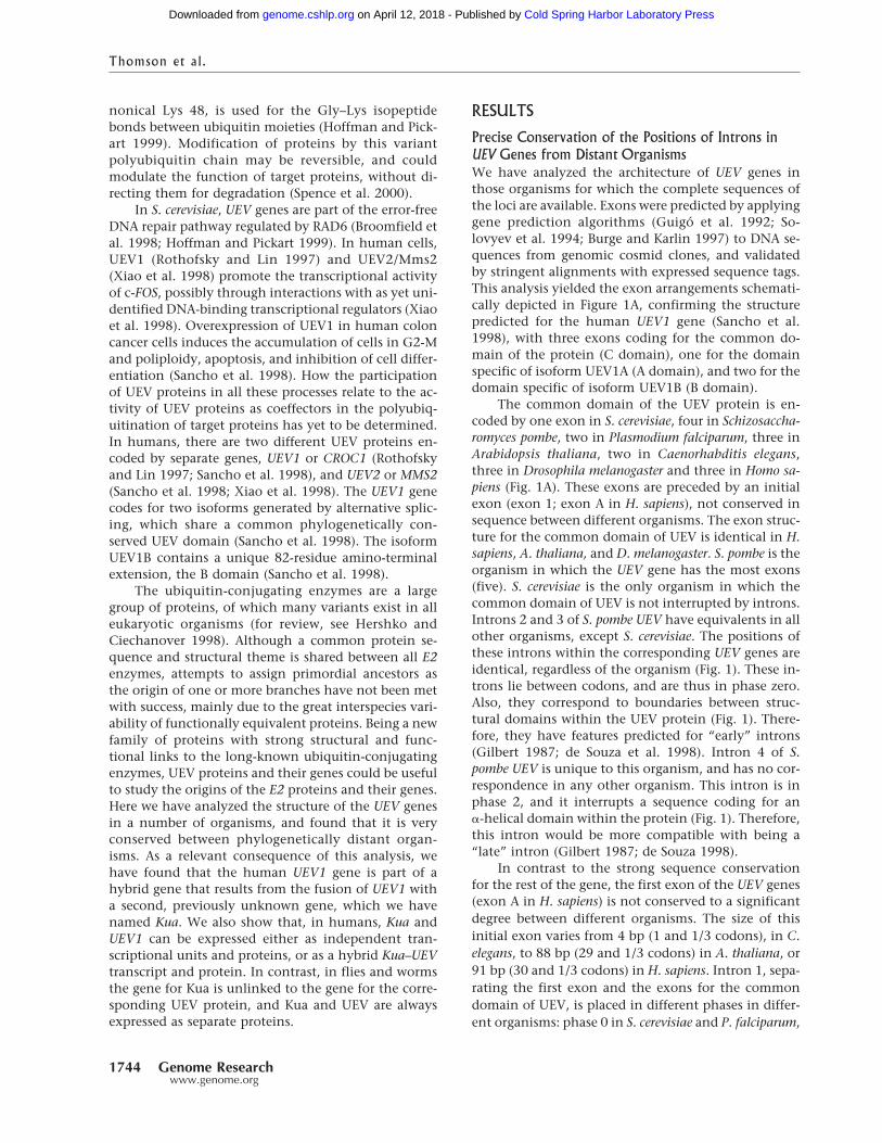
Precise Conservation of the Positions of Introns inUEV Genes from Distant OrganismsWe have analyzed the architecture of UEV genes inthose organisms for which the complete sequences ofthe loci are available. Exons were predicted by applyinggene prediction algorithms (Guigo et al. 1992; So-lovyev et al. 1994; Burge and Karlin 1997) to DNA se-quences from genomic cosmid clones, and validatedby stringent alignments with expressed sequence tags.This analysis yielded the exon arrangements schemati-cally depicted in Figure 1A, confirming the structurepredicted for the human UEV1 gene (Sancho et al.1998), with three exons coding for the common do-main of the protein (C domain), one for the domainspecific of isoform UEV1A (A domain), and two for thedomain specific of isoform UEV1B (B domain).
The common domain of the UEV protein is en-coded by one exon in S. cerevisiae, four in Schizosaccha-romyces pombe, two in Plasmodium falciparum, three inArabidopsis thaliana, two in Caenorhabditis elegans,three in Drosophila melanogaster and three in Homo sa-piens (Fig. 1A). These exons are preceded by an initialexon (exon 1; exon A in H. sapiens), not conserved insequence between different organisms. The exon struc-ture for the common domain of UEV is identical in H.sapiens, A. thaliana, and D. melanogaster. S. pombe is theorganism in which the UEV gene has the most exons(five). S. cerevisiae is the only organism in which thecommon domain of UEV is not interrupted by introns.Introns 2 and 3 of S. pombe UEV have equivalents in allother organisms, except S. cerevisiae. The positions ofthese introns within the corresponding UEV genes areidentical, regardless of the organism (Fig. 1). These in-trons lie between codons, and are thus in phase zero.Also, they correspond to boundaries between struc-tural domains within the UEV protein (Fig. 1). There-fore, they have features predicted for “early” introns(Gilbert 1987; de Souza et al. 1998). Intron 4 of S.pombe UEV is unique to this organism, and has no cor-respondence in any other organism. This intron is inphase 2, and it interrupts a sequence coding for an�-helical domain within the protein (Fig. 1). Therefore,this intron would be more compatible with being a“late” intron (Gilbert 1987; de Souza 1998).
In contrast to the strong sequence conservationfor the rest of the gene, the first exon of the UEV genes(exon A in H. sapiens) is not conserved to a significantdegree between different organisms. The size of thisinitial exon varies from 4 bp (1 and 1/3 codons), in C.elegans, to 88 bp (29 and 1/3 codons) in A. thaliana, or91 bp (30 and 1/3 codons) in H. sapiens. Intron 1, sepa-rating the first exon and the exons for the commondomain of UEV, is placed in different phases in differ-ent organisms: phase 0 in S. cerevisiae and P. falciparum,
Thomson et al.
1744 Genome Researchwww.genome.org
Cold Spring Harbor Laboratory Press on April 12, 2018 - Published by genome.cshlp.orgDownloaded from
phase 2 in S. pombe, and phase 1 in C. elegans, A.thaliana, D. melanogaster, and H. sapiens (Fig. 1). The 3�
boundaries set by this intron impose different 5� endsfor the common domain of UEV in different organ-isms, which results in one to three codon differences
between UEV proteins from these organisms at theamino-end of this domain (Fig. 1). Similarly, there areno sequences resembling the exons coding for the Bdomain of human UEV1 in the vicinity of the UEVgenes of any other organism. It has been suggested that
Figure 1 Conservation of introns in UEV genes from distant organisms. (A) Diagrammatic representation (not to scale) of the relativeexon–intron arrangement of the UEV gene in Saccharomyces cerevisiae, Schizosaccharomyces pombe, P. falciparum, Arabidopsis thaliana,Caenorhabditis elegans, Drosophila melanogaster, and Homo sapiens. Inset diagram for the two major isoforms described for human UEV1.Putative 5� untranslated (UTR) segments are represented as open boxes. Introns are designated by numbers that are specific for each UEVgene. Vertical dotted lines within exons define segments corresponding to the exons in S. pombe UEV. (B) Alignment of UEV genes andproteins showing precise conservation of the position of the second and third introns interrupting the C domain of UEV proteins. Splicedonor and acceptor sequences in intron boundaries are in lower case. The positions of introns are marked by arrowheads. Predictions ofsecondary structure are shown below the sequences as rods (helices) or arrows (strands).
Evolution of UEV Genes
Genome Research 1745www.genome.org
Cold Spring Harbor Laboratory Press on April 12, 2018 - Published by genome.cshlp.orgDownloaded from
the B domain of human UEV1B could confer specificfunctions to this isoform (Sancho et al. 1998). We thusset our efforts to explore the evolutionary origin of thesequences coding for the B domain of UEV1B.
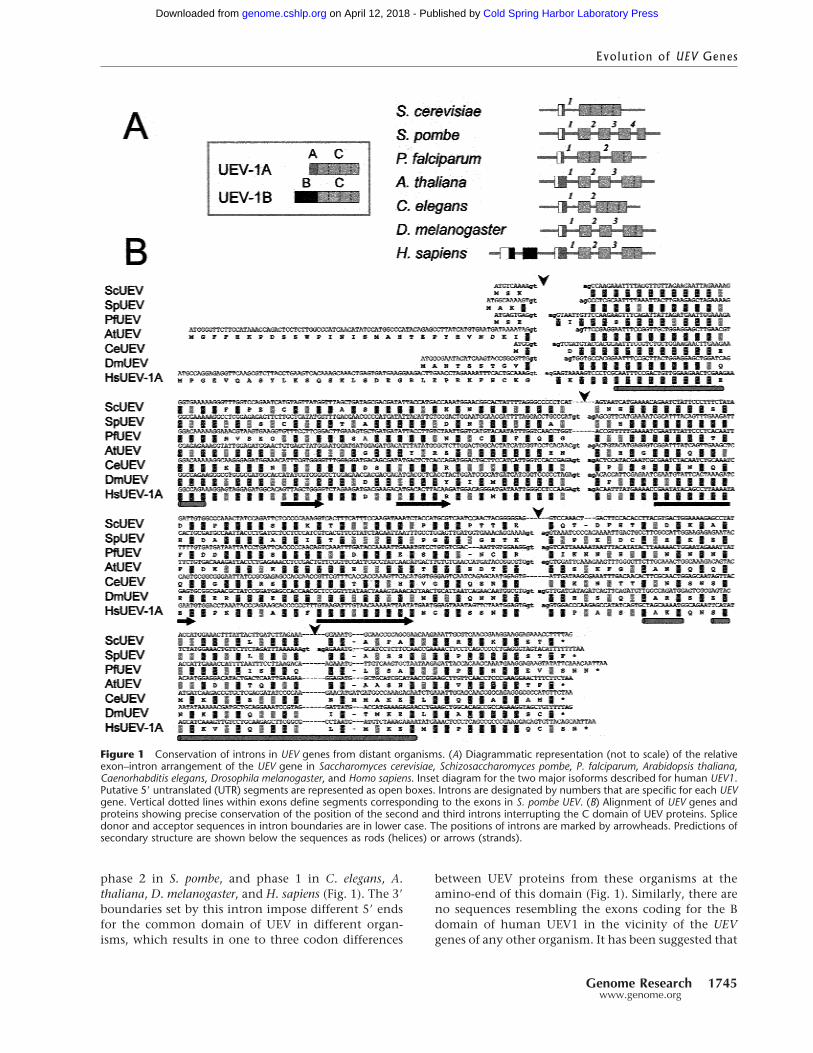
A New Gene in C. elegans and D. melanogaster Codingfor a Protein Containing a UEV1 BDomain-Like SequenceA search for B domain-like sequences in genomic DNAdatabases yielded two small fragments of significantsimilarity within C. elegans clone Y53C10 and D. me-lanogaster clone DS00863 (Fig. 2A). In the C. elegansgenome, the segment included in Y53C10 is in chro-mosome 1, ∼2.5 Mb away from the location of the UEVgene in clone F39B2, also in chromosome 1 (http://www.sanger.ac.uk). We have found no evidence forUEV-like sequences within C. elegans Y53C10 (86 Kb)or D. melanogaster DS00863 (78 Kb) genomic clones.Therefore, in contrast to the B domain sequences in H.sapiens, the B domain-like sequences in C. elegans andD. melanogaster do not appear to be part of the corre-sponding UEV genes.
We hypothesized that, in C. elegans and D. mela-nogaster, these sequences belonged to a second gene,unrelated to UEV. Mapping of known cDNAs and ESTsonto the genomic sequence indicated the presence ofknown genes upstream from position 17,000 anddownstream from position 29,000 in Y53C10, and up-stream from position 54,500 and downstream from po-sition 62,400 in DS00863 (data not shown). ReciprocalTBLASTX searches (Altschul et al. 1990) and dot-plotanalyses between the segments in Y53C10 andDS00863 that lie within these positions (denoted hereY53C10-B and DS00863-B) indicated that they sharedother conserved segments in the vicinity of the B do-main-like sequence, and delineated a tentative exonicstructure for a gene in Y53C10-B and DS00863-B (Fig.2B). The exonic structure of the putative gene inY53C10-B was further refined by means of stringentalignments with ESTs from a nonredundant database,and computational gene identification programs (Fig.2C). A recent addition to GenBank of a C. elegans EST(accession no. AV182903) provided strong support forthis analysis, and a confirmation of the existence of agene in this region. Thus, we predict in C. elegansY53C10-B a new gene consisting of seven exons, withthe potential to encode a 319-amino acid protein (Fig.2D). This prediction is compatible with that madeavailable at EMBL for AL033536, a genomic contig thatincludes Y53C10.
Similar procedures were used to refine the exonicstructure of the putative new gene containing a B do-main-like sequence gene in DS00863-B. Analysis ofDS00863-B with gene identification algorithms pro-duced a consistent gene structure, compatible with thestructure delinated by the regions conserved with C.
elegans Y53C10-B (Fig. 2E). This tentative structure wassupported by a number of ESTs with distant, but sig-nificative, similarity to sequence segments inDS00863-B (Fig. 2E). The exonic structure thus pre-dicted was highly compatible with that delineated bythe conserved sequence segments shared withY53C10-B for the 3� end of the putative gene. The finalanalysis resulted in a five-exon gene, with the potentialto encode a 326-amino acid protein (Fig. 2F).
The C. elegans EST confirmed that the gene pre-dicted is indeed expressed. However, evidence of thiskind was lacking for the D. melanogaster gene. There-fore, we designed primers specific for the exons pre-dicted for the D. melanogaster gene, for use in RT–PCRreactions. All sets of primers yielded specific amplifica-tion products from adult and larval RNA (Fig. 2G). Se-quencing of the products showed that they correspondto processed RNA, formed by joining of the predictedexons, of which exons 4 and 5 are the B domain-likesegments (Fig 2F). These experiments confirmed theexon structure for the new the gene, as predicted bycomputational methods. We have given the name Kuato this new gene (see Acknowledgments).
Clone DS00863, harboring the Drosophila Kuagene, is localized to segment 38B2–38C1, on chromo-some 2 (Hartl et al. 1994; http://flybase.bio.indi-ana.edu/). There is no experimental data for the cyto-genetic localization of the Drosophila UEV gene. There-fore, we performed in situ hybridization on polytenechromosomes, using a probe specific for DrosophilaUEV. This permitted the cytogenetic assignment forthe D. melanogaster UEV gene to 64D, on chromosome3 (Fig. 2H). With posteriority to this analysis, the se-quence of the D. melanogaster genome was released,confirming our assignment of D. melanogaster UEV to64D (Adams et al. 2000).
In conclusion, in both C. elegans and in D. mela-nogaster, the “B domain”-like sequences contained inclones Y53C10 and DS00863, respectively, correspondto exons located within a new gene, Kua, expressed inworms and in flies. In both organisms, the genes Kuaand UEV are in widely separated loci.
H. sapiens: Fusion of UEV1 with KuaThe entire human UEV1 gene, including exons codingfor the B domain, is contained within the genomicPAC clone dJ1185N5 from chromosome 20. Conservedsegments spanning a 32-Kb region within this clonewere found to correspond to most of D. melanogasterand C. elegans Kua. Identical or strongly related mouseand human EST matches fully covered this region.This, together with the application of gene predictionprograms allowed us to predict a six-exon human Kuagene (Fig. 3A) with the potential to code for a 270-amino acid protein. Exon 1 is predicted to contain a 5�
untranslated region (UTR) of unknown size, and exon
Thomson et al.
1746 Genome Researchwww.genome.org
Cold Spring Harbor Laboratory Press on April 12, 2018 - Published by genome.cshlp.orgDownloaded from
Figure 2 A new gene in Caenorhabditus elegans and Drosophila melanogaster coding for B domain-like sequences. (A) Alignment of thehuman UEV1 B domain with TBLASTN-identified segments from C. elegans Y53C10 and D. melanogaster DS00863 genomic clones. Thealigned segments are discontinuous in Y53C10, as indicated by the positions of nucleotides from the database entry. (B) Dot-plot analysisof reciprocal TBLASTX comparisons of genomic clones Y53C10-B (C. elegans) and DS00863-B (D. melanogaster). (Bottom) Scores ofsequence identities for each diagonal of conserved segments, shown as horizontal bars. (C) Predicted structure of the new gene in C.elegans (middle) supported by gene prediction algorithms (top) and alignment with ESTs (bottom). (D) Predicted C. elegans proteincontaining a B domain-like segment (shaded). (E) Predicted structure of the new gene in D. melanogaster (middle), supported by geneprediction algorithms (top) and alignment with ESTs (bottom). (F) Predicted D. melanogaster protein containing a B domain-like segment(shaded). (G) Expression analysis by RT–PCR, with forward primers corresponding to exons 1, 2, 3, and 4 of the predicted gene, and areverse primer corresponding to exon 5, using as templates embryo mRNAs. (H) Cytogenetic assignment of D. melanogaster UEV geneto chromosome 3 segment 64D. Digoxigenin-labeled cDNA probes were used for hybridization on wild-type Drosophila polytenechromosomes.
Genome Research 1747www.genome.org
Cold Spring Harbor Laboratory Press on April 12, 2018 - Published by genome.cshlp.orgDownloaded from
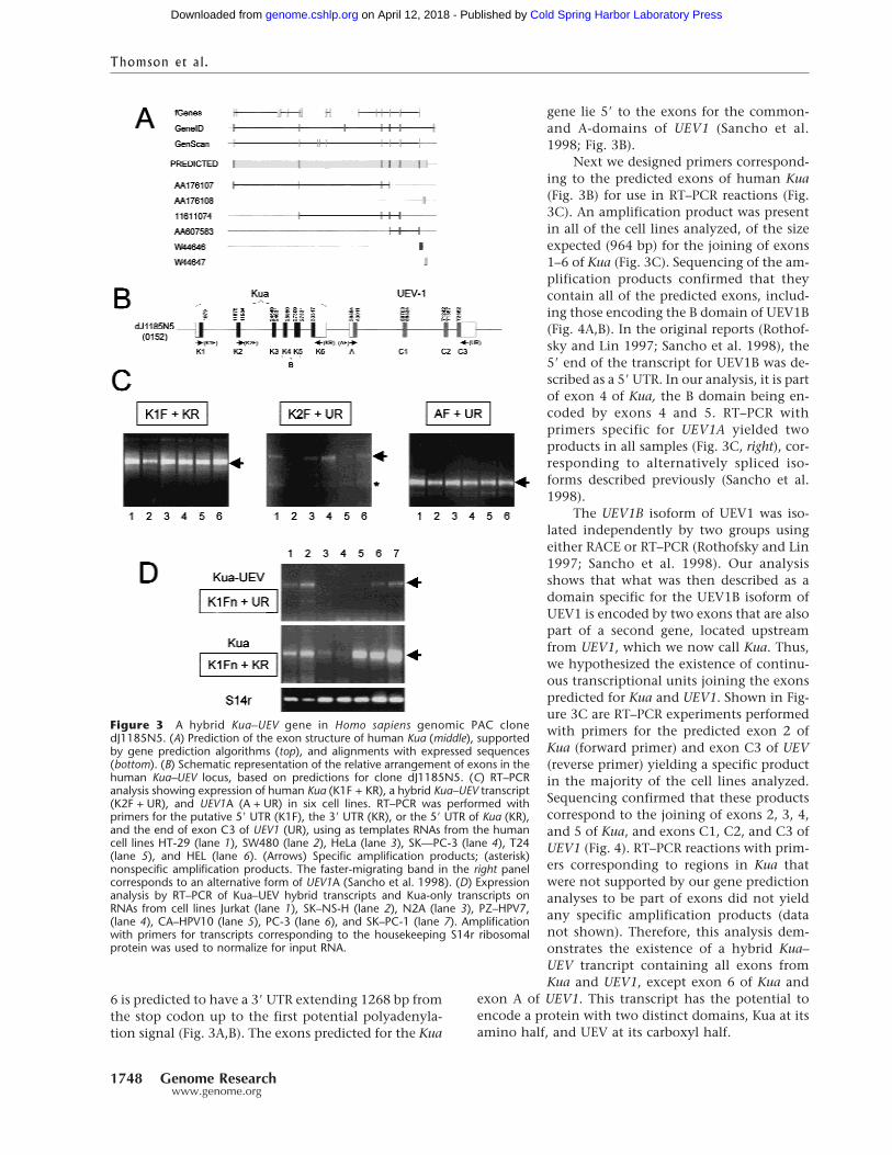
6 is predicted to have a 3� UTR extending 1268 bp fromthe stop codon up to the first potential polyadenyla-tion signal (Fig. 3A,B). The exons predicted for the Kua
gene lie 5� to the exons for the common-and A-domains of UEV1 (Sancho et al.1998; Fig. 3B).
Next we designed primers correspond-ing to the predicted exons of human Kua(Fig. 3B) for use in RT–PCR reactions (Fig.3C). An amplification product was presentin all of the cell lines analyzed, of the sizeexpected (964 bp) for the joining of exons1–6 of Kua (Fig. 3C). Sequencing of the am-plification products confirmed that theycontain all of the predicted exons, includ-ing those encoding the B domain of UEV1B(Fig. 4A,B). In the original reports (Rothof-sky and Lin 1997; Sancho et al. 1998), the5� end of the transcript for UEV1B was de-scribed as a 5� UTR. In our analysis, it is partof exon 4 of Kua, the B domain being en-coded by exons 4 and 5. RT–PCR withprimers specific for UEV1A yielded twoproducts in all samples (Fig. 3C, right), cor-responding to alternatively spliced iso-forms described previously (Sancho et al.1998).
The UEV1B isoform of UEV1 was iso-lated independently by two groups usingeither RACE or RT–PCR (Rothofsky and Lin1997; Sancho et al. 1998). Our analysisshows that what was then described as adomain specific for the UEV1B isoform ofUEV1 is encoded by two exons that are alsopart of a second gene, located upstreamfrom UEV1, which we now call Kua. Thus,we hypothesized the existence of continu-ous transcriptional units joining the exonspredicted for Kua and UEV1. Shown in Fig-ure 3C are RT–PCR experiments performedwith primers for the predicted exon 2 ofKua (forward primer) and exon C3 of UEV(reverse primer) yielding a specific productin the majority of the cell lines analyzed.Sequencing confirmed that these productscorrespond to the joining of exons 2, 3, 4,and 5 of Kua, and exons C1, C2, and C3 ofUEV1 (Fig. 4). RT–PCR reactions with prim-ers corresponding to regions in Kua thatwere not supported by our gene predictionanalyses to be part of exons did not yieldany specific amplification products (datanot shown). Therefore, this analysis dem-onstrates the existence of a hybrid Kua–UEV trancript containing all exons fromKua and UEV1, except exon 6 of Kua and
exon A of UEV1. This transcript has the potential toencode a protein with two distinct domains, Kua at itsamino half, and UEV at its carboxyl half.
Figure 3 A hybrid Kua–UEV gene in Homo sapiens genomic PAC clonedJ1185N5. (A) Prediction of the exon structure of human Kua (middle), supportedby gene prediction algorithms (top), and alignments with expressed sequences(bottom). (B) Schematic representation of the relative arrangement of exons in thehuman Kua–UEV locus, based on predictions for clone dJ1185N5. (C) RT–PCRanalysis showing expression of human Kua (K1F + KR), a hybrid Kua–UEV transcript(K2F + UR), and UEV1A (A + UR) in six cell lines. RT–PCR was performed withprimers for the putative 5� UTR (K1F), the 3� UTR (KR), or the 5� UTR of Kua (KR),and the end of exon C3 of UEV1 (UR), using as templates RNAs from the humancell lines HT-29 (lane 1), SW480 (lane 2), HeLa (lane 3), SK—PC-3 (lane 4), T24(lane 5), and HEL (lane 6). (Arrows) Specific amplification products; (asterisk)nonspecific amplification products. The faster-migrating band in the right panelcorresponds to an alternative form of UEV1A (Sancho et al. 1998). (D) Expressionanalysis by RT–PCR of Kua–UEV hybrid transcripts and Kua-only transcripts onRNAs from cell lines Jurkat (lane 1), SK–NS-H (lane 2), N2A (lane 3), PZ–HPV7,(lane 4), CA–HPV10 (lane 5), PC-3 (lane 6), and SK–PC-1 (lane 7). Amplificationwith primers for transcripts corresponding to the housekeeping S14r ribosomalprotein was used to normalize for input RNA.
Thomson et al.
1748 Genome Researchwww.genome.org
Cold Spring Harbor Laboratory Press on April 12, 2018 - Published by genome.cshlp.orgDownloaded from

To determine the relative levels of Kua and Kua–UEV transcripts, RT–PCR was performed under nonsat-urating conditions on RNAs from seven different celllines. For normalization, amplification was performedwith primers for transcripts for the ribosomal proteinS14r. In these analyses, the ratio of Kua–UEV to Kuaamplification products ranged from 0.1 (samples 1 and2) to 0.02 (samples 5 and 7), in those samples withvisible Kua–UEV amplification products (Fig. 3D). Twosamples did not yield measurable levels of Kua–UEVhybrid amplification products under these conditions
(30 cycles), although products were observed with 35amplification cycles or more (data not shown).
Kua: A New Family of Conserved ProteinsSeveral organisms express Kua transcripts, including H.sapiens, Mus musculus, D. melanogaster, C. elegans, Try-panosoma cruzi, each of which was predicted to expressone Kua protein, and the plant A. thaliana, in which wepredict three different Kua proteins corresponding todistinct genes (Fig. 5A). The three animal Kua proteinsshow identities of 94%, 67%, and 61% with respect tothe human protein (comparison with the sequence be-tween positions 83 and 238 of the human protein),whereas the three plant Kua proteins, strongly relatedto each other, show 29%–34% identities with respectto the human protein. Although yeasts have UEV pro-teins (Broomfield et al. 1998; Sancho et al. 1998), theydo not have potential Kua proteins.
A pattern of histidine residues conserved in all Kuaproteins is reminiscent of certain histidine-rich motifsin which specific residues are coordinated by transitionmetals (Shanklin et al. 1994). Membrane-bound fattyacid desaturases and hydroxylases contain several his-tidine-rich motifs, with the general pattern H-x(2,3)-(x-H)-H (Fox et al. 1993; Mitchell and Martin 1997).Kua proteins contain two of these motifs (Fig. 5A). Al-though all Kua proteins contain fatty acid hydroxylase-like His-rich motifs, only plant Kua proteins show amarginal degree of homology (E values in the order of10�2) to fatty acid hydroxylases within the segment ofthe protein containing such motifs. Nevertheless, aPHI–BLAST search of a nonredundant database with asequence profile generated based on the consensus se-quence for Kua proteins yielded bonafide fatty acid hy-droxylases (Fig. 5B).
In fatty acid hydroxylases and desaturases, theprotein segments containing these motifs are locatedon the cytoplasmic face of the ER membrane, such thatthe relevant histidine residues become closely apposedfor the coordination of a di-iron cluster (Mitchell andMartin 1997). A prediction of trasmembrane domainsfor the Kua proteins indicates an overall topology simi-lar to that of fatty acid hydroxylases (Mitchell and Mar-tin 1997) (Fig. 5C, left panel), especially when compar-ing the transmembrane prediction for the plant Kuaproteins (e.g., F19K23.12) with that for A. thalianaFah1 (Fig. 5C, left panel). The positions of the histi-dine-rich motifs relative to the predicted transmem-brane domains of Kua proteins and fatty acid hydroxy-lases are diagrammatically represented in Figure 5C(right panel).
Subcellular Localization of UEV1A, Kua,and Kua–UEVA partial UEV1 protein has been reported to localize tothe nucleus of mammalian cells (Rothofsky and Lin
Figure 4 Sequences of human Kua and Kua–UEV cDNAs andpredicted proteins. (A) Sequence shared by Kua and Kua–UEVtranscripts. (B) Sequence specific for Kua-only transcripts. Thissequence is continuous with A in Kua-only transcripts, and cor-responds to exon K6 (Fig. 3). (C) UEV C domain sequence fromKua–UEV transcripts. This sequence is continuous with A in Kua–UEV transcripts, and results from joining exons C1, C2, and C3(Fig. 3B). Exon boundaries, as predicted in Fig. 3, are marked byvertical bars. The sequences of primers used for RT–PCR in Fig. 3are boxed. The B domain is shaded.
Evolution of UEV Genes
Genome Research 1749www.genome.org
Cold Spring Harbor Laboratory Press on April 12, 2018 - Published by genome.cshlp.orgDownloaded from
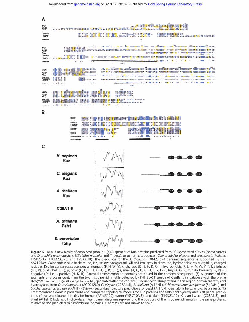
Figure 5 Kua, a new family of conserved proteins. (A) Alignment of Kua proteins predicted from PCR-generated cDNAs (Homo sapiensand Drosophila melanogaster), ESTs (Mus musculus and T. cruzi), or genomic sequences (Caenorhabditis elegans and Arabidopsis thaliana,F19K23.12, F10M23.370, and T20K9.10). The prediction for the A. thaliana F10M23.370 genomic sequence is supported by ESTAA712589. Color codes: blue background, His; yellow background, Gli and Pro; grey background, hydrophobic residues; blue, chargedresidues. Key for consensus sequences: a, aromatic (F, H, W, Y); c, charged (D, E, H, K, R); h, hydrophobic (F, L, M, V, W, Y, I); l, aliphatic(I, L, V); o, alcohol (S, T); p, polar (C, D, E, H, K, N, Q, R, S, T); s, small (A, C, D, G, N, P, S, T); u, tiny (A, G, S); x, helix breaking (G, P);�,negative (D, E); +, positive (H, K, R). Potential transmembrane domains are boxed in the consensus sequence. (B) Alignment of thesegments of proteins containing the two histidine-rich motifs detected by PHI–BLAST search of GenBank nr database with the profileH-x-(YWF)-x-H-x(8,25)-(RK)-x(2)-H-x(2)-H-H, generated after the consensus sequence for Kua proteins in this region. Shown are fatty acidhydroxylases from D. melanogaster (AC004280) C. elegans (C25A1.5), A. thaliana (AtFAH1), Schizosaccharomyces pombe (SpFAH1) andSaccharomyces cerevisiae (ScFAH1). (Bottom) Secondary structure predictions for yeast FAH (cylinders, alpha helix; arrow, beta sheet). (C)Transmembrane domain predictions and compared topological models for Kua proteins and fatty acid hydroxylases. Left panel, predic-tions of transmembrane domains for human (AF155120), worm (Y53C10A.5), and plant (F19K23.12), Kua and worm (C25A1.5), andplant (At Fah1) fatty acid hydroxylases. Right panel, diagrams representing the positions of the histidine-rich motifs in the same proteins,relative to the predicted transmembrane domains. Diagrams are not drawn to scale.
Cold Spring Harbor Laboratory Press on April 12, 2018 - Published by genome.cshlp.orgDownloaded from
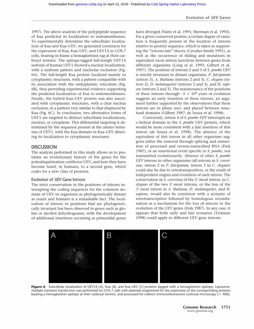
1997). The above analysis of the polypeptide sequenceof Kua predicted its localization to endomembranes.To experimentally determine the subcellular localiza-tion of Kua and Kua–UEV, we generated constructs forthe expression of Kua, Kua–UEV, and UEV1A in COS-7cells, bearing in-frame a hemagluttinin tag at their car-boxyl termini. The epitope-tagged full-lentgh UEV1Aisoform of human UEV1 showed a nuclear localization,with a uniform pattern and nucleolar exclusion (Fig.6A). The full-length Kua protein localized mainly tocytoplasmic structures, with a pattern compatible withits association with the endoplasmic reticulum (Fig.6B), thus providing experimental evidence supportingthe predicted localization of Kua to endomembranes.Finally, the hybrid Kua–UEV protein was also associ-ated with cytoplasmic structures, with a clear nuclearexclusion, in a pattern very similar to that displayed byKua (Fig. 6C). In conclusion, two alternative forms ofUEV1 are targeted to distinct subcellular localizations,nucleus, or cytoplasm. This differential targeting is de-termined by the sequence present at the amino termi-nus of UEV1, with the Kua domain in Kua–UEV direct-ing its localization to cytoplasmic structures.
DISCUSSIONThe analysis performed in this study allows us to pos-tulate an evolutionary history of the genes for thepolyubiquitination coeffector UEV, and how they havebecome fused, in humans, to a second gene, whichcodes for a new class of proteins.
Evolution of UEV Gene IntronsThe strict conservation in the positions of introns in-terrupting the coding sequences for the common do-main of UEV in organisms as phylogenetically distantas yeasts and humans is a remarkable fact. The local-ization of introns in positions that are phylogeneti-cally invariant has been observed in genes such as glo-bin or alcohol dehydrogenase, with the developmentof additional insertions occurring as primordial genes
have diverged (Naito et al. 1991; Sherman et al. 1992).For a given conserved protein, a certain degree of varia-tion is frequently present in the location of intronsrelative to protein sequence, which is taken as support-ing the “introns-late” theory (Cavalier-Smith 1991), aswell as the occurrence of sliding and mutations inequivalent exon–intron junctions between genes fromdifferent organisms (Long et al. 1995; Gilbert et al.1997). The position of introns 2 and 3 of S. pombe UEVis strictly invariant in distant organisms, P. falciparum(intron 3), A. thaliana (introns 2 and 3), C. elegans (in-tron 2), D. melanogaster (introns 2 and 3), and H. sapi-ens (introns 2 and 3). The maintenance of the positionsof these introns through ∼5 � 108 years of evolutionsuggests an early insertion of these introns, an argu-ment further supported by the observations that theseintrons are in phase zero, and placed between struc-tural domains (Gilbert 1987; de Souza et al. 1998).
Conversely, intron 4 of S. pombe UEV interrupts an�-helical domain in the S. pombe UEV protein, whichwould be more consistent with a late insertion of thisintron (de Souza et al. 1998). The absence of theequivalent of this intron in all other organisms sug-gests either the removal through splicing and reinser-tion of processed and reverse-transcribed RNA (Fink1987), or an insertional event specific to S. pombe, nottransmitted evolutionarily. Absence of other S. pombeUEV introns in other organisms (all introns in S. cerevi-siae, intron 2 in P. falciparum, intron 3 in C. elegans)could also be due to retrotransposition, or the result ofindependent origins and evolution of each intron. Theconservation in S. cerevisiae of the 5�-most intron, in C.elegans of the two 5�-most introns, or the loss of the3�-most intron in A. thaliana, D. melanogaster, and H.sapiens, would also be consistent with a scenario ofretrotranscription followed by homologous recombi-nation as a mechanism for the loss of introns in theevolution of the UEV genes (Fink 1987). In any case, itappears that both early and late scenarios (Trotman1998) could apply to different UEV gene introns.
Figure 6 Subcellular localization of UEV1A (A), Kua (B), and Kua–UEV (C) proteins tagged with a hemagluttinin epitope. Liposome-mediate transient transfection was performed on COS-7 cells with plasmids engineered for the expression of the corresponding proteinsbearing a hemagluttinin epitope at their carboxyl termini, and processed for indirect immunofluorescent confocal microscopy (� 400).
Evolution of UEV Genes
Genome Research 1751www.genome.org
Cold Spring Harbor Laboratory Press on April 12, 2018 - Published by genome.cshlp.orgDownloaded from
The analysis of intron 1 of the UEV genes suggeststhat it originated as a result of a process distinct fromthe ones discussed above for the introns that interruptthe common domain. Intron 1 is inserted in differentphases in different organisms, and it generates differ-ent carboxyl- and amino-ends in the flanking exons.Also, exon 1 (exon A in humans), placed 5� to thisintron, is different for all organisms. This suggests thatexon 1 evolved separately from the rest of the UEVgene.
Fusion of the Kua and UEV GenesWe also show that, in humans, one of the two UEVgenes in this organism, UEV1, is adjacent to an unre-lated gene, which we have named Kua, and can beexpressed as a hybrid Kua–UEV transcript and protein.In contrast, in D. melanogaster and C. elegans, Kua andUEV are independent genes, coding for separate tran-scripts and proteins. Therefore, the combination ofcomputational and experimental approaches used hereshow how two genes, which are unrelated and locatedin separate loci in worms and insects, converge into ahybrid gene and protein in humans. The identificationof Kua in C. elegans and D. melanogaster was possiblewithout any prior knowledge of expressed sequences,applying ab initio biocomputational methods on ge-nomic sequences. Of these methods, the use of dot-plot analyses of reciprocal TBLASTX alignments wassufficient to infer the structure of Kua in both organ-isms, later confirmed with alignments with ESTs andexperimental data. Therefore, this could be a very use-ful tool for the identification of genes when expressiondata are not available.
New genes are thought to originate through eventssuch as gene duplications (Ohno 1970; Ohta 1989),exon shuffling (Gilbert 1978), or the generation of pro-cessed genes (McCarrey and Thomas 1987). In metazo-ans, fusion of genes generally involves a number ofintermediate processes, such as duplication and shuf-fling of exons (Bazan et al. 1989; Simmer et al. 1990;Chen et al. 1997; Coppock et al. 1998). Secondaryevents, such as retrotransposition with exon capture(Long and Langley 1993) or genetic hitchhiking asso-ciated with selective sweeps can generate chimericgenes (Nurminsky et al. 1998; Long et al. 1999). Theseevents often involve extensive refashioning of codingand noncoding regions (Nurminsky et al. 1998). TheKua–UEV fusion does not appear to involve such pro-miscuous changes, and it rather suggests the occur-rence of a direct fusion of loci. The likely scenario inthe Kua–UEV fusion would be a two-step process, du-plication of the UEV gene, followed by fusion of theduplicated gene to Kua. This model is supported by thefact that, in humans, there are two UEV genes, ofwhich the UEV1 gene is fused to Kua in chromosome20, whereas the UEV2 gene is on chromosome 8, with-
out any evidence for this type of fusion (B. Cormandand T.M. Thomson, unpubl.). Therefore, UEV2 wouldcorrespond to the gene in the original locus, and UEV1to the duplicated gene, which would undergo subse-quent rearrangement with a head-to-tail fusion to Kua.
The generation of three different classes of tran-scripts from the Kua–UEV locus represents a uniquestrategy aimed at the modular expression of two genes,coding either for two separate polypeptides, or as acombination of both to yield a single two-domainpolypeptide. The generation of a Kua–UEV hybrid tran-script could be the result either of cis-splicing directedby canonical splice sites, or trans-splicing. Trans-splicing has been shown to occur in mammalian cells,either in artificial (Bruzik and Maniatis 1992) or natu-ral (Caudevilla et al. 1998; Kingzette et al. 1998; Ako-pian et al. 1999; Li et al. 1999; Zaphiropoulos 1999)settings. Although trans-splicing in mammalian cellsusually occurs between transcripts from genes in sepa-rate loci or chromosomes, it has been reported to occuralso between transcripts from clustered genes (Zaphi-ropoulos 1999). It has also been shown that both cis-and trans-splicing can be concomitant mechanisms forthe generation of hybrid transcripts from the samegenes in mammalian cells (Eul et al. 1995). Our obser-vations do not provide sufficient information to inferthe splicing mechanism prevalent in the generation ofhybrid Kua–UEV transcripts. However, there are indi-rect arguments against trans-splicing as the majormechanism for the generation of these transcripts.First, trans-splicing in mammalian transcripts appearsto be regulated by sequences at the acceptor exon, withthe consensus GAAGAAG(G/C) (Caudevilla et al.1998). Sequences fully compatible with this consensusare present in exon C1 of UEV1, and also at equivalentpositions in exon C1 of UEV2, but only UEV1, and notUEV2, is involved in hybrid Kua–UEV transcripts (T.M.Thomson, unpubl.). A more speculative argumentwould be based on the teleological nature of the fusionof Kua and UEV from the standpoint of the evolutionof these genes. The driving force for the evolutionaryrearrangement and fusion of Kua and UEV into a singlelocus would be stronger for cis-splicing being a majormechanism for the generation of hybrid transcriptsthan it would be for a trans-splicing mechanism.
The close proximity of Kua to UEV1 could raise thequestion whether the mere juxtaposition of two geneswith the same transcriptional direction is sufficient forthe generation of detectable run-off transcription fromthe upstream gene. To test whether this is a commonsituation, we have performed a survey of all genes onhuman chromosome 22 with a distance between genesof �25 Kb. Of 546 genes annotated on this chromo-some, 221 correspond to pairs that are within a dis-tance of �25 Kb and with the same transcriptional di-rection. Five of these pairs correspond to overlapping
Thomson et al.
1752 Genome Researchwww.genome.org
Cold Spring Harbor Laboratory Press on April 12, 2018 - Published by genome.cshlp.orgDownloaded from
genes. Of the remaining 216 gene pairs, BLASTNsearches of EST databases have identified two withtranscripts matching both genes in the pair, that couldcorrespond to transcripts bridging the two genes. Forone of these two gene pairs, PNUTL1 and GP1BB, ex-perimental evidence for the existence of hybrid tran-scripts, as well as single-gene transcripts, has been re-ported (Zieger et al. 1997; Yagi et al. 1998). The secondgene pair with a potential hybrid transcript has notbeen characterized, and corresponds to genes predictedfor a hypothetical protein (transcript dJ1194E15.3) andan EST cluster (dJ1104E15.5). The PNUTL1–GP1BB fu-sion transcript is predicted to contain two open read-ing frames (ORFs), and appears to result from defectivetruncation of the upstream PNUTL1 transcript due toan imperfect polyadenylation signal sequence (Ziegeret al. 1997). Therefore, the approach used in this analy-sis can detect potential gene-bridging hybrid tran-scripts in 1% of the gene pairs analyzed. This is prob-ably an underestimate of all instances of gene fusionsexpressing hybrid transcripts, because not all fusedgene pairs will be represented by ESTs matching bothgenes in expression databases. Also, some of the anno-tated genes could have been misrepresented as singlegenes, especially if they have been predicted on thebasis of matching ESTs. One conclusion of this type ofanalysis, relevant to the present study, is that transcriptfusions between two adjacent genes, although not in-frequent, are observed only in a subset of closely asso-ciated gene pairs.
In contrast to the Kua–UEV fusion, the PNUTL1–GP1BB gene fusion does not result in a fusion of pro-teins (Zieger et al. 1997). A second difference is that themature PNUTL1–BP1BB fusion includes the terminalexon from the upstream gene, with its transcriptionaltruncation signal. Because this signal is apparently in-efficient, the PNUTL1–BP1BB hybrid transcript couldbe the consequence of a genuine transcriptional run-off. In contrast, the mature processed transcripts of theKua–UEV fusion have spliced out the terminal exon ofthe upstream gene, Kua, as well as the first exon of thedownstream gene, UEV1A, which contains a 5� un-translated sequence. RT–PCR experiments aimed at de-tecting fused transcripts that contain these two exonshave failed to yield any amplification products. In theKua–UEV fusion, therefore, a continuous primary tran-script between both genes is subjected to specific splic-ing events that allow the expression of a two-domainprotein. This also implies that a truncation of tran-scripts at the terminal exon of Kua must proceed at arate that is sufficiently slow to allow the subsequentsplicing events for the maturation of the Kua–UEVtranscript. A carefully orchestrated balance betweensplicing and truncation of transcripts has been shownto occur in lower organisms (Ull et al. 1993).
A summary of the different classes of transcripts
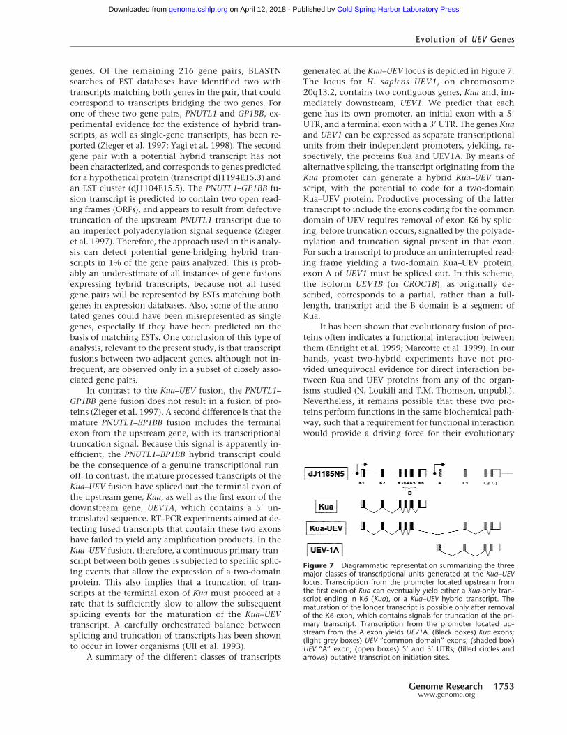
generated at the Kua–UEV locus is depicted in Figure 7.The locus for H. sapiens UEV1, on chromosome20q13.2, contains two contiguous genes, Kua and, im-mediately downstream, UEV1. We predict that eachgene has its own promoter, an initial exon with a 5�
UTR, and a terminal exon with a 3� UTR. The genes Kuaand UEV1 can be expressed as separate transcriptionalunits from their independent promoters, yielding, re-spectively, the proteins Kua and UEV1A. By means ofalternative splicing, the transcript originating from theKua promoter can generate a hybrid Kua–UEV tran-script, with the potential to code for a two-domainKua–UEV protein. Productive processing of the lattertranscript to include the exons coding for the commondomain of UEV requires removal of exon K6 by splic-ing, before truncation occurs, signalled by the polyade-nylation and truncation signal present in that exon.For such a transcript to produce an uninterrupted read-ing frame yielding a two-domain Kua–UEV protein,exon A of UEV1 must be spliced out. In this scheme,the isoform UEV1B (or CROC1B), as originally de-scribed, corresponds to a partial, rather than a full-length, transcript and the B domain is a segment ofKua.
It has been shown that evolutionary fusion of pro-teins often indicates a functional interaction betweenthem (Enright et al. 1999; Marcotte et al. 1999). In ourhands, yeast two-hybrid experiments have not pro-vided unequivocal evidence for direct interaction be-tween Kua and UEV proteins from any of the organ-isms studied (N. Loukili and T.M. Thomson, unpubl.).Nevertheless, it remains possible that these two pro-teins perform functions in the same biochemical path-way, such that a requirement for functional interactionwould provide a driving force for their evolutionary
Figure 7 Diagrammatic representation summarizing the threemajor classes of transcriptional units generated at the Kua–UEVlocus. Transcription from the promoter located upstream fromthe first exon of Kua can eventually yield either a Kua-only tran-script ending in K6 (Kua), or a Kua–UEV hybrid transcript. Thematuration of the longer transcript is possible only after removalof the K6 exon, which contains signals for truncation of the pri-mary transcript. Transcription from the promoter located up-stream from the A exon yields UEV1A. (Black boxes) Kua exons;(light grey boxes) UEV “common domain” exons; (shaded box)UEV “A” exon; (open boxes) 5� and 3� UTRs; (filled circles andarrows) putative transcription initiation sites.
Evolution of UEV Genes
Genome Research 1753www.genome.org
Cold Spring Harbor Laboratory Press on April 12, 2018 - Published by genome.cshlp.orgDownloaded from
fusion. One of the functional consequences of the fu-sion of Kua with UEV in humans is that the domainregulating polyubiquitination is redirected for localiza-tion to cytoplasmic structures, rather than the nucleus.As a consequence of their different subcellular localiza-tions, nuclear (UEV1A) and cytoplasmic (Kua–UEV)forms of UEV1 could target different substrates for vari-ant (K63) polyubiquitination. Thus, an endomem-brane-associated form of UEV1 could preferentially di-rect the variant polyubiquitination of substratesclosely associated with the cytoplasmic face of the ER,possibly, although not necessarily, in conjunctionwith membrane-bound ubiquitin-conjugating en-zymes (Sommer and Jentsch 1993). The substratescould be either proteins specifically targeted for ubiq-uitination by these UEV–E2 complexes, or misfoldedER-associated proteins that are dislocated into thecytoplasm for subsequent ubiquitination (Kopito1997).
METHODS
Biocomputational AnalysisPutative conserved coding regions between genomic se-quences were identified with reciprocal TBLASTX (Altschul etal. 1990), the output processed with MSPCrunch (Sonnham-mer and Durbin 1994), and conserved segments visualizedwith aplot (http://www1.imim.es/∼jabril/GFFTOOLS/APLOT.html). For prediction of protein coding genes, threedifferent ab initio gene prediction programs were used, Gen-scan (Burge and Karlin 1997), Geneid (Guigo et al. 1992), andFgenes (Solovyev et al. 1994). To support the resulting exonicstructures, BLASTN searches were performed against the ESTdivision of GenBank (Benson et al. 2000). A minimum subsetof ESTs covering the matches was selected, all other ESTmatches being either identical or included therein. Each ofthese ESTs was aligned with the matching genomic clone se-quence using the program est_genome (Mott 1997). Putativecoding domains were recorded using the gff format (http://www.sanger.ac.uk/Software/formats/GFF/GFF_Spec.shtml),and plotted with gff2ps (available at http://www1.imim.es/∼jabril/GFFTOOLS/GFF2PS.html). Protein sequences werealigned with ClustalW (Thompson et al. 1994), and secondarystructures predicted with the PHD package (Rost 1996). Trans-membrane domains were predicted with TMpred (http://www.ch.embnet.org/software/TMPRED_form.html). Searchesfor distant homologies were performed with PSI–BLAST(Altschul et al. 1997).
Expression Analysis by RT–PCRRNAs from human cell lines and mouse tissues were isolatedby the acid phenol procedure (Chomczynski and Sacchi1987). RNAs from D. melanogaster embryos were isolated byguanidium isothiocyanate extraction and CsCl gradients, andenriched for mRNA on oligo dT-cellulose columns (Sambrooket al. 1989). RNAs were resuspended in diethyl pyrocarbonate-treated H2O, and 20 µg were treated with 1 unit RNase-freeRQ1 DNase I (Promega, Madison, WI), in a reaction contain-ing 10 mM Tris-HCl at pH 8.0, 5 mM MgCl2, and 40 unitsRNasin (Promega). For Drosophila samples, RT–PCR was doneby a single-tube procedure (Life Technologies, Barcelona),
with forward primers 1 (5�-AATGACATCAACGAACGTC-3�), 2(5�-CTTAGTTTCGACTTCTCCGCGAT-3�), 3 (5�-CCTGTGCGGCATTATAACGG-3�), or 4 (5�-TGGCATACCAATTCTCGGCTA-3�), and reverse primer 5�-CGATGATGAGCTCGAATGTTGA-3� (see Fig. 2G). For human samples, a two-step RT–PCR procedure was used, with reverse transcription of 1 µgRNA in a reaction containing 1� first-strand buffer, 200 µMdNTPs, 500 ng oligo-dT(12–18), and 200 units RNase H(�) RT(Life Technologies) at 42°C for 1 h. Aliquots were used astemplates for hot-start PCR in 25-µl reactions containing 1�
buffer, 130 µM dNTPs, 5 pmol of each primer and 0.2 unitsTaq polymerase (Ecogen, Barcelona). Amplification productswere gel purified and aliquots used for nested or seminestedPCR, and products sequenced from both strands by cycle se-quencing and resolution in a ABI Prism 310 automatic se-quencer (Applied Biosystems). Forward primers used for RT–PCR and nested PCR were K1F (5�-GTCATTGGGCGTGATCT-3�) or KF1n (5�-GAGCTGGACGAGGACGAG-3�) for exon 1 ofhuman Kua, K2F (5�-CAGGCTCATCGCCCACAACC-3�) forexon 2, and K4F (5�-ATGGCCTACAAGTTCCGCACC-3�), forexon 4. Reverse primers were KR (5�-GGCAGATGGCTTCGGTTTGG-3�), for exon 6 of Kua, or UR (5�-CTAAGGGGAGAAGGCAGAGA-3�), for exon C3 of UEV1 (see Fig. 3B).Amplification products were gel purified and sequenced asabove. To determine relative levels of amplification of Kua–UEV and UEV transcripts, RT–PCR products were electropho-resed in ethidium bromide-containing agarose gels, and in-tensities (arbitrary units) determined for specific bands, nor-malized relative to the intensity of RT–PCR amplificationproducts of the same RNAs with primers for the ribosomalprotein gene S14r.
In Situ Hybridization on DrosophilaPolytene ChromosomesDrosophila polytene chromosome spreads were obtained fromthird instar wild-type larvae (Canton S strain) salivary glands.cDNA for D. melanogaster Kua was generated by RT–PCR withspecific primers (primers 1 and reverse; see above and Fig. 2G),in single-tube RT–PCR reactions (Life Sciences) using as a tem-plate mRNA from Drosophila embryos. cDNA for D. melano-gaster UEV corresponded to the insert in IMAGE cloneLD23138 (Research Genetics), cloned in pOT2 (plasmidpOT2/DmUEV). DNA probes were labeled by random-priming(Boehringer-Mannheim). Chromosomes were denatured in70 mM NaOH for 2 min, rinsed in 2� SSC and dehydrated ingraded ethanols. Hybridization was done at 58°C overnight.Biotinylated probes were detected with streptavidin-HRP anddiaminobenzidine (Sigma). Chromosomes were counter-stained with Giemsa (Pardue 1994).
Expression Constructs and TransientTransfection ExperimentsFull-length Kua, Kua–UEV, and UEV1A were amplified by RT–PCR using as a template total RNA from the cell lines HT-29 orJurkat, and Expand High Fidelity polymerase (Boehringer-Mannheim), and the products subcloned in pGEM-T (Pro-mega). The resulting inserts were amplified with primers forsubcloning into pGEM11Z-HA, bearing sequences coding forthe hemagluttining epitope, such that this sequence wasplaced in-frame at the carboxyl termini of the cDNAs. Theresulting HA-tagged cDNAs were subcloned into pcDNA3.1(Invitrogen). For transient transfection, 1 µg of plasmid DNAwas transfected with Lipofectamine Plus (Life Sciences) into
Thomson et al.
1754 Genome Researchwww.genome.org
Cold Spring Harbor Laboratory Press on April 12, 2018 - Published by genome.cshlp.orgDownloaded from
COS-7 cells grown on glass coverslips. As a control, pcDNA3.1vector DNA was used. Twenty-four hours after transfection,cells were washed, fixed in 4% paraformaldehyde/PBS, andpermeabilized with 1% saponin/2% BSA/PBS. Cells were in-cubated with rat monoclonal anti-HA antibody (Boehringer-Mannheim) for 2 h, washed, and further incubated for 1 hwith FITC-conjugated goat anti-rat Ig (Dako), washed,mounted in Immuno-Fluore (ICN), and observed under aLeica confocal microscope (Wetzlar, Germany). Transfectionefficiencies ranged from 10%–15%.
ACKNOWLEDGMENTSWe thank J. Rozas for critically reviewing the manuscript, andC. Harvey for helpful contributions. This work was funded bygrant PB97-1170 of the Ministerio de Educacion y Ciencia (toT.M.T), and supported in part by grants BIO98-0443-C02-01(to R.G.) and PB96-1253 of the MEC (to F.S. and M.C.), andgrant 18/99 from the Fundacion Marques de Valdecilla,Santander, Spain (to J.M.). R.C., M.V., V.M.D., J.A., and M.B.were supported by fellowships from the Fundacion Marquesde Valdecilla, the Fundacio per a la Recerca Vall d’Hebron, theMinisterio de Educacion y Ciencia, the Instituto de Salud Car-los III (99/9345), and the Ministerio de Educacion y Ciencia(FP95-38817943), respectively. The designation Kua is afterthe Catalan word cua, meaning “queue” or “tail”.
The publication costs of this article were defrayed in partby payment of page charges. This article must therefore behereby marked “advertisement” in accordance with 18 USCsection 1734 solely to indicate this fact.
REFERENCESAdams, M.D., Celniker, S.E., Holt, R.A., Evans, C.A., Gocayne, J.D.,
Amanatides, P.G., Scherer, S.E., Li, P.W., Hoskins, R.A., Galle,R.F., et al. 2000. The genome sequence of Drosophilamelanogaster. Science 287: 2185–2195.
Akopian, A.N., Okuse, K., Souslova, V., England, S., Ogata, N., andWood, J.N. 1999. Trans-splicing of a voltage-gated sodiumchannel is regulated by nerve growth factor. FEBS Lett.445: 177–182.
Altschul, S.F., Gish, W., Miller, W., Myers, E.W., and Lipman, D.1990. Basic Local Alignment Search Tool. J. Mol. Biol.215: 403–410.
Altschul, S.F., Madden, T.L., Schaffer, A.A., Zhang, J., Zhang, Z.,Miller, W., and Lipman, D.J. 1997. Gapped BLAST andPSI-BLAST: A new generation of protein database searchprograms. Nucleic Acids Res. 25: 3389–3402.
Bazan, J.F., Fletterick, R.J., and Pilis, S.J. 1989. Evolution of abifunctional enzyme: 6-phosphofructo-2-kinase/fructose-2,6-bisphosphatase. PNAS 86: 9642–9646.
Benson, D.A., Karsch-Mizrachi, I., Lipman, D.J., Ostell, J., Rapp, B.A.,and Wheeler, D.L. 2000. GenBank. Nucleic Acids Res. 28: 15–18.
Broomfield, S., Chow, B.L., and Xiao, W. 1998. MMS2, encoding aubiquitin-conjugating-enzyme-like protein, is a member of theyeast error-free postreplication repair pathway. PNAS95: 5678–5683.
Bruzik, J.P. and Maniatis, T. 1992. Spliced leader RNAs from lowereukaryotes are trans-spliced in mammalian cells. Nature360: 692–695.
Burge, C. and Karlin, S. 1997. Prediction of complete gene structuresin human genomic DNA. J. Mol. Biol. 268: 78–94.
Caudevilla, C., Serra, D., Miliar, A., Codony, C., Asins, G., Bach, M.,and Hegardt, F.G. 1998. Natural trans-splicing in carnitineoctanoyltransferase pre-mRNAs in rat liver. PNAS95: 12185–12190.
Cavalier-Smith, T. 1991. Intron phylogeny: A new hypothesis. TrendsGenet. 7: 145–148.
Chen, J.J., Janssen, B.J., Williams, A., and Sinha, N. 1997. A gene
fusion at a homeobox locus: Alterations in leaf shape andimplications for morphological evolution. Plant Cell9: 1289–1304.
Chomczynski, P. and Sacchi, N. 1987. Single-step method of RNAisolation by acid guanidinium thiocyanate-phenol-chloroformextraction. Anal. Biochem. 162: 156–159.
Coppock, D.L., Cina-Poppe, D., and Gilleran, S. 1998. The quiescinQ6 gene (QSCN6) is a fusion of two ancient gene families:Thioredoxin and ERV1. Genomics 54: 460–468.
de Souza, S.J., Long, M., Klein, R.J., Lin, S., and Gilbert, W. 1998.Toward a resolution of the introns early/late debate: Only phasezero introns are correlated with the structure of ancient proteins.PNAS 95: 5094–5099.
Enright, A.J., Illopoulos, I., Kyrpides, N.C., and Ousounis, C.A. 1999.Protein interaction maps for complete genomes based on genefusion events. Nature 402: 86–90.
Eul, J., Graessmann, M., and Graessmann, A. 1995. Experimentalevidence for RNA trans-splicing in mammalian cells. EMBO J.14: 3226–3235.
Fink, G.R. 1987. Pseudogenes In Yeast? Cell 49: 5–6.Fox, B.G., Shanklin, J., Somerville, C.R., and Munck, E. 1993.
Stearoyl-acyl carrier protein �9 desaturase from Ricinus communisis a diiron-oxo protein. PNAS 90: 2486–2490.
Gilbert, W. 1978. Why genes in pieces? Nature 271: 501.———. 1987. The exon theory of genes. Cold Spring Harbor Symp.
Quant. Biol. 52: 901–905.Gilbert, W., de Souza, S.J., and Long, M. 1997. Origin of genes. PNAS
94: 7698–7703.Guigo, R., Knudsen, S., Drake, N., and Smith, T. 1992. Prediction of
gene structure. J. Mol. Biol. 226: 141–157.Hartl, D.L., Nurminsky, D.I., Jones, R.W., and Lozovskaya, E.R. 1994.
Genome structure and evolution in Drosophila: Applications ofthe framework P1 map. PNAS 9: 6824–6829.
Hershko, A. and Ciechanover, A. 1998. The ubiquitin system. Annu.Rev. Biochem. 67: 425–479.
Hoffman, R.M. and Pickart, C.M. 1999. NoncanonicalMMS2-encoded ubiquitin-conjugating enzyme functions inassembly of novel polyubiquitin chains for DNA repair. Cell96: 645–653.
Kingzette, M., Spieker-Polet, H., Yam, P.C., Zhai, S.K., and Knight,K.L. 1998. Trans-chromosomal recombination within the Igheavy chain switch region in B lymphocytes. PNAS95: 11840–11845.
Koonin, E. and Abagyan, R.A. 1997. TSG101 may be the prototypeof a class of dominant negative ubiquitin regulators. Nat. Genet.16: 3331–3341.
Kopito, R.R. 1997. ER quality control: The cytoplasmic connection.Cell 88: 427–430.
Li, B.L., Li, X.L., Duan, Z.J., Lee, O., Lin, S., Ma, Z.M., Chang, C.C.,Yang, X.Y., Park, J.P., Mohandas, T.K., et al. 1999. Humanacyl-CoA:cholesterol acyltransferase-1 (ACAT-1) geneorganization and evidence that the 4.3-kilobase ACAT-1 mRNA isproduced from two different chromosomes. J. Biol. Chem.274: 11060–11071.
Long, M. and Langley, C.H. 1993. Natural selection and the originof jingwei, a chimeric processed functional gene in Drosophila.Science 260: 91–95.
Long, M., Rosenberg, C., and Gilbert, W. 1995. Intron phasecorrelations and the evolution of the intron/exon structure ofgenes. PNAS 92: 12495–12499.
Long, M., Wang, W., and Zhang, J. 1999. Origin of new genes andsource for N-terminal domain of the chimerical gene, jingwei, inDrosophila. Gene. 238: 135–141.
Marcotte, E.M., Pellegrini, M., Ng, H.L., Rice, D.W., Yeates, T.O., andEisenberg, D. 1999. Detecting protein function andprotein-protein interactions from genome sequences. Science285: 751–753.
McCarrey, J.R. and Thomas, K. 1987. Human testis-specific PGK genelacks introns and possesses characteristics of a processed gene.Nature 326: 501–505.
Mitchell, A.G. and Martin, C.E. 1997. Fah1p, a Saccharomyces
Evolution of UEV Genes
Genome Research 1755www.genome.org
Cold Spring Harbor Laboratory Press on April 12, 2018 - Published by genome.cshlp.orgDownloaded from
cerevisiae cytochrome b5 fusion protein, and its Arabidopsisthaliana homolog that lacks the cytochrome b5 domain bothfunction in the alpha-hydroxylation of sphingolipid-associatedvery long chain fatty acids. J. Biol. Chem. 272: 28281–28288.
Mott, R. 1997. EST_GENOME: A program to align spliced DNAsequences to unspliced genomic DNA. Comput. Appl. Biosci.13: 477–478.
Naito, Y., Riggs, C.K., Vanderbon, T.L., and Riggs, A.F. 1991. Originof a “bridge” intron in the gene for a two-domain globin. PNAS88: 6672–6676.
Nurminsky, D.I., Nurminskaya, M.V., De Aguiar, D., and Hartl, D.L.1998. Selective sweep of a newly evolved sperm-specific gene inDrosophila. Nature 396: 572–575.
Ohno, S. 1970. Evolution by gene duplication. Springer-Verlag, Berlin.Ohta, T. 1989. Role of gene duplication in evolution. Genome
31: 304–310.Rothofsky, M.L. and Lin, S.L. 1997. CROC-1 encodes a protein
which mediates transcriptional activation from the human FOSpromoter. Gene 195: 141–149.
Pardue, M.-L. 1994. Looking at polytene chromosomes. In Drosophilamelanogaster. Practical uses in cell and molecular biology (ed. L.S.B.Goldstein. and E.A. Fyrberg), pp. 333–351. Academic Press, SanDiego, CA.
Ponting, C.P., Cai, Y.-D., and Bork, P. 1997. The breast cancer geneproduct TSG101: A regulator of ubiquitination? J. Mol. Med.75: 467–469.
Rost, B. 1996. PHD: Predicting one-dimensional protein structure byprofile based neural networks. Methods Enzymol. 266: 525–539.
Sambrook, J., Fritsch, E.F., and Maniatis, T. 1989. Molecular cloning.Cold Spring Harbor Laboratory Press, Cold Spring Harbor, NY.
Sancho, E., Vila, M.R., Sanchez-Pulido, L., Lozano, J.J., Paciucci, R.,Nadal, M., Fox, M., Harvey, C., Bercovich, B., Loukili, N., et al.1998. Role of UEV1, an inactive variant of the E2ubiquitin-conjugating enzymes, in in vitro differentiation and cellcycle behavior of HT-29-M6 cells. Mol. Cell. Biol. 18: 576–589.
Shanklin, J., Whittle, E., and Fox, B.G. 1994. Eight histidine residuesare catalytically essential in a membrane-associated iron enzyme,stearoyl-CoA desaturase, and are conserved in alkane hydroxylaseand xylene monooxygenase. Biochemistry 33: 12686–12694.
Sherman, D.R., Kloek, A.P., Krishnan, B.R., Guinn, B., and Goldberg,D.E. 1992. Ascaris hemoglobin gene: Plant-like structure reflectsthe ancestral globin gene. PNAS 89: 11696–11700.
Simmer, J.P., Kelly, R.E., Rinker Jr., A.G., Scully, J.L., and Evans, D.R.1990. Mammalian carbamyl phosphate synthetase (CPS). DNAsequence and evolution of the CPS domain of the Syrianhamster multifunctional protein CAD. J. Biol. Chem.265: 10395–10402.
Solovyev, V.V., Salamov, A.A., and Lawrence, C.B. 1994. Predictinginternal exons by oligonucleotide composition and discriminantanalysis of spliceable open reading frames. Nucleic Acids Res.22: 5156–5163.
Sommer, T. and Jentsch, S. 1993. A protein translocation defectlinked to ubiquitin conjugation at the endoplasmic reticulum.Nature 365: 176–179.
Sonnhammer, E.L. and Durbin, R.A. 1994. A workbench forlarge-scale sequence homology analysis. Comput. Appl. Biosci.10: 301–307.
Spence, J., Gali, R.R., Dittmar, G., Sherman, F., Karin, M., and Finley,D. 2000. Cell cycle-regulated modification of the ribosome by avariant multiubiquitin chain. Cell 102: 67–76.
Thompson, J.D., Higgins, D.G., and Gibson, T.J. 1994. CLUSTAL W:Improving the sensitivity of progressive multiple sequencealignment through sequence weighting, position-specific gappenalties and weight matrix choice. Nucleic Acids Res.22: 4673–4680.
Thomson, T.M., Khalid, H., Sancho, E., and Arino, J. 1998. Role ofUEV1A, a homologue of the tumor suppressor protein TSG101,in protection from DNA damage. FEBS Lett. 423: 49–52.
Trotman, C.N.A. 1998. Introns-early: Slipping lately? Trends Genet.14: 132–134.
Ull, E., Matthews, K.R., and Tschudi, C. 1993. Temporal order ofRNA-processing reactions in trypanosomes: Rapid trans splicingprecedes polyadenylation of newly synthesized tubulintranscripts. Mol. Cell. Biol. 13: 720–725.
Xiao, W., Lin, S.L., Broomfield, S., Chow, B.L., and Wei, Y.F. 1998.The products of the yeast MMS2 and two human homologs(hMMS2 and CROC-1) define a structurally and functionallyconserved Ubc-like protein family. Nucleic Acids Res.26: 3908–3914.
Yagi, M., Zieger, B., Roth, G.J., and Ware, J. 1998. Structure andexpression of the human septin gene HCDCREL-1. Gene212: 229–236.
Zaphiropoulos, P.G. 1999. RNA molecules containing exonsoriginating from different members of the cytochrome P450 2Cgene subfamily (CYP2C) in human epidermis and liver. NucleicAcids Res. 27: 2585–2590.
Zieger, B., Hashimoto, Y., and Ware, J. 1997. Alternative expressionof platelet glycoprotein Ib� mRNA from an adjacent 5� gene withan imperfect polyadenylation signal sequence. J. Clin. Invest.99: 520–525.
Received March 10, 2000; accepted in revised form August 11, 2000.
Thomson et al.
1756 Genome Researchwww.genome.org
Cold Spring Harbor Laboratory Press on April 12, 2018 - Published by genome.cshlp.orgDownloaded from
10.1101/gr.GR-1405RAccess the most recent version at doi:2000 10: 1743-1756 Genome Res.
Timothy M. Thomson, Juan José Lozano, Noureddine Loukili, et al.
, a Newly Identified Gene KuaUEV1 with Fusion of the Human Gene for the Polyubiquitination Coeffector
License
ServiceEmail Alerting
click here.top right corner of the article or
Receive free email alerts when new articles cite this article - sign up in the box at the
http://genome.cshlp.org/subscriptionsgo to: Genome Research To subscribe to
Cold Spring Harbor Laboratory Press
Cold Spring Harbor Laboratory Press on April 12, 2018 - Published by genome.cshlp.orgDownloaded from