galaxy as an integraon and workflow pla6orm · pdf filegalaxy as an integraon and workflow...
TRANSCRIPT

Galaxy as an Integra.on and Workflow Pla6orm for Bio-‐medical Image Analysis and Image Processing Toolkit
COMPUTATIONAL SIMULATION SCIENCES TCP
Piotr Szul, Dadong Wang, Yulia Arzhaeva, Shiping Chen, Alex Khassapov, Neil BurdeF, Timur Gureyev, John Taylor, Tomasz Bednarz
https://www.nectar.org.au/cloud-based-image-analysis-and-processing-toolbox http://cloudimaging.blogspot.com.au/p/about-project.html

Cloud 50,000 concurrent tasks
Data Scalable @ 10-20 PB
Data @ 1-2 PB
HPC Capability @ > 1 Pf/s
HPC Specialised @ 100 Tf/s
Network Layer-1 @ Nx100 Gb/s
Network Layer-3 @ 10 Gb/s
The Australian eResearch
(Backbone) Infrastructure @ 2013
NeCTAR Research Cloud…
40 So&ware Tools and Virtual Laboratory Projects
250 Data improvement projects @ all insNtuNons
NeCTAR – slides by Dr Nigel Ward of NeCTAR

NeCTAR is funding four programs
Virtual laboratories eResearch Tools
Research Cloud NaNonal Server
Program
Research
software
Computational
platforms
NeCTAR – slides by Dr Nigel Ward of NeCTAR
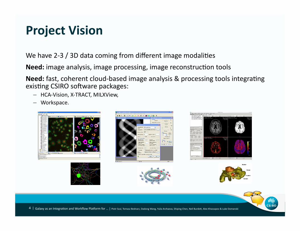
Project Vision
Galaxy as an IntegraNon and Workflow PlaTorm for … | Piotr Szul, Tomasz Bednarz, Dadong Wang, Yulia Arzhaeva, Shiping Chen, Neil BurdeF, Alex Khassapov & Luke Domanski 4 |
We have 2-‐3 / 3D data coming from different image modaliNes
Need: image analysis, image processing, image reconstrucNon tools
Need: fast, coherent cloud-‐based image analysis & processing tools integraNng exisNng CSIRO so]ware packages:
– HCA-‐Vision, X-‐TRACT, MILXView,
– Workspace.
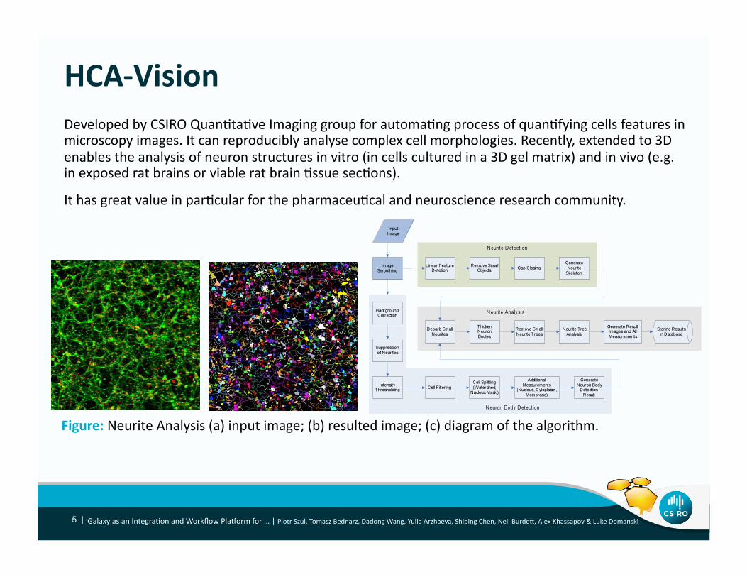
HCA-‐Vision
Galaxy as an IntegraNon and Workflow PlaTorm for … | Piotr Szul, Tomasz Bednarz, Dadong Wang, Yulia Arzhaeva, Shiping Chen, Neil BurdeF, Alex Khassapov & Luke Domanski 5 |
Developed by CSIRO QuanNtaNve Imaging group for automaNng process of quanNfying cells features in microscopy images. It can reproducibly analyse complex cell morphologies. Recently, extended to 3D
enables the analysis of neuron structures in vitro (in cells cultured in a 3D gel matrix) and in vivo (e.g. in exposed rat brains or viable rat brain Nssue secNons).
It has great value in parNcular for the pharmaceuNcal and neuroscience research community.
Figure: Neurite Analysis (a) input image; (b) resulted image; (c) diagram of the algorithm.

X-‐TRACT
Galaxy as an IntegraNon and Workflow PlaTorm for … | Piotr Szul, Tomasz Bednarz, Dadong Wang, Yulia Arzhaeva, Shiping Chen, Neil BurdeF, Alex Khassapov & Luke Domanski 6 |
A so]ware for advanced X-‐ray image analysis and Computed Tomography currently in use on the MASSIVE cluster at the Australian Synchrotron, ANU and at the Shanghai Synchrotron in China.
X-‐TRACT implements a large number of convenNonal and advanced algorithms for 2D and 3D X-‐ray image reconstrucNon and simulaNon.
Figure: (a) Insect, reconstrucNon and rendering by Sherry Mayo (CSIRO); (b) Acacia plant, sample (~1
mm across) provided by Mel Linton (CSIRO), collected, reconstructed and rendered by Sherry Mayo; (c)
Sample input Sinogram.

MILXView
Galaxy as an IntegraNon and Workflow PlaTorm for … | Piotr Szul, Tomasz Bednarz, Dadong Wang, Yulia Arzhaeva, Shiping Chen, Neil BurdeF, Alex Khassapov & Luke Domanski 7 |
A 3D medical imaging analysis and visualisaNon plaTorm increasingly popular with researchers and medical specialists working with MRI, PET and other types of medical images.
Figure: (a) Brain tumor -‐ PET scan and MRI overlaid; (b) CT scan of a prostate of a paNent
overlaid with radiaNon dose; (c) Generated 3D view of a brain allowing study of atrophy paFern
characterisNcs of diseases such as Alzheimer's disease.

Galaxy as an IntegraNon and Workflow PlaTorm for … | Piotr Szul, Tomasz Bednarz, Dadong Wang, Yulia Arzhaeva, Shiping Chen, Neil BurdeF, Alex Khassapov & Luke Domanski 8 |

Glue = Galaxy
Galaxy as an IntegraNon and Workflow PlaTorm for … | Piotr Szul, Tomasz Bednarz, Dadong Wang, Yulia Arzhaeva, Shiping Chen, Neil BurdeF, Alex Khassapov & Luke Domanski 9 |
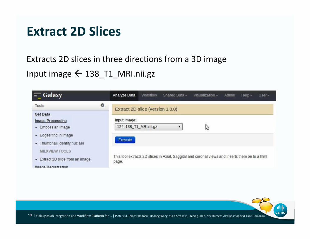
Extract 2D Slices
Galaxy as an IntegraNon and Workflow PlaTorm for … | Piotr Szul, Tomasz Bednarz, Dadong Wang, Yulia Arzhaeva, Shiping Chen, Neil BurdeF, Alex Khassapov & Luke Domanski 10 |
Extracts 2D slices in three direcNons from a 3D image
Input image 138_T1_MRI.nii.gz

Galaxy as an IntegraNon and Workflow PlaTorm for … | Piotr Szul, Tomasz Bednarz, Dadong Wang, Yulia Arzhaeva, Shiping Chen, Neil BurdeF, Alex Khassapov & Luke Domanski 11 |
It takes few seconds and a HTML is created to show the user the 2D slices and also lets the user download the images

Visualisa.on
Galaxy as an IntegraNon and Workflow PlaTorm for … | Piotr Szul, Tomasz Bednarz, Dadong Wang, Yulia Arzhaeva, Shiping Chen, Neil BurdeF, Alex Khassapov & Luke Domanski 12 |
WebGL based, Slice:Drop

Cellular imaging in Galaxy
Galaxy as an IntegraNon and Workflow PlaTorm for … | Piotr Szul, Tomasz Bednarz, Dadong Wang, Yulia Arzhaeva, Shiping Chen, Neil BurdeF, Alex Khassapov & Luke Domanski 13 |

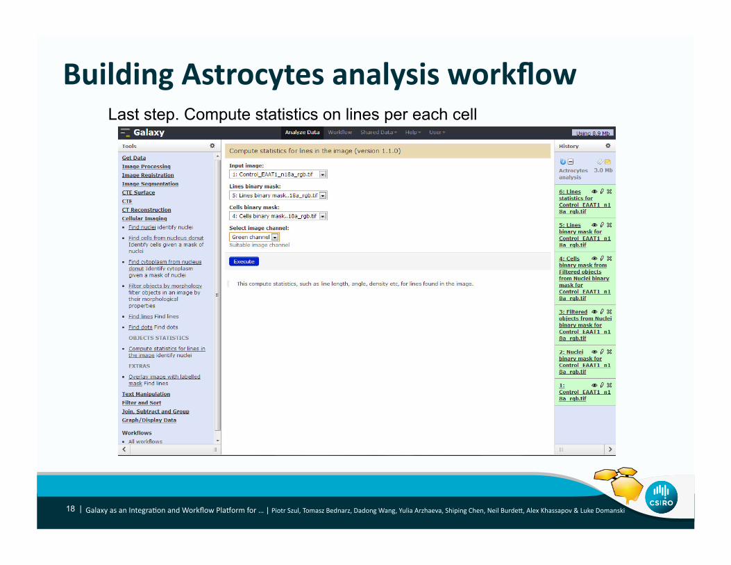
Building Astrocytes analysis workflow
Galaxy as an IntegraNon and Workflow PlaTorm for … | Piotr Szul, Tomasz Bednarz, Dadong Wang, Yulia Arzhaeva, Shiping Chen, Neil BurdeF, Alex Khassapov & Luke Domanski 14 |
Step 1. Find nuclei

Building Actrocytes analysis workflow
Galaxy as an IntegraNon and Workflow PlaTorm for … | Piotr Szul, Tomasz Bednarz, Dadong Wang, Yulia Arzhaeva, Shiping Chen, Neil BurdeF, Alex Khassapov & Luke Domanski 15 |
Output: nuclei binary mask

Building Astrocytes analysis workflow
Galaxy as an IntegraNon and Workflow PlaTorm for … | Piotr Szul, Tomasz Bednarz, Dadong Wang, Yulia Arzhaeva, Shiping Chen, Neil BurdeF, Alex Khassapov & Luke Domanski 16 |
Step 2. Find lines

Building Astrocytes analysis workflow
Galaxy as an IntegraNon and Workflow PlaTorm for … | Piotr Szul, Tomasz Bednarz, Dadong Wang, Yulia Arzhaeva, Shiping Chen, Neil BurdeF, Alex Khassapov & Luke Domanski 17 |
Output: lines binary mask
Building Astrocytes analysis workflow
Galaxy as an IntegraNon and Workflow PlaTorm for … | Piotr Szul, Tomasz Bednarz, Dadong Wang, Yulia Arzhaeva, Shiping Chen, Neil BurdeF, Alex Khassapov & Luke Domanski 18 |
Last step. Compute statistics on lines per each cell

Building Actrocytes analysis workflow
Galaxy as an IntegraNon and Workflow PlaTorm for … | Piotr Szul, Tomasz Bednarz, Dadong Wang, Yulia Arzhaeva, Shiping Chen, Neil BurdeF, Alex Khassapov & Luke Domanski 19 |
Output: lines statistics
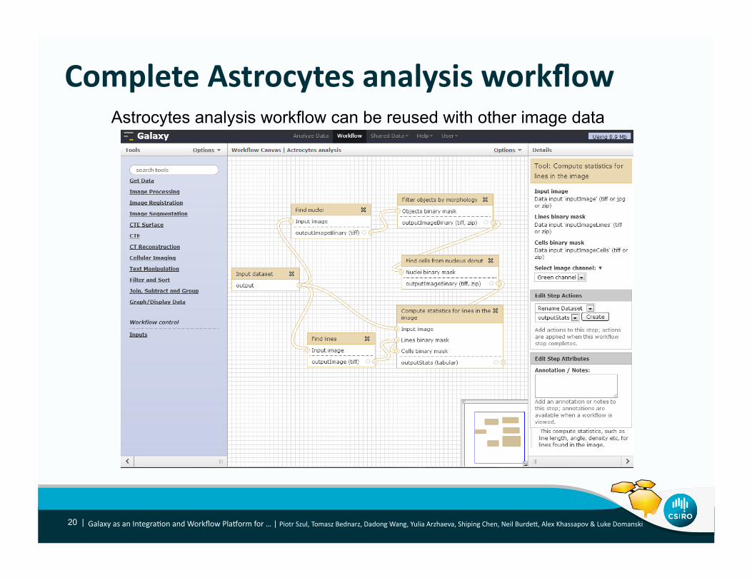
Complete Astrocytes analysis workflow
Galaxy as an IntegraNon and Workflow PlaTorm for … | Piotr Szul, Tomasz Bednarz, Dadong Wang, Yulia Arzhaeva, Shiping Chen, Neil BurdeF, Alex Khassapov & Luke Domanski 20 |
Astrocytes analysis workflow can be reused with other image data

NeCTAR Imaging Toolkit Produc.on Deploy
21 | Galaxy as an IntegraNon and Workflow PlaTorm for … | Piotr Szul, Tomasz Bednarz, Dadong Wang, Yulia Arzhaeva, Shiping Chen, Neil BurdeF, Alex Khassapov & Luke Domanski

Where are we …
Galaxy as an IntegraNon and Workflow PlaTorm for … | Piotr Szul, Tomasz Bednarz, Dadong Wang, Yulia Arzhaeva, Shiping Chen, Neil BurdeF, Alex Khassapov & Luke Domanski 22 |
So far:
• Migrated all the packages to Linux plaTorm
• Defined domain specific data types and developed most of the tools
• Small scale cloud deployments for pilot users
• Prepared training materials for the users
• Gathered iniNal feedback from the user communiNes
Next steps:
• Full scale deployment on the Research Cloud
• Bringing more users on and ongoing improvement of the plaTorm
• Create a ToolShed and refactor the toolkit
• Develop and share workflows

Cloud-‐Based Image Analysis and Processing Toolbox
Tomasz B Yulia A Dadong W Piotr S Shiping C Neil B Alex K Luke D
PROJECT TEAM
http://cloudimaging.blogspot.com.au
SAMPLE
GALAXY WORKFLOW
HCA-VISION
MILXVIEW
X-TRACT
Big Data Platform
Clouds Collaboration
Workflows Research community
Bio-Imaging
Visualisation
Galaxy as an IntegraNon and Workflow PlaTorm for … | Piotr Szul, Tomasz Bednarz, Dadong Wang, Yulia Arzhaeva, Shiping Chen, Neil BurdeF, Alex Khassapov & Luke Domanski
Thank you CMIS / CSS TCP Piotr Szul, Senior So]ware Engineer
t +61 2 xxxx xxx E [email protected] w www.csiro.au/cmis
CSS TCP

HCA-‐Vision High Level Func.onality
ID FUNCTION SHORT DESCRIPTION
H.01 Detect nuclei Detect nuclei in a 2D microscope image
H.02 Detect nuclei from cytoplasm holes
Detect nuclei from absence of stain of cytoplasm
H.03 Detect cells with nuclei Detect cells using nucleus image as a mask
H.04 Detect cells without nuclei Detect cells without using nucleus image as a mask
H.05 Detect neurons with nuclei Detect neurons from a neurite outgrowth image using nucleus image as a mask
H.06 Detect neurons without nuclei
Detect neurons from a neurite outgrowth image without using nucleus image as a mask
H.07 De-‐clump touching objects Separate any touching objects in an image, such as touching nuclei or cells
H.08 Label objects Label objects such nuclei or cells in a binary image
H.09 Get object stats Retrieve staNsNcal features of individual objects in a segmented image, including area, perimeter, origin, width and height of the bounding box, coordinate of the centroid, major and minor axis of best fit ellipse, approximate of area of convex hull etc.
H.10 Detect cell from nucleus donuts
Get an anisotropic doughnut which is a region around a cell nucleus that is not uniformly thick. The extension of the doughnut is larger along the major axis of the nucleus than perpendicular to it.
H.11 Detect dots Detect dots in a 2D image or a cell
H.12 Detect lines Detect line structures in a 2D image or a cell
H.13 Get dot stats Retrieve staNsNcal features of the detected dots, including area, perimeter etc.
H.14 Get line stats Retrieve the staNsNcal features of detected line structures
H.15 Cell Scoring Count negaNve and posiNve cells, measure integrated and average intensity of negaNve and posiNve cells.
IDs H.xx – Application area: cell features of microscopy images

X-‐TRACT High Level Func.onality ID FUNCTION SHORT DESCRIPTION
XP.01 Sinogram crea.on X-‐ray projecNon data must first be converted into sinograms before CT reconstrucNon can be carried out. Each sinogram contains data from a single row of detector pixels for each illuminaNng angles. This data is sufficient for the reconstrucNon of a single axial slice (at least, in parallel-‐beam geometry).
XP.02 Ring artefact removal
Ring artefacts are caused by imperfect detector pixel elements as well as by defects or impuriNes in the scinNllator crystals. Ring artefacts can be reduced by applying various image processing techniques on sinograms or reconstructed images.
XP.03 Dark current subtrac.on
Dark current subtracNon compensates for the readout noise, ADC offset, and dark current in the detector. The dark current images are collected before and/or a]er CT measurements with no radiaNon applied and with the same integraNon Nme as the one used during the measurements. The dark current image is subtracted from each CT projecNon.
XP.04 Flat field correc.on
Flat-‐field images are obtained under the same condiNons as the actual CT projecNons, but without the sample in the beam. They allow one to correct the CT projecNons for the unevenness of the X-‐ray illuminaNon.
XP.05 Posi.onal drid correc.on
The funcNon is used for correcNon of transverse dri] between related experimental images. Image dri] is assessed by cross-‐correlaNng pairs of images.
XP.06 Data normalisa.on Data normalisaNon Including normalisaNon to a user-‐defined region
XP.07 TIE-‐based phase extrac.on
The TIE algorithm allows the recovery of the opNcal phase of an electromagneNc wave (e.g. an X-‐ray beam) from a single near-‐field in-‐line image by solving the Transport of Intensity equaNon under the assumpNon that the phase shi] and absorpNon distribuNons are proporNonal to each other. This method is usually applied in propagaNon-‐based in-‐line CT imaging (PCI-‐CT).
XCT.01 FBP CT reconstruc.on
Filtered back-‐projecNon (FBP) parallel-‐beam CT reconstrucNon
XCT.02 FDK CT reconstruc.on
Feldkamp-‐Davis-‐Kress (FDK) cone-‐beam CT reconstrucNon
XCT.03 Centre of rota.on Automated calculaNon of the centre of sample rotaNon in a CT scan from experimental X-‐ray projecNons, sinograms or reconstructed axial slices.
XCT.04 CT Reconstruc.on Filters
The choice of available CT reconstrucNon filters will include at least the Liner-‐Ramp, Shepp-‐Logan, Cosine, Hamming and Hann filters.
XCT.05 ROI reconstruc.on This opNon enables the user to select a subset of axial slices to be reconstructed and/or limit the reconstrucNon area to a user-‐defined rectangular subarea of the axial slice. The opNon reduces the reconstrucNon Nme and the size of the output data.
IDs XP.xx – Application area: data processing functions IDs XCP.xx – Application area: CT reconstruction functions

MILXView High Level Func.onality ID FUNCTION SHORT DESCRIPTION
MC.01 Atlas registra.on Align an atlas image to a target image
MC.02 Segmenta.on Segment the MRI into grey maFer (GM), white maFer (WM) and cerebrospinal fluid (CSF)
MC.03 Bias Field Correc.on EsNmate and remove the noise on the image
MC.04 Par.al Volume es.ma.on QuanNfy the amount of parNal voluming inside each voxel
MC.05 Topology Correc.on Create the topology of the brain to ensure that it is genus zero
MC.06 Thickness Es.ma.on Compute the thickness of the cortex for each Grey maFer voxel
MS.01 Cor.cal surface extrac.on Extract a 3D mesh from the brain segmentaNon
MS.02 Topological correc.on Remove holes and handles from the mesh
MS.03 Biomarker mapping on cor.cal surface
Mapping of various values on the mesh i.e. thickness, PET values, MR intensity etc ...
MS.04 Surface registra.on Align the meshes of any given subject to a template to obtain a correspondence across subjects
MS.05 Transfer of biomarkers on template surface
Map all the values from all subjects to a common space where they can be compared
MP.01 PVC Registra.on RegistraNon of the PET image to its corresponding MRI
MP.02 Segmenta.on SegmentaNon of the MRI into GM, WM, and CSF
MP.03 Par.al Volume correc.on (PVC)
CorrecNon for spill in and spill over of the PET image using the MRI segmentaNon
MR.01 SUVR Registra.on RegistraNon of the PET image to its corresponding MRI
MR.02 Segmenta.on SegmentaNon of the MRI into GM, WM and CSF
MR.03 Atlas Registra.on RegistraNon of an atlas to the MRI to define a reference region on the MRI
MR.04 Image Normalisa.on Normalising the PET intensity with the intensity of the reference region
IDs MC.xx – Application area: neuro-imaging analysis, cortical thickness estimation (CTE) IDs MS.xx – Application area: neuro-imaging analysis, CTE surface
IDs MP.xx – Application area: neuro PET analysis, PET PVC IDs MR.xx – Application area: neuro PET analysis, PET SUVR