gga proteins mediate the recycling pathway of memapsin 2
TRANSCRIPT

GGA PROTEINS MEDIATE THE RECYCLING PATHWAY
OF MEMAPSIN 2 (BACE)*
Xiangyuan He‡, Feng Li‡§, Wanpin Chang‡ and Jordan Tang‡§٭٭
From the ‡Protein Studies Program Program, Oklahoma Medical Research
Foundation and §Department of Biochemistry and Molecular Biology, University of
Oklahoma Health Science Center, Oklahoma City, OK 73104
(Running title) GGA mediated Memapsin 2 Recycling
1
JBC Papers in Press. Published on December 21, 2004 as Manuscript M411296200
Copyright 2004 by The American Society for Biochemistry and Molecular Biology, Inc.
by guest on April 5, 2018
http://ww
w.jbc.org/
Dow
nloaded from

Summary:
Memapsin 2 (BACE, β-secretase) is a membrane-associated aspartic protease that
initiates the hydrolysis of β-amyloid precursor protein (APP) leading to the
production of amyloid- β (Aβ) and the progression of Alzheimer’s disease (AD). Both
memapsin 2 and APP are transported from the cell surface to endosomes where APP
is cleaved by memapsin 2. We described previously that the cytosolic domain of
memapsin 2 contains an acid-cluster-dileucine motif (ACDL) that binds the VHS
domain of Golgi-localized γ–ear-containing ARF binding (GGA) proteins (He et al.
Biochemistry. 42:12174-12180, 2003). Here we report that GGA proteins colocalize
in the trans-Golgi network and endosomes with memapsin 2 and a memapsin 2
chimera containing a cytosolic domain of a mannose-6-phosphate receptor. Depleting
cellular GGA proteins with RNA interference or mutation of serine498 to stop the
phosphorylation of ACDL resulted in the accumulation of memapsin 2 in early
endosomes. A similar change of memapsin 2 localization also was observed when a
retromer subunit VPS26 was depleted. These observations suggest that GGA proteins
function with the phosphorylated ACDL in the memapsin 2 recycling pathway from
endosomes to trans-Golgi on the way back to the cell surface.
2
by guest on April 5, 2018
http://ww
w.jbc.org/
Dow
nloaded from

Introduction
Memapsin 2 (EC 3.4.23.46) (1), also called β-secretase, BACE (2) and ASP-2
(3, 4), is the protease that initiates the cleavage of β-amyloid precursor protein (APP)1.
After a second cleavage by another protease, γ-secretase, a 40- or 42-residue peptide
called amyloid-β (Aβ) is produced. An excessive level of Aβ in the brain facilitates a
series of adverse events, including the accumulation of Aβ plaques, brain
inflammation and the death of neurons, and leads to the progression of Alzheimer’s
disease (AD) (5). In view of its pivotal position in the pathogenesis of AD, memapsin
2 is viewed as a promising target for the development of inhibitor drugs to treat AD.
In this connection, the understanding of molecular and cellular activity of memapsin
2 is of great scientific interest at the present.
Memapsin 2 is a type I transmembrane protein with a single transmembrane
segment linking a luminal catalytic ectodomain, which is homologous to other
mammalian aspartic proteases (6), to a C-terminal cytosolic tail of 21 residues. Newly
synthesized pro-memapsin 2 is processed to mature protease by furin during transit
through the secretory pathway to the cell surface (7-10). Memapsin 2 and its
substrate APP are both internalized from the cell surface to endosomes where APP is
cleaved by memapsin 2 in an acidic medium leading to the production of Aβ. The
memapsin 2 protease then is recycled to the cell surface (10, 11). Thus, the cellular
trafficking of memapsin 2 is intimately related to the mechanism and regulation of Aβ
production.
3
by guest on April 5, 2018
http://ww
w.jbc.org/
Dow
nloaded from
As in the case of many other membrane proteins, the cellular transport of
memapsin 2 is mediated by its cytosolic domain (10, 12). Recently, the region of
memapsin 2 cytosolic domain that mediates cellular trafficking has been traced to a
motif with the sequence DISLL. This motif, which belongs to the acidic-cluster-
dileucine (ACDL) sorting signal (DXXLL, where X denotes a nonconserved residue;
for review, see reference13), binds the VHS (Vps-27, Hrs, and STAM) domains of
GGA (Golgi-localized γ-ear-containing ARF binding) proteins (14, 15). GGA
proteins are known to bind the ACDL sorting signal in the cytosolic tails of mannose-
6-phosphate receptors (MPR’s) (16, 17) and other lysosomal membrane proteins (18)
as the initial sorting recognition in the transport from trans-Golgi to endosomes. By
analogy, the GGA/memapsin 2 interaction is thought to mediate the cellular transport
of memepsin 2 (14, 15), although direct cellular evidence and the specific route of
involvement are yet to be demonstrated. Several pieces of evidence, however, argue
against the involvement of GGA proteins in the endocytic pathway of memapsin 2.
First, GGA proteins are associated with trans-Golgi and endosome, but scarcely at the
cellular plasma membrane (19, 20). Second, memapsin 2 ectodomain devoid of GGA
binding motif is internalized by cells through the interaction with APP (21),
suggesting that the ACDL motif of memapsin 2 is not essential for its endocytosis.
Three, the native ACDL motif of memapsin 2 interacts about 20 times weaker than
the corresponding interaction with mannose-6-phosphate receptors. The
phosphorylation of Ser498 in the ACDL motif of memapsin 2 enhances GGA binding
to the intensity similar to those for MPR proteins (15). Phosphorylation of the
memapsin 2 ACDL motif is essential for its recycling pathway from early endosomes
4
by guest on April 5, 2018
http://ww
w.jbc.org/
Dow
nloaded from
to the cell surface (11). These observations open the possibility that the interaction of
GGA proteins with the phosphorylated memapsin 2 may mediate the recycling of the
protease from endosomes back to the cell surface. In order to further understand the
involvement of GGA proteins in intracellular transport of memapsin 2, we studied the
cellular trafficking of memapsin 2 and its chimera with MPR cytosolic domain. We
report in this paper that GGA proteins are involved in the cellular transport of
memapsin 2 from endosome to the cell surface in the recycle pathway.
Materials and Methods
cDNA vectors – The constructs of expression vectors of human wild type
memapsin 2 and Swedish mutant of APP cDNAs were previously described (1). PCR
based mutagenesis was used for the constructs of memapsin2 mutants. In M2-MPR,
memapsin 2 cytosolic domain from residue 481 to the C-terminus was replaced with
the cytosolic domain of CD-MPR (Fig. 1A) with the sequence
QRLVVGAKGMEQFPHLAFWQDLGNLVADGCDFVCRSKPRNVPAAYRGVGD
DQLGEESEERDDHLLPM. In mutant M2-L/A, Leu499 and Leu500 in the C-terminal
sequence –DDISLLK were replaced by alanines. In mutant M2-S/A, Ser498 was
replaced by alanine. The cDNA constructs encoding these memapsin 2 mutants were
inserted into the mammalian expression vector pSecTag2 (Invitrogen). Expression
vectors for the GGA proteins, HA-GGA1, GGA2-V5 and GGA3-V5, were previously
described (15).
5
by guest on April 5, 2018
http://ww
w.jbc.org/
Dow
nloaded from

Antibodies – The rabbit polyclonal antibodies (Covance, Denver, PA, USA) and
goat polyclonal antibodies against recombinant pro-memapsin2 were affinity-purified
using Affigel (Bio-Rad) immobilized recombinant memapsin 2 (1). Monoclonal
antibodies MAB1560 (specific to Aβ 1-17), rabbit polyclonal antibodies AB5352
(specific to APP C-terminal) were bought from CHEMICON (Temecula, CA). Mouse
monoclonal antibodies for early endosome antigen 1 (EEA1), TGN38, Bip/GRP78,
Lamp-1, GGA2 and GGA3 were purchased from BD Biosciences. Mouse monoclonal
antibodies for the Golgi marker Giantin were purchased from Calbiochem.
Monoclonal V5 antibody was purchased from Invitrogen. Monoclonal Anti-HA
antibodies and Cy3-conjugated secondary sheep anti-rabbit antibody were purchased
from Sigma. Alex 488 conjugated secondary donkey anti-mouse antibody was
purchased from Molecular Probes. Rabbit polyclonal anti-β-actin and goat polyclonal
anti-VPS26 were purchased from Novus Biologicals. Rabbit polycolonal anti-GGA1
was gift from Dr. Margaret S. Robinson (University of Cambridge, Cambridge, UK).
RNAi experiment for knockdowns – pSuper vectors of siRNA for GGA1-3 were
obtained from Dr. Stuart Kornfeld (Washington University, St. Louis) (22). HeLa
cells were grown in Dulbeco’s modification of minimal essential medium
supplemented with 10% FBS, 100 U/mL penicillin, and 100 µg/mL streptomycin at
37° C in the presence of 5% CO2. HeLa cells were cotransfected with pSuper GGA
RNAi vectors and pSectag vector of memapsin 2 or mutants using transfection agent
LipofecAMINE 2000 (Invitrogen). The ratio of p-Super/RNAi plasmid DNA to
pSectag was 4:1. Fifty six h after transfection, the cells were harvested for double-
6
by guest on April 5, 2018
http://ww
w.jbc.org/
Dow
nloaded from

labeled immunofluorescence to detect co-localization of memspin 2 with different
organelle markers. siRNA knockdown of VPS26 was performed as described by
Senman (23). siRNA oligonucleotides used to knock down VPS26 expression
(AAUGAUGGGGAAACCAGGAAA) and control Lamin A/C siRNA were obtained
from Dharmacon. HeLa cells were cultured as described above in 6-well dishes until
about 60-70% confluenc. At least 4 h after seeding the cells, the first transfection was
performed using OligofectAMINETM (Invitrogen). The cells were trypsinized the
following day. Two transfections were performed one at the start of the experiment
and the other after two days. The cells were trypsinized 24 h after second transfection
and were plated onto coverslips of 6-well dishes for memapsin 2 transient
overexpression and confocal microscopy oberservation.
Immuno-fluorescence Labeling of Cultured Cells – HeLa cells were seeded onto
6-well plates with glasses coverslips, and expressed the memapsin 2 or mutants
constructs for 56 h after transfection. Cells were gently fixed in 4% paraformaldehyde,
in phosphate-buffered saline, pH 7.4, at room temperature for 15 min. Following
fixation, coverslips were washed twice for 10 min in PBS and incubated for 1 h at
room temperature with the indicated combinations of primary antibodies diluted in
0.1% BSA, 0.1% sapoin, 0.02% sodium azide in PBS (immunofluorescence buffer).
At the end of this period, coverslips were washed twice with PBS followed by
incubation for 30 min with the indicated combinations of secondary antibodies
diluted in immunofluoresence buffer. Following incubation, coverslips were again
washed twice with PBS and mounted on slides using Vector-shield (Vector
7
by guest on April 5, 2018
http://ww
w.jbc.org/
Dow
nloaded from

Laboratories, Inc., Burlingame, CA). Images were obtained in an inverted confocal
laser scanning microscope (LSM 510, Carl Zeiss Inc.). Human neuroblastoma M17
Cells (ATCC) were grown in DMEM/F12 medium supplemented with 10% FBS, 100
U/mL penicillin, and 100 µg/mL streptomycin at 37° C in the presence of 5% CO2.
M17 Cells were seeded onto 6-well plates with glass coverslips; endogenous
memapsin 2 and GGA1 were observed as above.
Transient Transfection and IP/WB – Plasmids containing separately memapsin 2
or its mutants and APPsw cDNAs were transiently transfected into HEK 293 cells in
six-well plates using transfection reagent LipofecAMINE 2000 (Invitrogen). Cells
were grown in Dulbecco’s Modified Eagle’s medium (DMED) containing 10% fetal
bovine serum, 100 U/mL penicillin, and 100 µg/mL streptomycin (GibcoBUR). After
transfection, cells were changed to fresh medium (1.5 ml for a six-well plate)
conditioned for 48 h, subjected to immunopreciptitation (IP) and Western bottting
(WB).
Aβ peptides secreted into medium were immunoprecipitated at 4° C overnight
with monoclonal antibody MAB1560 (1:800) and protein G plus A-agarose (Sigma).
Immunoprecipitates were washed for 20 min at 4° C with lysis buffer (PBS with 1%
Nonidet P-40 with protease inhibitors) and then washed in lysis buffer containing
0.1% SDS. The samples were washed again in lysis buffer, eluted in Laemmli sample
buffer, separated by SDS-PAGE on 10-20% Tris-Tricine gels (Invitrogen), and
transferred to PVDF membrane. The membrane was blotted with the same antibody,
and detected with ECL (Amersham Pharmacia).
8
by guest on April 5, 2018
http://ww
w.jbc.org/
Dow
nloaded from

Immuno-electrophoresis of APP and fragments – After transfection, HEK 293
cells were solubilized in lysis buffer as described above and centrifuged at maximum
speed in an Eppendorf centrifuge for 10 min to remove cellular debris. The samples
were mixed with Laemmli buffer, resolved by SDS-PAGE on 10-20% Tris-Tricine
gels (Invitrogen), transferred to PVDF membrane and immunoblotted with AB5352.
Results
Subcellular localization of memapsin 2 and mutants – We compared the
intracellular distribution of wild-type memapsin 2 with that of two memapsin 2
mutants. One was a chimera, M2-MPR, in which the memapsin 2 cytosolic domain is
replaced by the cytosolic domain of CD-MPR (Fig. 1A) that also contains an ACDL
motif. Since GGA proteins (Fig. 1B) are known to be involved in the transport and
retrieval between trans-Golgi and the early endosome (16, 17, 22 and 24), this mutant
serves for comparison to the localization and trafficking of memapsin 2. In the second
mutant, M2-L/A, two leucines in the ACDL motif of memapsin 2 were replaced by
alanines (Fig. 1A), resulting in a mutant defective in endocytosis (10, 12) that served
as a negative control. HeLa cells expressing memapsin 2 and mutants were visualized
by immunochemical staining. The cellular localization pattern of wild-type memapsin
2 had a punctate distribution mainly in the perinuclear region (Fig. 1C), which
confirmed previous reports (2, 9, 10) that the protease was localized primarily in
TGN and early endosomes. Little memapsin 2 is seen on the cell surface. The co-
localization of memapsin 2 with TGN and early endosome markers was confirmed by
separate immunochemical staining with TGN38 (TGN marker) and EEA1 (early
9
by guest on April 5, 2018
http://ww
w.jbc.org/
Dow
nloaded from

endosome marker) (results not shown). Imunolableing pattern for cells expressing
M2-MPR, which showed a similar perinuclear distribution as well as peripheral
labeling (Fig. 1D), also co-localized well with TGN and early endosome markers
(data not shown). The cellular distribution pattern of this chimera is similar to that of
CD-MPR itself, which was found most abundantly in TGN, less in endosomes and
little on the cell surface (25). M2-L/A mutant showed a predominant localization on
the cell surface (Fig. 1, E) as reported previously (10, 12). These observations suggest
that wild-type memapsin 2 is localized mainly in endosomes and trans-Golgi similar
to MPR. The chimera of memapsin 2 with the cytosolic domain of CD-MPR also had
a similar intracellular distribution to that of CD-MPR.
Memapsin 2 and GGA proteins colocalize in the cell – Wild-type memapsin 2 and
individual GGA proteins expressed in HeLa cells were examined using
immunofluorescent staining and confocal microscopy. The distribution of GGA
proteins in the cells was similar to that previously reported (19, 20). Extensive
colocalization was seen for the wild-type memapsin 2 with each of the three GGA
proteins in the perinuclear region and some but much less extensive co-localization
was observed in the peripheral structure (Fig. 2). Similar experiments were also
performed on neuroblastoma M17 cells with the same results (Fig. 2J-L). M2-MPR
chimera also colocalizes with each of the three GGA proteins with the distribution
patterns similar to those observed for the wild-type memapsin 2 (Fig. 3). These
observations suggest that MPR cytosolic domain in the chimera interacted with GGA
proteins and directed the transport of M2-MPR to endosomes. Memapsin 2 mutant
10
by guest on April 5, 2018
http://ww
w.jbc.org/
Dow
nloaded from
M2-L/A, however, was mainly present on the cell surface and did not significantly
co-localize with any of the GGA proteins (Fig. 4). Very weak co-localization seen for
GGA2 and GGA3 in the merged images may have resulted from residual interaction
between M2-L/A and these two GGA proteins. These observations are consistent with
the idea that all three GGA proteins function in the cellular transport of memapsin 2
in a manner similar to the transport of MPR (22).
Change in Memapsin 2 intracellular distribution in siRNA mediated knockdown
of GGA proteins – To study further the involvement of GGA proteins in memapsin 2
intracellular trafficking, we determined the distribution of memapsin 2 in HeLa cells
in which the synthesis of GGA proteins were silenced by using plasmid-based siRNA
interference. Each of the three GGA proteins, but not the control protein, β-actin, was
greatly diminished by the transfection of specific GGA siRNA vectors (Fig. 5). In
mock-treated cells, the bulk of the wild-type memapsin 2 (Fig. 6B) and the memapsin
2-MPR chimera (Fig. 7B) were found in the juxtanuclaear area as seen in the
preceding figures. Silencing each GGA proteins significantly increased the presence
in the peripheral region of the cells and decreased at about the same level the
juxtanuclear localization for both M2-WT (Figs. 6E, 6H and 6K) and M2-MPR (Figs.
7E, 7H and 7K). These changes are similar to those observed for cation-independent
MPR (CI-MPR) when GGA proteins were individually depleted (22).
Immunostaining of these cells with different organelle markers revealed significant
colocalization of M2-WT (Figs. 6F, 6I and 6L) and M2-MPR (Figs. 7F, 7I and 7L)
with the early endosome marker EEA1 (only the co-localization with EEA1 is shown).
11
by guest on April 5, 2018
http://ww
w.jbc.org/
Dow
nloaded from

Significantly, the EEA1 co-localization with peripheral memapsin 2 is substantially
increased with GGA depletion, suggesting an increase of memapsin 2 in endosomes.
In contrast, M2-L/A in HeLa cells exhibited no significant change from predominant
cell surface localization upon the depletion of GGA proteins (data not shown). These
observations suggest that all three GGA proteins participate in intracellular trafficking
of native memapsin 2 and the M2-MPR chimera.
Change in Memapsin 2 intracellular distribution upon mutation of Ser498 to Ala –
We previously demonstrated that phosphorylation of Ser498 of memapsin2 within the
ACDL motif increased the binding affinity of memapsin 2 for the VHS domain of
GGA proteins (15). It was of interest then to determine if the phosphorylated
memapsin 2 ACDL is involved in the intracellular trafficking of the protease. We
mutated Ser498 to an alanine in order to preclude phoshporylation at this position and
compared the localization in cells expressing the wild-type and mutant memapsin 2.
Immunofluorescence microscopy study showed that the mutation of Ser498 changed
the distribution of memapsin 2 from perinuclear (Fig. 8, upper panels) to the
peripheral localization (Fig. 8, lower panels). Consequently, the co-localization of the
endosome marker EEA1 and the mutant M2-S/A is observed in the peripheral region
but is not predominantly in the perinuclear region as seen for the wild-type memapsin
2. These observations are reminiscent of the changes found for GGA depletion
discussed above and suggests that both GGA depletion and Ser498 mutation may block
the same retrieval pathway for memapsin 2 from endosomes back to the cell surface.
12
by guest on April 5, 2018
http://ww
w.jbc.org/
Dow
nloaded from

Change of Memapsin 2 intracellular distribution in siRNA mediated VPS26
knockdown – The retrieval of MPR from endosomes back to Golgi has been shown to
involve an intracellular protein complex called retromer (23). siRNA mediated
depletion of VPS26, a component protein of retromer, blocked the transport of CI-
MPRs retrieval from endosome to Golgi. Since memapsin 2 and MPR both utilize
ACDL/GGA interaction in cellular trafficking, it seemed possible that in its recycling
pathway memapsin 2 is transported from endosomes to Golgi, as an intermediate
compartment, before memapsin 2 reaches the cell surface. We therefore asked if the
depletion of VPS26 influenced the recycling of memapsin 2 from endosomes.
siRNA mediated depletion of VPS26 was nearly complete and did not affect the level
of a control siRNA vector for β-actin (Fig. 9A). Images from immunochemical
staining showed that the intracellular localization of memapsin 2 changed from a
predominantly perinucear localization with VPS26 expression (Fig. 9B-D) to a
peripheral localization (Fig. 9E-G) upon VPS26 depletion. Some memapsin 2 was
also found to be present at the plasma membrane. This may have resulted from the
backup of the trafficking due to serious disruption of endosome to Golgi transport. In
general, however, the similarity in intracellular distribution changes with VPS26
depletion, GGA depletion or Ser498 mutation is striking. Such a comparison, together
with the retromer’s known role in MPR retrieval, suggests that memapsin 2 recycles
from endosomes by way of the Golgi network.
13
by guest on April 5, 2018
http://ww
w.jbc.org/
Dow
nloaded from

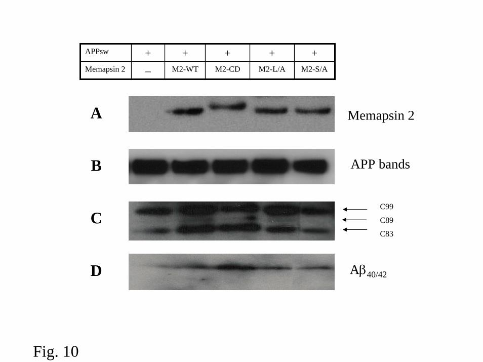
APP hydrolysis and Aβ production mediated by Memapsin2 mutants – Since
alterations in memapsin 2 distribution and trafficking were observed in the
experiments described above, it was of interest to assess the effect of these changes
on APP hydrolysis by memapsin 2. Therefore, we compared the memapsin 2
generated APP C-terminal fragment C99 in HEK293 cells cotransfected with Swedish
mutant of APP, APPsw, and each of the following: M2-WT, M2-MPR, M2-L/A and
M2-S/A. Forty eight h after transfection, the expression of memapsin 2 and APP was
in evidence by Western blots (Fig. 10A, B). Chimera M2-MPR and mutants M2-L/A
and M2-S/A each produced APP fragment C99 (Fig. 10C) and Aβ (Fig. 10D) at a
comparable level as that produced by the wild-type memapsin 2. The
immunoprecipitated soluble APP fragment resulting from β-secretase cleavage was
seen in M2-WT as well as in M2-MPR, M2-L/A and M2-S/A experiments (data not
shown). These observations suggest that both memapsin 2 mutants hydrolyzed APP
intracellularly to generate Aβ.
Discussion
Evidence reported in this study and previous reports quoted herein suggest an
intracellular transport route of memapsin 2 as shown schematically in Fig. 11. Cell
surface memapsin 2 and APP are internalized to endosomes where the hydrolysis of
APP and the production of Aβ take place. The ACDL motif in the cytosolic domain
of memapsin 2 is prsummably phosphorylated and interacts with GGA proteins to
facilitate its transport from endosomes via TGN to cell surface.
14
by guest on April 5, 2018
http://ww
w.jbc.org/
Dow
nloaded from

We previously demonstrated the interaction between GGA proteins and the
native and phosphorylated ACDL motif in the cytosolic domain of memapsin 2 (14,
15). These observations and the knowledge that a similar interaction of GGA proteins
with MPR mediates the transport of MPR between the trans-Golgi and endosomes
prompted the suggestion that GGA proteins were involved in the intracellular
transport of memapsin 2. However, the supporting cellular studies had not been
carried out and the specific point of GGA involvement in memapsin 2 trafficking was
unknown. GGA proteins are found predominantly in the TGN but are also present in
early endosomes (19, 20). We observed cellular co-localization of GGA proteins with
memapsin 2 and alteration of the memapsin 2 intracellular locations upon siRNA
mediated GGA depletion. These observations support the contention that GGA
proteins are involved in the cellular transport of memapsin 2. As observed in MPR
transport (22), all three GGA proteins are involved in the intracellular trafficking of
memapsin 2 since the depletion of any of the three GGA proteins caused a significant
redistribution of memapsin 2. In the transport of CI-MPR, three GGA proteins are
interdependent in their functions (22). Although we did not provide evidence for this
aspect in memapsin 2 transport, it seems probable that the mechanism of involvement
of GGA in the transport of the two MPRs and memapsin 2 are the same, namely the
recognition of the ACDL motif facilitates packaging the proteins into the transporting
vesicles. Our results also support the contention that GGA proteins interact with the
phosphorylated ACDL motif of memapsin 2 in the recycling pathway from
endosomes to TGN. It is also interesting that the ACDL mutated to preclude
phosphorylation resulted in a redistribution of cellular memapsin 2 similarly as
15
by guest on April 5, 2018
http://ww
w.jbc.org/
Dow
nloaded from

observed on depletion of GGA proteins. This similarity suggests that the peripherally
localized memapsin 2 resulted from blockage is achieved by siRNA or by mutation of
ACDL. The similarity of the redistribution of memapsin 2 and CI-MPR affected by
siRNA for GGA proteins is also striking. The routes for memapsin 2 and CI-MPR to
reach endosomes are presumably different. CI-MPR is predominantly transported to
the endosome from the Golgi and memapsin 2 is known to be endocytosed to
endosomes from cell surface. The similarity can be explained from the GGA
involvement in the recycling pathway of both proteins, i.e. the depletion of GGA
would result in the accumulation of both proteins in endosomes.
In its recycling pathway, memapsin 2 is likely transported from endosomes to
TGN as an intermediate stop before returning to cell surface by the usual secretory
pathway. Recent evidence suggests that retromer protein complex is specifically
involved in the endosome to TGN transport of MPR (23). Depletion of VPS26, a
subunit of retromer complex, caused the redistribution of memapsin 2 from
predominantly perinuclear to peripherally localized endosomes. These observations
are consistent with the contention that the loss of retromer functions blocks the
trafficking from endosomes to TGN and thus causes accumulation of memapsin 2 in
the endosomes. We did observe, however, the accumulation of memapsin 2 at the cell
surface upon retromer depletion. It is not clear if this is due to the back up of the
memapsin 2 traffic or the involvement of retromer in other trafficking routes of
memapsin 2. Whichever is the cause, the complication does not appear to change the
main conclusion that memapsin 2 recycles through TGN. The overall evidence
16
by guest on April 5, 2018
http://ww
w.jbc.org/
Dow
nloaded from

discussed above is consistent with the notion that phosphorylated memapsin 2 is
recycled via TGN on its way back to cell surface. However, identification of specific
subcellular compartments along this recycling route would require detailed studies in
the future.
Current evidence does not support the involvement of GGA proteins in the
endocytosis of memapsin 2. We did not observe significant colocalization of GGA
and memapsin 2 at the cell surface (Fig. 2). The depletion of GGA proteins did not
cause a significant accumulation of memepsin 2 on cell surface (Fig. 6). Since the
cytosolic GGA proteins should be accessible for memapsin 2 located both in
endosomes and at cell surface, the specificity of recycling involvement may be
determined by the phosphorylation of the memapsin 2 ACDL motif. We have shown
that the phosphorylated memapsin 2 ACDL binds GGA proteins with about 20 times
lower Kd than the non-phosphorylated ACDL. This tighter binding may be sufficient
to provide specificity for the memapsin 2/GGA interaction. If such a mechanism is
operative, it would predict the presence of a memapsin 2 ACDL kinase in the
endosomes and a phosphatase in the TGN or beyond.
The above discussion raises another interesting question about the identity of
the endocytic signal for memapsin 2 endocytosis. We have confirmed the previous
report (10, 12) that two leucines within the ACDL motif appear to be essential for
memeapsin 2 endocytosis, since the replacement of these residues with alanines
resulted in the accumulation of the mutant protease on cell surface (Fig. 4). Dileucine
17
by guest on April 5, 2018
http://ww
w.jbc.org/
Dow
nloaded from

is a well established endocytotic signal (26) and in this case, it appears to function
independent of the GGA/ACDL interaction. Another possible internalization route is
the APP-dependent endocytosis. We reported recently (21) that the exogenously
added memapsin 2 ectodomain is endocytosed by way of an interaction with APP.
This process is dependent on the endocytic signal GYENPTY in the cytosolic domain
of APP. Full length memapsin 2 appears to also interact with APP suggesting that the
APP dependent endocytosis of memapsin 2 may be a viable pathway.
In all experiments describe herein, M2-MPR chimera respond to all different
challenges in almost the same way as the native memapsin 2. Superficially, this
suggests that the cytosolic domain of MPR may contain the same targeting
information as memapsin 2. The comparison of the two in the current studies is
restricted to the recycling pathway that the two native proteins share. However the
pathway leading to endosomes should be different for these two proteins, although it
is know that small fraction of MPR is endocytosed from cell surface.
We observed that all the memapsin 2 constructs, including M2-MPR chimera,
produced Aβ and the C99, the C-terminal fragment of APP (Fig. 10). However, the
subcellular locales where the cleavage takes place are difficult to dissect. Although
the main subcellular site for the generation of Aβ is endosomes, the production of
significant amount of Aβ in the secretory pathway has been documented (27-29). In
the case of the dileucine to alanine mutant of memapsin 2 (M2-L/A in Fig. 10), the
Aβ generated should predominantly come from the secretory pathway since this
18
by guest on April 5, 2018
http://ww
w.jbc.org/
Dow
nloaded from

mutant is devoid of endocytic ability. In the case of memapsin 2-MPR chimera (M2-
CD in Fig. 10), the presence of C83 (Fig. 10), an APP fragment from the cleavage by
cell surface α-secretaase, suggests that M2-MPR chimera is capable of reaching cell
surface and experiences endocytosis. However, the robust production of Aβ by the
chimera raises another interesting question on whether the chimera is transported
directly from Golgi to endosomes where the bulk of Aβ is generated. The absence of
a C89 band for the non-phosphorylated M2-S/A mutant (Fig. 10) is consistent with
the predominant endosomal localization of this memapsin 2 mutant and perhaps the
cell surface exposure of this protein is a transient one.
Acknowledgements – The authors thank Dr. S. Kornfeld for the plasmids of siRNA
for GGA proteins, Dr. M.S. Robinson for antibody against GGA1, Dr. Gerald
Koelsch for helpful discussions and antibodies against memapsin 2, Dr. J.A. Hartsuck
for critical reading of this manuscript and the Cell Imaging Core Facility at Oklahoma
Medical Research Foundation for excellent assistance.
References
1. Lin, X., Koelsch, G., Wu, S., Downs, D., Dashti, A. and Tang, J. (2000) Proc.
Natl. Acad. Sci. 97, 1456-1460.
2. Vassar, R., Bennett, B. D., Babu-khan, S., Mendiaz. E. A., Denis, P., Teplow, D.
B., Ross, s., Amarante, P., Loeloff, R., Luo, Y., Fisher, S., Fuller, J., Edenson, S.,
19
by guest on April 5, 2018
http://ww
w.jbc.org/
Dow
nloaded from

Lile, J., Jaronsinski, M., A., Biere, A. L., Curran, E., Burgess, T., Louis, J. C.,
Collins, F., Treanor, J., Rogers, G., and Citron, M. (1999) Science 268, 735-741.
3. Yan, R., Bienkowski, M. J., Shuck, M. E., Miao, H., Tory, M. C., Pauley, A. M.,
Brashier, J. R., Stratman, N. C., Mathews, W. R., Buhl, A. E., Carter, D. B.,
tomasselli, A. G., Parodi, L. A., Heinrikson, R. Lp, and Gurney, M. E. (1999)
Nature 402, 533-537.
4. Hussain, J., Powell, D., Howlett, D. R., Tew, D. G., Meek, T. D., Chapman, C.,
Gloger, I. S., Murphy, K. E., Southan, C. D., Ryan, D. M., Smith, T. S., Simmons,
D. L., Walsh, F. S., Dingwall, C., and Christie, G. (1999) Mol. Celll Neurosci. 14,
419-427.
5. Selkoe, D. (2001) Physiol. Rev. 81, 741-766.
6. Hong, L., Koelsch, G., Lin, X., Wu, S., Terzyan, S., Ghosh, A., Zhang, X. C., and
Tang, J. (2000) Science 290, 150-153.
7. Bennett, B. D., Denis, P., Haniu, R., Teplow, D. B., Kahn, S., Louis, J. C., citron,
M., and Vassar, R. (2000) J. Biol. Chem. 275, 37712-37717.
8. Creemers, J., w., Dominguez, D. I., Plets, E., Serneels, L., Taylor, N. A.,
Multharp, G., Craessaerts, K., Annaert, W., and De Strooper, B. (2000) J. Biol.
Chem. 276, 4211-4217.
20
by guest on April 5, 2018
http://ww
w.jbc.org/
Dow
nloaded from
9. Capell, A., Steiner, H., Willem, M., Kaiser, H., Meyer, C., Walter, J., Lammich,
S., Multhaup, G., and Haass, C. (2000) J. Biol. Chem. 275, 30849-30854
10. Huse, J. T., Pijak, D. S., Leslie, G. J., Lee, V. M.-Y., and Doms, R. W. (2000) J.
Biol. Chem. 275, 33729-33737.
11. Walter, J., Fluhrer, R., Hartung, B, Willem, M., Kaether, C., Capell, A., Lammich,
S., Multhaup, G., and Haass, C. (2001) J. Biol. Chem. 276, 14634-14641
12. Pastorino, L., Ikin, A.F., Narin, A.C., Pursnani, A. and Buxbaum, J.D. (2002) Mol
Cell Neurosci. 19,175-185.
13. Bonifacino, J.S. (2004) Nat Rev Mol Cell Biol. 5, 23-32.
14. He, X., Chang, W.P., Koelsch, G. and Tang, J. (2002) FEBS Lett. 524,183-7.
15. He, X., Zhu, G., Koelsch, G., Rodgers, K.K., Zhang, X.C. and Tang, J. (2003)
Biochemistry. 42,12174-12180.
16. Puertollano, R., Aguilar, R.C., Gorshkova, I., Crouch, R.J. and Bonifacino, J.S.
(2001) Science. 292,1712-1716.
21
by guest on April 5, 2018
http://ww
w.jbc.org/
Dow
nloaded from

17. Zhu, Y., Doray, B., Poussu, A., Lehto, V.P. and Kornfeld, S. (2001) Science
292,1716-1718.
18. Takatsu, H., Katoh, Y., Shiba, Y. and Nakayama, K. (2001) J Biol Chem. 276,
28541-28545.
19. Boman, A.L., C. Zhang, X. Zhu, and R.A. Kahn. (2000) Mol. Biol. Cell. 11,1241-
1255.
20. Hirst, J., W. W. Lui, N.A. Bright, N. Totty, M.N. Seamen, and M.S. Robinson.
(2000) J. Cell Biol. 149,67-80.
21. Huang, X.P., Chang, W.P., Koelsch, G., Turner, R., 3rd, Lupu, F., Tang, J. (2004)
J. Biol. Chem. 279,37886-37894.
22. Ghosh, P., Griffith, J., Geuze, H. J., and Kornfeld, S. (2003) J. Cell Biol. 163,
755-766.
23. Seaman, M. N. J. (2004) J. Cell Biol. 165,111-122.
24. Puertollano, R., and Bonifacino, J. S. (2004) Nature Cell Biol. 6, 244-251.
22
by guest on April 5, 2018
http://ww
w.jbc.org/
Dow
nloaded from

25. Mathews, P.M., Guerra, C.B., Jiang, Y., Grbovic, O.M., Kao, B.H., Schmidt, S.D.,
Dinakar, R., Mercken, M., Hille-Rehfeld, A., Rohrer, J., Mehta, P., Cataldo, A.M.,
Nixon RA. (2002)J Biol Chem. 277, 5299-307.
26. Bonifacino, J.S. and Traub, L.M. (2003) Annu Rev Biochem.72, 395-447.
27. Citron, M., Taplow, D.B. and Selkoe, D.J. (1995) Neuron 14, 661-670.
28. Huse, J.T., Liu, K., Pijak, D.S., Carlin, D., Lee, V.M. and Doms, R.W. (2002) J.
Biol. Chem. 277, 16278-16284.
29. Steinhilb, M.L., Turner, R.,S. and Gaut, J.R. (2002) J. Neurochem. 80, 1019-1028.
FOOTNOTES
This work was supported in part by NIH Grant AG-18933 and Alzheimer’s ٭
Association Pioneer Award to J.T.
Holder of J. G. Puterbaugh Chair in Medical Research at the Oklahoma Medical ٭٭
Research Foundation. To whom correspondence should be addressed: Protein Studies
Program, Oklahoma Medical Research Foundation, 825 NE 13th Street, Oklahoma
23
by guest on April 5, 2018
http://ww
w.jbc.org/
Dow
nloaded from
City, OK 73104. Tel: 405-271-7291; Fax: 405-271-7249; e-mail: jordan-
1The abbreviations used are: AD, Alzheimer’s disease; ACDL, acid cluster dileucine
motif; Aβ, amyloid β, APP, amyloid precursor protein; APPsw, Swedish mutant of
APP; CI-M6PR, cation-independent mannose-6-phosphate receptor; GGA proteins,
Golgi-localized γ–ear-containing ARF binding proteins; PAGE, polyacrylamide gel
electrophoresis; TGN, trans-Golgi network.
Legends to the Figures:
Fig. 1. A. Schematic presentation of the domain structure of wild-type memaspin 2
(top line), memapsin 2-CD-MPR chimera (second line) and memapsin 2 mutants in
which two leucines in the cytosolic domain have been replaced by alanines (third line,
see C-terminal sequence with bold A’s). B. Schematic diagram of the domain
organization of human GGA proteins. C-E. Morphology of the HeLa cells separately
expressing three vectors shown in A. The cells were fixed and permeabilized prior to
immunochemical staining. The first antibody is the affinity purified rabbit anti-
promemapsin 2 and the secondary antibody is the cy3-conjugated Sheep anti-rabbit
antibody. Scale bar represents 20 µm.
24
by guest on April 5, 2018
http://ww
w.jbc.org/
Dow
nloaded from

Fig. 2. Cellular localization of wild-type memapsin 2, M2-WT, and GGA proteins.
HeLa cells (A-I) expressing memspin 2 and one of the GGA proteins were
immunolabeled with polyclonal antibody directed against the memapsin 2
ectodomain and followed by Cy3 conjugated sheep anti-rabbit secondary antibody,
then observed in confocal microscopy (red). GGA proteins in the cells were first
immunostained by monoclonal antibodies against V5 or HA tag, which was then
recognized by Alex-488 conjugated donkey anti-mouse secondary antibody (green).
Human neuroblastoma cell M17 (J-L) were immunolabeled with goat polyclonal
antibodies against memapsin 2 and rabbit polyclonal antibodies against GGA1, and
then were recognized by Cy3 and Alex-488 conjugated second antibodies. Scale bar
represents 20 µm.
Fig. 3. Cellular localization of chimera M2-MPR and GGA proteins. HeLa cells
expressing memspin 2/CD-MPR chimera M2-MPR and one of the GGA proteins
were immunolabeled as described in the legend of Fig. 2. M2-MPR was observed in
confocal microscopy as red florescence and GGA proteins in green florescence. Scale
bar represents 20 µm.
Fig. 4. Cellular localization of memapsin 2 mutant M2-L/A and GGA proteins. HeLa
cells expressing M2-L/A and one of the GGA proteins were immunolabeled as
described in the legend of Fig. 2. M2-L/A was observed in confocal microscopy as
red florescence and GGA proteins in green florescence. Scale bar represents 20 µm.
25
by guest on April 5, 2018
http://ww
w.jbc.org/
Dow
nloaded from

Fig. 5. Post-transcriptional silencing of GGA1, GGA2 and GGA3 with siRNA. HeLa
cells were transfected with either empty vector (control) or plasmid DNA (A. GGA1
SiRNA, B. GGA2 SiRNA, C. GGA3 SiRNA) encoding antisense SiRNAs targeting
the three GGA proteins. Cells were harvested 56 h after transfection and equal
aliquots of cells extracts were subjected to SDS-PAGE and Western blot with
appropriate antibodies.
Fig. 6. Depletion of GGAs 1, 2 and 3 affects the localization of memapsin 2-WT.
HeLa cells were transfected with M2-WT and RNAi pSuper vectors, immunostained
for memapsin 2 (red) and EEA1 (green) examined by confocal fluorescence
microscopy. Scale bar represents 20 µm.
Fig. 7. Depletion of GGAs 1, 2 and 3 affects the localization of memapsin 2-CD.
HeLa cells were transfected with M2-WT and RNAi pSuper vectors, immunostained
for memapsin 2 (red) and EEA1 (green) examined by confocal fluorescence
microscopy. Scale bar represents 20 µm.
Fig. 8. Change of intracellular distribution of memapin 2 from mutation of Ser498 to
alanine. HeLa cells were separately transfected with vectors of wild-type memepsin 2,
M2-WT (A-C) and memapsin 2 mutant, M2-S/A (D-F), immunostained for
memapsin 2 (red) and EEA1 (green) and examined by confocal fluorescence
microscopy. Scale bar represents 20 µm.
26
by guest on April 5, 2018
http://ww
w.jbc.org/
Dow
nloaded from

Fig. 9. Depletion of VPS26 affects the intracellular distribution of memapsin 2. A.
Specific depletion of VPS26. HeLa Cells were twice transfected with either control
siRNA (lamin A/C) or siRNA VPS26 within 72 h. After harvest of cells, equal
aliquots of cells extracts were subjected SDS-PAGE and Western blotting with
appropriate antibodies to evaluate the efficiency of knockdown achieved. B to G.
Depletion of VPS26 affects the localization of memapsin 2. HeLa cells tranfected
with either control siRNA (B-D) or siRNA VPS26 (E-G) were transfected with M2-
WT, immunostained for memapsin 2 (red) and EEA1 (green) examined by confocal
fluorescence microscopy.
Fig. 10. Western blot analysis of APP fragments in HEK 293 cells transfected with
APPsw and different memapsin 2 constructs. Each experiment was repeated at least
three times, the representative data were shown here. A. Memapsin 2 or mutants were
overexpressed in 293 Cells. B. The representative Western blot of full-length APPsw
overexpressed in 293 Cells. C. APP C-terminal fragments in HEK 293 Cells lysates.
Cells were lysed in 1% Nonidet P-40 buffer 48 h after transfection and proteins were
separated on 10-20% Tricine gels, transferred to PVDF, and blotted with the AB5352.
D. Total Aβ from conditioned medium of various transfected HEK 293 cells was
immunoprecipitated with MAB 1560, separated on 10-20% Tricine gel, transferred to
PVDF, and blotted with MAB 1560. Note that some Aβ on the right side of lane one
on the left is the result of leakage from the next lane.
27
by guest on April 5, 2018
http://ww
w.jbc.org/
Dow
nloaded from

Fig. 11. Schematic presentation of intracellular transport of memapsin 2 (blue arrows)
and a comparison to that of M6PR mediated transport (black arrows) of lysosomal
enzymes (purple dots). From the cell surface, memapsin 2 (light blue) and APP (red)
are endocytosed to endosomes, where APP is hydrolyzed by memapsin 2 and the
resulting Aβ is secreted outside of the cell. The ACDL motif in the cytosolic domain
of memapsin 2 is presumably phosphorylated (+P) at this stage, which facilitates the
interaction with GGA proteins and transport of the protease to TGN (Golgi) then to
cell surface upon the dephosphorylation of ACDL motif (-P). In comparison, the
ACDL motif of M6PR interacts with GGA proteins to transfer lysosomal enzymes to
endosomes and ultimately lysosomes. The heavy arrows show GGA mediated steps.
The direct transport of memapsin 2 from TGN to endosome has not been
demonstrated (?).
28
by guest on April 5, 2018
http://ww
w.jbc.org/
Dow
nloaded from

A Clone NameMemapsin 2 ectodomain M2 TM
Memapsin 2M2 cytosolic domain
M2-WT
Memapsin 2-MPR chimera
M2-MPR
CM-MPR cytosolic domain
Memapsin 2 mutant
M2-L/A
---DFADDISAAK
B
GGA proteins
VHS domain GAT domain Hinge domain EAR domain
Fig. 1
by guest on April 5, 2018 http://www.jbc.org/ Downloaded from

M2-WT M2-MPR M2-L/A
C D E
Fig. 1
by guest on April 5, 2018 http://www.jbc.org/ Downloaded from

GGAs MergeM2-WT
A B C
GGA1+M2-WT
D E F
GGA2+M2-WT
G H IGGA3+M2-WT
Fig. 2
by guest on April 5, 2018 http://www.jbc.org/ Downloaded from

GGA1 M2-WT Merge
J LK
Fig.2
by guest on April 5, 2018 http://www.jbc.org/ Downloaded from

GGAs M2-MPR Merge
A B CGGA1+M2-MPR
D E F
GGA2+M2-MPR
H IG
GGA3+M2-MPR
Fig. 3
by guest on April 5, 2018 http://www.jbc.org/ Downloaded from

GGAs M2-L/A Merge
A B C
GGA1+M2-L/A
D E F
GGA2+M2-L/A
G H I
GGA3+M2-L/A
Fig. 4
by guest on April 5, 2018 http://www.jbc.org/ Downloaded from

si-GGAs-
vecto
r
Blank
vecto
rsi-
GGAs-ve
ctor
Blank
vecto
rA GGA1 β-actin
B GGA2 β-actin
C β-actinGGA3
Fig. 5
by guest on April 5, 2018 http://www.jbc.org/ Downloaded from

Blank vector GGA1 siRNA GGA2 siRNA GGA3 siRNA
A D G J
EEA1
E H KB
M2-WT
C F I L
Merge
Fig. 6
by guest on April 5, 2018 http://www.jbc.org/ Downloaded from

Blank vector GGA1 siRNA GGA2 siRNA GGA3 siRNA
A G JD
EEA1
H KEB
M2-MPR
C I LF
Merge
Fig. 7
by guest on April 5, 2018 http://www.jbc.org/ Downloaded from

EEA1 M 2 Merge
A CB
M2-WT+EEA1
FD E
M2-S/A+EEA1
Fig. 8
by guest on April 5, 2018 http://www.jbc.org/ Downloaded from

siRNA
VPS 26
β-actin
siRNA
VPS 26
Contro
l siR
NA
Contro
l siR
NA
VPS26A
MergeEEA1 M2-WTB DC
siRNA LaminA/C (control)
F GE
siRNA VPS26
Fig. 9
by guest on April 5, 2018 http://www.jbc.org/ Downloaded from
APPsw + + + + +Memapsin 2 – M2-WT M2-CD M2-L/A M2-S/A
A Memapsin 2
B APP bands
C99
C89
C83C
D Aβ40/42
Fig. 10
by guest on April 5, 2018 http://www.jbc.org/ Downloaded from

M2
APP
Endocytosis
Endosome
Cell
mRNA ER
-PLE
Lysosome
TGN
M6PR
?
Aβ
GGA
Aβ
+P
Ret
rom
er
Fig. 11
by guest on April 5, 2018 http://www.jbc.org/ Downloaded from

Xiangyuan He, Feng Li, Wanping Chang and Jordan TangGGA proteins mediate the recycling pathway of memapsin 2 (BACE)
published online December 21, 2004J. Biol. Chem.
10.1074/jbc.M411296200Access the most updated version of this article at doi:
Alerts:
When a correction for this article is posted•
When this article is cited•
to choose from all of JBC's e-mail alertsClick here
by guest on April 5, 2018
http://ww
w.jbc.org/
Dow
nloaded from