green synthesis and antibacterial activities...
TRANSCRIPT

1st Mae Fah Luang University International Conference 2012 1
GREEN SYNTHESIS AND ANTIBACTERIAL ACTIVITIES OF SILVER NANOPARTICLES Natthawan Phuphansri, Ampa Jimtaisong*, Sirirat Mookriang School of Cosmetic Science, Mae Fah Luang University, 333 M.1, Thasud, Muang, ChiangRai, 57100 Thailand *e-mail: [email protected] Abstract Silver ions and silver compounds have been known to have strong antibacterial activities. Using silver nanoparticles lead to an increase in number of particles per unit area thus, anti-bacterial effects can be maximized. In this study, silver nanoparticles were prepared by treating a solution of AgNO3 dissolved in deionized water with Vitamin C rich fruits (Guava, grape and tomato) as reducing agent. The factors that affect the synthesis of silver nanoparticles (reaction time, temperature and concentration of silver nitrate) were investigated. The nanoparticles were characterized by UV-vis spectrophotometer, X-ray diffraction and Fourier transform infrared (FT-IR). A suitable condition for silver nanoparticle synthesis is using 0.001M silver nitrate and guava extract at 60 °C for 8 hours. The UV-vis spectrophotometer show peak located of silver nanoparticles at 420 nm. X-ray diffraction analysis also showed functional structure and pattern of silver nanoparticles. The FT-IR spectra indicated the role of different functional groups of reducing agent and silver nanoparticles. Antibacterial activities of the prepared nanoparticles against Gram-positive (S. aureus) and Gram-negative (E. coli, P. aeruginosa and S. typhimurium) were evaluated by disc diffusion method. At concentration of 5µg/ml, silver nanoparticles have better antibacterial activities than that of AgNO3. However, at concentrations of 20, 50 and 200 µg/ml, the antibacterial activities of silver nanoparticles and AgNO3 were relatively close to each other. Keywords: silver nanoparticles, green synthesis, Vitamin C rich fruit, antibacterial activities Introduction The medicinal and preservative properties of silver have been known for over 2,000 years. Silver based compounds have been widely used in bactericidal applications, in burn, wound therapy and healthcare product (Klasen 2000). They have been widely used as disinfectants that inhibit bacteria growth by inhibiting the essential enzymatic functions of the microorganism and attribution to reactive oxygen species mediated bactericidal activity (Bragg and Rainnie 1974; Messner and Imlay 1999). Currently silver nanoparticles are wildly used as antibacterial and antifungal agents in a diverse range of consumer products: air sanitizer sprays, detergents, soaps, shampoos, toothpastes and washing machine (Buzea et al. 2007). Nanoparticles exhibit new or improved properties depending upon their size, morphology and distribution (Song and Kiml 2009; Nalwa 2000). Many techniques of silver nanoparticles synthesis are extremely expensive and also involve the use of toxic, hazardous chemicals, which may pose potential environmental and biological risks. Bioinspired
1st Mae Fah Luang University International Conference 2012 2
synthesis of nanoparticles provides advancement over chemical and physical methods as it is a cost effective and environment friendly and in this method there is no need to use high pressure, energy, temperature and toxic chemicals (Goodsell 2004). The plant contains a variety of phytochemical compounds such as phenols, amino acids, flavones and these molecules are expected to self assemble and cap the metal nanoparticles formed in their presence and thereby induce some shape control during metal ion reduction (Ahmad et al. 2010). Lately, ascorbic acid or Vitamin C has been reported as a reducing agent for gold nanoparticles synthesis (Goia and Matijevic, 1999). Thus, it is of interest to study the use of the high Vitamin C fruit extracts for synthesis of silver nanoparticles in order to search for the high efficiency natural source for silver nanoproperties preparation. In addition, antibacterial activity of the synthesized silver nanoparticles was also determined and reported. Methodology Preparation of fruit extract and determination of Vitamin C content Juice extract was prepared using juice extractor. Fruits were squeezed to separate between juice and fiber. Vitamin C content was determined by an iodometric titration method. Briefly, fruit extract (25 ml) was added into a 100 ml Erlenmeyer flask and 10 drops of 1% starch solution was then added. The titration was performed using vitamin C standard until the endpoint is reached. Synthesis of silver nanoparticles Silver nitrate solution (50 ml, 0.001M) and fruit extract (2 ml) were mixed and the mixture was constant stirred. The experiment was performed at different conditions (reaction time, temperature and concentration of silver nitrate). UV-Vis spectrophotometer Silver nanoparticles solution was prepared as 25% by volume in distilled water. The wavelength was scanned at the range of 300-600 nm. Fourier transformed infrared spectroscopy (FT-IR) The FT-IR spectra were obtained by using FT-IR spectrophotometer (Perkin Elmer/ FT-IR Spectrum GX). The samples were mixed uniformly with potassium bromide at 1:100 (sample: KBr) ratio respectively and incubated at 110 °C overnight. After that, the mixture was cooled down in desiccators. The KBr discs were prepared by compressing the powders (mixture of sample and KBr) in a hydraulic press. The discs were scanned in the range of 500 – 4000 cm−1 to obtain FT-IR spectra. X-Ray diffraction (XRD) XRD patterns were obtained using XRD diffractometer (PANalytical/X’Pert Pro MPD), with Cu Kα target tube, NaI detector, variable slits, a 0.050 step size, operated at a voltage of 30 kV, 15 mA current, at 2 theta (2θ)/min scanning speed, and scanning angles ranged from 0 to 80°. Determination of antibacterial activities The agar disc diffusion method was employed for determination of the antimicrobial activities. Nutrient agar or broth and other equipments such as filter paper discs, tips, plates, tubes, etc. were sterilized by autoclaving at 121 °C for 15 min. Briefly, a suspension of the

1st Mae Fah Luang University International Conference 2012 3
tested microorganism (0.1 ml of 108 CFU/ml) was spread by a glass spreader on nutrient agar used as a medium for the testing of S. aureus, E. coli, P. aeruginosa and S. typhimurium with 20 µl of 2% (w/v) aqueous solution of silver nanoparticles concentrations (5, 20, 50 and 200 µg/ml) compared with silver nitrate in a same concentrations, 20 µl negative control (sterile DI water) and 20 µl positive control (tetracycline) were added on the filter paper discs (5 mm in diameter) and placed on the inoculated plates. After that, the plates were incubated at 37 °C for 24 h. The diameters of the inhibition zones were then measured in millimeters (mm). All experiments were performed in triplicate, results were averaged. Results and discussion Preparation of fruit extracts. The Vitamin C rich fruits (Guava, Red grape, Green grape, Tomato (srida), Tomato (cherry) and Tomato (rashinee)) were used as reducing agent in this study. Fruits were squeezed to separate between juice and fiber using a juice extractor. The fruit extracts have a different color and final volume. Vitamin C concentration of fruit extracts was determined by iodometric titration method. Guava extract has highest Vitamin C content (3.90 mM) and the lowest concentration of Vitamin C was found in green grape extract (0.51 mM) as shown in Table 1. Table 1 Physical properties of fruit extracts
Sciencetific name Common name
Weight before
squeezed (g)
Volume of extract
(ml)
Vitamin C content (mM)
Fruit extracts
Psidium guajava Linn.
Guava (phansrithong) 541.70 275 3.90 ±0.20
Vitis amurensis Rupr. Red grape 193.51 83 0.83 ±0.14
Vitis vinifera Linn. Green grape 184.10 85 0.51 ±0.09
Lycopersicon esculuentum Mill.
Tomato (srida) 202.70 78 1.19 ±0.08
Lycopersicon esculentum var. cerasiforme
Tomato (cherry) 103.54 18 1.59 ±0.13
Solanum lycopersicum L. var. cerasiforme
Tomato (rashinee) 119.39 10.5 1.46 ±0.04
Effect of reducing agent and silver nitrate concentration on preparation of silver nanoparticles.

1st Mae Fah Luang University International Conference 2012 4
The synthesis of silver nanoparticles using a mixture of AgNO3 (0.1M, 50 ml) and 2 ml fruit extract at room temperature for 1 h was preliminary studied. The change of color was observed and shown in Figure 1. The solutions color changed to brown and black shade. Guava extract changed the colorless of AgNO3 solution to black color in 5 min). It is the fastest reaction which may due to its high Vitamin C content. The high Vitamin C fruit has antioxidant activity, acting as reducing agent by reduction of Ag+ from silver nitrate to Ag0. When, the concentration of reducing agent was increased, the intensity of color solution increased indicating increased in nanoparticles in the preparation. The color solution of the synthesized silver nanoparticles when using AgNO3 0.1M and 0.001M were different. Reaction of AgNO3 0.1M is faster than that of 0.001M. Higher concentration of silver nitrate provided darker color than that of lower concentration (Figure 2)
Figure 1 Synthesis of silver nanoparticles using 0.1M AgNO3 and different fruit extract at room temperature for 1 h

1st Mae Fah Luang University International Conference 2012 5
Figure 2 Synthesis of silver nanoparticles using different AgNO3 concentration Effect of reaction time and temperature on preparation of silver nanoparticles. Guava, red grape and cherry tomato extracts were selected to react with 0.001M of AgNO3 at different reaction time and the color changes which indicate the formation of metal nanoparticles was monitored. The progress of reaction as function of time was followed by UV-visible spectroscopy. The peak intensity related to the concentration of colloidal silver is enhanced when the reaction time extended. The absorption band of red grape is at about 440 nm, cherry tomato is at 433 nm and guava at about 420 nm (Figure 3). The plasmon bands are broadened with an absorption tail in the longer wavelength due to the size distribution of the particles (Jha and Prasad 2011). In order to determine the effect of temperature reaction synthesis of silver nanoparticles, AgNO3 0.001M was to react with fruit extract at 60 °C. Guava and cherry tomato extracts which showed better reaction at room temperature were selected for the study. When the temperature is increased, the reactant is consumed rapidly leading to the formation of smaller particles (Bankar et al. 2010). The synthesis of silver nanoparticles at 60 °C by guava extract had higher intensity absorption band (λmax 432 nm and cherry tomato λmax 426 nm) for 6, 8 and 24 hours (Figure 4).

1st Mae Fah Luang University International Conference 2012 6
Figure 3 UV-visible spectra of silver nanoparticles prepared using 0.001M AgNO3 and fruit extracts for 24 h at room temperature (a) Red grape (b) Cherry tomato (c) Guava

1st Mae Fah Luang University International Conference 2012 7
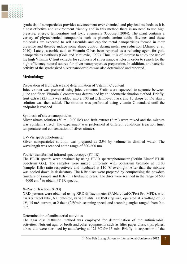
Figure 4 UV-visible spectra of silver nanoparticles prepared using 0.001M AgNO3 at different time (a) guava juice at room temperature (b) guava juice at 60 °C (c) cherry tomato juice at room temperature (d) cherry tomato juice at 60 °C Fourier transforms infrared spectroscopy (FT-IR). Silver nanoparticles synthesized using guava extract was used for FT-IT analysis. FT-IR spectroscopy measurements are carried out to identify the biomolecules that bound specifically on the silver surface and the local molecular environment of capping agent on the nanoparticles. The spectrum of silver nitrate (Figure 5) showed an intense peak at 1383 cm-1 due to nitrate ions (Martin et al. 2011). The peaks at 1730, 1604, 1412, 1261 and 1056 cm-1 in guava extract (Figure 6) match with that of silver nanoparticles (Figure 7). Thus, amount of guava extract remains in the synthesized sample. The peaks at 3301 cm-1of guava extract, 3342 cm-1 of silver nanoparticles are broad and strong assigned to the hydroxyl groups stretching vibration (Panicker et al. 2006). The band at 2925 cm-1 may assign to be a C-H stretching of -CH2- (Figure 7). The peak located at 1723 and 1642 cm-1 assigned to C=O stretching in carboxyl or amino group which is common in several biological compounds. The 1642 cm-1 peak is accompanied of a signal at 1431 cm-1 which is common in this group because they correspond to the asymmetric and symmetric stretching vibration (Bankar et al. 2010). A vibration observed at 1247 cm-1 indicates the presence of stretching C-O bond of ester, which is common in several natural compounds (Figure 7). The intense signal 1059 cm-
1 in both guava extract and silver nanoparticles synthesized by the extract can be attributed to stretching vibrations of C-O-C bonds (Shankar et al. 2003). The FT-IR spectroscopic study confirmed that the guava extract has the ability to perform both reduction and capping functions on the silver nanoparticles.
(c) (a)
(b)(d)

1st Mae Fah Luang University International Conference 2012 8
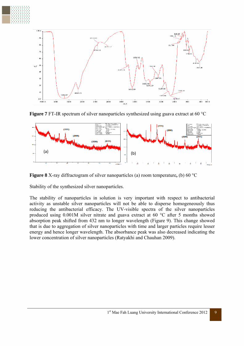
X-ray diffractometry (XRD). The XRD clearly indicates the silver nanoparticles formed by reduction of silver ions by Psidium guajava Linn. (Guava extract). The synthesis of silver nanoparticles at 60 °C showed obvious clear peaks structure of silver at 111, 200, 220 and 311 (Figure 8). The sharpness of peak clearly indicated that the particles are in nanoregime. The diffraction peak of condition 60 °C is sharp and higher than that synthesized at room temperature due to effect of the high temperature which made it easier to grow in a higher concentration of silver ion solution (Nianxin et al. 2011).
Figure 5 FT-IR spectrum of silver nitrate
Figure 6 FT-IR spectrum of guava extract
1st Mae Fah Luang University International Conference 2012 9
Figure 7 FT-IR spectrum of silver nanoparticles synthesized using guava extract at 60 °C
Figure 8 X-ray diffractogram of silver nanoparticles (a) room temperature, (b) 60 °C Stability of the synthesized silver nanoparticles. The stability of nanoparticles in solution is very important with respect to antibacterial activity as unstable silver nanoparticles will not be able to disperse homogeneously thus reducing the antibacterial efficacy. The UV-visible spectra of the silver nanoparticles produced using 0.001M silver nitrate and guava extract at 60 °C after 5 months showed absorption peak shifted from 432 nm to longer wavelength (Figure 9). This change showed that is due to aggregation of silver nanoparticles with time and larger particles require lesser energy and hence longer wavelength. The absorbance peak was also decreased indicating the lower concentration of silver nanoparticles (Ratyakhi and Chauhan 2009).
(a) (b)

1st Mae Fah Luang University International Conference 2012 10
Figure 9 UV-visible spectra of silver nanoparticles at initial and after 5 months at room temperature storage Antibacterial activities of the synthesized silver nanoparticles. The agar disc diffusion method was employed for screening of antibacterial activities of silver nanoparticles compared with silver nitrate at the concentrations of 5, 20, 50 and 200 µg/ml. DI water is a negative control with no inhibition zone and tetracycline as a positive control with clear zone inhibition. The inhibition zone was observed after 24 h incubation at 37 °C (Figure 10). The inhibition zone of samples against Gram-positive (S. aureus) and Gram-negative (E. coli, P. aeruginosa and S. typhimurium) are shown in Table 2. Nanoparticles may infiltrate the cells causing intracellular loss leading to cell death and this inhibition depends on the concentration of the silver nanoparticles (Sondi and Salopek 2004). E. coli and S. aureus are higher sensitivity to silver nanoparticles due to the high lipopolysaccharide and thick peptidoglycan layer of the microorganisms. A gram positive bacterium is made up of rigid peptidoglycan layer and is without the outer membrane (Yoksan and Chirachanchai 2010). The concentration increased the area of inhibition is increased in both AgNO3 and silver nanoparticles. At the low concentration 5 µg/ml silver nanoparticles has antibacterial activity better than and AgNO3. At the concentration 20, 50 and 200 µg/ml silver nanoparticles and AgNO3 is close to each other. However, silver nitrate may not be the choice due to its corrosive black silver stain on the skin when used.

1st Mae Fah Luang University International Conference 2012 11
Figure 10 antibacterial effects varying the concentration of samples (1) 200 µgmL-1 (2) 5 µgmL1 (3) 20 µg/ml (4) 50 µg/ml (5) tetracycline (6) DI water Table 2 The antibacterial activities of silver nanoparticles and silver nitrate at different concentrations
Conclusions Silver nanoparticles have been synthesized using a high Vitamin C fruit extract. Guava contains the highest concentration of Vitamin C and also reacted rapidly with silver ion to produce silver nanoparticles. Reaction time, temperature, concentration of silver nitrate and concentration of reducing agents affect the yield and particle size of the synthesized silver nanoparticles. The FT-IR analysis of silver nanoparticles showed peaks which also found in both spectrum of guava extract and the synthesized nanoparticles, thus amount of guava extract remains in silver nanoparticles and it also confirmed that the guava extract has ability to perform both reducing agent and capping functions on the silver nanoparticles. The XRD analysis showed clear sharp diffraction peaks of silver nanoparticles. The antibacterial activity test examined by disc diffusion method showed that at 5µg/ml concentration, silver nanoparticles have better antibacterial activities than that of AgNO3. However, at higher concentrations the antibacterial activities of silver nanoparticles and AgNO3 were relatively close to each other.
Samples Conc. ( µgmL-1)
Diameter of inhibit zone (mm) E. coli S. aureus P. aeruginosa S. typhimurium
DI water - 0 0 0 0 Tetracycline - 10.0 22.5 16.7 30.0 Silver nanoparticles 200 8.9 8.5 7.0 6.0 50 8.0 8.0 7.0 6.0 20 7.3 8.0 8.0 6.0 5 6.6 7.3 8.0 5.0 Silver nitrate 200 9.0 10.0 10.0 7.3 50 8.0 8.0 6.0 6.6 20 7.0 7.5 5.0 6.0 5 5.3 6.3 0 5.0

1st Mae Fah Luang University International Conference 2012 12
Acknowledgements The authors are grateful to Mae Fah Luang University for support this work. References
1. Ahmad N, Sharma S, Singh VN, Shamsi SF (2010) Rapid synthesis of silver nanoparticles using dried medicinal plant of basil. Colloids and Surfaces B: Biointerfaces. 81-86.
2. Bankar A, Joshi B, Kumar A, Zinjarde S (2010) Banana peel extract mediated route for the synthesis of silver nanoparticles, Colloids and Surfaces A: Physicochemical and Engineering Aspects. 368(1-3): 58-63.
3. Bragg PD, Rainnie DJ (1974) The effect of silver ions on the respiratory chain of Escherichia coli. Canadian Journal of Microbiology 20:883–889.
4. Buzea C et al. (2007) Nanomaterials and nanoparticles: sources and toxicit Biointerphases 2 (4):17–71.
5. Goodsell DS (2004) Bionanotechnology. Lessons from Nature. John Wiley & Sons Inc. Publication.
6. Jha A, Prasad K (2011) Green fruit of chili (Capsicum annum L.) Synthesize nano silver. Digest Journal of Nanomaterials and Biostructures. 6(4): 1717-1723.
7. Jorge L, Jose R, Jason G, Ntebogene S, Guadalupe R (2008) Production of metal nanoparticles by plants and plant-derived materials. Metal Nanoclusters in Catalysis and Material Science. 401-410.
8. Goia DV, Matijevic E (1999) Tailoring the particle size of monodispersed colloidal gold, Colloids and Surfaces A: Physicochemical and Engineering Aspects 146: 139–152.
9. Klasen H (2000) A historical review of the use of silver in the treatment of burns. Renewed interest for silver Burns 26 (2):131–138.
10. Martin J, Cardamone J, Irwin P, Brown E (2011) Keratin capped silver nanoparticles Synthesis and characterization of a nanomaterial with desirable handling properties, Colloids and Surfaces B. Biointerfaces. 88(1): 354-361.
11. Messner KR, Imlay JA (1999) The identification of primary sites of superoxide and hydrogen peroxide formation in the aerobic respiratory chain and sulfite reductase complex of Escherichia coli. The Journal of biological chemistry 274:10119–28.
12. Nalwa HS (2000) Handbook of Nanostructure materials and Nanotechnology. New York: Academic Press.
13. Nianxin X, Yurong C, Tian J, Juming Y (2011) Green synthesis of silver nanoparticles by chemical reduction with hyaluronan. Carbohydrate polymer. 86:956-961.
14. Panicker C, Varghese H, Daizy P (2006) FT-IR, FT-Raman and SERS spectra of Vitamin C, Spectrochimica Acta Part A 65:802-804.
15. Ratyakhi R, Chauhan R (2009) Colloidal synthesis of silver nanoparticles. Asian Journal of Chemistry. 21: 113-116.
16. Shankar S, Ahmad A, Sastry M (2003) Geranium leaf assisted biosynthesis of silver nanoparticles. Biotechnology Progress. 19(6): 1627-1631.
17. Sondi I, Salopek B (2004) Silver nanoparticles as antimicrobial agent: a case study on E. coli as a model for Gram-negative bacteria. Journal of Colloid Interface Science. 275:177–182.

1st Mae Fah Luang University International Conference 2012 13
18. Song JY, Kiml BS (2009) Rapid biological synthesis of silver nanoparticles using plant leave extract. The journal Bioprocess and Biosystems Engineering. 32:79–84.
19. Varna RS, Hoag GE, Collins JB (2011) Green Synthesis of nanometals using fruit extract and use thereof, U.S. Pat. US2011/0110723A1.
20. Yoksan R, Chirachanchai S (2010) Silver nanoparticle-loaded chitosan–starch based films Fabrication and evaluation of tensile barrier and antimicrobial properties. Materials Science and Engineering: 30(6), 891-897.