green synthesis of silver nanoparticles: a review · agnps, green synthesis, silver nano, plant...
TRANSCRIPT
Green and Sustainable Chemistry, 2016, 6, 34-56 Published Online February 2016 in SciRes. http://www.scirp.org/journal/gsc http://dx.doi.org/10.4236/gsc.2016.61004
How to cite this paper: Srikar, S.K., Giri, D.D., Pal, D.B., Mishra, P.K. and Upadhyay, S.N. (2016) Green Synthesis of Silver Nanoparticles: A Review. Green and Sustainable Chemistry, 6, 34-56. http://dx.doi.org/10.4236/gsc.2016.61004
Green Synthesis of Silver Nanoparticles: A Review Sista Kameswara Srikar1,2*, Deen Dayal Giri1,3, Dan Bahadur Pal1, Pradeep Kumar Mishra1, Siddh Nath Upadhyay1* 1Department of Chemical Engineering & Technology, Indian Institute of Technology (BHU) Varanasi, Varanasi, Uttar Pradesh, India 2Tata Steel, Jamshedpur, Jharkhand, India 3Department of Botany, IFTM University, Moradabad, Uttar Pradesh, India
Received 30 October 2015; accepted 26 February 2016; published 29 February 2016
Copyright © 2016 by authors and Scientific Research Publishing Inc. This work is licensed under the Creative Commons Attribution International License (CC BY). http://creativecommons.org/licenses/by/4.0/
Abstract The bio-molecules from various plant components and microbial species have been used as po-tential agents for the synthesis of silver nanoparticles (AgNPs). In spite of a wide range of bio-mol- ecules assisting in the process, synthesizing stable and widely applicable AgNPs by many re-searchers still poses a considerable challenge to the researchers. The biological agents for synthe-sizing AgNPs cover compounds produced naturally in microbes and plants. More than 100 differ-ent biological sources for synthesizing AgNPs are reported in the past decade by various authors. Reaction parameters under which the AgNPs were being synthesized hold prominent impact on their size, shape and application. Available published information on AgNPs synthesis, effects of various parameters, characterization techniques, properties and their application are summa-rised and critically discussed in this review.
Keywords AgNPs, Green Synthesis, Silver Nano, Plant Extract, Microbe
1. Introduction Materials in the nano dimensions (1 - 100 nm) have remarkable difference in the properties compared to the same material in the bulk. These differences lie in the physical and structural properties of atoms, molecules and
*Corresponding author.
S. K. Srikar et al.
35
bulk materials of the element due to difference in physiochemical properties and surface to volume ratio [1]. With advancement in nanotechnology, a large number of nanomaterials are appearing with unique properties, opening spectrum of applications and research opportunities [2].
About 5000 years ago, many Greeks, Romans, Persians and Egyptians used silver in one form or other to store food products [3]. Use of silver ware during ancient period by various dynasties was common across the globe utensils for drinking and eating and storing various drinkable and eatable items probably due to the know-ledge of antimicrobial action [4]. There are records regarding therapeutic application of silver in literature as earlier as 300 BC. In the Hindu religion, till date silver utensils are preferred for the “panchamrit” preparation using curd, Ocimum sanctum and other ingredients. The therapeutic potentials of various metals are mentioned in ancient Indian Aurvedic medicine book medicinal literature named “Charak Samhita” [5]. Until the discovery of antibiotics by Alexzander Flemming, silver was commonly used as antimicrobial agent.
In the recent past, silver nano particles (AgNps) have received enormous attention of the researchers due to their extraordinary defense against wide range of microorganisms and also due to the appearance of drug resis-tance against commonly used antibiotics [2]. The exceptional characteristics of AgNPs have made them appli-cable in various fields like biomedical [6], drug delivery [7], water treatment [8], agricultural etc. [9]. AgNps are applied in inks, adhesives, electronic devises, pastes etc. due to high conductivity [10]. AgNps have been syn-thesized by physio-chemical techniques such as chemical reduction [11], gamma ray radiation [12], micro emul-sion [13], electrochemical method [14], laser ablation [15], autoclave [16], microwave [17] and photochemical reduction [18]. These methods have effective yield, but they are associated with the limitations like use of toxic chemicals and high operational cost and energy needs. Considering the drawbacks of physio-chemical methods, cost-effective and energy efficient new alternative for AgNP synthesis using microorganisms [2], plant extracts [19] and natural polymers [20] as reducing and capping agents are emerging very fast. The association of nano-technology and green chemistry will unfold the range of biologically and cytologically compatible metallic na-noparticles [21] [22].
Over the past decade, few reviews focusing on green synthesis of AgNPs were published [23]-[27]. Most of these reviews focused on several plant and microbial sources for synthesis, several characterization techniques for analysis, certain tabular data representing source, shape and size and information regarding various applica-tions. The present review, unlike the earlier ones, summarizes the synthesis procedure, parameters, characteriza-tions, applications and predicted antibacterial mechanism in a systematic manner, focusing on various green routes for AgNPs synthesis.
2. Green Synthesis The primary requirement of green synthesis of AgNPs is silver metal ion solution and a reducing biological agent. In most of the cases reducing agents or other constituents present in the cells acts as stabilizing and cap-ping agents, so there is no need of adding capping and stabilizing agents from outside.
2.1. Metal Ion Solution The Ag+ ions are primary requirement for the synthesis of AgNPs which can be obtained from various water so-luble salts of silver. However, the aqueous AgNO3 solution with Ag+ ion concentration range between 0.1 - 10 mm (most commonly 1 mm) has been used by the majority of researchers.
2.2. Biological Reducing Agents The reducing agents are widely distributed in the biological systems. The AgNPs have been synthesized using different organisms belonging to four kingdom out of five kingdom of living organisms i.e. Monera (prokaryotic organisms without true nucleus) Protista (unicellular organisms with true nucleus), fungi (eukaryotic, sapro-phyte/parasite), plantae (eukaryotic, autotrophs) and animalia (eukaryotic, heterotrophs). Data are not available regarding use of animal materials for the synthesis of AgNP’ till date to the best of our knowledge. Due to this limitation, green synthesis of AgNPs has been discussed under headings microorganisms, plants, and bio-poly- mers.
Green syntheses of AgNPs have been performed using plant extracts, microbial cell biomass or cell free growth medium and biopolymers. The plants used for AgNps synthesis range from algae to angiosperms; how-ever, limited reports are available for lower plants and the most suitable choice are the angiosperm plants. Parts
S. K. Srikar et al.
36
like leaf, bark, root, and stem have been used for the AgNP synthesis. The medicinally important plants like Boerhaavia diffusa [28], Tinospora cordifolia [29], Aloe vera [30], Terminalia chebula [31] Catharanthus ro-seus [32], Ocimum tenuiflorum [33], Azadirachta indica [34], Emblica officinalis [35], Cocos nucifera [36], common spices Piper nigrum [37]), Cinnamon zeylanicum [38]. Some exotic weeds like Parthenium hystero-phorus [39] growing in uncontrolled manner due to lack of natural enemies and causing health problems have also been used for AgNP’s synthesis. The other group includes alkaloids (Papaver somniferum) and essential oils (Mentha piperita) producing plants. All the plant extracts played dual role of potential reducing and stabi-lizing agents with an exception in few cases where external chemical agents like sodium-do-decyl sulphate were used for stabilization the AgNPs [40]). Metabolites, proteins [41] and chlorophyll [42] present in the plant ex-tracts were found to be acting as capping agents for synthesized AgNPs.
The preferred solvent for extracting reducing agents from the plant is water in most of the cases however, there are few reports regarding the use of organic solvents like methanol [43]-[46], ethanol [47] [48] and ethyl acetate [49]. Some researchers pre-treated the plants materials in saline [39] or acetone [50] atmospheres before extraction. On the whole, even though the extracting solvents differed, the nanoparticle suspensions have made in aqueous medium only. Synthesis using plant extracts generate nanoparticles of well-defined shape, structure and morphology in compared to those obtained through the utilization of bark, tissue and whole plant [51].
The AgNPs synthesis by microbes is strenuous compared to the use of plant extracts and biopolymers as re-ducing and capping agents mainly due to the difficulty in growth, culture maintenance, and inoculums size standardization. Several fungal and bacterial species have been successfully used in the synthesis. The AgNPs synthesis mainly followed one of the two distinct routes, one utilizing extracellular materials secreted in the growth medium whereas the other utilizing microbial cell biomass directly. The microbes synthesize AgNP intracellularly as well as extracellularly. The Intracellular synthesis of AgNPs was observed by few researchers [52].
AgNPs synthesis supports better control on size and shape of AgNPs, due to easy down streaming and larger adaptability to nano systems. However, extracellular AgNP synthesis is been widely reported [53] [54]. One of the commonly used fungal genera for synthesizing AgNPs is Fusarium [53] [55]-[57]. No special capping agent was used in the work of many researchers for stabilizing synthesized AgNPs, except Perni et al. [58] and Shah-verdi et al. [59] who used L-cystine and piperitone as stabilizing agents, respectively. Among the wide varieties off bio-polymers used for AgNP synthesis, almost all played the dual role of reducing and stabilizing agents with an exception of using starch as a capping agent [60].
3. Separation of AgNPs Centrifugation technique is mostly used by researchers to obtain the pellet or powder form of synthesized silver nanoparticles. The AgNPs suspensions were also oven dried to obtain the product in powder form [44].
Some common characterizations of AgNPs include UV-Vis Spectra, SEM, TEM, FTIR, XRD and EDAX or EDX/EDS. DLS study is mostly used for AgNPs synthesized from bio-polymers rather than plant extracts and microorganisms. Zeta potential values indicate the stability of synthesized AgNPs. Thermo-Gravimetric Analysis (TGA) is used to find the effect of AgNO3 and L-cystine on the organic composition of AgNPs [58] to find out the amount of organic material in synthesized AgNPs [61] and predict the thermal stability of AgNPs [62]. Induc-tive Coupled Plasma (ICP) analysis was performed to analyze the concentration and conversion of AgNPs [19].
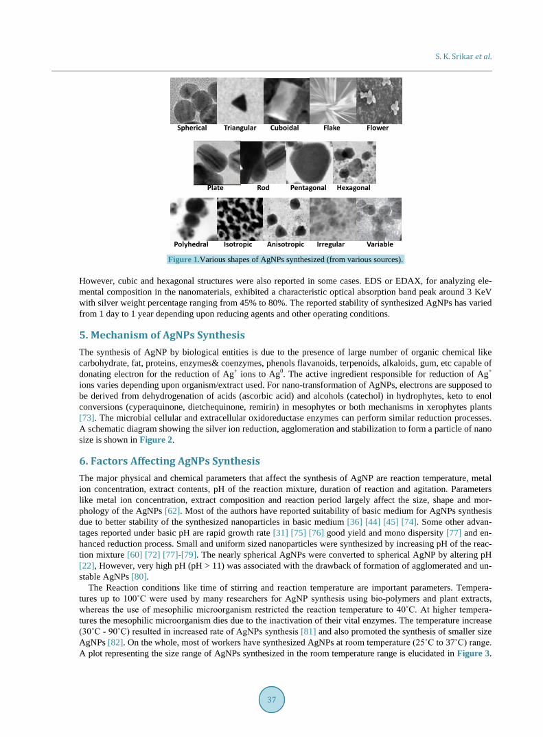
4. Monitoring of AgNPs The appearance of yellow to slight brownish-yellow color in the colorless solution has been taken as indicative of AgNPs synthesis by almost all the researchers. The SPR peak of the synthesized AgNPs was witnessed in the range of 400 - 450 nm, the significant range for AgNPs [63]. The UV-Vis spectral analyses have been used to analyze the dependency of pH, metal ion concentration, extract content on the formation of AgNPs and reveal the size-stability of synthesized AgNPs by exhibiting red shift in the SPR peak with increase in size of nanopar-ticles and blue shift for decrease in size. The SEM morphological analysis in most of the studies revealed spher-ical AgNPs, whereas few authors reported irregular [64], triangular [65], hexagonal [66], isotropic [67], polyhe-dral [60], flake [68], flower [69], pentagonal [70], anisotropic [71] and rod like structures [72]. A pictorial re-presentation of SEM/TEM images of AgNPs with different shapes is shown in Figure 1. Using XRD studies of almost all the researchers reported the formation of face centered cubic (FCC) crystalline structured AgNPs.
S. K. Srikar et al.
37
Figure 1.Various shapes of AgNPs synthesized (from various sources).
However, cubic and hexagonal structures were also reported in some cases. EDS or EDAX, for analyzing ele-mental composition in the nanomaterials, exhibited a characteristic optical absorption band peak around 3 KeV with silver weight percentage ranging from 45% to 80%. The reported stability of synthesized AgNPs has varied from 1 day to 1 year depending upon reducing agents and other operating conditions.
5. Mechanism of AgNPs Synthesis The synthesis of AgNP by biological entities is due to the presence of large number of organic chemical like carbohydrate, fat, proteins, enzymes& coenzymes, phenols flavanoids, terpenoids, alkaloids, gum, etc capable of donating electron for the reduction of Ag+ ions to Ag0. The active ingredient responsible for reduction of Ag+
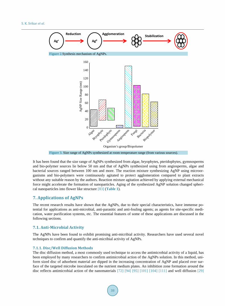
ions varies depending upon organism/extract used. For nano-transformation of AgNPs, electrons are supposed to be derived from dehydrogenation of acids (ascorbic acid) and alcohols (catechol) in hydrophytes, keto to enol conversions (cyperaquinone, dietchequinone, remirin) in mesophytes or both mechanisms in xerophytes plants [73]. The microbial cellular and extracellular oxidoreductase enzymes can perform similar reduction processes. A schematic diagram showing the silver ion reduction, agglomeration and stabilization to form a particle of nano size is shown in Figure 2.
6. Factors Affecting AgNPs Synthesis The major physical and chemical parameters that affect the synthesis of AgNP are reaction temperature, metal ion concentration, extract contents, pH of the reaction mixture, duration of reaction and agitation. Parameters like metal ion concentration, extract composition and reaction period largely affect the size, shape and mor-phology of the AgNPs [62]. Most of the authors have reported suitability of basic medium for AgNPs synthesis due to better stability of the synthesized nanoparticles in basic medium [36] [44] [45] [74]. Some other advan-tages reported under basic pH are rapid growth rate [31] [75] [76] good yield and mono dispersity [77] and en-hanced reduction process. Small and uniform sized nanoparticles were synthesized by increasing pH of the reac-tion mixture [60] [72] [77]-[79]. The nearly spherical AgNPs were converted to spherical AgNP by altering pH [22], However, very high pH (pH > 11) was associated with the drawback of formation of agglomerated and un-stable AgNPs [80].
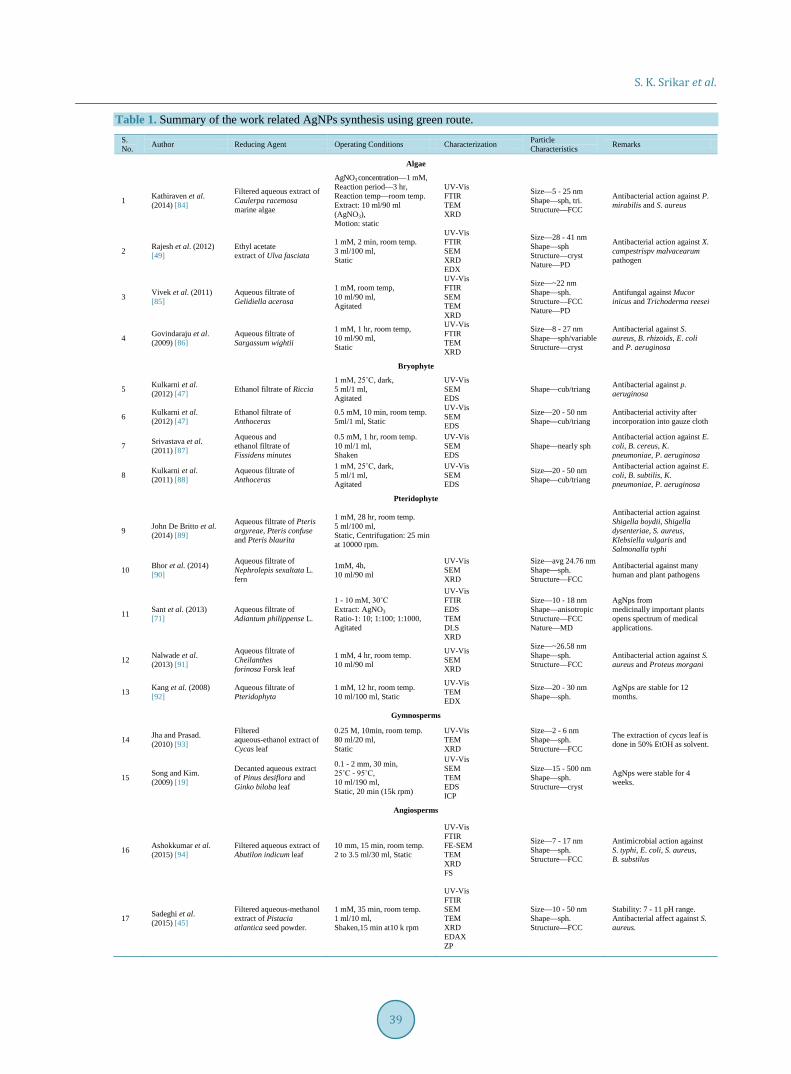
The Reaction conditions like time of stirring and reaction temperature are important parameters. Tempera-tures up to 100˚C were used by many researchers for AgNP synthesis using bio-polymers and plant extracts, whereas the use of mesophilic microorganism restricted the reaction temperature to 40˚C. At higher tempera-tures the mesophilic microorganism dies due to the inactivation of their vital enzymes. The temperature increase (30˚C - 90˚C) resulted in increased rate of AgNPs synthesis [81] and also promoted the synthesis of smaller size AgNPs [82]. On the whole, most of workers have synthesized AgNPs at room temperature (25˚C to 37˚C) range. A plot representing the size range of AgNPs synthesized in the room temperature range is elucidated in Figure 3.
Spherical Triangular FlakeCuboidal
Plate
Flower
PentagonalRod Hexagonal
IrregularIsotropic VariableAnisotropicPolyhedral
S. K. Srikar et al.
38
Figure 2.Synthesis mechanism of AgNPs.
Figure 3. Size range of AgNPs synthesized at room temperature range (from various sources).
It has been found that the size range of AgNPs synthesized from algae, bryophytes, pteridophytes, gymnosperms and bio-polymer sources lie below 50 nm and that of AgNPs synthesized using from angiosperms, algae and bacterial sources ranged between 100 nm and more. The reaction mixture synthesizing AgNP using microor-ganisms and bio-polymers were continuously agitated to protect agglomeration compared to plant extracts without any suitable reason by the authors. Reaction mixture agitation achieved by applying external mechanical force might accelerate the formation of nanoparticles. Aging of the synthesized AgNP solution changed spheri-cal nanoparticles into flower like structure [83] (Table 1).
7. Applications of AgNPs The recent research results have shown that the AgNPs, due to their special characteristics, have immense po-tential for applications as anti-microbial, anti-parasitic and anti-fouling agents; as agents for site-specific medi-cation, water purification systems, etc. The essential features of some of these applications are discussed in the following sections.
7.1. Anti-Microbial Activity The AgNPs have been found to exhibit promising anti-micribial activity. Researchers have used several novel techniques to confirm and quantify the anti-micribial activity of AgNPs.
7.1.1. Disc/Well Diffusion Methods The disc diffusion method, a most commonly used technique to access the antimicrobial activity of a liquid, has been employed by many researchers to confirm antimicrobial action of the AgNPs solution. In this method, uni-form sized disc of adsorbent material are dipped in the increasing concentration of AgNP and placed over sur-face of the targeted microbe inoculated on the nutrient medium plates. An inhibition zone formation around the disc reflects antimicrobial action of the nanomaterials [72] [94] [95] [101] [104] [111] and well diffusion [29]
Ag+ Ag0
Reduction Agglomeration Stabilization
Organism’s group/Biopolumer
AgN
PSi
ze R
ange
(nm
)
160
140
120
100
80
60
40
20
0
S. K. Srikar et al.
39
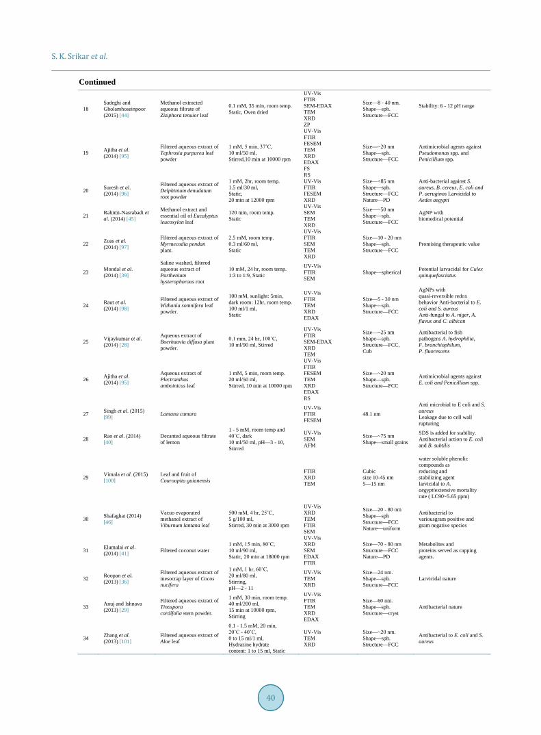
Table 1. Summary of the work related AgNPs synthesis using green route. S. No. Author Reducing Agent Operating Conditions Characterization Particle
Characteristics Remarks
Algae
1 Kathiraven et al. (2014) [84]
Filtered aqueous extract of Caulerpa racemosa marine algae
AgNO3 concentration—1 mM, Reaction period—3 hr, Reaction temp—room temp. Extract: 10 ml/90 ml (AgNO3), Motion: static
UV-Vis FTIR TEM XRD
Size—5 - 25 nm Shape—sph, tri. Structure—FCC
Antibacterial action against P. mirabilis and S. aureus
2 Rajesh et al. (2012) [49]
Ethyl acetate extract of Ulva fasciata
1 mM, 2 min, room temp. 3 ml/100 ml, Static
UV-Vis FTIR SEM XRD EDX
Size—28 - 41 nm Shape—sph Structure—cryst Nature—PD
Antibacterial action against X. campestrispv malvacearum pathogen
3 Vivek et al. (2011) [85]
Aqueous filtrate of Gelidiella acerosa
1 mM, room temp, 10 ml/90 ml, Agitated
UV-Vis FTIR SEM TEM XRD
Size—~22 nm Shape—sph. Structure—FCC Nature—PD
Antifungal against Mucor inicus and Trichoderma reesei
4 Govindaraju et al. (2009) [86]
Aqueous filtrate of Sargassum wightii
1 mM, 1 hr, room temp, 10 ml/90 ml, Static
UV-Vis FTIR TEM XRD
Size—8 - 27 nm Shape—sph/variable Structure—cryst
Antibacterial against S. aureus, B. rhizoids, E. coli and P. aeruginosa
Bryophyte
5 Kulkarni et al. (2012) [47] Ethanol filtrate of Riccia
1 mM, 25˚C, dark, 5 ml/1 ml, Agitated
UV-Vis SEM EDS
Shape—cub/triang Antibacterial against p. aeruginosa
6 Kulkarni et al. (2012) [47]
Ethanol filtrate of Anthoceras
0.5 mM, 10 min, room temp. 5ml/1 ml, Static
UV-Vis SEM EDS
Size—20 - 50 nm Shape—cub/triang
Antibacterial activity after incorporation into gauze cloth
7 Srivastava et al. (2011) [87]
Aqueous and ethanol filtrate of Fissidens minutes
0.5 mM, 1 hr, room temp. 10 ml/1 ml, Shaken
UV-Vis SEM EDS
Shape—nearly sph Antibacterial action against E. coli, B. cereus, K. pneumoniae, P. aeruginosa
8 Kulkarni et al. (2011) [88]
Aqueous filtrate of Anthoceras
1 mM, 25˚C, dark, 5 ml/1 ml, Agitated
UV-Vis SEM EDS
Size—20 - 50 nm Shape—cub/triang
Antibacterial action against E. coli, B. subtilis, K. pneumoniae, P. aeruginosa
Pteridophyte
9 John De Britto et al. (2014) [89]
Aqueous filtrate of Pteris argyreae, Pteris confuse and Pteris blaurita
1 mM, 28 hr, room temp. 5 ml/100 ml, Static, Centrifugation: 25 min at 10000 rpm.
Antibacterial action against Shigella boydii, Shigella dysenteriae, S. aureus, Klebsiella vulgaris and Salmonalla typhi
10 Bhor et al. (2014) [90]
Aqueous filtrate of Nephrolepis sexaltata L. fern
1mM, 4h, 10 ml/90 ml
UV-Vis SEM XRD
Size—avg 24.76 nm Shape—sph. Structure—FCC
Antibacterial against many human and plant pathogens
11 Sant et al. (2013) [71]
Aqueous filtrate of Adiantum philippense L.
1 - 10 mM, 30˚C Extract: AgNO3 Ratio-1: 10; 1:100; 1:1000, Agitated
UV-Vis FTIR EDS TEM DLS XRD
Size—10 - 18 nm Shape—anisotropic Structure—FCC Nature—MD
AgNps from medicinally important plants opens spectrum of medical applications.
12 Nalwade et al. (2013) [91]
Aqueous filtrate of Cheilanthes forinosa Forsk leaf
1 mM, 4 hr, room temp. 10 ml/90 ml
UV-Vis SEM XRD
Size—~26.58 nm Shape—sph. Structure—FCC
Antibacterial action against S. aureus and Proteus morgani
13 Kang et al. (2008) [92]
Aqueous filtrate of Pteridophyta
1 mM, 12 hr, room temp. 10 ml/100 ml, Static
UV-Vis TEM EDX
Size—20 - 30 nm Shape—sph.
AgNps are stable for 12 months.
Gymnosperms
14 Jha and Prasad. (2010) [93]
Filtered aqueous-ethanol extract of Cycas leaf
0.25 M, 10min, room temp. 80 ml/20 ml, Static
UV-Vis TEM XRD
Size—2 - 6 nm Shape—sph. Structure—FCC
The extraction of cycas leaf is done in 50% EtOH as solvent.
15 Song and Kim. (2009) [19]
Decanted aqueous extract of Pinus desiflora and Ginko biloba leaf
0.1 - 2 mm, 30 min, 25˚C - 95˚C, 10 ml/190 ml, Static, 20 min (15k rpm)
UV-Vis SEM TEM EDS ICP
Size—15 - 500 nm Shape—sph. Structure—cryst
AgNps were stable for 4 weeks.
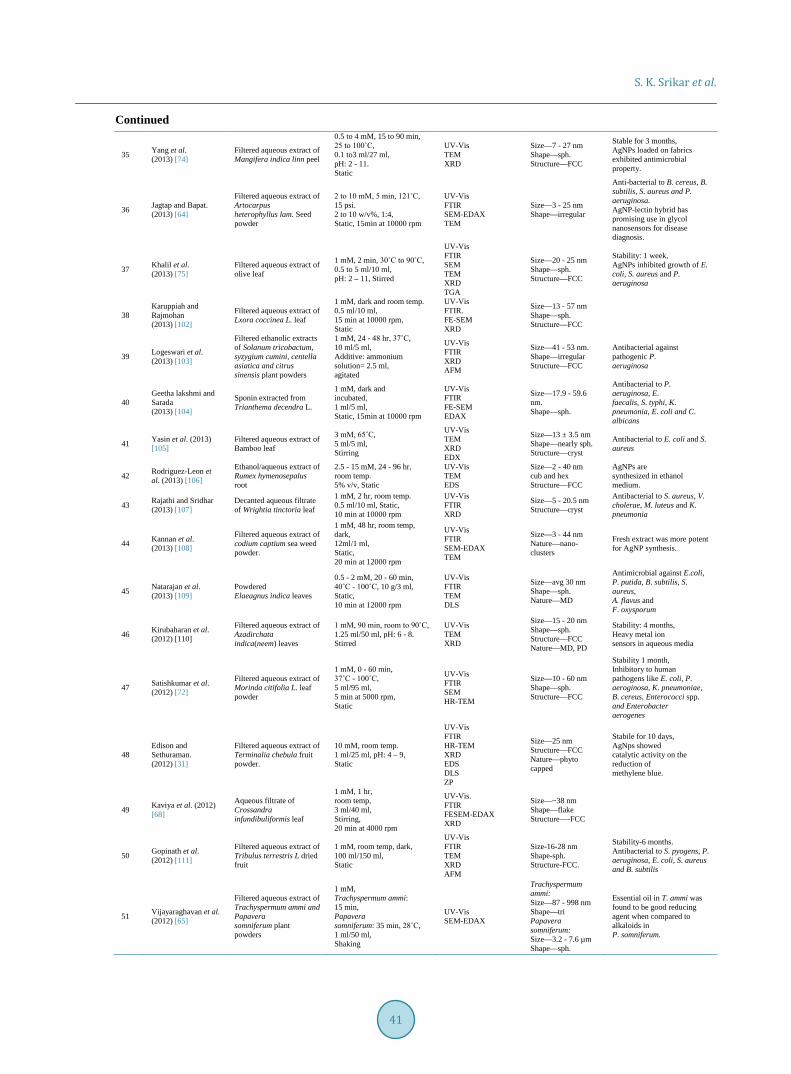
Angiosperms
16 Ashokkumar et al. (2015) [94]
Filtered aqueous extract of Abutilon indicum leaf
10 mm, 15 min, room temp. 2 to 3.5 ml/30 ml, Static
UV-Vis FTIR FE-SEM TEM XRD FS
Size—7 - 17 nm Shape—sph. Structure—FCC
Antimicrobial action against S. typhi, E. coli, S. aureus, B. substilus
17 Sadeghi et al. (2015) [45]
Filtered aqueous-methanol extract of Pistacia atlantica seed powder.
1 mM, 35 min, room temp. 1 ml/10 ml, Shaken,15 min at10 k rpm
UV-Vis FTIR SEM TEM XRD EDAX ZP
Size—10 - 50 nm Shape—sph. Structure—FCC
Stability: 7 - 11 pH range. Antibacterial affect against S. aureus.
S. K. Srikar et al.
40
Continued
18 Sadeghi and Gholamhoseinpoor (2015) [44]
Methanol extracted aqueous filtrate of Ziziphora tenuior leaf
0.1 mM, 35 min, room temp. Static, Oven dried
UV-Vis FTIR SEM-EDAX TEM XRD ZP
Size—8 - 40 nm. Shape—sph. Structure—FCC
Stability: 6 - 12 pH range
19 Ajitha et al. (2014) [95]
Filtered aqueous extract of Tephrosia purpurea leaf powder
1 mM, 5 min, 37˚C, 10 ml/50 ml, Stirred,10 min at 10000 rpm
UV-Vis FTIR FESEM TEM XRD EDAX FS RS
Size—~20 nm Shape—sph. Structure—FCC
Antimicrobial agents against Pseudomonas spp. and Penicillium spp.
20 Suresh et al. (2014) [96]
Filtered aqueous extract of Delphinium denudatum root powder
1 mM, 2hr, room temp. 1.5 ml/30 ml, Static, 20 min at 12000 rpm
UV-Vis FTIR FESEM XRD
Size—<85 nm Shape—sph. Structure—FCC Nature—PD
Anti-bacterial against S. aureus, B. cereus, E. coli and P. aeruginos Larvicidal to Aedes aegypti
21 Rahimi-Nasrabadi et al. (2014) [45]
Methanol extract and essential oil of Eucalyptus leucoxylon leaf
120 min, room temp. Static
UV-Vis SEM TEM XRD
Size—~50 nm Shape—sph. Structure—FCC
AgNP with biomedical potential
22 Zuas et al. (2014) [97]
Filtered aqueous extract of Myrmecodia pendan plant.
2.5 mM, room temp. 0.3 ml/60 ml, Static
UV-Vis FTIR SEM TEM XRD
Size—10 - 20 nm Shape—sph. Structure—FCC
Promising therapeutic value
23 Mondal et al. (2014) [39]
Saline washed, filtered aqueous extract of Parthenium hysterophorous root
10 mM, 24 hr, room temp. 1:3 to 1:9, Static
UV-Vis FTIR SEM
Shape—spherical Potential larvacidal for Culex quinquefasciatus
24 Raut et al. (2014) [98]
Filtered aqueous extract of Withania somnifera leaf powder.
100 mM, sunlight: 5min, dark room: 12hr, room temp. 100 ml/1 ml, Static
UV-Vis FTIR TEM XRD EDAX
Size—5 - 30 nm Shape—sph. Structure—FCC
AgNPs with quasi-reversible redox behavior Anti-bacterial to E. coli and S. aureus Anti-fungal to A. niger, A. flavus and C. albican
25 Vijaykumar et al. (2014) [28]
Aqueous extract of Boerhaavia diffusa plant powder.
0.1 mm, 24 hr, 100˚C, 10 ml/90 ml, Stirred
UV-Vis FTIR SEM-EDAX XRD TEM
Size—~25 nm Shape—sph. Structure—FCC, Cub
Antibacterial to fish pathogens A. hydrophilia, F. branchiophilum, P. fluorescens
26 Ajitha et al. (2014) [95]
Aqueous extract of Plectranthus amboinicus leaf
1 mM, 5 min, room temp. 20 ml/50 ml, Stirred, 10 min at 10000 rpm
UV-Vis FTIR FESEM TEM XRD EDAX RS
Size—~20 nm Shape—sph. Structure—FCC
Antimicrobial agents against E. coli and Penicillium spp.
27 Singh et al. (2015) [99] Lantana camara
UV-Vis FTIR FESEM
48.1 nm
Anti microbial to E coli and S. aureus Leakage due to cell wall rupturing
28 Rao et al. (2014) [40]
Decanted aqueous filtrate of lemon
1 - 5 mM, room temp and 40˚C, dark 10 ml/50 ml, pH—3 - 10, Stirred
UV-Vis SEM AFM
Size—~75 nm Shape—small grains
SDS is added for stability. Antibacterial action to E. coli and B. subtilis
29 Vimala et al. (2015) [100]
Leaf and fruit of Couroupita guianensis
FTIR XRD TEM
Cubic size 10-45 nm 5—15 nm
water soluble phenolic compounds as reducing and stabilizing agent larvicidal to A. aegyptiextensive mortality rate ( LC90~5.65 ppm)
30 Shafaghat (2014) [46]
Vacuo evaporated methanol extract of Viburnum lantana leaf
500 mM, 4 hr, 25˚C, 5 g/100 ml, Stirred, 30 min at 3000 rpm
UV-Vis XRD TEM FTIR SEM
Size—20 - 80 nm Shape—sph Structure—FCC Nature—uniform
Antibacterial to variousgram positive and gram negative species
31 Elumalai et al. (2014) [41] Filtered coconut water
1 mM, 15 min, 80˚C, 10 ml/90 ml, Static, 20 min at 18000 rpm
UV-Vis XRD SEM EDAX FTIR
Size—70 - 80 nm Structure—FCC Nature—PD
Metabolites and proteins served as capping agents.
32 Roopan et al. (2013) [36]
Filtered aqueous extract of mesocrap layer of Cocos nucifera
1 mM, 1 hr, 60˚C, 20 ml/80 ml, Stirring, pH—2 - 11
UV-Vis TEM XRD
Size—24 nm. Shape—sph. Structure—FCC
Larvicidal nature
33 Anuj and Ishnava (2013) [29]
Filtered aqueous extract of Tinospora cordifolia stem powder.
1 mM, 30 min, room temp. 40 ml/200 ml, 15 min at 10000 rpm, Stirring
UV-Vis FTIR TEM XRD EDAX
Size—60 nm. Shape—sph. Structure—cryst
Antibacterial nature
34 Zhang et al. (2013) [101]
Filtered aqueous extract of Aloe leaf
0.1 - 1.5 mM, 20 min, 20˚C - 40˚C, 0 to 15 ml/1 ml, Hydrazine hydrate content: 1 to 15 ml, Static
UV-Vis TEM XRD
Size—~20 nm. Shape—sph. Structure—FCC
Antibacterial to E. coli and S. aureus
S. K. Srikar et al.
41
Continued
35 Yang et al. (2013) [74]
Filtered aqueous extract of Mangifera indica linn peel
0.5 to 4 mM, 15 to 90 min, 25 to 100˚C, 0.1 to3 ml/27 ml, pH: 2 - 11. Static
UV-Vis TEM XRD
Size—7 - 27 nm Shape—sph. Structure—FCC
Stable for 3 months, AgNPs loaded on fabrics exhibited antimicrobial property.
36 Jagtap and Bapat. (2013) [64]
Filtered aqueous extract of Artocarpus heterophyllus lam. Seed powder
2 to 10 mM, 5 min, 121˚C, 15 psi. 2 to 10 w/v%, 1:4, Static, 15min at 10000 rpm
UV-Vis FTIR SEM-EDAX TEM
Size—3 - 25 nm Shape—irregular
Anti-bacterial to B. cereus, B. subtilis, S. aureus and P. aeruginosa. AgNP-lectin hybrid has promising use in glycol nanosensors for disease diagnosis.
37 Khalil et al. (2013) [75]
Filtered aqueous extract of olive leaf
1 mM, 2 min, 30˚C to 90˚C, 0.5 to 5 ml/10 ml, pH: 2 – 11, Stirred
UV-Vis FTIR SEM TEM XRD TGA
Size—20 - 25 nm Shape—sph. Structure—FCC
Stability: 1 week, AgNPs inhibited growth of E. coli, S. aureus and P. aeruginosa
38 Karuppiah and Rajmohan (2013) [102]
Filtered aqueous extract of Lxora coccinea L. leaf
1 mM, dark and room temp. 0.5 ml/10 ml, 15 min at 10000 rpm, Static
UV-Vis FTIR. FE-SEM XRD
Size—13 - 57 nm Shape—sph. Structure—FCC
39 Logeswari et al. (2013) [103]
Filtered ethanolic extracts of Solanum tricobactum, syzygium cumini, centella asiatica and citrus sinensis plant powders
1 mM, 24 - 48 hr, 37˚C, 10 ml/5 ml, Additive: ammonium solution= 2.5 ml, agitated
UV-Vis FTIR XRD AFM
Size—41 - 53 nm. Shape—irregular Structure—FCC
Antibacterial against pathogenic P. aeruginosa
40 Geetha lakshmi and Sarada (2013) [104]
Sponin extracted from Trianthema decendra L.
1 mM, dark and incubated, 1 ml/5 ml, Static, 15min at 10000 rpm
UV-Vis FTIR FE-SEM EDAX
Size—17.9 - 59.6 nm. Shape—sph.
Antibacterial to P. aeruginosa, E. faecalis, S. typhi, K. pneumonia, E. coli and C. albicans
41 Yasin et al. (2013) [105]
Filtered aqueous extract of Bamboo leaf
3 mM, 65˚C, 5 ml/5 ml, Stirring
UV-Vis TEM XRD EDX
Size—13 ± 3.5 nm Shape—nearly sph. Structure—cryst
Antibacterial to E. coli and S. aureus
42 Rodriguez-Leon et al. (2013) [106]
Ethanol/aqueous extract of Rumex hymenosepalus root
2.5 - 15 mM, 24 - 96 hr, room temp. 5% v/v, Static
UV-Vis TEM EDS
Size—2 - 40 nm cub and hex Structure—FCC
AgNPs are synthesized in ethanol medium.
43 Rajathi and Sridhar (2013) [107]
Decanted aqueous filtrate of Wrightia tinctoria leaf
1 mM, 2 hr, room temp. 0.5 ml/10 ml, Static, 10 min at 10000 rpm
UV-Vis FTIR XRD
Size—5 - 20.5 nm Structure—cryst
Antibacterial to S. aureus, V. cholerae, M. luteus and K. pneumonia
44 Kannan et al. (2013) [108]
Filtered aqueous extract of codium captium sea weed powder.
1 mM, 48 hr, room temp, dark, 12ml/1 ml, Static, 20 min at 12000 rpm
UV-Vis FTIR SEM-EDAX TEM
Size—3 - 44 nm Nature—nano- clusters
Fresh extract was more potent for AgNP synthesis.
45 Natarajan et al. (2013) [109]
Powdered Elaeagnus indica leaves
0.5 - 2 mM, 20 - 60 min, 40˚C - 100˚C, 10 g/3 ml, Static, 10 min at 12000 rpm
UV-Vis FTIR TEM DLS
Size—avg 30 nm Shape—sph. Nature—MD
Antimicrobial against E.coli, P. putida, B. subtilis, S. aureus, A. flavus and F. oxysporum
46 Kirubaharan et al. (2012) [110]
Filtered aqueous extract of Azadirchata indica(neem) leaves
1 mM, 90 min, room to 90˚C, 1.25 ml/50 ml, pH: 6 - 8. Stirred
UV-Vis TEM XRD
Size—15 - 20 nm Shape—sph. Structure—FCC Nature—MD, PD
Stability: 4 months, Heavy metal ion sensors in aqueous media
47 Satishkumar et al. (2012) [72]
Filtered aqueous extract of Morinda citifolia L. leaf powder
1 mM, 0 - 60 min, 37˚C - 100˚C, 5 ml/95 ml, 5 min at 5000 rpm, Static
UV-Vis FTIR SEM HR-TEM
Size—10 - 60 nm Shape—sph. Structure—FCC
Stability 1 month, Inhibitory to human pathogens like E. coli, P. aeroginosa, K. pneumoniae, B. cereus, Enterococci spp. and Enterobacter aerogenes
48 Edison and Sethuraman. (2012) [31]
Filtered aqueous extract of Terminalia chebula fruit powder.
10 mM, room temp. 1 ml/25 ml, pH: 4 – 9, Static
UV-Vis FTIR HR-TEM XRD EDS DLS ZP
Size—25 nm Structure—FCC Nature—phyto capped
Stabile for 10 days, AgNps showed catalytic activity on the reduction of methylene blue.
49 Kaviya et al. (2012) [68]
Aqueous filtrate of Crossandra infundibuliformis leaf
1 mM, 1 hr, room temp, 3 ml/40 ml, Stirring, 20 min at 4000 rpm
UV-Vis. FTIR FESEM-EDAX XRD
Size—~38 nm Shape—flake Structure—-FCC
50 Gopinath et al. (2012) [111]
Filtered aqueous extract of Tribulus terrestris L dried fruit
1 mM, room temp, dark, 100 ml/150 ml, Static
UV-Vis FTIR TEM XRD AFM
Size-16-28 nm Shape-sph. Structure-FCC.
Stability-6 months. Antibacterial to S. pyogens, P. aeruginosa, E. coli, S. aureus and B. subtilis
51 Vijayaraghavan et al. (2012) [65]
Filtered aqueous extract of Trachyspermum ammi and Papavera somniferum plant powders
1 mM, Trachyspermum ammi: 15 min, Papavera somniferum: 35 min, 28˚C, 1 ml/50 ml, Shaking
UV-Vis SEM-EDAX
Trachyspermum ammi: Size—87 - 998 nm Shape—tri Papavera somniferum: Size—3.2 - 7.6 µm Shape—sph.
Essential oil in T. ammi was found to be good reducing agent when compared to alkaloids in P. somniferum.
S. K. Srikar et al.
42
Continued
52 Sreekanth et al. (2012) [69]
Dioscorea batatas rhizome powder
1 mM, 25 and 80˚C Static, 20 min at 5000 rpm
UV-Vis FTIR SEM XRD
Shape—circular and flower Structure—FCC Nature—MD
53 Chaudhary et al. (2012) [112]
Aqueous filtrate of Vitis viniera fruit
1 mM, 10 hr, room temp. 10 ml/90 ml, Static.15 min at 2000 rpm
UV-Vis SEM XRD
Size—10 - 880 nm Shape—sph Structure—FCC, cubic and hexl
Antibacterial to B. subtilis, E. coli, P. aeruginosa and S. pnemoniae
54 Ashok kumar (2012) [113]
Aqueous filtrate of Prathemium hysterophorus plant
1 mM, 24 hr, room temp. 1 ml/9 ml, Static, 20 min at 5000 rpm
UV-Vis FTIR SEM XRD
Size—avg 10 nm Shape—nearly sph Structure—FCC
55 Patil et al. (2012) [33]
Filtered aqueous extract of Ocimum tenuiflorum leaf
1 mM, 10 min, room temp. 2 ml/20 ml, Static
UV-Vis TEM PS ZP
Size—15-25 nm Shape-sph Structure—FCC
Antibacterial against E. coli, C. bacterium, B. subtilis
Arunachalam et al. (2013) [114]
Indigofera aspalathoides, aqueos leaf t extracts
UV Vis SEM EDAX FTIR
Size—20 - 50 nm
Water-soluble organics leaf extract responsible to reduction. Wound healing applications
56 Mubarakali et al. (2011) [115]
Filtered aqueous extract of Mentha piperita plant powder
1 mM, 24 hr, 28˚C, 1.5 ml/30 ml, Static, 10 min at 6000 rpm
UV-Vis FTIR SEM EDS
Size—90 nm Shape—sph.
Active against clinically isolated human pathogens like E. coli and S. aureus.
57 Mukunthan et al. (2011) [32]
Aqueous extract of Catharanthus roseus leaf
1 mM, 15 min, 80˚C, 10 ml/90 ml, Static
UV-Vis SEM XRD EDAX
Size—48 - 67 nm Structure—FCC Nature—uniform
Antibacterial activity against S. aureus, E. coli, K. pneumoniae, B. aureus and P. aeruginosa
58 Rajakumar and Abdul Rahuman (2011) [70]
Filtered aqueous extract of Eclipta prostrate leaf
1 mM, 1 hr, room temp. 12 ml/88 ml, 45 min at 10000 rpm, Static
UV-Vis FTIR SEM TEM XRD
Size—35 - 60 nm Shape—TEM: sph. SEM: triang, hex and pentagon Structure— crystalline Nature—biphasic
Stabile for 6 hr Larvicidal to filariasis vector C. quinquefasciatus and malarial vector A. subpictus
59 Kumar and Yadav. (2011) [116]
Filtered aqueous extract of Lonicera japonica L leaf.
1 to 9 mM, 24 hr, 40˚C - 80˚C, 5% to 40% (v/v), Static, 5 min at 10000 rpm
UV-Vis FTIR SEM TEM AFM ZP
Size—36 - 72 nm Shape—sph, plate, and other shaped
Stability: zeta potential—41mV
60 Gnanadesigan et al. (2011) [117]
Filtered aqueous extract of Rizophora mucronata leaf
1 M, 10 min, room temp. 10 ml/90 ml, 20 min at 12000 rpm, Static
UV-Vis FTIR XRD AFM
Size—60 - 95 nm Shape—sph. Structure—cryst
Larvicidal to Ae. aegypti and Cx. quinquefasciatus
61 Rani and Reddy (2011) [118]
Decanted aqueous extract of Piper betel L. leaf
1 mM, 1 min to 2 hr, room temp, sunlight, 10 ml/190 ml, Static, 15min at 6000 rpm.
UV-Vis FTIR TEM XRD
Sunlight: 5min. Size—~120 nm Shape—irregular Structure—FCC Nature— agglomerated Sunlight: 10 - 80 min Size—28 - 17 nm Shape—sph. Structure—FCC Nature—shelled AgNP
AgNP toxic to aquatic plant D. magna. Biosynthesized AgNP less toxic compared to chemically synthesized ones
62 Veerasamy et al. (2011) [119]
Aqueous filtrate of Garcinia mangostana leaf
0.25 - 5 mM, 0 - 70 min, 37˚C - 90˚C, 5 ml/95 ml, Static, pH—4, 7, 8 30 min (5k rpm)
UV-Vis FTIR TEM
Size—avg 35 nm Shape—sph
Stable for 30 days, Antibacterial against E. coli and S. aureus
63 Santoshkumar et al. (2011) [120]
Decanted aqueous filtrate of Nelumbo nucifera leaf
1 mM, 10 min, room temp. 12 ml/8 ml, Static
UV-Vis FTIR TEM XRD
Size—25 - 80 nm Shape—sph, tri and dec Structure—FCC
Larvicidal against A. subpictus and C. quinquefasciatus
64 Ahmad et al. (2011) [121]
Aqueous extract of Desmodium triflorum
0.025 M, 1 hr UV-Vis TEM XRD
Size—5 - 20 nm Structure—cryst
Antibacterial against S. spp, E. coli, B. subtilis
65 Prathna et al. (2011) [122]
Filtered and centrifuged juice of Citruslimon fruit
0.1 - 10 mM, 4 hr, 30˚C, 1:4 to 4:1, Shaken, 10 min at 10000 rpm
UV-Vis XRD TEM FTIR AFM DLS ZP
Size—~50 nm Shape—nearly sph. Structure—cryst Nature—PD
AgNPs were stable for 14 days. Size-XRD-18.306 nm AFM—<100 nm TEM—25 - 50 nm DLS—153.68 nm
66 Bankar et al (2010) [50]
Acetone treated, aqueous extracted, filtered and precipitated powder of Banana peel
0.125 to 1mM, 3 min, 40˚C to 100˚C, 0.5 to 10 mg/2 ml, pH: 2 – 5, Static
UV-Vis FTIR. SEM-EDS XRD
Size—< 100 nm Structure—FCC
Antifungal and antibacterial action
67 Njagi et al. (2010) [123]
Filtered aqueous extract of Sorghum bran
0.1 M, 1min, room temp. 2:1 volume ratio, Shaken
UV-Vis FE-SEM HR-TEM-EDS XRD
Size—10 nm Shape—sph. Structure—FCC Nature—uniform nano clusters
AgNP of smaller size at 50˚C of extraction temperature compared to 25˚C and 80˚C
S. K. Srikar et al.
43
Continued
68 Kumar et al. (2010) [124]
Filtered aqueous extract of Syzygium cumini leaf (LE) and seed (SE) powder
1 mM, 24 hr, room temp. 10% (v/v), Static, 20 min, 12k rpm
UV-Vis FTIR SEM AFM
Size—LE: 30nm, Water content of LE: 29 nm, SE: 92 nm, Water content of LE: 73nm.
SE have higher synthesis rates and larger size AgNP compared to LE.
69 Dubey et al. (2010) [79]
Filtered aqueous extract of Tanaetum vulgare fruit.
1 - 3 mM, 10 min - 5hr, 25˚C - 150˚C, 0.5 - 4.8 ml/50 ml, pH: 2 - 10, Static
UV-Vis FTIR TEM XRD EDAX
Size—10 - 40 nm Shape—sph. Structure—FCC
AgNP more stable in basic compared to acidic medium
70 Shukla et al. (2010) [37]
Filtered aqueous extract of Piper nigrum (black pepper)
10 mM, room temp. 1 ml/100 ml, Stirred, 10 min at 3000 rpm
UV-Vis TEM XRD
Size—20 - 50 nm Shape—sph. Structure—FCC Nature—large grain, WD, uniform and polycrystalline
71 Krishnaraj et al. (2010) [125]
Aqueous filtrate of Acalypha indica leaf
1 mM, 30 min, 37˚C, dark 12 ml/100 ml, Static, 30 min at 75000 g
UV-Vis SEM TEM EDS XRD
Size—20 - 30 nm Structure—cub
Antimicrobial against water borne pathogens E. coli and Vibrio cholera
72 Satish kumar et al. (2009) [38]
Aqueous bark and powder extracts of Cinnamon zeylanicum plant
1 mM, 25˚C, 1 to 5 ml/50 ml, Powder content: 0.1 to 1 g/50 ml, pH: 1 - 11, Shaken
UV-Vis TEM XRD EDX
Size—powder: 31 nm, Extract: 40 nm Shape—quasi sph and R, Structure— cub and hex Nature—bi-phasic
Stable for 3 months, Served as antimicrobial agents
73 Tripathi et al. (2009) [34]
Aqueous filtrate of Azadirachta indica leaves
10 mM, 24 hr, 28˚C, 1:4. 15 min at 10,000 rpm Shaken
UV-Vis TEM SEM FTIR
Size—50 - 100 nm Shape—irregular Nature—PD
AgNPs loaded on cotton disks shown antibacterial activity.
74 Leela and Vivekanandan. (2008) [126]
Aqueous extract of Helianthus annus plant
UV-Vis XRD SEM
Structure-cryst
75 Chandran et al. (2006) [30]
Aqueous extract of Aloe vera leaf
1 mM, 24 hr, room temp. 5 ml/5 ml, Static
UV-Vis XRD TEM
Size—15.2 ± 4.2 nm Shape—sph. Structure—FCC
76 Ankamwar et al. (2005) [35]
Emblica Officinalis fruit extract UV-Vis
TEM Size—10 - 20 nm Transmetallation reaction promoted the AgNPs synthesis
77 Shankar et al. (2004) [127]
Decanted aqueous extract of Azadirachta indica leaf
1 mM, 24 hr 5 ml/45 ml, 15 min at 10000 rpm. Static
UV-Vis XRD TEM FTIR
Size—5 - 35 nm Shape—Sph Structure—cryst Nature—PD
AgNPs stable for 4 weeks
78 Shankar et al. (2003) [42]
Decanted aqueous broth of Pelargonium graveolens leaf
1 mM, 24 hr 5 ml/100 ml, 15 min at 10000 rpm. Static
UV-Vis XRD FTIR TEM EDAX
Size—16 - 20 nm. Shape—nearly sph Structure—FCC Nature—PD
Chlorophyll of leaf extract formed 5 nm capping around the AgNP.
Fungi
79 Das et al. (2012) [76]
Mycelia of Rhizopus oryzae
1 to 5 mM, 72 hr, 30˚C, 0.2 g/25 ml. pH—2 to 8, Shaken
UV-Vis FTIR HRTEM EDAX
Size—~15 nm Shape—sph. Structure—FCC
Stable for 3 months, Antimicrobial to E. coli and B. subtilis, Used for treating contaminated water and adsorption of pesticides
80 Naveen et al (2010) [128]
Aqueous cell filtrate of Penicillium Sp. fungi
1 mM, 24 hr, room temp, dark 50 ml/50 ml, Agitated, Lyophilized
UV-Vis FTIR AFM
Size—52 - 104 nm
81 Balaji et al (2009) [129]
Cladosporium clado sporioides fungal aqueous filtrate
78 h, 27˚C. 10 ml, Shaken
UV-Vis TEM XRD FTIR
Size—Avg: 35 nm Shape—Sph. Structure—FCC Nature—PD
82 Shaligram et al (2009) [130]
Penicillium brevicompatum WA 2315 fungal aqueous filtrate
1 mM, 72 hr, 25˚C, Shaken
UV-Vis FTIR TEM XRD
Size—58.35 ± 17.8 nm Structure—FCC
83 Fayaz et al. (2009) [82]
Harvested cell aqueous filtrate of Trichoderma viride fungus
1 mM, dark, 10˚C - 40˚C. Shaken.
UV-Vis XRD TEM FTIR
10˚C: 2 - 4 nm, sph. 27˚C: 10 - 40 nm, sph. 40˚C: 80 - 100 nm, Plate like, Structure: Cryst, Nature: MD
Increase in temperature led to blue shift in UV-Vis peak, decreased size and increased dispersity
84 Kathiresan et al. (2009) [131]
Aqueous Cell filtrate of Penicillum fellutanum fungus
0.5 - 2.5 mM, 0 - 48 hr, 0˚C - 40˚C, dark, pH: 5 - 7.5. Salinity-1% - 5% NaCl, Shaken
UV-Vis TEM
Size—5 -2 5 nm Shape—Sph.
(NH4)2SO4 solid used for precipitation and phosphate buffer (pH-8) for dissolution of nanoparticles
85 Ingle et al (2009) [57]
Aqueous cell filtrate of Fusarium solani fungus
1mM, room temp. Static, 10 min, 10000 g
UV-Vis FTIR TEM
Size—5 - 35 nm Shape—Sph.
86 Basavaraja et al (2008) [132]
Aqueous filtrate of Fusarium semitetum fungus
1 mM, 48 hr, 27˚C, Shaken
UV-Vis XRD TEM FTIR
Size—10 - 60 nm Shape—Sph. Structure—cryst Nature—PD
AgNP stable for 6 - 8 weeks
S. K. Srikar et al.
44
Continued
87 Vigneswaran et al. [66]
Asphergillus flavus fungal cells
1 mM, 24 hr, 37˚C, Dark. 5 g/100 ml, Shaken
UV-Vis TEM XRD FTIR FS
Size—8.92 ± 1.61 nm Shape—Isotropic Structure—FCC Nature—MD
AgNP stable for 3 months
88 Bhainsa and D’souza (2006) [54]
Aspherillus fumigates aqueous cell filtrate
1 mM, 1 hr, 25˚C, Dark, Shaken
UV—Vis TEM XRD
Size—5 - 25 nm Shape—Sph and Tri. Structure—Crystal Nature—WD
No precipitation of AgNP observed upto 72 hrs
89 Vigneswaran et al. [67]
Phaenerochaete chrysosporium mycelium
1 mM, 24 hr, 37˚C, Dark, Shaken
UV-Vis XRD SEM TEM FS
Size—50 - 200 nm Shape—sph. and hex. Structure—FCC Nature—non uniform
AgNP formed on the surface of mycelium
90 Duran et al (2005) [56]
Aqueous filtrate and biomass of Fusarium oxysporum species.
1 mM, 28 hr, 28˚C. 10 g/100 ml Static.
UV-Vis SEM
Size—20 - 50 nm Shape—sph.
Nitrate based reductase promoted the AgNP synthesis
91 Senapati et al. (2004) [133]
Verticillium and F. oxysporum UV-Vis
SEM/TEM
Size—Verticillium 25 ± 8 nm, F. oxysporum—5 - 50 nm
Verticillium (intracellular) and F. oxysporum—extracellular synthesis.
92 Ahmad et al. (2003) [55]
Fusarium oxysporum biomass
1 mM, 72 hr, room temp, dark 10 g/100 ml, Static
UV-Vis XRD TEM FTIR FS
Size—5 - 50 nm Shape—sph/tri. Structure—FCC
93 Mukherjee et al. (2001) [52]
Harvested mycelia of Verticillium sp. fungi
0.2 mM, 72 hr, 28˚C, 10 g/100 ml, Shaken, pH: 5.5 - 6
UV-Vis SEM TEM EDAX
Size—25 ± 12 nm Shape—nearly sph, Nature— monodispersed
AgNPs were synthesized on intracellular bases.
Gram positive Bacteria
94 Zhang et al. (2014) [134]
Lactobacillus fermentum.LMG 8900 cells
10 g/L, 24 hr, 30˚C, 10 g/L, Shaken, 6 min at 5000 rpm and 10 min at 6000 rpm
UV-Vis TEM XRD ZP
Size—~6 nm Shape—sph. Structure—FCC
Stable for 3 months. Resist growth of E. coli, S. aureus and P. aeruginosa Act as promising anti-biofouling agent
95 Zonnoz and Salouti (2011) [83]
Aqueous cell filtrate of Streptomyces sp. ERI-3
1 mM, 48 hr, 28˚C. Dark. Shaken.
UV-Vis XRD TEM SEM
Size—10 - 100 nm Shape—Spherical
After 3 months, nanoparticles developed floret shape
96 Deepak et al. (2011) [135]
Fibrinolytic URAK enzyme produced by Bacillus cereus NK1
1 mM, 24 hr without NaOH and 5 min with NaOH, 37˚C, URAK content: 1 mg, additives: 10 ml of Tris-Hcl buffer of pH 9
UV-Vis TEM XRD AFM
Size—50 - 80 nm Shape—sph. Structure—FCC Nature—WD
AgNP with mmobilized enzyme
97 Kalishwarlal et al (2010) [136]
Brevibacterium casei harvested cells
1 mM, 24 hr, 37˚C, 1 g, Shaken, 30 min at 16000 g
UV-Vis TEM XRD FTIR FS
Size—10 - 50 nm. Shape—Sph. Structure—FCC
AgNP act as stable anti-coagulant
98 Ganeshbabu and Gunasekaran (2009) [137]
Isolated and harvested Bacillus cereus PGN1 cells.
1 mM, 120 hr, 37˚C. 10 g/100 ml. 15 min at 15000 rpm. Shaken.
UV-Vis FTIR XRD TEM
Size-4-5 nm Shape-Sph. Structure-FCC. Nature-MD.
Tris Buffer (pH-7) as suspension media for nanoparticles
99 Nanda et al (2009) [138]
Staphylococcus aureus supernatant 1 mM, 5 min UV-Vis
AFM Size—160 - 180 nm Nature—PD.
AgNP antibacterial action against human pathogenic bacteria MRSA, MRSE, S. pyogenes
100 Kalimuthu et al (2008) [139]
Bacillus icheniormis cells
1 mM, 24 hr, 37˚C, 30 min at 15000 rpm. Shaken
UV-Vis SEM EDX XRD
Size—50 nm Structure—Crystal Nature—WD
Gram negative bacteria
101 Perni et al (2013) [58] Escherichia coli cells
1 or 5 mM, 24 hr, 30˚C, Ratio of AgNO3: L-cysteine = 1:5, Shaken, 10 min at 1851 g
UV-Vis FTIR TEM TGA
Size—~5 nm Capping agent: L-cysteine, Antimicrobial against E. coliand S. aureus
102 Juibari et al. (2011) [140]
Ureibacillus thermo sphaerius supernatant
1 - 100 mM, 24 hr, 60˚C - 80˚C, Dark, 15 min (13 k, rpm Static
UV-Vis DLS XRD FTIR TEM
Size—10 - 100 nm Shape—Sph. Structure—FCC Nature—PD
Temperature around 80˚C stands possible because of thermophilic nature of bacteria
103 Gurunathan et al. (2009) [141] E. coli supernatant
1 - 10 mM, 24 hr, 20˚C - 90˚C, pH: 5 - 12, 10 min at 10k rpm Static
UV-Vis DLS TEM FTIR
Size—10 - 90 nm Shape—Sph. Structure—Crystal Nature—Uniform
Nitrate medium (pH-8) is used for culture.
104 Shahverdi et al (2007) [59]
K. pneumonia (Enterobacteria) supernatant
1 mM, 5 min, Room temp. UV-Vis TEM EDS
Size—Avg: 52.25 nm Shape—Sph.
Nanoparticles are unstable after 5 min. Addition of piperitone resisted the nanoparticle growth.
Bio-polymers
S. K. Srikar et al.
45
Continued
105 Cheng et al. (2014) [142] Chondrotin sulfate
1 and 6.25 mM, 3 - 120 hr, 25˚C and 80˚C, 0.8 to 20 mg/l, 10 min at 5000 g, Stirred
UV-Vis FTIR TEM DLS
Size—<20 nm Shape—sph.
Stable for 2 months, Served as nano carrier for drug delivery
106 Chen et al. (2014) [143]
Chitosan biopolymer
UV-Vis FTIR TEM DLS
Size—~218.4 nm Shape—oval and sph. Nature—Ag/ chitosan nano hybrids
Antimicrobial to E. coli, S. choleraesuis, S. aureus and B. subtilis
107 Tagad et al. (2013) [80]
Locust bean gum polysaccharide.
1 - 5 mM, 6 hr, 60˚C, 0.1 to 0.4 (w/v)/25 ml, pH: 4 to 12, Static
UV-Vis AFM Size—18 - 51 nm
Stability: 7 months, AgNP served in development of H2O2 sensor
108 El-Rafie et al. (2013) [144]
Crude hot water soluble polysaccharide extracted from different marine algae
0.1 mM, 20 min, 70˚C, 0.3 (mg/ml)/1 ml, pH: 10.10 min at 5000 rpm, Stirring
UV-Vis FTIR TEM
Size—7 - 20 nm Shape—sph
Stability: 6 months, AgNP treated cotton fibers antibacterial to E. coli and S. aureus
109 Ashraf et al. (2013) [77]
Casein milk protein
1 mM, 5 - 10 min, 50˚C - 60˚C, 1-c10 ml/25 ml, pH: 10 - 14, vigorous stirring
UV-Vis FTIR SEM TEM DLS ZP
Size—pH > 7: 3 - 18 nm, pH < 6: 60 - 80 nm. Shape—sph.
Cytotoxocity and cellular uptake of AgNP was studied.
110 Dehnavi et al. (2013) [78] Fructose
10 - 100 ppm, 11 - 100 min, 55˚C - 95˚C, 1(g/L)/9.35 ml, Other contents: Diammonium hydrogen citrate, 1 M ammonium solution, pH: 8.5 to 11.5, stirring
UV-Vis FE-SEM TEM XRD DLS
Size—36 nm Shape—sph. Structure— crystalline Nature—WD and homogenous
Stability for 1 month, Antibacterial to E. coli and S. aureus
111 Ortega-arroyo et al. (2013) [60]
D-glucose
0.13 to 0.97 M, 1 min, 26˚C - 94˚C, 150 µL (0.1 M)/100 µL, Capping agent-6ml of 1.7 wt%, pH: 7 to 13, Stirred
UV-Vis TEM XRD RS
Size—2 - 24 nm Shape—sph and polyhedral Structure—FCC Nature— homogenous WD
Smaller particle range of silver nanoparticles are observed at 0.55M D-glucose, pH-11 and temperature > 70˚C.
112 Lu et al. (2012) [145] Egg white extract
10 mM, 72 hr, room temp. 1 ml/2 ml, Vigorous stirring, 15 min, 15k rpm
UV-Vis FTIR TEM DLS
Size—~20 nm Shape—sph Structure—Cryst
Silver nanoparticle conjugate is used in cancer radiation therapy.
113 Guidelli et al. (2012) [146] DL-Alanine
Ag/alanine ratio (%): 0.045 to 0.36, 40 min, 100˚C. vigorous stirring.
UV-Vis FTIR TEM XRD
Size-~7.5 nm Shape-sph. Structure-FCC
Nanoparticle stands applicable for ESR-Dosimetry.
114 Tanvir et al. (2012) [147] Co-enzyme (β-NADPH)
0.31 - 10 mM, 20˚C. 1:1 to 3:1. Stirring, 30 min at 15000 rpm
UV-Vis TEM XRD DLS ZP EDAX
Size—20.77 ± 0.67 nm Shape—sph. Structure—FCC Nature—narrow and MD
Stabile for 2 months, The reagent used for the synthesis of nanoparticles can be regenerated.
115 Bankura et al. (2012) [148] Dextran
0.01 M, room temp. 5%, Additive: 0.4 ml of 0.001 M NaOH, static
UV-Vis TEM XRD EDAX AFM
Size—5 - 60 nm Shape—sph. Structure—FCC Nature—WD
Stable for 1 months, Antimicrobial to B. subtilis, B. cereus, E. coli, S. aureus, P. aeruginosa
116 Sasikala et al. (2012) [149] Soyabean protein
1 mM, 24 hr, room temp. 1 g/100 ml, 10 min at 10000 rpm, Static
UV-Vis FTIR HR-SEM HRTEM XRD EDAX
Size—7 - 29 nm Shape—sph. Structure—FCC Nature—WD
Protein of 51 kDa was responsible for the formation of AgNP formation.
117 Morales-Sanchez et al. [61] Albumin
30 mM, 24 min, room temp. Additive: Ammonium hydroxide (pH: 11), Stirred
UV-Vis TEM TGA DLS
Size—~26 nm Shape—sph. Stable for 6 months
118 El-rafie et al. (2011) [81] Hydropropyl starch
100 - 750 ppm, 15 - 90 min, 30˚C - 90˚C, 9 g/l with 0.84 molar substitutions, pH: 2 - 12, Stirring
UV-Vis TEM Size-6-8 nm
Stable for 6 months, More reduction at higher pH, rate increased rate with temp; particle aggregation with time
119 Philip (2010) [21] Honey
1 mM, 1 min, 15 ml/20 ml, pH: 6.5 - 8.5, Stirred
UV-Vis FTIR HR-TEM XRD
Size—4 nm Shape—sph. Structure—FCC Nature—MD
Stabile for 6 months, NaOH is added for pH adjustment
120 Kora et al. (2010) [62]
Gum kondagogu (Cochlospermum gossypium)
1 - 5 mM, 10 - 60 min, 121˚C, 15 psi, 0.1 - 0.5(w/v), gum mean particle size: 30 - 300µm, Static
UV-Vis TEM XRD TGA
1 mM AgNO3, (0.1) and (0.5) w/v% gum: Size—30 min—(55) and (11.2) nm; 60 min—(18.9) and (4.5) nm Shape—(R, hex) and (sph). Structure—FCC Nature—PD, WD
Anti-bacterial to S. aureus, E. coli, and P. aeruginosa
Note: DLS—Dynamic light scattering, EDAX/EDS Energy Dispersive X-ray Analysis/Energy Dispersive Spectroscopy; FTIR—Fourier transform infrared spectroscopy, HRTEM—High Resolution Transmission Electron Microscopy; SEM—Scanning Electron Microscopy, TGA—Thermogra- vimetric analysis, UV-Vis—Ultra violet-visible spectroscopy; XRD—X Ray Diffraction, DEC—decahedral, sph—spherical, Tri—Triangular, R— Rod, Hex—Hexagonal, PD—Polydispersed, MD—monodispersed, WD—Well Dispersed, Cryst—Crystalline.
S. K. Srikar et al.
46
[32] [62] [75] [104] [115] [148]. In the Well diffusion method instead of using discs, small disc shaped pits are created on the agar plate for filling the test solution. In both the techniques, the microbe inoculated plates are incubated under standard condition for the formation of clear inhibition zone. The inhibition zone diameter around the disc or well, directly relates the effects of AgNPs on the chosen microbe.
7.1.2. Minimum Inhibitory Concentration (MIC)/Minimum Bactericidal Concentration (MBC)
The MIC is defined as the minimum concentration of the analyte which inhibit 100% visible growth of the tar-geted microbe after 24 hours. The MIC is determined by monitoring growth of bacteria in culture tubes inocu-lated with the same amount of bacterial culture but increasing concentration of AgNPs in the growth medium. The minimum concentration of AgNP which checks growth of bacteria is called the minimum inhibitory con-centration. For the determination of MBC, fixed AgNP concentration greater than MIC value is added to the nu-trient mediums containing increasing bacterial inoculum and bacterial growth is monitored, using UV-Vis spec-troscopy or plate analyzer, for change in the optical density of the samples [58] [134] [142]. The broth dilution test is also used to conduct MIC and MBC analysis, in which the results after experimentation are compared with a standard data [96] [98].
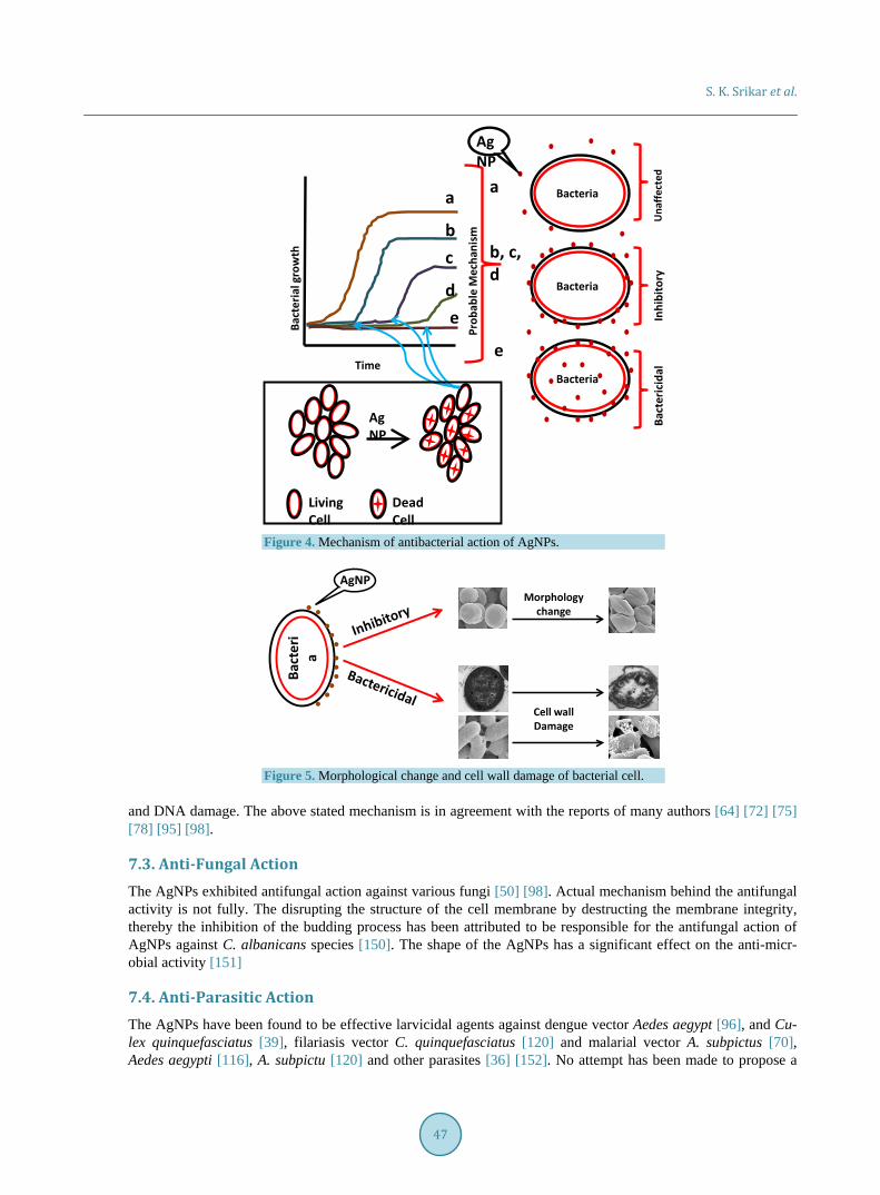
7.1.3. Analysis of SEM and TEM Micrographs The SEM and TEM analyses have been used to monitor changes in the morphology of the bacterial cell before and after treatment with “AgNPs”; The visible alterations in the cell shape and perforations in the cell wall have been reported and used as indicator of the antimicrobial action of AgNPs by several workers [45] [134] [142].
7.2. Antibacterial Action The AgNPs have potent antibacterial action against gram positive bacteria, Lactobacillus fermentum [134], Streptomyces sp. [83]. Bacillus cereus [135] Brevibacterium casei [136], S. aureus [138] B. licheniromis [139], and gram negative bacteria, E. coli [58] Entrobacteria [59] and Ureibacillus thermo sphaerius [140]. The anti-bacterial action of AgNPs on gram positive and gram negative bacterial strains is not the same but competes one over the other. There are contradictory reports regarding antibacterial action against gram positive and gram negative bacteria. According to some researchers the gram negative bacteria are reported to be more sensitive to AgNPs compared to gram positive bacteria [32] [78] [111] [134] whereas reverse results were observed by other researchers [62] [75] [76] [98]. The reported differential sensitivity of both the bacterial species could be attri-buted to the difference in structural characteristics of the bacterial species [62] [111] as well as shape and size of AgNP, bacterial inoculum size, exposure time and nutrient medium used during analysis of antibacterial action [98].
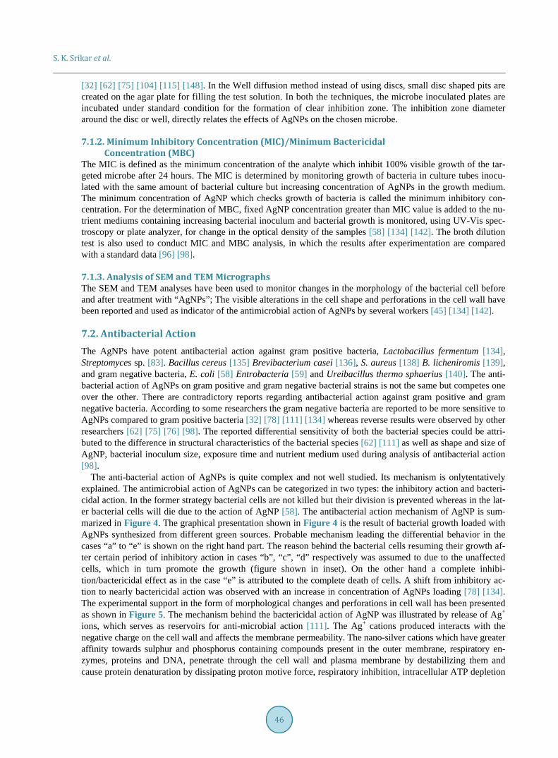
The anti-bacterial action of AgNPs is quite complex and not well studied. Its mechanism is onlytentatively explained. The antimicrobial action of AgNPs can be categorized in two types: the inhibitory action and bacteri-cidal action. In the former strategy bacterial cells are not killed but their division is prevented whereas in the lat-er bacterial cells will die due to the action of AgNP [58]. The antibacterial action mechanism of AgNP is sum-marized in Figure 4. The graphical presentation shown in Figure 4 is the result of bacterial growth loaded with AgNPs synthesized from different green sources. Probable mechanism leading the differential behavior in the cases “a” to “e” is shown on the right hand part. The reason behind the bacterial cells resuming their growth af-ter certain period of inhibitory action in cases “b”, “c”, “d” respectively was assumed to due to the unaffected cells, which in turn promote the growth (figure shown in inset). On the other hand a complete inhibi-tion/bactericidal effect as in the case “e” is attributed to the complete death of cells. A shift from inhibitory ac-tion to nearly bactericidal action was observed with an increase in concentration of AgNPs loading [78] [134]. The experimental support in the form of morphological changes and perforations in cell wall has been presented as shown in Figure 5. The mechanism behind the bactericidal action of AgNP was illustrated by release of Ag+ ions, which serves as reservoirs for anti-microbial action [111]. The Ag+ cations produced interacts with the negative charge on the cell wall and affects the membrane permeability. The nano-silver cations which have greater affinity towards sulphur and phosphorus containing compounds present in the outer membrane, respiratory en-zymes, proteins and DNA, penetrate through the cell wall and plasma membrane by destabilizing them and cause protein denaturation by dissipating proton motive force, respiratory inhibition, intracellular ATP depletion
S. K. Srikar et al.
47
Figure 4. Mechanism of antibacterial action of AgNPs.
Figure 5. Morphological change and cell wall damage of bacterial cell.
and DNA damage. The above stated mechanism is in agreement with the reports of many authors [64] [72] [75] [78] [95] [98].
7.3. Anti-Fungal Action The AgNPs exhibited antifungal action against various fungi [50] [98]. Actual mechanism behind the antifungal activity is not fully. The disrupting the structure of the cell membrane by destructing the membrane integrity, thereby the inhibition of the budding process has been attributed to be responsible for the antifungal action of AgNPs against C. albanicans species [150]. The shape of the AgNPs has a significant effect on the anti-micr- obial activity [151]
7.4. Anti-Parasitic Action The AgNPs have been found to be effective larvicidal agents against dengue vector Aedes aegypt [96], and Cu-lex quinquefasciatus [39], filariasis vector C. quinquefasciatus [120] and malarial vector A. subpictus [70], Aedes aegypti [116], A. subpictu [120] and other parasites [36] [152]. No attempt has been made to propose a
a
b
d
c
e
Prob
able
Mec
hani
sm
Una
ffect
edIn
hibi
tory
Bact
eric
idal
Bacteria
Bacteria
BacteriaTime
a
b, c, d
e
Bact
eria
l gro
wth
AgNP
Dead Cell
AgNP
Living Cell
Bact
eri
a
Morphology change
Cell wall Damage
AgNP
S. K. Srikar et al.
48
proper mechanism for anti-parasitic action of AgNPs. Denaturation of sulfur containing proteins and phosphorus containing DNA by AgNPs, leading to denaturation of organelles and enzymes is believed to be responsible for the larvicidal activity [117].
7.5. Anti-Fouling Action The AgNPs synthesized from Rhizopus oryzae fungal species have been used for treating contaminated water and adsorption of pesticides [76] and that from Lactobacillus fermentum cells have been used as anti-bio fouling agent [134]. The AgNPs are being used to treat many environmental concerns like; air disinfection, water disin-fection, ground water and biological water disinfection and surface disinfection [153].
7.6. Other Applications There have been several reports on the use of AgNPs in the field of medicine. The AgNPs have been used as therapeutic agents [97], as glyconano sensors for disease diagnosis [63] and as nano carriers for drugs delivery [142]. Reports are also available on the use of AgNPs in radiation therapy [145], in H2O2 sensor [80], in ESR- Dosimetry [146], as heavy metal ion sensors [110] and as catalyst for reduction of dyes such as methylene blue [31].
8. Conclusion Sufficient volume of published literature is available on the synthesis of AgNPs through green routes. Among plants, angiosperm species has been widely used in comparison with the other sources. Several characterizations methods and techniques have been used for AgNPs synthesis and confirmation. The AgNPs synthesized using biological reducing and capping agents have shown wide variation in shape and size. Among applications, the anti-microbial action of AgNPs has been widely studied. Various methods used to carry out antibacterial study and elucidate mechanism of anti-microbial have been developed. The results, however, are conflicting and there is a need for more work to resolve this issue. The potential of AgNPs for their use as drug carriers in cancer therapy, as biosensors for metabolites and pollutants, as catalyst etc. is quite high and requires intensive and integrated research activity for harnessing it.
Acknowledgements One of the Authors (SNU) is grateful to the Department of Atomic Energy, GoI, Mumbai for the award of Raja Ramanna fellowship. The financial support to DDG in the form of Dr DS Kothari Postdoc fellowship from the UGC, New Delhi is gratefully acknowledged. Authors are also grateful to Head of the Department of Chem-ical Engineering and Technology, IIT (BHU) for providing necessary encouragement and facilities.
References [1] Mody, V.V., Siwale, R., Singh, A. and Mody H.R. (2010) Introduction to Metallic Nanoparticles. Journal of Pharmacy
and Bioallied Sciences, 2, 282-289. http://dx.doi.org/10.4103/0975-7406.72127 [2] Sharma. V.K., Yngard, R.A. and Lin, Y. (2009) Silver Nanoparticles: Green Synthesis and Their Antimicrobial Activi-
ties. Advances in Colloid and Interface Science, 145, 83-96. http://dx.doi.org/10.1016/j.cis.2008.09.002 [3] Grier, N. (1968) Silver and Its Compounds. In: Block, S.S., Ed., Disinfection, Sterilization and Preservation, Lee and
Febiger, Philadelphia, 375-398. [4] Hill, W.R. and Pillsbury, D.M. (1939) Argyria—The Pharmacology of Silver. Williams and Williams, Baltimore. [5] Galib, B.M., Mashru, M., Jagtap, C., Patgiri, B.J. and Prajapati, P.K. (2011) Therapeutic Potentials of Metals in An-
cient India: A Review through Charaka Samhita. Journal of Ayurveda and Integrative Medicine, 2, 55-63. http://dx.doi.org/10.4103/0975-9476.82523
[6] Chaloupka, K., Malam, Y. and Seifalian, A.M. (2010) Nanosilver as a New Generation of Nanoproduct in Biomedical Applications. Trends in Bioethanology, 28, 580-588.
[7] Prow, T.W., Grice, J.E., Lin, L.L., Faye, R., Butler, M., Becker, W., Wurm, E.M.T., Yoong, C., Robertson, T.A., Soyer, H.P. and Roberts, M.S. (2011) Nanoparticles and Microparticles for Skin Drug Delivery. Advanced Drug Delivery Re-views, 63, 470-491. http://dx.doi.org/10.1016/j.addr.2011.01.012
S. K. Srikar et al.
49
[8] Dankovich, T.A. and Gray, D.G. (2011) Bactericidal Paper Impregnated with Silver Nanoparticles for Point-of-Use Water Treatment. Environmental Science & Technology, 45, 1992-1998. http://dx.doi.org/10.1021/es103302t
[9] Nair, R., Varghese, S.H., Nair. B.G., Maekawa. T., Yoshida, Y. and Sakthi Kumar, D. (2010) Nanoparticulate Material Delivery to Plants. Plant Science, 179, 154-163. http://dx.doi.org/10.1016/j.plantsci.2010.04.012
[10] Park, K., Seo, D. and Lee, J. (2008) Conductivity of Silver Paste Prepared from Nanoparticles. Colloids and Surfaces A, 313, 351. http://dx.doi.org/10.1016/j.colsurfa.2007.04.147
[11] Khan, Z., Al-Thabaiti, S.A., Obaid, A.Y. and Al-Youbi, A.O. (2011) Preparation and Characterization of Silver Nano-particles by Chemical Reduction Method. Colloids and Surfaces B: Biointerfaces, 82, 513-517. http://dx.doi.org/10.1016/j.colsurfb.2010.10.008
[12] Chen, P., Song, L.Y. and Liu, Y.K. (2007) Synthesis of Silver Nanoparticles by Gamma-Ray Irradiation in Acetic Wa-ter Solution Containing Chitosan. Radiation Physics and Chemistry, 76, 1165-1168. http://dx.doi.org/10.1016/j.radphyschem.2006.11.012
[13] Zhang, W.Z., Qiao, X.L. and Chen, J.G. (2006) Synthesis and Characterization of Silver Nanoparticles in AOT Mi-cro-Emulsion System. Chemical Physics, 300, 495-500. http://dx.doi.org/10.1016/j.chemphys.2006.09.029
[14] Reicha, F.M., Sarhan, A., Abdel-Hamid, M.I. and El-Sherbiny, I.M. (2012) Preparationof Silver Nanoparticles in the Presence of Chitosan by Electrochemical Method. Carbohydrate Polymers, 89, 236-244. http://dx.doi.org/10.1016/j.carbpol.2012.03.002
[15] Abid, J.P., Wark, A.W., Brevetm, P.F. and Girault, H.H. (2002) Preparation of Silver Nanoparticles in Solution from a Silver Salt by Laser Irradiation. Chemical Communications, 7, 792-793. http://dx.doi.org/10.1039/b200272h
[16] Yang, J. and Pan, J. (2012) Hydrothermal Synthesis of Silver Nanoparticles by Sodium Alginate and Their Applica-tions in Surface-Enhanced Raman Scattering and Catalysis. Acta Materialia, 60, 4753-4758. http://dx.doi.org/10.1016/j.actamat.2012.05.037
[17] Khan, A., El-Toni, A.M., Alrokayan, S., Alsalhi, M., Alhoshan, M. and Aldwayyan, A.S. (2011) Microwave-Assisted Synthesis of Silver Nanoparticles Using Poly-N Isopropyl Acrylamide/Acrylic Acid Microgel Particles. Colloids and Surfaces A: Physicochemical and Engineering Aspects, 377, 356-360. http://dx.doi.org/10.1016/j.colsurfa.2011.01.042
[18] Alarcon, E.I., Udekwu, K., Skog, M., Pacioni, N.L., Stamplecoskie, K.G., González-Béjar, M., et al. (2012) The Bio-compatibility and Antibacterial Properties of Collagen-Stabilized, Photochemically Prepared Silver Nanoparticles. Biomaterials, 33, 4947-4956. http://dx.doi.org/10.1016/j.biomaterials.2012.03.033
[19] Song, J.Y. and Kim, B.S. (2009) Rapid Biological Synthesis of Silver Nanoparticles Using Plant Leaf Extracts. Bio-process and Biosystems Engineering, 32, 79-84. http://dx.doi.org/10.1007/s00449-008-0224-6
[20] Huang, H. and Yang, X. (2004) Synthesis of Polysaccharide-Stabilized Gold and Silver Nanoparticles: A Green Me-thod. Carbohydrate Research, 339, 2627-2631. http://dx.doi.org/10.1016/j.carres.2004.08.005
[21] Philip, D. (2010) Honey Mediated Green Synthesis of Silver Nanoparticles. Spectrochimica Acta Part A: Molecular and Biomolecular Spectroscopy, 75, 1078-1081. http://dx.doi.org/10.1016/j.saa.2009.12.058
[22] Philip, D. (2010) Rapid Green Synthesis of Spherical Gold Nanoparticles Using Mangifera indica Leaf. Spectrochimi-ca Acta Part A: Molecular and Biomolecular Spectroscopy, 77, 807-810. http://dx.doi.org/10.1016/j.saa.2010.08.008
[23] Mittal, A.K., Chisti, Y. and Banerjee, U.C. (2013) Synthesis of Metallic Nanoparticles Using Plant Extracts. Biotech-nology Advances, 31, 346-356. http://dx.doi.org/10.1016/j.biotechadv.2013.01.003
[24] Prabhu, S. and Polusu, E.K. (2012) Silver Nanoparticles: Mechanism of Antimicrobial Action, Synthesis, Medical Ap-plications and Toxicity Affects. International Nano Letters, 2, 32. http://dx.doi.org/10.1186/2228-5326-2-32
[25] Gopinath, S.M., Saha, N.S., John, V.J., Khanum, N.S., Ganesh, S. and Patil, G.M.A. (2013) Biological Synthesis, Characterization and Application of Silver Nanoparticles. International Journal of Pharmaceutical Applications, 4, 19-28.
[26] Geoprincy, G., Srri, B.N.V., Poonguzhali, U., Gandhi, N.N. and Renganthan, S.A. (2013) Review on Green Synthesis of Silver Nanoparticles. Asian Journal of Pharmaceutical and Clinical Research, 6, 8-12.
[27] Kaler, A., Patel, N. and Banerjee, U.C. (2010) Green Synthesis of Silver Nanoparticles. Current Research and Infor-mation on Pharmaceutical Science, 11, 68-71.
[28] Vijaykumar, P.P.N., Pammi, S.V.N., Kollu, P., Satyanarayana, K.V.V. and Shameem, U. (2014) Green Synthesis and Characterization of Silver Nanoparticles Using Boerhaavia diffusa Plant Extract and Their Antibacterial Activity. In-dustrial Crops and Products, 52, 562-566. http://dx.doi.org/10.1016/j.indcrop.2013.10.050
[29] Anuj, S.A. and Ishnava, K.B. (2013) Plant Mediated Synthesis of Silver Nanoparticles Using Dried Stem Powder of Tinospora cordifolia, Its Antibacterial Activity and Its Comparison with Antibiotics. International Journal of Phar-macy and Biological Sciences, 4, 849-863.
[30] Chandran, S.P., Chaudhary, M., Pasricha, R., Ahmad, A. and Sastry, M. (2006) Synthesis of Gold Nanotriangles and
S. K. Srikar et al.
50
Silver Nanoparticles Using Aloe vera Plant Extract. Biotechnology Progress, 22, 577-583. http://dx.doi.org/10.1021/bp0501423
[31] Edison, T.J.I. and Sethuraman, M.G. (2012) Instant Green Synthesis of Silver Nanoparticles Using Terminalia chebula Fruit Extract and Evaluation of Their Catalytic Activity on Reduction of Methylene Blue. Process Biochemistry, 47, 1351-1357. http://dx.doi.org/10.1016/j.procbio.2012.04.025
[32] Mukunthan, K.S., Elumalai, E.K., Patel, E.N. and Murty, V.R. (2011) Catharanthus roseus: A Natural Source for Synthesis of Silver Nanoparticles. Asian Pacific Journal of Tropical Biomedicine, 1, 270-274. http://dx.doi.org/10.1016/S2221-1691(11)60041-5
[33] Patil, R.S., Kokate, M.R. and Kolekar, S.S. (2012) Bioinspired Synthesis of Highly Stabilized Silver Nanoparticles Using Ocimum tenuiflorum Leaf Extract and Their Antibacterial Activity. Spectrochimica Acta Part A: Molecular and Biomolecular Spectroscopy, 91, 234-238. http://dx.doi.org/10.1016/j.saa.2012.02.009
[34] Tripathi, A., Chandrasekaran, N., Raichur, A.M. and Mukherjee, A. (2009) Antibacterial Applications of Silver Nano-particles Synthesized by Aqueous Extract of Azadirachta indica (Neem) Leaves. Journal of Biomedical Nanotechnol-ogy, 5, 93-98. http://dx.doi.org/10.1166/jbn.2009.038
[35] Ankamwar, B., Damle, C., Ahmad, A. and Sastry, M. (2005) Biosynthesis of Gold and Silver Nanoparticles Using Emblica officinalis Fruit Extract, Their Phase Transfer and Transmetallation in an Organic Solution. Journal of Nanos-cience and Nanotechnology, 5, 1665-1671. http://dx.doi.org/10.1166/jnn.2005.184
[36] Roopan, S.M., Rohit, Madhumitha, G., Rahuman, A.A., Kamraj, C., Bharathi, A. and Surendra, T.V. (2013) Low-Cost and Eco-Friendly Phyto-Synthesis of Silver Nanoparticles Using Coos nucifera Coir Extract and Its Larvicidal Activi-ty.Industrial Crops and Products, 43, 631-635. http://dx.doi.org/10.1016/j.indcrop.2012.08.013
[37] Shukla, V.K., Singh, R.P. and Pandey, A.C. (2010) Black Pepper Assisted Biomimetic Synthesis of Silver Nanopar-ticles. Journal of Alloys and Compounds, 507, L13-L16. http://dx.doi.org/10.1016/j.jallcom.2010.07.156
[38] Satishkumar, M., Sneha, K., Won, S.W., Cho, C.W., Kim, S. and Yun, Y.S. (2009)Cinnamon zeylancium Bark Extract and Powder Mediated Green Synthesis of Nano-Crystalline Silver Particles and Its Antibacterial Activity. Colloids and Surfaces B: Biointerfaces, 73, 332-338. http://dx.doi.org/10.1016/j.colsurfb.2009.06.005
[39] Mondal, N.K., Chaudhury, A., Mukhopadhya, P., Chatterjee, S., Das, K. and Datta, J.K. (2014) Green Synthesis of Silver Nanoparticles and Its Application for Mosquito Control. Asian Pacific Journal of Tropical Disease, 4, S204-S210. http://dx.doi.org/10.1016/s2222-1808(14)60440-0
[40] Rao, P., Chandraprasad, M.S., Lakshmi, Y.N., Rao, J., Aishwarya, P. and Shetty, S. (2014) Biosynthesis of Silver Na-noparticles Using Lemon Extract and Its Antibacterial Activity. International Journal of Multidisciplinary and Current Research, 2, 165-169.
[41] Elumalai, E.K., Kayalvizhi, K. and Silvan, S. (2014) Coconut Water Assisted Green Synthesis of Silver Nanoparticles. Journal of Pharmacy & Bioallied Sciences, 6, 241-245. http://dx.doi.org/10.4103/0975-7406.142953
[42] Shankar, S.S., Ahmad, A. and Sastry, M. (2003) Geranium Leaf Assisted Biosynthesis of Silver Nanoparticles. Bio-technology Progress, 19, 1627-1631. http://dx.doi.org/10.1021/bp034070w
[43] Sadeghi, B., Rostami, A. and Momei, S.S. (2015) Facile Green Synthesis of Silver Nanoparticles Using Seed Aqueous Extract of Pistacia atlantica and Its Antibacterial Activity.Spectrochimica Acta Part A: Molecular and Biomolecular Spectroscopy, 134, 326-332. http://dx.doi.org/10.1016/j.saa.2014.05.078
[44] Sadeghi, B. and Gholamhoseinpoor, F. (2015) A Study on Stability and Green Synthesis of Silver Nanoparticles Using Ziziphora tenuior (Zt) Extract at Room Temperature. Spectrochimica Acta Part A: Molecular and Biomolecular Spec-troscopy, 134, 310-315. http://dx.doi.org/10.1016/j.saa.2014.06.046
[45] Rahimi-Nasrabadi, M., Pourmortazavi, S.M., Shandiz, S.A.S., Ahmadi, F. and Batooli, H. (2014)Green Synthesis of Silver Nanoparticles Using Eucalyptus leucoxylon Leaves Extract and Evaluating the Antioxidant Activities of the Ex-tract. Natural Product Research, 28, 1964-1969. http://dx.doi.org/10.1080/14786419.2014.918124
[46] Shafaghat, A. (2014) Synthesis and Characterization of Silver Nanoparticles by Phytosynthesis Method and Their Bio-logical Activity. Synthesis and Reactivity in Inorganic, Metal-Organic, and Nano-Metal Chemistry, 45, 381-387. http://dx.doi.org/10.1080/15533174.2013.819900
[47] Kulkarni, A.P., Srivastava, A.A., Nagalgaon, R.K. and Zunjarrao, R.S. (2012) Phytofabrication of Silver Nanoparticles from a Novel Plant Source and Its Application. International Journal of Biological & Pharmaceutical Research, 3, 417-421.
[48] Logeswari, P., Silambarasan, J. and Abraham, J. (2015) Synthesis of Silver Nanoparticles Using Plants Extract and Analysis of Their Antimicrobial Property. Journal of Saudi Chemical Society, 19, 311-317. http://dx.doi.org/10.1016/j.jscs.2012.04.007
[49] Rajesh, S., Raja, D.P., Rathi, J.M. and Sahayaraj, K. (2012) Biosynthesis of Silver Nanoparticles Using Ulva fasciata (Delile) Ethyl Acetate Extract and Its Activity against Xanthomonas campestris pv. Malvacearum. Journal of Biopesti-
S. K. Srikar et al.
51
cides, 5, 119-128. [50] Bankar, A., Joshi, B., Kumar, A.R. and Zinjarde, S. (2010) Banana Peel Extract Mediated Novel Route or the Synthe-
sis of Silver Nanoparticles. Colloids and Surfaces A: Physicochemical and Engineering Aspects, 368, 58-63. http://dx.doi.org/10.1016/j.colsurfa.2010.07.024
[51] Patete, J.M., Peng, X., Koenigsmann, C., Xu, Y., Karn, B. and Wong, S.S. (2011) Viable Methods or the Synthesis of High Quality Nanostructures. Green Chemistry, 13, 482-519. http://dx.doi.org/10.1039/c0gc00516a
[52] Mukherjee, P., Ahmad, A., Mandal, D.D., Senapati, S., Sainkar, S.R., Khan, M.I., Parishcha, R., Ajaykumar, P.V., Alam, M., Kumar, R. and Sastry, M. (2001) Fungus-Mediated Synthesis of Silver Nanoparticles and Their Immobiliza-tion in the Mycelial Matrix: A Novel Biological Approach to Nanoparticle Synthesis. Nano Letters, 1, 515-519. http://dx.doi.org/10.1021/nl0155274
[53] Ajitha, B., Redd, Y.A.K. and Reddy, P.S. (2014) Biosynthesis of Silver Nanoparticles Using Plectranthus amboinicus Leaf Extract and Its Antimicrobial Activity. Spectrochimica Acta Part A: Molecular and Biomolecular Spectroscopy, 128, 257-262. http://dx.doi.org/10.1016/j.saa.2014.02.105
[54] Bhainsa, K.C. and D’souza, S.F. (2006) Extracellular Biosynthesis of Silver Nanoparticles Using the Fungus Aspergil-lus fumigates. Colloids and Surfaces B: Biointerfaces, 47, 160-164. http://dx.doi.org/10.1016/j.colsurfb.2005.11.026
[55] Ahmad, A., Mukherjee, P., Senapati, S., Mandal, D., Kham, M.I., Kumar, R. and Sastry, M. (2003) Extracellular Bio-synthesis of Silver Nanoparticles Using the Fungus Fusarium oxysporum. Colloids and Surfaces B: Biointerfaces, 28, 313-318. http://dx.doi.org/10.1016/S0927-7765(02)00174-1
[56] Duran, N., Marcato, P.D., Alves, O.L., De Souza, D.I.H. and Esposito, E. (2005) Mechanistic Aspect of Biosynthesis of Silver Nanoparticles by Several Fusarium oxysporum Species. Journal of Nanobiotechnology, 3, 8. http://dx.doi.org/10.1186/1477-3155-3-8
[57] Ingle, A., Rai, M., Gade, A. and Bawaskar, M. (2009) Fusarium solani: A Novel Biological Agent for the Extracellular Synthesis of Silver Nanoparticles. Journal of Nanoparticle Research, 11, 2079-2085. http://dx.doi.org/10.1007/s11051-008-9573-y
[58] Perni, S., Hakala, V. and Prokopovich, K. (2014) Biogenic Synthesis of Antimicrobial Silver Nanoparticles Caped with L-Cystine. Colloids and Surfaces A: Physicochemical and Engineering Aspects, 460, 219-224.
[59] Shahverdi, A.R., Minaeian, S., Shahverdi, H.R., Jamalifar, H. and Nohi, A.-A. (2007) Rapid Synthesis of Silver Nano-particles Using Cultural Supernatants of Enterobacteria: A Novel Biological Approach. Process Biochemistry, 42, 919-923. http://dx.doi.org/10.1016/j.procbio.2007.02.005
[60] Ortega-Arroyo, L., Martin-Martinez, E.S., Aguilar-Mendez, M.A., Cruz-Orea, A., Hernandez-Perez, I. and Glorieux, C.(2013) Green Synthesis Method of Silver Nanoparticles Using Starch as Capping Agent Applied the Methodology of Surface Response. Starch/Starke, 65, 814-821. http://dx.doi.org/10.1002/star.201200255
[61] Morales-Sanchez, E., Guajardo-Pacheco, J., Noriega-Trevino, M., Quintero-Gonzalez, C., Compean-Jasso, M., Lopez- Salinas, F., Gonzalez-Hernandez, J. and Ruiz, F. (2011) Synthesis of Silver Nanoparticles Using Albumin as Reducing Agent. Materials Sciences and Applications, 2, 578-581. http://dx.doi.org/10.4236/msa.2011.26077
[62] Kora, A.J., Sashidhar, R.B. and Arunachalam, J. (2010) Gum Kondagogu (Cochlospermum gossypium): A Template for Green Synthesis and Stabilization of Silver Nanoparticles with Antibacterial Application. Carbohydrate Polymers, 82, 670-679. http://dx.doi.org/10.1016/j.carbpol.2010.05.034
[63] Sastry, M., Mayyaa, K.S. and Bandyopadhyay, K. (1997) pH Dependent Changes in the Optical Properties of Carbox-ylic Acid Derivatized Silver Colloid Particles. Colloids and Surfaces A: Physicochemical and Engineering Aspects, 127, 221-228. http://dx.doi.org/10.1016/S0927-7757(97)00087-3
[64] Jagtap, U.B. and Bapat, V.A. (2013) Green Synthesis of Silver Nanoparticles Using Artocarpus heterophyllus Lam. Seed Extract and Its Antibacterial Activity. Industrial Crops and Products, 46, 132-137. http://dx.doi.org/10.1016/j.indcrop.2013.01.019
[65] Vijayaraghavan, K., Nalini, S.P.K., Prakash, N.U. and Madhankumar, D. (2012) One Step Green Synthesis of Silver nano/Micro Particles Using Extracts of Trahyspermum ammi and Papaver somniferum. Colloids and Surfaces B: Bio-interfaces, 94, 114-117. http://dx.doi.org/10.1016/j.colsurfb.2012.01.026
[66] Vigneswaran, N., Ashtaputre, N.M., Varadarajan, P.V., Nachane, R.P., Paralikar, K.M. and Balasubramanya, R.H. (2007) Biological Synthesis of Silver Nanoparticles Using the Fungus Aspergillus flavus. Materials Letters, 61, 1413- 1418. http://dx.doi.org/10.1016/j.matlet.2006.07.042
[67] Vigneshwaran, N., Kathe, A.A., Varadarajan, P.V., Nachane, R.P. and Balasubramanya, R.H. (2006) Biomimitics of Silver Nanoparticles by White Rot Fungus. Colloids and Surfaces B: Biointerfaces, 53, 55-59. http://dx.doi.org/10.1016/j.colsurfb.2006.07.014
[68] Kaviya, S., Santhanalakshmi, J. and Viswanathan, B. (2012) Biosynthesis of Silver Nanoflakes by Crossandra infun-dibuliformis Leaf Extract. Materials Letters, 67, 64-66. http://dx.doi.org/10.1016/j.matlet.2011.09.023
S. K. Srikar et al.
52
[69] Sreekanth, T.V.M., Nagajyothi, P.C. and Lee, K.D. (2012) Dioscorea Batatas Rhizome-Assisted Rapid Biogenic Syn-thesis of Silver and Gold Nanoparticles. Synthesis and Reactivity in Inorganic, Metal-Organic, and Nano-Metal Che-mistry, 42, 567-572. http://dx.doi.org/10.1080/15533174.2011.613886
[70] Rajakumar, G. and Rahuman, A.A. (2011) Larvaidal Activity of Synthesized Silver Nanoparticles Using Eclipta pro-strata Leaf Extract against Filariasis and Malaria Vectors. Acta Tropica, 118, 196-203. http://dx.doi.org/10.1016/j.actatropica.2011.03.003
[71] Sant, D.G., Gujarathi, T.R., Harne, S.R., Ghosh, S., Kitture, R., Kale, S., Chopade, B.A. and Pardesi, K.R. (2013) Adiantum phillipenseL. Frond Assisted Rapid Green Synthesis of Gold and Silver Nanoparticles. Journal of Nanopar-ticles, 2013, Article ID: 182320.
[72] Satishkumar, G., Gobinath, G., Karpagam, K., Hemamalini, V., Premkumar, K. and Sivaramakrishna, S. (2012) Phy-to-Synthesis of Silver Nanoscale Particles Using Morinda citifolia L. and Its Inhibitory Action against Human Patho-gens. Colloids and Surfaces B: Biointerfaces, 95, 235-240. http://dx.doi.org/10.1016/j.colsurfb.2012.03.001
[73] Jha, A.K., Prasad, K., Prasad, K. and Kulkarni, A.R. (2009) Plant System: Natures Nanofactory. Colloids and Surfaces B: Biointerfaces, 73, 219-223. http://dx.doi.org/10.1016/j.colsurfb.2009.05.018
[74] Yang, N. and Li, W.H. (2013) Mango Peel Extract Mediated Novel Route or Synthesis of Silver Nanoparticles and An-tibacterial Application of Silver Nanoparticles Loaded onto Non-Woven Fibers. Industrial Crops and Products, 48, 81-88. http://dx.doi.org/10.1016/j.indcrop.2013.04.001
[75] Khalil, M.M.H., Ismail, E.H., El-Bhaghdady, K.Z. and Mohamed, D. (2014) Green Synthesis of Silver Nanoparticles Using Olive Leaf Extract and Its Antibacterial Activity. Arabian Journal of Chemistry, 7, 1131-1139.
[76] Das, S.K., Khan, M.M.R., Guha, A.K., Das, A.K. and Mandal, A.B. (2012) Silver-NanoBiohybrid Material: Synthesis, Characterization and Application in Water Purification. Bioresource Technology, 124, 495-499. http://dx.doi.org/10.1016/j.biortech.2012.08.071
[77] Ashraf, S., Abbasi, A.Z., Pfeiffer, C., Hussain, S.Z., Khalid, Z.M., Gil, P.R., Parak, W.J. and Hussain, I. (2013) Pro-tein-Mediated Synthesis, pH-Induced Reversible Agglomeration, Toxicity and Cellular Interaction of Silver Nanopar-ticles. Colloids and Surfaces B: Biointerfaces, 102, 511-518. http://dx.doi.org/10.1016/j.colsurfb.2012.09.032
[78] Dehnavi, A.S., Raisi, A. and Aroujalian, A. (2013) Control Size and Stability of Colloidal Silver Nanoparticles with Antibacterial Activity Prepared by a Green Synthesis Method. Synthesis and Reactivity in Inorganic, Metal-Organic, and Nano-Metal Chemistry, 43, 543-551. http://dx.doi.org/10.1080/15533174.2012.741182
[79] Dubey, S.P., Lahtinen, M. and Sillianpaa, M. (2010) Tansy Fruit Mediated Greener Synthesis of Silver and Gold Na-noparticles. Process Biochemistry, 45, 1065-1071.http://dx.doi.org/10.1016/j.procbio.2010.03.024
[80] Tagad, C.K., Dugasani, S.R., Aiyer, R., Park, S., Kulkarni, A. and Sabharwal, S. (2013)Green Synthesis of Silver Na-noparticles and Their Application for the Development of Optical Fiber Based Hydrogen Peroxide Sensor. Sensors and Actuators B: Chemical, 183, 144-149. http://dx.doi.org/10.1016/j.snb.2013.03.106
[81] El-Rafie, M.H., El-Nagger, M.E., Ramadan, M.A., Fouda, M.M.G., Al Deyab, S.S. and Hebeish, A. (2011) Environ-mental Synthesis of Silver Nanoparticles Using Hydroxypropyl Starch and Their Characterization. Carbohydrate Po-lymers, 86, 630-635. http://dx.doi.org/10.1016/j.carbpol.2011.04.088
[82] Fayaz, A.M., Balaji, K., Kalaichelvan, P.T. and Venkatesan, R. (2009) Fungal Based Synthesis of Silver Nanopar-ticles—An Effect of Temperature on the Size of Particles. Colloids and Surfaces B: Biointerfaces, 74, 123-126. http://dx.doi.org/10.1016/j.colsurfb.2009.07.002
[83] Zonnoz, N.F. and Salouti, M. (2011) Extracellular Biosynthesis of Silver Nanoparticles Using Cell Filtrate of Streptomyces sp. ERI-3. Scientia Iranica, 18, 1631-1635. http://dx.doi.org/10.1016/j.scient.2011.11.029
[84] Kathiraven, T., Sundaramanickam, A., Shanmugam, N. and Balasubramanian, T. (2014) Green Synthesis of Silver Nanoparticles Using Marine Algae Caulerpa resmosa and Their Anti-Baterial Activity against Some Human Patho-gens. Applied Nanoscience, 5, 499-504. http://dx.doi.org/10.1007/s13204-014-0341-2
[85] Vivek, M., Kumar, P.S., Steffi, S. and Sudha, S. (2011) Biogenic Silver Nanoparticles by Gelidiella acerosa Extract and Their Antifungal Effects. Avicenna Journal of Medical Biotechnology, 3, 143-148.
[86] Govindaraju, K., Kiruthiga, V., Kumar, V.G. and Singaravelu, G.(2009) Extracellular Synthesis of Silver Nanoparticles by Marine Algae, Sargassum wightii Grevilli and Their Antibacterial Effects. Journal of Nanoscience and Nanotech-nology, 9, 5497-5501. http://dx.doi.org/10.1166/jnn.2009.1199
[87] Srivastava, A.A., Kulkarni, A.P., Harpale, P.M. and Zunjarrao, R.S. (2011) Plant Mediated Synthesis of Silver Nano-particles Using a Bryophyte: Fissidens minutes and Its Anti-Microbial Activity. International Journal of Engineering Science and Technology, 3, 8342-8347.
[88] Kulkarni, A.P., Srivastava, A.A., Harpale, P.M. and Zunjarrao, R.S. (2011) Plant Mediated Synthesis of Silver Nano-particles—Tapping the Unexploited Resources. Journal of Natural Product and Plant Resources, 1, 100-107.
[89] Britto, J.D., Gracelin, D.H.S. and Kumar, P.B.J.R. (2014) Antibacterial Activity of Silver Nanoparticles Synthesized
S. K. Srikar et al.
53
from a Few Medicinal Ferns. International Journal of Pharmaceutical Research and Development, 6, 25-29. [90] Bhor, G., Maskare, S., Hinge, S., Singh, L. and Nalwade, A. (2014) Synthesis of Silver Nanoparticles Using Leaflet
Extract of Nephrolepi sexaltata L. and Evaluation Antibacterial Activity against Human and Plant Pathogenic Bacteria. Asian Journal of Pharmaceutical Technology and Innovation, 2.
[91] Nalwade, A.R., Badhe, M.N., Pawale, C.B. and Hinge, S.B. (2013) Rapid Biosynthesis of Silver Nanoparticles Using Fern Leaflet Extract and Evaluation of Their Antibacterial Activity. International Journal of Biological Technology, 4, 12-18.
[92] Kang, K.C., Kim, S.S., Baik, M.H., Choi, J.W. and Kwon, S.H. (2008) Synthesis of Silver Nanoparticles Using Green Chemical Method. Applied Chemistry, 12, 281-284.
[93] Jha, A.K. and Prasad, K. (2010) Green Synthesis of Silver Nanoparticles Using Cycas Leaf. International Journal of Green Nanotechnology: Physics and Chemistry, 1, 110-117. http://dx.doi.org/10.1080/19430871003684572
[94] Ashokkumar, S., Ravi, S., Kathiravan, V. and Velmurugan, S. (2015) Synthesis of Silver Nanoparticles Using A. indi-cum Leaf Extract and Their Antibacterial Activity. Spectrochimica Acta Part A: Molecular and Biomolecular Spec-troscopy, 134, 34-39. http://dx.doi.org/10.1016/j.saa.2014.05.076
[95] Ajitha, A., Reddy, Y.A.K. and Reddy, P.S. (2014) Biogenic Nano-Scale Silver Particles by Tephrosia purpurea Leaf Extract and Their Inborn Antimicrobial Activity. Spectrochimica Acta Part A: Molecular and Biomolecular Spectros-copy, 121, 164-172. http://dx.doi.org/10.1016/j.saa.2013.10.077
[96] Suresh, P., Gunasekar, P.H., Kokila, D., Prabhu, D., Dinesh, D., Ravichandran, N., Ramesh, B., Koodalingam, A. and Siva, G.V.(2014) Green Synthesis of Silver Nanoparticles Using Delphinium denundatum Root Extract Exhibits Anti-bacterial and Mosquito Larvicidal Activities. Spectrochimica Acta Part A: Molecular and Biomolecular Spectroscopy, 127, 61-66. http://dx.doi.org/10.1016/j.saa.2014.02.030
[97] Zuas, O., Hamim, N. and Sampora, Y.(2014) Bio-Synthesis of Silver Nanoparticles Using Water Extract of Myrmeco-dia pendan (Sarang Semut Plant). Materials Letters, 123, 156-159. http://dx.doi.org/10.1016/j.matlet.2014.03.026
[98] Raut, R.W., Mendhulkar, V.D. and Kashid, S.B. (2014) Photosensitized Synthesis of Silver Nanoparticles Using Wi-thania somnifera Leaf Powder and Silver Nitrate. Journal of Photochemistry and Photobiology B: Biology, 132, 45-55. http://dx.doi.org/10.1016/j.jphotobiol.2014.02.001
[99] Singh, P.K., Bhardwaj, K., Dubey, P. and Prabhune, A. (2015) UV-Assisted Size Sampling and Antibacterial Screen-ing of Lantana camara Leaf Extract Synthesized Silver Nanoparticles. RSC Advances, 5, 24513-24520. http://dx.doi.org/10.1039/C4RA17233G
[100] Vimala, R.T.V., Satishkumar, G. and Sivramakrishnan, S. (2015) Optimization of Reaction Conditions to Fabricate Nano Silover Using Couroupita guianensis Aubel(Leaf & Fruit) and Its Enhanced Larvicidal Effects. Spectrochimica Acta Part A: Molecular and Biomolecular Spectroscopy, 135, 110-115. http://dx.doi.org/10.1016/j.saa.2014.06.009
[101] Zhang, Y., Cheng, X., Zhang, Y., Xue, X. and Fu, Y.(2013) Biosynthesis of Silver Nanoparticles at Room Temperature Using Aqueous Aloe Leaf Extract and Antibacterial Properties. Colloids and Surfaces A: Physicochemical and Engi-neering Aspects, 423, 63-68. http://dx.doi.org/10.1016/j.colsurfa.2013.01.059
[102] Karuppiah, M. and Rajmohan, R. (2013) Green Synthesis of Silver Nanoparticles Using Ixora coccinea Leaves Extract. Materials Letters, 97, 141-143. http://dx.doi.org/10.1016/j.matlet.2013.01.087
[103] Logeswari, P., Silambarasan, S. and Abraham, J. (2013) Ecofriendly Synthesis of Silver Nanoparticles from Commer-cially Available Plant Powders and Their Antibacterial Properties. Scientia Iranica, 20, 1049-1054.
[104] Geethalakshmi, R. and Sarada, D.V.L.(2013) Characterization and Antimicrobial Activity of Gold and Silver Nanopar-ticles Synthesized Using Saponin Isolated from Trianthema decendra L. Industrial Crops and Products,51, 107-115. http://dx.doi.org/10.1016/j.indcrop.2013.08.055
[105] Yasin, S., Liu, L. and Yao, J. (2013) Biosynthesis of Silver Nanoparticles by Bamboo Leaves Extract and Their Anti-microbial Activity. Journal of Fiber Bioengineering and Informatics, 6, 77-84. http://dx.doi.org/10.3993/jfbi03201307
[106] Rodriguez, L.E., Iniguez, P.R., Navarro, R.E., Herrera, U.R., Tanori, J., Iniguez, P.C. and Maldonado, A. (2013) Syn-thesis of Silver Nanoparticles Using Reducing Agents Obtained from Natural Sources (Rumex hymenosepalus Ex-tracts).Nanoscale Research Letters, 8, 318. http://dx.doi.org/10.1186/1556-276X-8-318
[107] Rajathi, K. and Sridhar, S. (2013) Green Synthesis of Silver Nanoparticles from the Medicinal Plant Wrightia tinctoria and Its Antimicrobial Potential. International Journal of ChemTech Research, 5, 1701-1713.
[108] Kannan, R.R.R., Strik, W.A. and Staden, V. (2013) Synthesis of Silver Nanoparticles Using the Seaweed Codium ca-pitatum P.C. Silva (Chlorophyceae). South African Journal of Botany, 86, 1-4. http://dx.doi.org/10.1016/j.sajb.2013.01.003
[109] Natarajan, R.K., John Nayagam, A.A., Gurunagarajan, S., Muthukumar, Ekambaram, N. and Manimaran, A. (2013) Elaeagnus indica Mediated Green Synthesis of Silver Nanoparticles and Its Potent Toxicity against Human Pathogens.
S. K. Srikar et al.
54
Global Journal of Pharmacology, 7, 222-231. [110] Kirubaharan, C.J., Kalpana, D., Lee, Y.S., Kim, A.R., Yoo, D.J., Nahm, K.S. and Kumar, G.G.(2012) Biomediated
Silver Nanoparticles for the Highly Selective Copper(II) Ion Sensor Applications. Industrial and Engineering Chemi-stry Research, 51, 7441-7446. http://dx.doi.org/10.1021/ie3003232
[111] Gopinath, V., Mubarakali, D., Priyadarshini, S., Priyadharsshini, N.M., Thajuddin, N. and Velusamy, P. (2012) Bio-synthesis of Silver Nanoparticles from Tribulus terrestris and Its Antimicrobial Activity: A Novel Biological Approach. Colloids and Surfaces B: Biointerfaces, 96, 69-74. http://dx.doi.org/10.1016/j.colsurfb.2012.03.023
[112] Chaudhary, S., Paul, S. and Sagar, S. (2012) Biosynthesis of Silver Nanoparticles Using Vitis vinifera Extract and Evaluation of Their Antimicrobial Activity. International Journal of Bio-Technology and Research, 2, 1-12.
[113] Ashok Kumar, D. (2012) Rapid and Green Synthesis of Silver Nanoparticles Using the Leaf Extracts of Parthenium hysterophorus: A Novel Biological Approach. International Research Journal of Pharmacy, 3, 169-173.
[114] Arunachalam, K.D., Annamalai, S.K., Arunachalam, A.M. and Kennedy, S. (2013) Green Synthesis of Crystalline Sil-ver Nanoparticles Using Indigofera Aspalathoides-Medicinal Plant Extract for Wound Healing Applications. Asian Journal of Chemistry, 25, S311-S314.
[115] Mubarakali, D., Thajuddin, N., Jeganathan, K. and Gunasekaran, M. (2011) Plant Extract Mediated Synthesis of Silver and Gold Nanoparticles and Its Antibacterial Activity against Clinically Isolated Pathogens. Colloids and Surfaces B: Biointerfaces, 85, 360-365. http://dx.doi.org/10.1016/j.colsurfb.2011.03.009
[116] Kumar, V. and Yadav, S.K. (2011) Synthesis of Stable, Polyshaped Silver and Gold Nanoparticles Using Leaf Extract of Lonicera japonica L. International Journal of Green Nanotechnology, 3, 281-291. http://dx.doi.org/10.1080/19430892.2011.633474
[117] Gnanadesigan, M., Anand, M., Ravikumar, S., Maruthupandy, M., Vijaykumar, V., Selvam, S., et al. (2011) Biosyn-thesis of Silver Nanoparticles Using Mangrove Plant Extract and Their Potential Mosquito Larvicidal Property. Asian Pacific Journal of Tropical Disease, 4, 799-803. http://dx.doi.org/10.1016/S1995-7645(11)60197-1
[118] Rani, P.U. and Rajasekharreddy, P.(2011) Green Synthesis of Silver-Protein (Core-Shell) Nanoparticles Using Piper betel L. Leaf Extract and Its Ecotoxicological Studies on Daphnia manga.Colloids and Surfaces A: Physicochemical and Engineering Aspects, 389, 188-194. http://dx.doi.org/10.1016/j.colsurfa.2011.08.028
[119] Veerasamy, R., Xin, T.Z., Gunasagaran, S., Xiang, T.F.W., Yang, E.F.C., Jeyakumar, N. and Dhanaraj, S.A. (2011) Biosynthesis Silver Nanoparticles Using Mangosteen Leaf Extract and Evaluation of Their Antimicrobial Activities. Journal of Saudi Chemical Society, 15, 113-120. http://dx.doi.org/10.1016/j.jscs.2010.06.004
[120] Santoshkumar, T., Rahuman, A.A., Rajakumar, G., Marimuthu, S., Bagavan, A., Jayaseelan, C., Zahir, A.A., Elango, G. and Kamaraj, C. (2011) Synthesis of Silver Nanoparticles Using Nelumbo nucifera Leaf Extract and Its Larvicidal Ac-tivity against Malaria and Filariasis Vectors.Parasitology Research, 108, 693-702. http://dx.doi.org/10.1007/s00436-010-2115-4
[121] Ahmad, N., Sharma, S., Singh, V.N., Shamsi, S.F., Fatma, A. and Mehta, B.R. (2011) Biosynthesis of Silver Nanopar-ticles from Desmodium triflorum: A Novel Approach towards Weed Utilization. Biotechnology Research International, 2011, Article ID: 454090. http://dx.doi.org/10.4061/2011/454090
[122] Prathna, T.C., Chandrasekaran, N., Raichur, A.M. and Mukherjee, A. (2011) Biomimetic Synthesis of Silver Nanopar-ticles by Citrus limon (Lemon) Aqueous Extract and Theoretical Prediction of Particle Size. Colloids and Surfaces B: Biointerfaces, 82, 152-159. http://dx.doi.org/10.1016/j.colsurfb.2010.08.036
[123] Njagi, E.C., Huang, H., Stafford, L., Genuino, H., Galindo, H.M., Collins, J.B., Hoag, G.E. and Suib, S.L. (2011) Bio-synthesis of Iron and Silver Nanoaparticles at Room Temperature Using Aqueous Sorghum Bran Extracts. Langmuir, 27, 264-271. http://dx.doi.org/10.1021/la103190n
[124] Kumar, V., Yadav, S.C. and Yadav, S.K. (2010) Syzygium cumini Leaf Extract and Seed Extract Mediated Biosynthe-sis of Silver Nanoparticles and Their Characterization. Journal of Chemical Technology and Biotechnology, 85, 1301-1309. http://dx.doi.org/10.1002/jctb.2427
[125] Krishnaraj, C., Jagan, E.G., Rajasekar, S., Selvakumar, P., Kalaichelvan, P.T. and Mohan, N. (2010) Synthesis of Sil-ver Nanoparticles Using Aclypha indica Extracts and Its Antibacterial Activity against Water Borne Pathogens. Collo-ids and Surfaces B: Biointerfaces, 76, 50-56.http://dx.doi.org/10.1016/j.colsurfb.2009.10.008
[126] Leela, A. and Vivekanandan, M. (2008) Tapping the Unexploited Plant Resources for the Synthesis of Silver Nanopar-ticles. African Journal of Biotechnology, 7, 31662-3165.
[127] Shankar, S.S., Rai, A., Ahmad, A. and Sastry, M. (2004) Rapid Synthesis of Au, Ag and Bimetallic Au Core-Ag Shell Nanoparticles Using Neem (Azadirachta indica) Leaf Broth. Journal of Colloid and Interface Science, 275, 496-502. http://dx.doi.org/10.1016/j.jcis.2004.03.003
[128] Naveen, K.S.H., Kumar, G., Karthik, L. and Rao, K.V.B. (2010) Extracellular Biosynthesis of Silver Nanoparticles Using the Filamentous Fungus Penicillium sp. Archives of Applied Science Research, 2, 161-167.
S. K. Srikar et al.
55
[129] Balaji, D.S., Basavaraja, S., Deshpande, R., Mahesh, D.B., Prabhakar, B.K. and Venkataraman, A. (2009) Extracellular Biosynthesis of Functionalized Silver Nanoparticles by Strains of Cladosporium cladosporioides Fungus. Colloids and Surfaces B: Biointerfaces, 68, 88-92. http://dx.doi.org/10.1016/j.colsurfb.2008.09.022
[130] Shaligram, N.S., Bule, M., Bhambure, R., Singhal, R.S., Singh, S.K., Szakacs, G. and Pandey, A. (2009) Biosynthesis of Silver Nanoparticles Using Aqueous Extract from the Compactin Producing Fungal Strain. Process Biochemistry, 44, 939-943. http://dx.doi.org/10.1016/j.procbio.2009.04.009
[131] Kathiresan, K., Manivannan, S., Nabeel, M.A. and Dhivya, B. (2009) Studies on Silver Nanoparticles Synthesized by Marine Fungus, Penicillium fellutanum Isolated from Coastal Mangrove Sediment. Colloids and Surfaces B: Biointer-faces, 71, 133-137. http://dx.doi.org/10.1016/j.colsurfb.2009.01.016
[132] Basavaraja, S., Balaji, S.D., Lagashetty, A., Rajasab, A.H. and Venkataraman, A. (2008) Extracellular Biosynthesis of Silver Nanoparticles Using the Fungus Fusarium semitectum. Materials Research Bulletin, 43, 1164-1170. http://dx.doi.org/10.1016/j.materresbull.2007.06.020
[133] Senapati, S., Mandal, D., Ahmad, A., Khan, M.I., Sastry, M. and Kumar, R. (2004) Fungus Mediated Synthesis of Sil-ver Nanoparticles: A Novel Biological Approach. Indian Journal of Physics, 78, 101-105.
[134] Zhang, M., Zhang, K., Gusseme, B.D., Verstraete, W. and Field, R. (2014) The Antibacterial and Anti-Biofouling Per-formance of Biogenic Silver Nanoparticles by Lactobacillus fermentum. Biofouling: The Journal of Bioadhesion and Biofilm Research, 30, 347-357.http://dx.doi.org/10.1080/08927014.2013.873419
[135] Deepak, V., Umamaheshwaran, P.S., Guhan, K., Nanthini, R.A., Krithiga, B., Jaithoon, N.M.H. and Gurunathan, S. (2011) Synthesis of Gold and Silver Nanoparticles Using Purified URAK. Colloids and Surfaces B: Biointerfaces, 86, 353-358.http://dx.doi.org/10.1016/j.colsurfb.2011.04.019
[136] Kalishwaralal, K., Deepak, V., Pandian, S.R.K., Kottaisamy, M., Barathmanikanth, S., Kartikeyan, B. and Gurunathan, S. (2010) Biosynthesis of Silver and Gold Nanoparticles Using Brevibacterium casei. Colloids and Surfaces B: Bioin-terfaces, 77, 257-262. http://dx.doi.org/10.1016/j.colsurfb.2010.02.007
[137] Babu, M.M.G. and Gunasekaran, P. (2009) Production and Structural Characterization of Crystalline Silver Nanopar-ticles from Bacillus cereus Isolate. Colloids and Surfaces B: Biointerfaces, 74, 191-195. http://dx.doi.org/10.1016/j.colsurfb.2009.07.016
[138] Nanda, A. and Saravanan, M. (2009) Biosynthesis of Silver Nanoparticles from Staphylococcus aureus and Its Antimi-crobial Activity against MRSA and MRSE. Nanomedicine: Nanotechnology, Biology and Medicine, 5, 452-456. http://dx.doi.org/10.1016/j.nano.2009.01.012
[139] Kalimuthu, K., Babu, R.S., Venkataraman, D., Bilal, M. and Gurunathan, S. (2008) Biosynthesis of Silver Nanocrys-tals by Bacillus licheniformis. Colloids and Surfaces B: Biointerfaces, 65, 150-153. http://dx.doi.org/10.1016/j.colsurfb.2008.02.018
[140] Juibari, M.M., Abbasalizadeh, S., Jouzani, G.S. and Noruzi, M. (2011) Intensified Biosynthesis of Silver Nanoparticles Using a Native Extremophilic Ureibacillus thermosphaerius Strain. Materials Letters, 65, 1014-1017. http://dx.doi.org/10.1016/j.matlet.2010.12.056
[141] Gurunathan, S., Kalishwaralal, K., Vaidhyanathan, R., Deepak, V., Pandian, S.R.K., Muniyandi, J., Hariharan, N. and Eom, S.H.(2009) Biosynthesis, Purification and Characterization of Silver Nanoparticles Using Escherichia coli. Col-loids and Surfaces B: Biointerfaces, 74, 328-335. http://dx.doi.org/10.1016/j.colsurfb.2009.07.048
[142] Cheng, K.M., Hung, Y.W., Chen, C.C., Lin, C.C. and Yong, J.J. (2014) Green Synthesis of Chondroitin Sulfate- Capped Silver Nanoparticles: Characterization and Surface Modification. Carbohydrate Polymers, 110, 195-202. http://dx.doi.org/10.1016/j.carbpol.2014.03.053
[143] Chen, Q., Jiang, H., Ye, H., Li, J. and Huang, J. (2014) Preparation, Antibacterial, and Antioxidant Activities of Sil-ver/Chitosan Composities. Journal of Carbohydrate Chemistry, 33, 298-312.
[144] El-Rafie, H.M., El-Rafie, M.H. and Zahran, M.K. (2013) Green Synthesis of Silver Nanoparticles Using Polysaccha-rides Extracted from Marine Macro Algae. Carbohydrate Polymers, 96, 403-410. http://dx.doi.org/10.1016/j.carbpol.2013.03.071
[145] Lu, R., Yang, D., Cui, D., Wang, Z. and Guo, L. (2012) Egg White-Mediated Green Synthesis of Silver Nanoparticles with Excellent Biocompatibility and Enhanced Radiation Effects on Cancer Cells. International Journal of Nanomedi-cine, 7, 2101-2107. http://dx.doi.org/10.2147/IJN.S29762
[146] Guidelli, E.D., Ramos, A.P., Zaniquelli, M.E.D., Nicolucci, P. and Baffa, O. (2012) Synthesis of Silver Nanoparticles Using DL-Alanine for ESR Dosimetry Applications. Radiation Physics and Chemistry, 81, 301-307. http://dx.doi.org/10.1016/j.radphyschem.2011.10.020
[147] Tanvir, S., Oudet, F., Pulvin, S. and Anderson, W.A.(2012) Coenzyme Based Synthesis of Silver Nanorystals. Enzyme and Microbial Technology, 51, 231-236. http://dx.doi.org/10.1016/j.enzmictec.2012.07.002
[148] Bankura, K.P., Maity, D., Mollick, M.M.R., Mondal, D., Bhowmick, B., Bain, M.K., Chakraborty, A., Sarkar, J.,
S. K. Srikar et al.
56
Acharya, K. and Chattopadhyay, D. (2012) Synthesis, Characterization and Antimicrobial Activity of Dextran Stabi-lized Silver Nanoparticles in Aqueous Medium. Carbohydrate Polymers, 89, 1159-1165. http://dx.doi.org/10.1016/j.carbpol.2012.03.089
[149] Sasikala, D., Govindaraju, K., Tamilselvan, K. and Singaravelu, G. (2012) Soyabean Protein: A Natural Source for the Production of Green Silver Nanoparticles. Biotechnology and Bioprocess Engineering, 17, 1176-1181. http://dx.doi.org/10.1007/s12257-012-0021-6
[150] Kim, K.J., Sung, W.S., Suh, B.K., Moon, S.K., Choi, J.S., Kim, J.G. and Lee, D.G. (2009) Antifungal Activity and Mode of Action of Silver Nanoparticles on Candida albicans. Biometals, 22, 235-242. http://dx.doi.org/10.1007/s10534-008-9159-2
[151] Rai, M.K., Deshmukh, S.D., Ingle, A.P. and Gade, A.K. (2012) Silver Nanoparticles: The Powerful Nanoweapon against Multidrug-Resistant Bacteria. Journal of Applied Microbiology, 112, 841-852. http://dx.doi.org/10.1111/j.1365-2672.2012.05253.x
[152] Marimuthu, S., Rahuman, A.A., Rajakumar, G., Santhoshkumar, T., Kirthi, A.V., Jayaseelan, C., Bagavan, A., Zahir, A.A., Elango, G. and Kamaraj, C. (2011) Evaluation of Green Synthesized Silver Nanoparticles against Parasites. Pa-rasitology Research, 108, 1541-1549. http://dx.doi.org/10.1007/s00436-010-2212-4
[153] Tran, Q.H., Nguyen, V.Q. and Le, A.T. (2013) Silver Nanoparticles: Synthesis, Properties, Toxicology, Applications and Perspectives. Advances in Natural Sciences: Nanoscience and Nanotechnology, 4, Article ID: 033001. http://dx.doi.org/10.1088/2043-6262/4/3/033001