handbook of musculoskeletal pain management
TRANSCRIPT

HANDBOOK OF MUSCULOSKELETAL PAIN MANAGEMENT
A Pocketbook for Physicians and Practitioners by Physicians and Practitioners
Richard A. Gasalberti MD and Isaac J. Kreizman MD, Editors
Supported by an Educational Grant fromAcutis Diagnostics and Premiere Genetics
The New York State Pain Society – 1st Edition - March 2017

HANDBOOK OF MUSCULOSKELETAL PAIN MANAGEMENT
A Pocketbook for Physicians and Practitioners by Physicians and Practitioners
Supported by an Educational Grant fromAcutis Diagnostics and Premiere Genetics
The New York State Pain Society – 1st Edition - March 2017
ABOUT THE NEW YORK STATE PAIN SOCIETY:Established in 2011, the New York State Pain Society’s mission is to advance the art and science of pain medicine by promoting and maintaining the highest standards of professional practice through education and research; by aiding and encouraging the education of medical students, residents, fellows, practicing physicians, and other health care providers in pain management and by obtaining and publishing scientific information in pain medicine and management.
The concept of this Handbook was to provide a digital “pocket-sized” reference to be consulted by physicians and practitioners when considering treatment options to manage musculoskeletal pain. The goal is individualized, integrative management of common syndromes. The Editors ask that the reader recognize that the opinions contained in this Handbook are those of the authors and should be considered a resource: a place to begin the exploration of how to best treat your patient who suffers from musculoskeletal pain. This Handbook should not be the only resource consulted; it should be one of many from which the reader draws conclusions using his or her independent professional medical judgment.
The Editors wish to thank the authors, including the trainees who volunteered to complete editorial and research tasks to keep this endeavor on schedule. It was through many hours of collaboration by all the authors that this Handbook concept became reality.
The Editors wish to thank Acutis Diagnostics and Premiere Genetics for educational grant support. Each company evolved to deliver products and diagnostics that support quality pain care. Without educational support, this Handbook would still be just a concept.

ABOUT THE EDITORS:
Richard A. Gasalberti MDRichard A. Gasalberti MD is the Co-Founding President of the New York State Pain Society. With over 25 years of experience as a Board-certified physical medicine and rehabilitation specialist, Dr. Gasalberti not only owns and operates comprehensive pain practice in Forest Hills, NY, but is also a clinical instructor at NYU Langone Medical Center.
Isaac J. Kreizman MD Isaac J. Kreizman MD is the Chair of the Education Committee for the New
York State Pain Society. With over 20 years of experience as a Board-certified physical medicine and rehabilitation specialist, Dr. Kreizman is affiliated with
New York Presbyterian Brooklyn Methodist Hospital, Maimonides Medical Center, Mount Sinai Queens and Richmond University Medial Center. He has a
private comprehensive pain practice in Brooklyn, New York.
ABOUT THE CHAPTER AUTHORS:
Aziz Abdurakhimov MD Azizjon Abdurakhimov MD is medical student graduate from Russia. Has experience in pain management in Brooklyn, New York.
Charles E. Argoff MD Charles E. Argoff MD is a Founding Director of the New York State Pain
Society. With over 25 years of experience as a Board-certified neurology and pain medicine specialist, Dr. Argoff is a Professor of Neurology at Albany
Medical College and serves as Director of the Comprehensive Pain Program at Albany Medical Center, Albany, New York. Dr. Argoff is Internationally recognized as a pain expert and is an Editorial Board Member /Author/
Investigator for several nationally recognized journals, treatises, and research studies.
Bradley Cash MDBradley Cash MD is a Founding Director of the New York State Pain Society. With over 20 years of experience as a Board-certified physical medicine and rehabilitation specialist, Dr. Cash is the Medical Director and Founder of Spine Options in White Plains, New York.
Andrew Dubin MD, MSAndrew Dubin MD, MS is a frequent lecturer at the New York State Pain Society scientific programs. With over 30 years of experience as a Board-certified physical medicine and rehabilitation specialist and electrodiagnostic medicine specialist, Dr. Dubin is a Professor of PMR of the Department of Physical Medicine and Rehabilitation at Albany Medical Center and is responsible for the PM&R Residency Program at Albany Medical Center.

Robert Duarte MDRobert Duarte MD is President and a Founding Director of the New York State Pain Society. With over 25 years of experience as a Board certified neurology, headache medicine and pain medicine specialist, Dr. Duarte is the Director of the Pain Institute of Neurology for Northwell Health. He is an Assistant Professor of Neurology and serves on the Admissions Committee for Hofstra Northwell School of Medicine.
Jeffrey Fudin BS, PharmD, FCCP, FASHPJeffrey Fudin BS, PharmD, FCCP, FASHP is a frequent lecturer at the New
York State Pain Society scientific programs. Dr. Fudin is a Diplomate of the American Academy of Pain Medicine, Founder and Chair of PROMPT
(Professionals for Rational Opioid Monitoring and Pharmacotherapy), Owner and Managing Editor of PainDr.com, and is the Director of the PGY2 Pain
Residency at Stratton VA Medical Center, Albany, NY.
David Gasalberti MD Dr. David Gasalberti is currently a PGY-2 Radiation Oncology Resident at Hahnemann University Hospital/Drexel College of Medicine in Philadelphia, PA. He graduated from Drexel University College of Medicine in 2015.
Lawrence Kobak DPM JDLawrence Kobak DPM JD is General Counsel to the New York State Pain
Society. A graduate of New York College of Podiatric Medicine and Touro Law School, Dr. Kobak is a frequent lecturer at the New York State Pain Society
scientific programs. He was the international president of the Academy of Ambulatory Food and Ankle Surgery; is a Diplomate of the American
Board of Podiatric Surgery, Ambulatory Division; is a Diplomate of the American Board of Quality Assurance and Utilization Review Physicians; and
a Diplomate of the American Institute of Foot Medicine and the American Academy of Pain Management.
Marina Kokova MD Marina Kokova MD is certified obstetrics/gynecology specialist from Russia.
Carlisle St. Martin MDCarlisle St. Martin MD is a Founding Member of the New York State Pain
Society. With over 35 years of experience as a Board-certified neurology and pain medicine specialist, Dr. St. Martin has a private practice in Forest Hills,
New York. He is also affiliated with Mt. Sinai Hospital of Queens and New York Presbyterian/Weill Cornell Medical Center.
Todd Schlifstein DOTodd Schlifstein DO is a Founding Director of the New York State Pain Society. With over 15 years of experience as a Board-certified physical medicine and rehabilitation specialist, Dr. Schlifstein is the Medical Director and Founder of Fountain Medical Group in New York, New York. He is a member of the Interdisciplinary pain management team at Rusk Institute for Rehabilitation; is an attending physiatrist at NYU Langone Medical Center, and is a consulting physician at Manhattan Orthopedics at Lennox Hill Hospital.

Frank Schirripa DO Frank V. Schirripa DO is a new member and faculty of the New York State Pain Society. Dr. Schirripa is a resident physician at Northwell Health Staten Island University Hospital completing his internship in Internal Medicine. He will then continue to embark on his specialty, Physical Medicine and Rehabilitation at New York Presbyterian Columbia and Cornell Hospitals, NYC. Dr. Schirripa had abstracts accepted to New York & New Jersey Societies Interventional Pain Physicians, New York State Pain Society, and Association of Academic Physiatrists, which were presented at their annual conferences as poster presentations. Dr. Schirripa is also a trainee member of the New York State Pain Society. He recently graduated Edward Via College of Osteopathic Medicine and was inducted to the prestigious Sigma Sigma Phi Honor Society. Dr. Schirripa has a keen interest in Pain Medicine and is very appreciative to have the opportunity to participate in this book.
Sekhar Upadhyayula MDSekhar Upadhyayula MD is a Founding Member of the New York State Pain Society. With over 30 years of experience as a Board-certified anesthesiology and pain management specialist, Dr. Upadhyayula is the Medical Director of a private practice in Forest Hills, New York. He is affiliated with Forest Hills Hospital and New York Medical College is the Director of Fellowship at St. Vincent’s Catholic Medical Centers. During 9/11, Dr. Upadhyayula served the City and State in various capacities including Director of the Burn Unit, Ground Zero search and rescue, medical care and cleanup.
Erica L. Wegrzyn BS, PharmDErica L. Wegrzyn BS, PharmD, received her Doctor of Pharmacy degree from
Western New England University College of Pharmacy. She completed a PGY-2 pain and palliative care pharmacy residency under the direction of Dr.
Jeffrey Fudin, at the Stratton VA Medical Center in Albany, NY. At the completion of her residency, Dr. Wegrzyn will continue at the Stratton VA as
a Clinical Pharmacy Specialist in Pain Management.
Alexander Weingarten MDAlexander Weingarten MD is Co-Founding President of the New York State Pain Society. With over 30 years of experience as a Board-certified anesthesiologist and pain medicine specialist, Dr. Weingarten has a private practice in Syosset, New York. He is a member of the Department of Anesthesiology at Northwell Health and Nassau University Medical Center where he trains resident physicians in comprehensive pain management.
Shakira Shanker MDShakira Shanker, MD is a current resident at Northwell Hospital who received her M.D. from
Ross University School of Medicine. She currently serves as the Chief Resident for the Neurology Program and Co-Chair Resident for the New York State Pain Society. She is also an
active member of the American Academy of Neurology

TABLE OF CONTENTS
ANATOMY, PHYSIOLOGY, PATHOPHYSIOLOGY (including initial Patient examination)
INTRODUCTION Richard A. Gasalberti MD
CHAPTER 1Richard A. Gasalberti MD Isaac J. Kreizman MD Aziz Abdurakhimov MD
CHAPTER 2Frank Schirripa DO Isaac J. Kreizman MD Richard A. Gasalberti MD
COMMON SYNDROMES
CHAPTER 3
Charles E. Argoff MD Andrew Dubin MD, MS Jeffrey Fudin BS, PharmD Erica L. Wegrzyn BS, PharmD
PHARMACOLOGICAL TREATMENT
CHAPTER 4 Alexander Weingarten MD Sechar Upadhyayula MD
INTERVENTIONAL TREATMENT
CHAPTER 5 Bradley Cash MDREHABILITATION TECHNIQUES
CHAPTER 6Isaac J. Kreizman MD Richard A. Gasalberti MD Aziz Abdurakhimov MD
STEM CELLS
CHAPTER 7
Marina Kokova MD Richard A. Gasalberti MD Isaac J. Kreizman MD Frank Schirripa DO
COMPLIMENTARY THERAPIES
CHAPTER 8 Robert Duarte MD Todd Schlifstein DO
EMERGING AND NOVEL THERAPIES
CHAPTER 9 Carlisle St. Martin MDTELEMEDICINE
CHAPTER 10David Gasalberti MD Richard A. Gasalberti MD Isaac J. Kreizman MD
THE ROLE OF RADIATION IN PAIN MANAGEMENT
CHAPTER 11 Lawrence Kobak DPM JDGUIDELINES AND NY LAW
HANDBOOK OF MUSCULOSKELETAL PAIN MANAGEMENT
A Pocketbook for Physicians and Practitioners by Physicians and Practitioners
Supported by an Educational Grant fromAcutis Diagnostics and Premiere Genetics
The New York State Pain Society – 1st Edition - March 2017

1C H A P T E R 1 . A N AT O M Y, P H Y S I O L O G Y, P AT H O P H Y S I O L O G Y
CHAPTER 1 . ANATOMY, PHYSIOLOGY, PATHOPHYSIOLOGY
(including initial Patient examination)Richard A. Gasalberti MD, Isaac J. Kreizman MD, Aziz Abdurakhimov MD.
UPPE R LIMBThe upper extremity consists the shoulder girdle formed by the clavicle and scapulae, the arm formed by the humerus, the forearm composed of the ulna and radius, the wrist composed of the carpal bones, and the hand formed by the metacarpals and phalanges. The upper limb is supported and stabilized by muscles and ligaments. Table 2 describes muscles involved in a particular motion and nerves innervating these muscles (see Table 2). Blood supply occurs from brachiocephalic trunk and left subclavian artery.
Acromioclavicular Joint InjuryAnatomy. The acromioclavicular joint is a diarthrodial articulation between the articular surfaces of the acromial process and the clavicle, covered by the hyaline cartilage. The joint is safely stabilized by coracoclavicular ligament and acromioclavicular ligament, which covers the acromioclavicular joint capsule. (see Fig. 1)
Pathophysiology. AC joint injuries are due to direct force applied (fall directly) to the superior aspect of the acromion that may cause the acromioclavicular and coracoclavicular ligaments disruption, so called shoulder disruption. Overuse AC joint injury (aka wear and tear injury) is most common in in-dividuals who are involved in sports. Anterior or posterior AC shear test, forced adduction test on hanging arm, Dugas test, AC distraction (bad cop) may be performed for diagnosing. [1]
Fig. 1 Anterior aspect of the right shoulder. Subacromial bursa is located below the acromion and superiorly to the tendon of supraspinatus muscle separating it from acromion and deltoid. (by Aziz Abdurakhimov MD.)

2 N Y S P S H A N D B O O K O F M U S C U L O S K E L E TA L P A I N M A N A G E M E N T
Shoulder DislocationAnatomy. Shoulder stability is maintained by the glenohumeral ligaments, the joint capsule, the rotator cuff muscles (see Fig. 2), the negative intra-articular pressure, and the bony/cartilaginous anatomy.
Pathophysiology. Anterior dislocations are usually due to fall with a combination of abduction, ex-tension of the arm and a force directed posteriorly. Also, anterior dislocations are associated with fractures (head of the humerus, greater tuberosity, clavicle or acromion can be involved). Posterior dislocation is generally caused by forceful contractions of the internal rotators with the shoulder inter-nally rotated and adducted that may result from seizures, electrical shock, or lightning injury. Inferior dislocations arise from an axial force directed on an abducted shoulder.
Presentation. The patient with anterior dislocation holds the arm slightly abducted, in external rota-tion. Abduction and internal rotation are limited. The shoulder loses its usual round shape and the humeral head is palpable anteriorly, in the front of the shoulder. Posterior dislocation present with the arm internally rotated and adducted. External rotation and attempted abduction are painful. Inferior dislocation leads to a condition known as luxatio erecta. [2][3]
Fig. 2 The dorsal scapula muscles of the right side. (by Aziz Abdurakhimov MD.)
Rotator Cuff DiseaseAnatomy. Shoulder muscles that form rotator cuff are innervated primary by C5-C6. Supraspinatus (subscapular nerve) abducts arm. Infraspinatus (suprascapular nerve) laterally rotates arm; pitching injury. Teres minor (axillary nerve) adducts and laterally rotates arm. Subscapular (subscapular nerve) medially rotates and adducts arm. (see Fig. 2)
Pathophysiology. Rotator cuff disease etiology is multifactorial. A combination of extrinsic, intrin-sic, and biomechanical factors plays a major role in development of rotator cuff injury. The extrinsic factors include repeated impingement of the rotator cuff tendon against different structures of the glenohumeral joint. The conditions owing to these factors include the anterosuperior impingement syndrome, posterosuperior impingement syndrome and anterointernal impingement syndrome. Pro-gressive age-related degeneration of the tendon may lead to “degenerative rotator cuff tear”.
Presentation. Pain and weakness of the upper extremity, decreased range of motions, clicking, catch-ing, stiffness, crepitus. Rotator cuff tests include the impingement and topographic tests, combination of which allows determination of whether or not a patient’s symptoms are caused by rotator cuff dis-ease [4][5]. Neer impingement test, Hawkins-Kennedy test, Yocum test and posterior impingement test are useful in confirmation of impingement syndromes. The topographic tests such as the Jobe test, full can test, Patte test, infraspinatus isolation test, Gerber lift-off test and speed palm up test are considered to be relatively sensitive but not specific.

3C H A P T E R 1 . A N AT O M Y, P H Y S I O L O G Y, P AT H O P H Y S I O L O G Y
Range of motion. Subacromial impingement syndrome (pain in elevation of the upper extremity be-tween 45-120°); Acromioclavicular joint disorder (pain persists and worsens after 120° elevation); Frozen shoulder (when abduction is initiated by the scapulothoracic joint).
Adhesive CapsulitisAdhesive capsulitis (aka frozen shoulder) is an idiopathic, benign, self-limiting condition characterized by pain, limited active and passive motions in the glenohumeral joint with capsular contracture.
Pathophysiology. Adhesive capsulitis is considered to be primary if its etiology is unknown. Second-ary capsulitis develops in a set of a known disease such as systemic, extrinsic or intrinsic conditions. Affected capsule has no actual adhesions; rather it shows signs of synovitis. The pathologic process involves the anteriosuperior joint capsule, axillary recess, and the coracohumeral ligament. [4][6]
Presentation. Most frequently lost motions are shoulder abduction and external rotation. Progressive-ly growing sharp pain at extremities. Also, pain at night with sleep interruption, which may last from 3-9 months.
Subacromial Bursitis (Subdeltoid Bursitis, Supraspinatus Tendinitis)
Pathophysiology. Most commonly occurs as a result of repetitive or prolonged activities placing strain on the subacromial bursa (see Fig. 1). Direct blow to the shoulder or due to a fall onto the shoulder, elbow or outstretched hand may also be a cause of this condition. Complication includes Frozen shoulder (also known as adhesive capsulitis).
Presentation. Patients complain of aching shoulder pain aggravated by using the arm above the hor-izontal level (painful abduction, internal rotation). Sleeping on the affected shoulder aggravates the pain. [7]
Bicipital TendinitisAnatomy. The biceps brachii has two heads located anterior to the humerus, with no attachment to the humerus itself. Tendon of the long head is exposed on the anterior shoulder as it passes through the humeral bicipital groove and inserts onto the superior aspect of the labrum of the glenohumeral joint. The bicipital tendinitis is an inflammatory process of the long head of the biceps tendon. (see Fig. 1)
Pathophysiology. Long standing repeated use or heavy strain on tendon are the primary causes. The transverse humeral ligament covers the intertubercular sulcus of the humerus, where the long head of the biceps tendon runs encased in its synovial sheath. If this ligament ruptures it may cause the tendon to slide back and forth, thus predisposing it to damage (wear and tear).
Presentation. Patients most often present with a specific history chronic overuse from repeated over-head activities. Main complaint is pain in anteromedial shoulder. Palpation over the bicipital groove usually provokes or exacerbates the pain. Biceps tendon instability may cause anterior shoulder “click-ing” or “popping” sensation[8]
Medial Epicondylitis (Golfer’s Elbow)
Anatomy. The medial epicondyle is the common origin of the forearm flexor and pronator muscles (see Fig. 3). Golfer’s elbow is an overuse tedinopathy leading to microtearing, causing tendon degen-eration. Pathology involves the flexor carpi radialis and pronator teres. Large diffuse tears can also occur in the palmaris longus, flexor digitorum superficialis and flexor carpi ulnaris.
Pathophysiology. Golfer’s elbow is the result of wear and tear injury leads to tissue degeneration. The pathology occurs as result of high-energy valgus stress on the medial elbow created by the overhead throw. Gradually the collagen fibers of the tendons lose strength and it becomes fragile and can break or be easily injured.

4 N Y S P S H A N D B O O K O F M U S C U L O S K E L E TA L P A I N M A N A G E M E N T
Presentation. Patients suffer from painful sensations in the elbow with the most sensitive region locat-ed near the origin of the wrist flexors on the medial epicondyle. Also, present with local tenderness over the medial epicondyle and the tendon of the flexor group, without swelling or erythema. [9][10]
Fig. 3 Medial aspect of the right elbow showing flexor muscles and ulnar nerve. (by Aziz Abdurakhimov MD.)
Lateral EpicondylitisIs an overuse injury involving the extensor muscles of the forearm originating on the lateral epicondyle of the distal humerus. (see Fig. 4)
Pathophysiology. Commonly involves the extensor carpi radialis brevis muscle (macroscopic tearing) and less commonly the extensor carpi radialis longus, extensor digitorum, and extensor carpi ulnaris. Any activity involving wrist extension or supination can be associated with overuse of the muscles originating at the lateral epicondyle. The radial nerve is also in close proximity to this region, and di-vides into the superficial radial nerve and the posterior interosseous nerve.
Presentation. Patients typically present with pain just distal to the lateral epicondyle, elbow stiffness. Localized tenderness over the lateral epicondyle also commonly present. Patients will commonly have pain with palpation of the lateral epicondyle. Pain can be increased with resisted wrist, second or third finger extension (Cozen’s sign, Mill’s Test). [9][10]
Fig. 4 Lateral aspect of the right elbow region showing extensor muscles. (by Aziz Abdurakhimov MD.)

5C H A P T E R 1 . A N AT O M Y, P H Y S I O L O G Y, P AT H O P H Y S I O L O G Y
Ulnar Nerve InjuryAnatomy. Ulnar nerve is a terminal nerve of a brachial plexus that supplies innervation to muscles in the forearm and hand. Also, carries sensory innervation from skin of the hypothenar eminence and medial 1,5 digits. Muscles innervated by ulnar nerve at the forearm include flexor carpi ulnaris and ulnar half of the flexor digitorum profundus which function is to flex wrist and digits 4 and 5. In hand ulnar innervates muscles of hypothenar compartment, central compartment [11] (palmar and dorsal interossei muscles, lumbricals, and adductor pollicis). (see Fig. 4 and Fig. 5)
Pathophysiology. The ulnar nerve can be damaged at 3 most common sites. At the level of medial epicondyle of the humerus, at the level of the wrist (wrist lacerations; entrapment in Guyon canal or Cubital canal), and at the hand (fractured hook of hamate). Ulnar nerve entrapment may cause de-nervation and paralysis of the muscles supplied by the nerve, and most commonly occurs in Cubital canal and Guyon’s canal.
De Quervain TenosynovitisIs a stenosing tenosynovitis of extensor pollicis brevis and abductor pollicis longus muscles contained within the first dorsal compartment at the wrist.
Pathophysiology. Non-inflammatory thickening of the tendons limits sliding of the tendons through the sheath. Histological specimens in De Quervain tenosynovitis shows a thickening and myxoid de-generation consistent with a chronic degenerative process. Repetitive motion or sustained a direct blow to the area of the first dorsal compartment usually leads to inflammatory lesion of tendon sheath.
Presentation. Severe aching and shooting pain resulting from thumb and wrist motion. Tenderness and thickening at the first dorsal compartment over the radial styloid. Usually, skin in thickened area forms a visible fusiform. Spasms, occasional burning sensation in the hand, difficulty gripping with the affected side of the hand is also present.
Finkelstein’s sign test: the patient’s thumb is folded into a clenched hand and then the wrist is deviated down to the ulnar side causes pain. [12]
Carpal Tunnel SyndromeAnatomy. The carpal tunnel is a narrow fibro-osseous tunnel through which 9 tendons passes with the median nerve. Within the carpal tunnel median nerve runs between flexor digitorum superficialis and flexor digitorum profundus. Carpal tunnel syndrome is the most common of the median nerve entrap-ments with a collection of characteristic symptoms and signs. (see Fig. 5)
Pathophysiology. Anything that increases the volume of the tunnel contents such as the swelling of lubrication tissue around the flexor tendons or decreases the size of the tunnel can lead to compres-sion of the median nerve. The median nerve entrapment undergoes demyelination followed by axonal degeneration.
Presentation. Patients with carpal tunnel syndrome have preserved flexion of the 2/3 digits and nor-mal wrist sensation over the thenar eminence as the braches responsible for these functions arise more proximally. [11][13]

6 N Y S P S H A N D B O O K O F M U S C U L O S K E L E TA L P A I N M A N A G E M E N T
Fig. 5 Volar aspect of the right hand showing superficial palmar arch, median and ulnar nerves distri-bution. (by Aziz Abdurakhimov MD.)
LOWE R LIMBThe lower extremity consists the hip bone which is formed by fusion of the ilium, ischium and pubis, the thigh formed by femur, the patella, the leg formed by tibia and fibula, and the foot composed of the tarsal bones, metatarsals and phalanges. The lower extremity is supported and stabilized by mus-cles and ligaments. Table 3 describes muscles involved in a particular motion and nerves innervating these muscles. Blood supply occurs from femoral artery continuation of external iliac artery.
Trochanter BursitisPathophysiology. Certain preexisting conditions such as activities, possibility of falls, lateral hip sur-geries are potentially associated with trochanteric bursitis. Pathology of hip abductor muscles can secondary inflame trochanteric bursa. Secondary inflammation of the bursa located between the glu-teus medius and minimus (trochanter bursa) present specific clinical symptoms. As well iliotibial band, which is tight and runs over the bursa can irritate and inflame trochanteric bursa. (see Fig. 6)
Presentation. Primary symptom is pain at the trochanteric region of the lateral hip with or without radiation down the lateral thigh. Pain limits range of motions within affected lower extremity. Muscle weakness is also presented in-patient. Positive Patrick (FAbER) and Trendelenburg tests. [14][7]
Fig. 6 Anterior aspect of the hip joint with bursae. (by Aziz Abdurakhimov MD.)

7C H A P T E R 1 . A N AT O M Y, P H Y S I O L O G Y, P AT H O P H Y S I O L O G Y
SciaticaPathophysiology. Sciatica is caused by the compression of L4-S3 nerves or sciatic nerve itself (herni-ated intervertebral disc, spondylolisthesis, degenerated discs) (see Fig. 7). Herniation causing inflam-mation, numbness, or excruciating pain. Inflammation in the spinal canal can also spread to adjacent facet joints and cause lower back pain and/or referred pain in the posterior thigh. [11][15]
Fig. 7 Anterior aspect of the pelvis showing sacral plexus. (by Aziz Abdurakhimov MD.)
Medial Collateral And Lateral Collateral Ligament InjuriesAnatomy. Medial collateral ligament is located on the medial side of the knee join. Deep inner layer firmly attached to the medial meniscus and covers the inferior medial genicular vessels and nerve. Superficial layer of the MCL proximally attached to the medial epicondyle of the femur immediately below the adductor tubercle and distally attached to the medial condyle of the tibia. Main function of MCL is to resists forces that push the knee medially. Lateral collateral ligament also called fibular collateral ligament is located on the lateral side of the knee. (see Fig. 8)
Pathophysiology. Excessive valgus force across the knee joint is the primary cause of MCL injury. A common involved injury is called “Unhappy triad” where MCL, the medial meniscus, and the anteri-or cruciate ligament are involved. Lateral collateral ligament (LCL) injuries result from a varus force applied to the knee. Also during excessive lateral rotation to the knee pathophysiologic changes can occur to both MCL and LCL.
Presentation. Patients present with acute knee pain, stiffness and/or signs of joint instability. Erythema and swelling over the knee can appear after several days. Rapid-onset hemarthrosis and knee swelling indicates ACL tear. If the disease accompanied by peroneal nerve injury the clinical presentation will also include foot drop and paresthesia. [16](by Aziz Abdurakhimov MD.)
Fig. 8 Anterior aspect of the right knee joint in full flexion. (by Aziz Abdurakhimov MD.)

8 N Y S P S H A N D B O O K O F M U S C U L O S K E L E TA L P A I N M A N A G E M E N T
Anterior Cruciate Ligament InjuryAnatomy. ACL originates on lateral femoral condyle. It proceeds anteriorly and medially to insert on the anterior intercondylar area of the tibia. The primary function of the ACL is to prevent anterior motion of the tibia with respect to the femur, abnormal external rotation of the tibia and femur and knee hyperextension. The middle geniculate artery provides the primary blood supply to the ACL. (see Fig. 8)
Pathophysiology. The ACL is most commonly injured in noncontact injuries involving sudden deceler-ations and pivots on an extended knee. A popping heart or felt at the time of injury. Contact traumatic injuries often are associated with “terrible triad” (MCL, ACL, medial meniscus injuries).
Presentation. Physical examination shows swelling of the affected knee. Rapid-onset hemarthrosis also may be seen (middle geniculate artery). The knee will show laxity with the tibia able to be easily pulled forward relative to the femur (Lachman test, anterior drawer test). [16]
Prepatellar BursitisAnatomy. The subcutaneous prepatellar bursa is a fluid-filled synovial sac located between the over-lying skin and the patella. It contains minimal amount of fluid and does not communicates with joint space. Main function of the bursa is to alleviate pressure and friction at bony prominences and liga-mentous attachments, and allow maximum ROM.
Pathophysiology. Bursa is susceptible to injury from acute trauma or chronic repetitive pressure. Prepatellar bursitis is inflammation of prepatellar bursa most commonly due to repetitive anterior knee trauma from kneeling, called “housemaid’s knee.”
Presentation. Physical features of bursitis may include swelling over the lower pole of the patella and erythema of the knee. Crepitation and sharp localized pain on palpitation are present. Active ROM is often decreased or painful, but passive ROM is usually normal as it results in less pressure on the inflamed bursa. [7]
Knee CystsRelated to chronic joint disease and are benign. There are several subtypes of cysts seen in and around the knee joint: synovial cysts, ganglion cysts, meniscal cysts and intraosseous cysts. The most common examples of synovial cysts in the knee are the popliteal cyst aka Baker’s cyst (fluid collec-tion of the gastrocnemius- semimembranosus bursa) and the proximal tibiofibular joint synovial cyst (believed to represent a joint capsule herniation, due to increased intra-articular pressure). Ganglion cysts are benign cystic mass that is surrounded by dense connective tissue, without a synovial lining and is filled with a gelatinous fluid, rich in hyaluronic acid and other mucopolysaccharides. Can have intra-, extra-articular and interosseous location. Meniscal cysts are associated with meniscal tears. [17]
Plantar FasciitisAnatomy. The plantar fascia is a thick fibrous band of connective tissue that originates from the medial tubercle and anterior aspect of the heel bone. From there, the fascia extends along the sole of the foot before inserting at the base of the toes, and supports the arch of the foot. (see Fig. 9)
Pathophysiology. The pathology is traditionally believed to be secondary to the development of mi-crotrauma (microtears), with resulting damage at the calcaneal-fascial interface secondary to repeti-tive stressing of the arch with weight bearing.
Presentation. The pain caused by degenerative irritation at the insertion of the plantar fascia on the medial process of the calcaneal tuberosity. [18]

9C H A P T E R 1 . A N AT O M Y, P H Y S I O L O G Y, P AT H O P H Y S I O L O G Y
Fig. 9 Medial aspect of the right ankle with ligaments. (by Aziz Abdurakhimov MD.)
Ankle Impingement Syndrome Pathophysiology. It may be congenital or occur as a result of synovial or capsular irritation secondary to traumatic injuries, infection, or rheumatologic or degenerative disease states. (see Fig. 10)
Anterior ankle impingement “footballer’s ankle”. Caused by repetitive forced dorsiflexion ankle inju-ries that leads to subsequent bone spur formation (anterior tibiotalar spurs). Present as anterior ankle pain with felling of stiffness usually after an ankle sprain. Dorsiflexion is limited and painful.
Anterolateral ankle impingement caused by repetitive inversion ankle injuries, which may lead to syno-vitis, scarring, hypertrophy and finally impingement. Present as chronic unclear pain over the antero-lateral ankle. Pivoting movement is painful. Commonly seen in basketball players.
Syndesmosis impingement caused by chronic instability and extrusion of the anterolateral talus sec-ondary to tearing of the syndesmosis. Present as severe pain along the syndesmosis and interosseous membrane. External rotation is painful.
Posterior impingement caused by repetitive plantarflexion. Present as lasting pain, swelling, and catching of a synovial nodule. Plantarflexion is painful. [19]
Fig. 10 Lateral aspect of the left ankle with ligaments. (by Aziz Abdurakhimov MD.)

10 N Y S P S H A N D B O O K O F M U S C U L O S K E L E TA L P A I N M A N A G E M E N T
Intermetatarsal Neuroma (Morton’s Neuroma)
Anatomy. At the base of the toes, intermetatarsal nerve splits forming a “Y” and enters the toes. In this area the nerve gets pinched and swells, forming the neuroma. Intermetatarsal neuroma is the enlarge-ment of the tissues surrounding sensory nerve that runs between the metatarsal bones (usually third and fourth). Despite the name, it is a benign condition.
Pathophysiology. Occurs as a result of a compression or trauma or surgery to the nerve or surround-ing tissues. The most common cause is tight and high heeled shoes that press the metatarsal bones together exposing the nerve to excessive irritation during walking, producing pain. [20]
HEADOccipital NeuralgiaAnatomy. Occipital nerves take origin from the C1-C3 nerve root. The greater occipital nerve passes through the trapezius muscle and goes vertically to the scalp at the back and the top of the head, skin over the ear and the parotid glands. The lesser occipital nerve innervates the scalp in the lateral area of the head posterior to the ear. [11] (see Fig. 11)
Pathophysiology. Due to damage to the occipital nerves, repetitive neck contraction, osteochondro-ma, multiple sclerosis, spondylosis of the upper cervical spine. The nerve may be entrapped beneath the attachments of the trapezius and semispinalis capitis muscles to the occipital bone.
Presentation. Is characterized by chronic pain in the upper neck, back of the head and behind the ears. May cause typical migraine symptoms, dizziness.
Fig. 11 Posterior view of the head showing occipital and auricular nerves distribution. Greater occipital nerve (dorsal ramus of C2); third occipital nerve (dorsal ramus C3); lesser occipital nerve (cervical plexus C2); greater auricular nerve (cervical plexus C2, C3). (by Aziz Abdurakhimov MD.)
Trigeminal NeuralgiaAnatomy. The trigeminal nerve is a mixed cranial nerve which carries general somatic afferent (GSA, sensory) and special visceral efferent (SVE, motor) fibers. Composed of ophthalmic (V1), maxillary (V2) and mandibular (V3) branches (see Fig. 12). Trigeminal neuralgia is a distinctive facial pain syn-drome that may become chronic. [11]

11C H A P T E R 1 . A N AT O M Y, P H Y S I O L O G Y, P AT H O P H Y S I O L O G Y
Pathophysiology. No structural lesion is present. Vascular compression, typically at the trigeminal nerve entry into the pons, results in focal trigeminal nerve demyelination. The etiology of trigeminal neuralgia may be central or peripheral. [21]
Presentation. Characterized by unilateral sharp pain following the sensory distribution of cranial nerve V accompanied by a brief facial spasm or tic. Pain is aggravated by light touch, eating or talking. Sus-pect multiple sclerosis or posterior cranial fossa mass if bilateral.
Fig. 12 The right ophthalmic, maxillary, and mandibular branches of trigeminal nerve. Lateral aspect. (by Aziz Abdurakhimov MD.)
AXIAL SKELETONThe axial skeleton includes the vertebral column, sacrum, coccyx, ribs, and sternum. The vertebral column usually consists of 33 vertebrae. 7 cervical C1 to C7, 12 thoracic T1 to T12 and 5 lumbar L1 to L5. Sacrum consists of 5 fused sacral vertebrae S1 to S5. Coccyx is composed of 4 frequently fused coccygeal vertebrae C1 to C4. Blood supply to the cervical spine occurs from vertebral arteries and ascending cervical arteries, to the thoracic spine from segmental arteries of the trunk and posterior intercostal arteries, to the lumbar spine from subcostal arteries and lumbar arteries, to the sacrum and coccyx from iliolumbar lateral and medial sacral arteries.
Cervical RadiculopathyAnatomy. The nerve root is named from the lower segment that it runs between. C1 innervates the neck muscles. C2 carries sensation from the back of the head and scalp, along with motor innervation to several muscles in the neck. C3-C5 contribute to the formation of the phrenic nerve and innervate the diaphragm. The cervical enlargement C5-T1 gives the rise to the rootlets that form the brachial plexus, which innervates the upper limbs. [21]
Pathophysiology. Cervical radiculopathy is a dysfunction of a nerve root of the cervical spine. Most commonly radicular pain is attributable to disc herniation, degenerative conditions (rheumatoid ar-thritis, ankylosing spondylitis, Seronegative Spondylarthropathy), metabolic bone diseases (osteo-porosis, hyperparathyroidism, Paget’s disease of bone), neoplasm, cervical bone fracture, infections, congenital vertebral anomalies, diabetes can cause lack of blood flow to nerves.
Presentation see Table 1 on the next page.

12 N Y S P S H A N D B O O K O F M U S C U L O S K E L E TA L P A I N M A N A G E M E N T
LEVEL SYMPTOMS
Pain and/or weakness in the shoulders and upper arms; discom-fort around the shoulder blades; rarely numbness or tingling.C5
Pain and/or weakness from the neck to the hand and can include the triceps and the middle finger.
C7
Pain and/or weakness along the length of the arm, including the biceps, wrists, and the thumb and index finger.C6
Pain from the neck to the hand; may cause weakness in handgrip, and pain and numbness can radiate along the inner side of the arm, ring, and little fingers.
C8
Table 1. Clinical presentation of cervical radiculopathy regarding to nerve root disfunction of the cervical spine.
Cervical Facet RadiculopathyAnatomy. Facet (zygapophysial) joint is a synovial joint with fibrous capsule, which located between articular processes of two adjacent vertebrae (see Fig. 13). Recurrent meningeal nerve innervates facet joint. Facet join is essential in stabilizing the cervical spine and prevention of excessive anterior translation. The mechanoreceptors from facet joint provide proprioception and pain sensation that can modulate protective muscular reflexes that can prevent joint instability and degeneration. [21]
Pathophysiology. Caused by trauma to the cervical spine, intervertebral disc injury or secondary to degenerative disc disease. When the intervertebral disc is damaged, more stress is placed onto the facet joints, as there is less space between their articulating surfaces. This in turn may result in degen-eration.
Presentation. Neck and shoulder pain, stiffness with some degree of loss in the neck muscle flexibility, frequent headaches. It may also present with point tenderness overlying the inflamed facet joints. Pain radiates locally or into the shoulders or upper back.
Fig. 13 Superior aspect of the seventh cervical vertebra showing herniation of nucleus pulposus. (by Aziz Abdurakhimov MD.)

13C H A P T E R 1 . A N AT O M Y, P H Y S I O L O G Y, P AT H O P H Y S I O L O G Y
Thoracic RadiculopathyAnatomy. Thoracic spine consists of twelve vertebrae separated by intervertebral discs. The stability of the thoracic discs is specified by stabilizing effect of the rib articulations. Thoracic spine joints in-clude fibrocartilaginous joint, zygapophysial (Facet) joint, costo-vertebral joint and costo-transverse joint (see Fig. 14). The thoracic spinal cord is composed of 12 thoracic segments and located inside the vertebral canal. The spinal nerve branches emerge below their corresponding vertebrae and go directly to the paravertebral ganglia of the autonomic nervous system. Ischemic injure of the thoracic spine is common due to fine blood supply at the watershed area (T4-T9). [21]
Pathophysiology. The pain arises as a result of chemical or mechanical irritation of the nerve root or arises as a result of excessive stresses caused by injury, deformity or other disease within the affected segment or adjacent segments. Annular tears, even in the absence of disc herniation, may contribute to thoracic pain. Thoracic intervertebral discs can herniate into the adjacent vertebral bodies and through the vertebral body endplate (Schmorl nodes or cartilaginous nodes). Central herniation may result in spinal cord compression, which presents with myelopathic symptoms, reduced sensation, tin-gling and burning. Centrolateral herniation may result in a presentation similar to Brown-Sequard syn-drome. Lateral herniation may result in nerve root compression, which presents with a radiculopathy.
Presentation. Radicular pain due to herniated disc is described as electric, burning, or shooting pain. Numbness is commonly reported. Lesions at T9 and T10 can produce the Beevor’s sign because lower abdominal muscles are paralyzed.
Fig. 14 Antero-lateral aspect of the eighth and ninth thoracic vertebrae. (by Aziz Abdurakhimov MD.)
Lower Back PainAnatomy. Spinal nerve fibers from L2-S2 provide motor control to lower extremities and related mus-cles (see Fig. 15). The conus medullaris is the termination of the spinal cord, which ends at vertebral levels L1-L2. The cauda equina is the collection of lumbar and sacral spinal nerve roots that run cau-dally and exit at their respective intervertebral foramina. The pia mater continues caudally as the filum terminale through the dural sac and attaches to the coccyx. The coccyx has only 1 spinal segment. Lesions affecting only the cauda equina cause polyradiculopathy in the lumbosacral area. Lesions affecting only the conus medullaris cause early disturbance of bowel and bladder functions. Lower back pain syndromes include lumbar discogenic pain syndrome, zygapophysial joint pain syndrome, sacroiliac joint pain syndrome, internal disc disruption, posterior sacrococcygeal joint pain syndrome, and etc.
Pathophysiology. Radiculopathy is a condition that results from nerve root impingement and/or in-flammation that cause radicular pain, weakness, numbness in the areas that are supplied by the affect-ed nerve root. LBP is most commonly associated with degeneration of the lumbar disc, degenerative facet arthritis, and spinal stenosis in the aging population. Patient may have worsening of their radicu-lar pain when the symptomatic leg is extended at the knee and examiner passively flexes the hip. [22]

14 N Y S P S H A N D B O O K O F M U S C U L O S K E L E TA L P A I N M A N A G E M E N T
Fig. 15 Posterior aspect of the lumbar spine showing exiting and traversing roots (medial protrusion of intervertebral disc at level L4-L5 affects spinal nerve root at level L5). (by Aziz Abdurakhimov MD.)
Sacroiliac Joint Pain Syndrome.Anatomy. Sacroiliac (SI) joint is a large diarthrodial synovial joint that joints the sacrum with the pelvis. SI joint supported and stabilized by ligamentous and muscle structures which surround and attach to the joint. Posterior SI joint is innervated from lateral branches of the L4-S3 dorsal rami, anterior joint from L2-S2. [23]
Pathophysiology. The mechanism of SI joint injury is due to abrupt rotation and high loading [24]. Etiology of joint pain varies. Inflammation, arthritis, fractures, ligamentous injury, pregnancy and my-ofascial pain are the main sources of pain.
ARM AND FOREARM
Movement Muscle Innervation Root
Elbow flexion Biceps brachii
Brachialis
Brachioradialis
Musculocutaneous
Musculocutaneous
Radial
C5, C6
C5, C6
C5, C6
Elbow extention Triceps brachii Radial nerve C6, C7, C8
Forearm supination Supinator
Biceps brachii
Posterior interosseous
Musculocutaneous
C5, C6, C7
C5, C6
Forearm pronation Pronator teres
Pronator quadratus
Median
Anterior interosseous
C6, C7
C8, T1
Table 2. Upper extremity muscle function and innervation.

15C H A P T E R 1 . A N AT O M Y, P H Y S I O L O G Y, P AT H O P H Y S I O L O G Y
SHOULDER
Movement Muscle Innervation Root
Shoulder abduction Middle deltoid
Supraspinatus
Axillary
Suprascapular
C5, C6
C5, C6
Shoulder adduction Pectoralis major
Latissimus dorsi Thoracodorsal C6, C7, C8
Shoulder flexion Anterior deloid
Caracobrachialis
Axillary
Musculocutaneous
C5
C6
Shoulder extention Latissimus dorsi
Teres major
Pectoralis deltoid
Thoracodorsal
Inferior subscapular
Axillary
C6, C7, C8
C5, C6
C5, C6
Shoulder external rota-tion
Infraspinatus
Teres major
Suprascapular C5, C6
Shoulder internal ro-tation
Subscapularis
Pectoralis major
Latissimus dorsi
Teres major
Superior/inferior subscapular
Thoracodorsal
Inferior subscapular
C5, C6
C5-T1
C6, C7, C8
C5, C6
Shoulder shrug (scap-ular elevation)
Trapezius
Levator scapulae
Spinal accessory
C3,C4,dorsal scapular C5
Scapular protraction and rotation
Serratus anterior Long thoracic n. C5, C6, C7
Table 2. cont.

16 N Y S P S H A N D B O O K O F M U S C U L O S K E L E TA L P A I N M A N A G E M E N T
WRIST
Movement Muscle Innervation Root
Wrist flexion Flexor carpi radialis
Flexor carpi ulnaris
Median
Ulnar
C6, C7, C8
C7, C8, C8
Wrist extension Ext carpi rad longus
Ext carpi rad brevis
Ext carpi ulnaris
Radial
Radil
Posterior interosseous
C6, C7
C6, C7
C7, C8
MCP flexion Lumbricals
Dorsal and Palmal interossei
Median, ulnar
Ulnar
C8, T1
C8, T1
PIP flexion Flexor digitorum superficialis
Flexor digitorum profundus
Median
Median, ulnar
C7, C8, T1
C7, C8, T1
DIP flexion Flexor digitorum profundus Median, ulnar C7, C8, T1
MCP, finger extension Extensor digitorum
Extensor inditis
Extensor digiti minimi
Posterior interosseous C7, C8
Finger abduction Dorsal interossei
Abductor digiti minimi
Ulnar C8, T1
Finger adduction Palmar interossei Ulnar C8, T1
Thumb abduction Abductor pollicis longus
Abductor pollicis brevis
Posterior interosseous
Median
C7, C8
C8, T1
Thumb adduction Adductor pollicis Ulnar C8, T1
Thumb flexion Flexor pollicis brevis
Flexor pollicis longus
Median, ulnar
Anterior interosseus
C8, T1
C7, C7, T1
Thumb extension Extensor pollicis brevis
Extensor pollicis longus
Posterior interossei C7, C8
Table 2. cont.

17C H A P T E R 1 . A N AT O M Y, P H Y S I O L O G Y, P AT H O P H Y S I O L O G Y
KNEE
Movement Muscle Innervation Root
Knee flexion Semitendinosus
Semimembranosus
Biceps femoris
Sciatic (tibial division)
Long head: Sciatic n. (tibial division)
Short head: Sciatic n. (common fibular division)
L5,S1,S2
L4-S2
L5,S1,S2
Knee extension Quadriceps femoris Femoral L2, L3, L4
Ankle eversion Peroneus longus
Peroneus brevis
Superficial peroneal L4, L5, S1
Toe extension Extensor hallucis longus
Extensor digitorum brevis
Deep peroneal L4, L5, S1
L5, S1
FOOT
Movement Muscle Innervation Root
Ankle dorsiflexion Tibialis anterior Deep peroneal L4, L5, S1
Ankle plantarflexion Gastrocnemius
Soleus
Tibial L5, S1, S2
Ankle inversion Tibialis posterior Tibial L4, L5, S1
Table 3. Lower extremity muscle functions and innervation.

18 N Y S P S H A N D B O O K O F M U S C U L O S K E L E TA L P A I N M A N A G E M E N T
HIP
Movement Muscle Innervation Root
Hip abduction Gluteus medius
Gluteus minimus
Superior gluteal L4,L5,S1
Hip adduction Adductor longus
Adductor magnus
Obturator
Obturator and sciatic
L2,L3,L4
L2-S1
Hip flexion Iliopsoas Femoral L2,L3,L4
Hip extension Gluteus maximus Inferior gluteal L5,S1,S2
Hip external rota-tion
Obturator internus
Obturator externus
Quadratus femoris
Piriformis
Superior gamellus
Inferior gamellus
Gluteus maximus
Nerve to obturator internus
Obturator
Nerve to quadratus femoris
Ventral rami of L5, S1, S2
Nerve to obturator internus
Nerve to quadratus femoris
Inferior gluteal
L3-S2
L2,L3,L4
S1, S2
L4-S2
L5, S1, S2
Hip internal rota-tion
Gluteus minimus
Gluteus medius
Tensor fascia latae
Superior gluteal L4,L5,S1
Table 3. Cont.

19C H A P T E R 1 . A N AT O M Y, P H Y S I O L O G Y, P AT H O P H Y S I O L O G Y
REFERENCES
[1] Rockwood CA Jr, G. D. (1996). Fractures in Adults. Philadelphia: Lippincott-Raven.
[2] Westin CD, G. E. (1995 , May-Jun). Anterior shoulder dislocation. A simple and rapid method for reduction. Am J Sports Med.
[3] Marx JA. Marx JA, H. R. (2002). Medicine: Concepts and Clinical Practice. St. Louis, MO: Mosby.
[4] EA., C. (1934). The shoulder. Boston, MA.
[5] G, W., JP, L., P, B., & E, N. (n.d.). Postero-superior glenoid impingement. Another shoulder impingement. Revue de chirurgie orthopedique et reparatrice de l’appareil moteur .
[6] BJ., L. (1969). The frozen shoulder. Acta Orthop Scand Suppl.
[7] Kristine M Lohr, M. (2015, Oct 13). Bursitis. (M. Harris Gellman, Editor) Retrieved from Medscape: http://emedicine.medscape.com/article/2145588-overview#a5
[8] Safran MR, M. D. (1998). Biceps tendon injuries. Manual of Sports Medicine. Philadelphia, PA: Lippincott Williams & Wilkins.
[9] Hannah GA, W. J. (1994). The elbow in athletics. Sports Medicine Secrets. Philadelphia, PA: Hanley & Belfus.
[10] RP., N. (1993). The Elbow and Its Disorders. Philadelphia, PA: WB Saunders Co.
[11] Thompson, J. C. (2010). Netter’s concise orthopedic anatomy. Philadelphia, PA: Saunders.
[12] Ilyas A, A. M. (2007). De quervain tenosynovitis of the wrist. J Am Acad Orthop Surg.
[13] AAOS. (2009, 12). Carpal tunnel syndrome. Retrieved from American Academy of Orthopaedic Surgeons.: http://orthoinfo.aaos.org/topic.cfm?topic=a00005
[14] Douglas D Dean, D. (n.d.). Trochanteric Bursitis. Retrieved from Emedicine: http://emedicine.medscape.com/article/309286-overview
[15] Ropper, A., & Zafonte, R. (2015, 03). “Sciatica.”. The New England Journal of Medicine.
[16] Pedowitz, R. A., O’Connor, J. J., & Akeson, W. H. (2003). Daniel’s Knee Injuries: Ligament and Cartilage Structure, Function, Injury, and Repair. Philadelphia, PA: Lippincott Williams & Wilkins.
[17] Sansone V, d. P. (1995). Popliteal cysts and associated disorders of the knee. International orthopaedics.
[18] Rosenbaum AJ, (2014). “Plantar Heel Pain”. Med Clin North Am.
[19] Sanders TG, R. S. (2008). Impingement syndromes of the ankle. Magn Reson Imaging Clin N Am.
[20] The Center for Morton’s Neuroma. (2014). mortons-neuroma. Retrieved from mortonsneuroma: http://www.mortonsneuroma.com/mortons-neuroma/
[21] Elsevier Ltd. (2005). Gray’s anatomy. The anatomical basis of clinical practice. Elsevier.
[22] NINDS. (2014, 12). Low Back Pain Fact Sheet. Retrieved from National institute of neurological disorders and stroke: http://www.ninds.nih.gov/disorders/backpain/detail_backpain.htm
[23] Bernard TN, Cassidy J.D. (1991). The sacroiliac syndrome. Pathophysiology, diagnosis and management. In: Frymoyer JW, ed. The adult spine: principles and practice. Raven.;2107–30.
[24] Dreyfuss P, Cole AJ, Pauza K. (1995). Sacroiliac joint injection techniques. Phys Med Rehabil Clin North Am.;6:785–813.

20 N Y S P S H A N D B O O K O F M U S C U L O S K E L E TA L P A I N M A N A G E M E N T

21C H A P T E R 2 . C O M M O N S Y N D R O M E S
CHAPTER 2 . COMMON SYNDROMES
Frank Schirripa DO, Isaac J. Kreizman MD, Richard A. Gasalberti MD.
Plantar FasciitisBackground. The plantar fascia attaching to the medial side of the calcaneal tuberosity is the main location of irritation causing pain in plantar fasciitis (see Fig. 9 on Chapter 1). It is worse during weight-bearing activities and in the morning but less when at rest. It is more common in females com-pared to males. [1]
Pathogenesis. The pain is generated due to chronic inflammation of the plantar fascia that has been under stressful tension for extended periods of time. Tight Achilles tendon, pes cavus, pes planus, bone spurs, improper footwear, high activity, and obesity are common risk factors.
Diagnosis. A physical exam, patient history, and risk factors are crucial for diagnosis. Plain X-ray films are ordered to rule out bone spurs or fracture.
Treatment. The patient is first advised to undergo conservative therapy. This includes weight loss, rest, ice massage, heel cups, orthotics, NSAIDS, Achilles and plantar fascia stretching, corticosteroid/anesthetic injection, extracorporeal shock wave therapy, and dorsiflexion night splints. Conservative treatment is mostly used and very effective in resolving plantar fasciitis. Plantar fascia release, a sur-gical procedure, is saved for failed conservative therapy. This form of treatment can be performed by a needle or an incision aiming to relieve tension that caused tissue damage. [2]
Carpal TunnelBackground/Pathogenesis. The median nerve is compressed in the carpal tunnel causing a neuropa-thy. The carpal tunnel contains 4 flexor digitorum superficialis tendons, 4 flexor digitorum profundus tendons, 1 flexor pollicis longus tendon and 1 median nerve (see Fig. 5 on Chapter 1). The median nerve can be aggravated by increased carpal tunnel volume from CHF, renal disease, thyroid disease, preg-nancy, mass or decreased tunnel volume from rheumatoid tenosynovitis, fracture, arthritis. The nerve can also be damaged from an idiopathic cause. The first 3 ½ fingers are affected with sensation except the base of the thumb. Muscle weakness occurs in the first two lumbricals, abductor pollicis brevis, opponens pollicis 4 heads of the flexor digitorum superficialis. [3]
Diagnosis. History and physical exam is a big part of the diagnosis. Complaints of numbness, pares-thesias and pain to the first three and a half digits waking the patient up at night can be considered mild carpal tunnel syndrome. Continuous median nerve sensory complaints is considered moderate carpal tunnel syndrome. Severe sensation loss and atrophy is classified as severe carpal tunnel syn-drome. Tinel’s sign is when symptoms are elicited when the median nerve is tapped at the wrist. Pha-len’s test is positive when the wrist is held in 90 degrees of flexion for 1 minute and symptoms arise. Reverse Phalen’s test is when symptoms ensure after the wrist is held in 90 degrees of extension for 1 minute. These helps further support the diagnosis. Electrodiagnostic testing helps further support the diagnosis of carpal tunnel syndrome.
Treatment. With no muscle atrophy, weakness, EMG abnormalities, a hand splint 0-30 degrees neu-tral to extension is indicated. Vitamin B6, thyroid, pregnancy, CHF may all contribute to carpal tunnel symptoms. All levels should be checked before proceeding with treatment. Levothyroxine, diuretics, and Vitamin B6 may help correct the symptoms if any abnormalities are found in the lab studies. NSAIDS or a steroid injection can help reduce inflammation and crowding in the carpal tunnel if re-lated to an inflammatory process. Surgical intervention, transverse ligament release, is indicated with conservative therapy failure, muscle atrophy and persistent pain.

22 N Y S P S H A N D B O O K O F M U S C U L O S K E L E TA L P A I N M A N A G E M E N T
Greater Trochanteric BursitisBackground/Pathogenesis. This condition is caused by inflammation of the bursa that overlies the greater trochanter and deep to gluteus minimus, medius, and tensor fasciae lata. This phenomenon occurs more often in patients with obesity, hemiparesis, muscle asymmetry, osteoarthritis, leg length discrepancy, overuse, and trauma which ultimately alters gait. The altered gait will then cause the bur-sa to become aggravated. [4][5] (see Fig. 6 on Chapter 1)
Diagnosis. Physical and history is the mainstay of diagnosis. On exam, the patient is tender to pal-pation on the greater trochanter and during extension to flexion of the hip. There is pain often when patient walks up stairs or getting out of a car. The history explains that the patient has trouble sleeping on the affected side due to pain. X-ray of the hip is important to rule out fracture or any other bony pathology.
Treatment. Conservative therapy such as NSAIDS, iliotibial band stretching, and strengthening of the hip muscles are the first-line treatment. Corticosteroid injection into the trochanteric bursa is utilized for patients who failed conservative therapy.
Lateral EpicondylitisBackground/Pathogenesis. There is tearing at the extensor carpi radialis brevis from common overuse activities such as tennis, golf, and use of a screwdriver. The repetitive forearm supination and wrist extension aggravate the supinator and extensor tendons (see Fig. 4 on Chapter 1). Poor biomechanics in these activities also play a major role. The patient usually complains of weak grip strength coupled with pain distal to the lateral epicondyle at extensor tendon origin. [6]
Diagnosis. History and physical exam should be enough to diagnose lateral epicondylitis. Resisted flexion of the wrist and passive extension of the elbow can display pain at the lateral epicondyle. Also, when the elbow is stabilized, pain can be elicited in the lateral epicondyle when the patient pronates the forearm, extends, and radially deviates against force. X-rays can be performed to rule out bony pathology. [7]
Treatment. Conservative treatment includes ice, rest, NSAIDS for 2 weeks. Physical therapy is im-plemented to strengthen muscles supporting the elbow as well as teaching better biomechanics in elbow usage. A forearm brace can also reduce stress on the painful elbow. Corticosteroid injection, platelet-rich plasma injection, prolotherapy, and dry-needling can serve as treatment. Surgery can be performed if conservative treatment fails after 6-12 months. This includes extensor carpi radialis brevis debridement through a large incision or through multiple smaller incisions. Rehabilitation is important post-surgery to increase muscle strength and proper biomechanics.
De Quervain’s TenosynovitisBackground/Pathogenesis. Patient experiences radial sided wrist pain with movement. The mecha-nism of injury is due to repetitive trauma to the sheath holding the abductor pollicis longus and exten-sor pollicis brevis tendons. Repetitively picking up a baby or playing a specific sport using the wrist may facilitate this pain pattern. Scar tissue due to an old injury or rheumatoid arthritis can be possible causes of De Quervain’s tenosynovitis. [8]
Diagnosis. History and physical is the mainstay for diagnosis. Finklestein’s test is positive when the patient flexes the thumb into the hand and ulnar deviates the wrist and experiences pain. Imaging can be done to rule out fracture or bony pathology but is usually not needed
Treatment. Conservative measures such as NSAIDS to reduce inflammation and pain, a thumb spica splint to stabilize and reduce aggravation of the thumb. An occupational therapist may sometimes be consulted to teach the patient how to avoid stressful hand positions and also strengthen muscles in the area. Providers may offer corticosteroid injection into the tendon sheath if previous methods fail to help reduce inflammation. Surgery is performed when conservative methods fail and the surgeon cuts the sheath that holds the tendons. Rehabilitation with occupational therapy of the hand motions and muscles are important after surgery. [9]
23C H A P T E R 2 . C O M M O N S Y N D R O M E S
Trigger PointBackground/Pathogenesis. A distinct location of irritable tissue that causes referred pain in a specific distribution. The trigger point can be palpated as a band of muscle or a nodule and when pressure applied, referred pain is felt. Chronic muscle contraction or overload, poor posture, trauma, and incon-sistent exercise can cause trigger points. It is not only in myofascial structures but can also be found in ligamentous, cutaneous, and periosteal tissues. [10]
Diagnosis. History and physical exam easily diagnoses trigger points.
Treatment. Trigger points should be treated through a rehabilitation approach to try and resolve what is causing the trigger points. Massage therapy, stretching, cooling spray, range of motion exercises, pulsed ultrasound, electro stimulation can be among the conservative approaches. The more invasive tool would be the trigger point injection. The trigger point injection has many different approaches. Dry needling is one without anesthetic or irritant. Anesthetic can be administered combined with cor-ticosteroid or alone. Botulinum can be injected which works quickly and used when other methods fail. Anesthetics can cause tissue necrosis while corticosteroids and botulinum can cause myositis and tissue damage. [11]
Sacroiliac Joint PainBackground/Pathogenesis. The common thought for the mechanism of injury is hyper or hypomobil-ity of the sacroiliac joint. Also, direct or repetitive trauma, overload to the joint, and capsular injury are common ways to induce SI dysfunction. The sacrum and the ilium share a syndesmosis posteriorly and a synovial joint anteriorly. Patient can present with leg, buttock, groin, and back pain. Positional changes and direct pressure on the joint can elicit pain. [12]
Diagnosis. Fabere, Gaenslen, Yeoman, Gillet’s tests can further support the diagnosis of SI dysfunc-tion. An X-ray is important to look for fracture, arthritis, and other types of bony pathology. CT will show greater bony detail and will be order if needed. MRI will look for soft tissue pathology such as labral tears or if there is still difficulty in coming up with a diagnosis. Blood tests may be ordered to rule out specific arthropathies. [13]
Treatment. Sacroiliac dysfunction is primarily treated with rest, NSAIDS, physical therapy, and a SI support belt. Physical therapists can help relax muscle spasms in the area, assess for ankylosing spon-dylitis, perform massages, and even use manual mobilization to the sacroiliac joint. They can even fix muscle asymmetries, strengthen the core, and stretch muscles of the hip which can provide some pain relief. Sacroiliac fluoroscopic-guided steroid injections are implemented when conservative therapy fails.
Facet ArthropathyBackground/Pathogenesis. The facet joints are significant weight-bearing joints that tend to bear more weight as time progresses and intervertebral discs reduce in size. They are synovial joints that are subject to capsular, meniscal, and synovial injury (see Fig. 15 on Chapter 1). Overloaded facets as well as pathologies previously listed can lead to osteoarthritis of the facet joint and ultimately induce pain.
Diagnosis. On physical examination, back pain is exacerbated with extension and rotation of the spine to the side of the facet pathology. This maneuver is called “facet loading.” The back pain can also radiate down that same side. Facet arthropathy is typically diagnosed if there is pain reduction with a medial branch block or a facet joint injection. X-ray and CT of the spine may show degenerative changes but has low diagnostic capability. MRI can rule out disc pathology as well as interpreting facet changes. [14]
Treatment. Physical therapy rehabilitation is recommended after pain medication or an interventional procedure controlled the pain. Lumbar spine stabilization and correct biomechanics are the goals of rehabilitation. Fluoroscopic-guided steroid/local anesthetic facet joint injections or medial branch blocks are interventional procedures that precede radiofrequency ablation if the injections reduce pain.

24 N Y S P S H A N D B O O K O F M U S C U L O S K E L E TA L P A I N M A N A G E M E N T
OsteoarthritisBackground/Pathogenesis. Osteoarthritis is the most common form of arthritis which progresses with age. The population of people who experience repetitive overload or trauma to the joints will be at higher risk. It is more common in women greater than fifty-five years old but of equal prevalence from ages forty-five to fifty-five. This disorder is non-inflammatory compared to rheumatoid arthritis which is another common arthritis. Osteoarthritis can be described as the breakdown of articular cartilage in the joint space which normally serves as a cushion between bones. [15]
Diagnosis. History and physical help diagnose OA. Important historical statements include relief at rest and pain with activity, joint stiffness regresses throughout the day, crepitus on movement of joints and stiffness lasting less than 30 minutes. On physical exam, palpation of affected joints cause pain and are typically monoarticular. On X-ray, there is joint space narrowing due to loss of cartilage. Os-teophytes and loose bodies are found in and around the joint space. Osseous cysts are another man-isfestation of osteoarthritis which may break up into tiny pieces of bone. On X-ray, the most common sites displaying osteoarthritis include the hip, knee, distal interphalangeal joints, and joints of Luschka. New bone formation is often seen with a white appearance near joint space.
Treatment. It is crucial to educate patients that weight loss can slow the osteoarthritic process and reduce pain. Patients that are highly active may need to modify their lifestyle in order to reduce painful episodes associated with degenerative arthritis. Physical and occupational therapy introduce assistive devices such as canes and muscle strengthening to protect the joints from further damage. Medications are recommended depending on the patient. NSAIDS works well for relief of pain but is not appropriate for patients with renal insufficiency or gastrointestinal bleeds. Tylenol is another option but must not exceed the recommended dose. More invasive measures include hyaluronic acid injections, PRP, and corticosteroid injections into the joint space. This can be offered every 6 months for moderate knee arthritis if it decreases pain. [16][17]
Adhesive CapsulitisBackground/Pathogenesis. Adhesive capsulitis, also known as frozen shoulder, is characterized with pain and stiffness that gradually worsens over time but then resolves. It is more common in diabetics, thyroid disease, tuberculosis, Parkinson’s disease, cardiovascular disease, depression, women over forty years old, and long periods of inactivity of the shoulder. The shoulder bursa and shoulder cap-sule synovial tissue stick together and becomes a thick connective tissue casing that constricts the shoulder. [18]
Diagnosis. A history and physical exam typically gives the physician the diagnosis. Adhesive capsu-litis decreases active and passive range of motion which is displayed on physical exam. Abduction and external rotation of the shoulder is usually decreased first with adduction, flexion and extension following. An X-ray or MRI may be ordered to rule out other pathology. Contrast can be injected into the shoulder arthroscopically which will be of decreased amount further supporting the diagnosis of frozen shoulder.
Treatment. Initially, the goal of treatment is regain range of motion. Physical therapists can teach the patient specific exercises to improve range of motion. However, in order to regain range of motion and to perform effective rehabilitation, pain needs to managed appropriately. Corticosteroid injec-tions into the shoulder and NSAIDS can control pain. The shoulder can be manually moved in different directions under general anesthesia to improve motion of the shoulder if conservative therapy fails. Patients who do not respond to the previous treatment may need an arthroscopic procedure to re-move scar tissue.

25C H A P T E R 2 . C O M M O N S Y N D R O M E S
REFERENCES
[1] Plantar Fasciitis. (2017). Retrieved January 9, 2017, from http://sutherlandpodiatry.com.au/services/plantar-fasciitis/
[2] Goff, J. D., DO, & Crawford, R., MD. (2011). Diagnosis and Treatment of Plantar Fasciitis. American Family Physician, 15(84), 6th ser., 676-682. Retrieved January 8, 2017, from http://www.aafp.org/afp/2011/0915/p676.html
[3] Carpal Tunnel Syndrome Explanation. (n.d.). Retrieved January 9, 2017, from http://www.carpal-tunnel-symptoms.com/carpal-tunnel-syndrome-explanation.html
[4] Trochanteric Bursitis of the Hip. (n.d.). Retrieved January 9, 2017, from http://eorthopod.com/trochanteric-bursitis-of-the-hip/
[5] Trochanteric Bursitis: Hip Bursitis. (2015). Retrieved January 8, 2017, from http://my.clevelandclinic.org/health/articles/trochanteric-bursitis
[6] Tennis elbow, Mayo Clinic. (2016, June 15). Retrieved January 8, 2017, from http://www.mayoclinic.org/diseases-conditions/tennis-elbow/diagnosis-treatment/diagnosis/dxc-20206090
[7] Evaluating concomitant lateral epicondylitis and cervical radiculopathy. (2010, March 6). Retrieved January 9, 2017, from http://www.rheumatologynetwork.com/pain/evaluating-concomitant-lateral-epicondylitis-and-cervical-radiculopathy
[8] De Quervain’s tenosynovitis, Mayo Clinic. (2015, June 13). Retrieved January 7, 2017.
[9] Treatment for De Quervain Tenosynovitis in Augusta GA. (n.d.). Retrieved from http://www.georgia-clinic.com/blog/2014/10/treatment-for-de-quervain-tenosynovitis-in-augusta-ga/
[10] Myofascial trigger point, Wikipedia. (2016, November 14). Retrieved January 7, 2017, from https://en.wikipedia.org/wiki/Myofascial_trigger_point
[11] Trigger Point Injections, Arizona Pain Specialists. (n.d.). Retrieved January 9, 2017, from https://www.preferredpaincenter.com/trigger-point-injections.html
[12] Sacroiliac Joint Pain, Sports Injury Clinic. (n.d.). Retrieved January 6, 2017, from http://www.sportsinjuryclinic.net/sport-injuries/low-back-pain/sacroiliac-joint-pain
[13] Sacroiliac Joint Disorder. (n.d.). Retrieved January 9, 2017, from http://www.orthopaedicsurgeon.com.sg/patients-education/spine-lumbar/sacroiliac-joint-disorder/
[14] Shin, C. H., MD, & Kishner, S., MD, MHA. (2016, May 26). Lumbar Facet Arthropathy Treatment & Management: Rehabilitation Program. Retrieved January 6, 2017, from http://emedicine.medscape.com/article/310069-treatment#d9
[15] Osteoarthritis. (2016, April 22). Retrieved January 8, 2017, from http://www.mayoclinic.org/diseases-conditions/osteoarthritis/home/ovc-20198248
[16] Osteoarthritis. Causes, symptoms, treatment. (n.d.). Retrieved January 8, 2017, from http://dxline.info/diseases/osteoarthritis
[17] Platelet-rich Plasma (PRP) Treatment Shows Potential for Knee Osteoarthritis. (2013, February 12). Retrieved January 8, 2017, from https://www.hss.edu/newsroom_prp-treatment-potential-for-knee-osteoarthritis.asp
[18] Frozen Shoulder, Mayo Clinic. (2015, March 10). Retrieved January 8, 2017, from http://www.mayoclinic.org/diseases-conditions/frozen-shoulder/basics/treatment/con-20022510

26 N Y S P S H A N D B O O K O F M U S C U L O S K E L E TA L P A I N M A N A G E M E N T

27C H A P T E R 3 . P H A R M A C O L O G I C A L M A N A G E M E N T
CHAPTER 3. PHARMACOLOGICAL MANAGEMENT
Charles E. Argoff MD; Andrew Dubin MD, MS; Jeffrey Fudin BS, PharmD; Erica L. Wegrzyn BS, PharmD.
Acetaminophen (acetoacetic acid-p-phenetidide, APAP)
APAP has long been considered a first line oral agent for the management of the pain associated with osteoarthritis (OA). Notwithstanding, recent data supports that APAP has no advantages for OA compared to placebo [1]. The advantages of APAP include the overall safety profile, although it may elevate liver enzymes with usual doses when used regularly beyond 2 weeks. In general APAP is not associated with significant gastrointestinal risk, and renal toxicity is not generally problematic except for higher than recommended doses. Caution should be exercised as there is a clear risk for hepat-ic-toxicity with long term exposure at high doses. Previously acceptable dosing guidelines for APAP included doses of up to 4 grams per day. With newer data showing an increased risk for liver toxicity as doses increase, more recent suggestions to limit the total dose to no more than 3 grams per day for over-the-counter dosing and 4 grams per day under the direction of a prescribing clinician. Monitoring of liver function enzymes (LFT’s) is appropriate for patients utilizing chronic APAP. When compared to nonsteroidal anti-inflammatories (NSAIDS), APAP offers advantages of avoiding adverse impact to platelet function. This can be of particular benefit in patient’s status post-surgery, trauma, or in a neurosurgical ICU status-post intracranial bleed. More recently the use of intravenous (IV) APAP has become a popular mode of intervention for pain control for several reasons. The IV form offers advan-tages of rapid absorption, higher peak serum concentrations when compared to orals and does not undergo the classic hepatic first pass effect. Whereas oral APAP absorption will be slowed in patients on concomitant opioids due to delayed gastric emptying, this is bypassed with IV APAP. While all of the above would seem to make IV APAP an attractive option for pain control, its major utility when compared to oral (PO) APAP is more rapid onset of action and twice the maximum concentration with an equal area under the curve compared to equal doses by the oral route. Data does indicate better pain control within the first 30 minutes of dosing by IV but no difference between 1 to 6-hours post dose when PO and IV dosing are compared. Furthermore, the overall use of opioids was not different between the groups. A particular and somewhat unique issue associated with the use of IV APAP is the potential for symptomatic hypotension status post use. Clearly this limits the utility of the med-ication in a more mobile pain patient population where the risk for orthostatic hypotension may be compounded by the use of IV APAP. [2][3][4]
Nonsteroidal anti-inflammatory drugs (NSAIDs)
NSAIDs encompass a broad group of medications. Salicylates have a long history in the manage-ment of both Rheumatoid and OA. Propionic acid derivatives including but not limited to ibuprofen, flurbiprofen, naproxen, and ketoprofen have also been used for years. Acetic acid derivatives such as sulindac, indomethacin, and tolmetin can also be used. Failure to respond to one class of NSAID does not mean that they are ineffective. Changing class from an acetic acid derivative to a propionic acid derivative or visa versa, or even within the same class may at times prove effective.

28 N Y S P S H A N D B O O K O F M U S C U L O S K E L E TA L P A I N M A N A G E M E N T
Carboxylic Acids Enoloic Acids
Non-Acidic Acids
Acetic Acids
Salicylic Acids
Carbo- and Heterocylic Acids
Salicylic Acids
Propionic Acids
Fenamic Acid
Oxicams
Aspirin
Diflunisal
Ketorolac
Etodolac
Sulindac
Indomethacin
Tolmentin
Aspirin
Diflunisal
Ibuprofen
Ketoprofen
Naproxen
Flurbiprofen
Fenoprofen
Oxaprozin
Mefenamic Acid
Meloxicam
Piroxicam
Nabumetone
Table 1. NSAID Chemical Classes
Traditional NSAIDs mechanistically block prostaglandin synthesis through the inhibition of cycloox-ygenase-1 (COX-1) and cyclooxygenase-2 (COX-2). COX-1 is considered constitutional under normal circumstances and is in part responsible for platelet activity by activating thromboxane synthesis and produces protective prostaglandins within the gut. COX-2 activity is generally inducible and following an injury or illness is responsible for pain, inflammation, and fever. COX-2 is also readily present as a constitutional enzyme in the bowel and macula densa of the kidney.
Traditional NSAIDs block COX-1 and COX-2 and therefore, provide analgesia but also can cause GI dis-tress and increased bleeding risk which is attributed to COX-1 activity. Selective COX-2 inhibitors, are associated with fewer GI side effects due to limited COX-1 inhibition. Additionally, through a negative feedback loop, COX-2 specific inhibitors stimulate prostacyclin which in turn results in more clotting therefore mitigating the increased bleeding risk associated with traditional NSAIDs. Notwithstanding, this pharmacological mechanism have the unfortunate outfall of increasing risk for thromboembolism.
The major drawbacks to NSAIDS, as a class of medications, include the well documented risks of both GI and renal toxicity. These risks increase with long term use and patient age. The risk for renal toxicity is further increased in patients with underlying diabetes and hypertension. COX -2 specific NSAIDs are associated with fewer GI risks compared to classic NSAIDs, but this has only been demonstrated in short term studies. The most COX-2 specific NSAIDs in order are: etodolac, then meloxicam, fol-lowed by celecoxib. There are no well controlled studies to show this holds for long term use. The risk for renal toxicity does not appear to be any different. COX-2 specific inhibitors carry the same black box warning as NSAIDs. As such caution should be the watch word with the long-term use of NSAIDs including cyclooxygenase inhibitors. [5][6][7]

29C H A P T E R 3 . P H A R M A C O L O G I C A L M A N A G E M E N T
Figure 1. Reprinted with permission, Dr. Jeffrey Fudin. http://paindr.com/wp-content/uploads/2015/12/Relative-Selectivity-of-NSAIDs-as-Inhibitors_edit.pdf
Questions have been raised as to whether or not NSAIDS are independently associated with increas-ing blood pressures. Proposed mechanisms include possible impact on prostaglandin synthesis, car-diac output, or renal function. Other questions regarding NSAIDs and the potential impact on blood pressure in hypertensive patients is whether the impact, if there is one, is across all classes of anti-hypertensive or unique to specific classes of antihypertensive medications. Data from the literature indicates that NSAIDS as a broad class of medications is implicated in raising supine mean pressures on average 5mm of Hg. In general, NSAID use was associated with a more significant increase in BP in treated hypertensive patients who were on beta blockers and/or vasodilators. When diuretics were used the increase in BP was significantly blunted. [8][9]

30 N Y S P S H A N D B O O K O F M U S C U L O S K E L E TA L P A I N M A N A G E M E N T
Studies Comparing NSAID Mortality
Singh, 1999(24) Cryer, 2005(26) Tarone, 2004(25)
Number of NSAID Deaths
16,500 3200 48/1,000 per-son-years
Data Source Arthritis, Rheuma-tism, and Aging Medical Information System (ARAMIS)1
Based on US mortal-ity data accumulated from 1990s
Medicare beneficia-ries with a diagnosis of rheumatoid arthri-tis or osteoarthritis
Study Type 1999 observational study
2004 observational study
2010 observational study
Accuracy/Flaws Questionable accu-racy; based on small number; extrapolated inappropriately
Annual deaths of NSAID-induced GI bleeding
Based on US mortali-ty a decade earlier
Coxibs had elevated risk of cardiovascular disease, and less GI bleed c/t traditionals
Coxibs did not raise the risk of all-cause mortality
Table 2. Represents studies comparing NSAID mortality.
NSAIDs are also associated with increased risk for adverse cardiovascular events and stroke. A sys-tematic review of community based controlled observational studies by McGettigan and Henry out of the UK sheds light on potential risks of NSAIDs while also highlighting the uniqueness of each NSAID. While the propionic acid derivatives on a whole seem to be associated with lower risk, naproxen was consistently associated with significantly less cardiovascular risk, especially for stroke, than ibuprofen despite both being propionic acid derivatives. The risk for adverse outcome also appears to be dose dependent with ibuprofen, not with naproxen again another point of divergence within the same class of drug. [10][11]
More recently data suggests that chronic use of NSAIDs could be associated with an increased risk for second hip fracture after a primary hip fracture in both men and women. Data, however, is mixed and largely dependent on animal studies with randomized control studies in humans lacking. While fracture healing in animals has been examined, some studies may have examined healing early at only 21 days after fracture, and animal data does not necessarily predict a direct human correlation. Several explanations for a possible NSAID impact on bone health can be posited. It has been suggested that NSAIDs as a broad class may directly and negatively impact on bone remodeling. COX-2 is involved in cortical bone remodeling and by inhibiting the production of needed inflammatory prostaglandins to drive bone remodeling. In essence the balance between resorption and remodeling could be skewed. It is also possible that the population of patients who take NSAIDs chronically may be at higher risk to fall at baseline which in turn independently increases their risk for second hip fracture and other non-vertebral as well as vertebral fractures. [12][13]
Controversy also exists in the use of NSAIDs for post-operative orthopedic procedures with many providers avoiding use due to concerns for healing time. Presently, there are no randomized controlled trials in humans to support such an avoidance of NSAIDs post-operatively, with only limited short term studies completed in animal models. There is, however, evidence that poor pain control post-oper-atively is associated with an increased risk for the development of complex regional pain syndrome (CRPS). [13][14]

31C H A P T E R 3 . P H A R M A C O L O G I C A L M A N A G E M E N T
Another frequently asked questions regarding NSAIDs and surgery is when should an NSAID be dis-continued pre-operatively. As previously discussed COX-2 specific NSAIDs are associated with lower bleeding risks compared to traditional NSAIDs and therefore the answer to this question is not gen-eralizable to the NSAID class as a whole. As such, it is important for providers to consider bleed risk of the planned procedure, and COX-2 specificity of the NSAID as well as the half-life, as agents with increased COX-2 specificity and shorter half-lives may allow for discontinuation closer to surgery. [15]
Data from the UK makes note that out of 8 million people in the UK with OA, approximately half of that group takes NSAIDs on a regular basis, and that this contributes to an annual estimated 2000 deaths from NSAID side effects in the UK. An increased risk for adverse cardiac events led to the withdrawal of several COX-2 specific NSAIDs and broad class restrictions and precautions in patients with car-diovascular pathology. Given these concerns the role of NSAIDs in the long-term management of OA needs to be carefully considered and other alternatives need to be fully explored. [5][6][7]
Given the trepidation over using systemic anti-inflammatory medications as a long-term agent, other options have been considered in terms of delivery and ways to limit systemic load. Topical NSAIDs are an option that has gained traction.
Topical NSAIDsTopical NSAIDs offer the potential advantage of the focal targeted application of an anti-inflammato-ry analgesic agent with the advantage of a decreased side effect profile. Other types of topical agents will be discussed later in this chapter. Topical NSAIDs may have utility in managing the pain of OA, both as an analgesic agent and an anti-inflammatory agent. Recent research has revealed that OA while not a classic inflammatory arthropathy does have a clear component of inflammation associated with it typically manifesting as a secondary synovitis in response to such factors as joint trauma, mal alignment, obesity etc. Topical NSAIDs have been shown in a meta-analysis to reduce joint pain and improve function, with loss of efficacy with use beyond 4-weeks duration. Given that the effectiveness of topical NSAIDs appears to wane after 4 weeks, they may have their greatest utility for management of flares of pain. A major benefit of topical NSAIDs when compared to oral agents is their side effect profile. [16][17][18]
While topical and oral preparations both carry the same black box warning, which is applied to the broad class of NSAIDS, the significantly lower plasma concentrations clearly decrease the risk for systemic toxicity. Data from Roth and Fuller indicates that the use of topical diclofenac preparations at a concentration of 1.5% applied QID was associated with a statistically significant lower incidence of gastrointestinal adverse events, as well as significantly lower incidence of adverse cardiovascular events, as well as laboratory abnormalities in terms of LFT elevation or changes in serum creatinine when compared to oral dosing. Work by Holt et al clearly demonstrates the marked differences be-tween systemic loads seen with application of topical diclofenac both at 1.5% and 2 % compared with 75mg BID oral diclofenac. At steady state, peak exposure from oral diclofenac resulted in systemic exposure states that were approximately 60-80 fold greater than topical preparations. Moreover, and potentially very interesting was that topical preparations of diclofenac were noted to be eliminated 4-6 times slower when compared to oral diclofenac. In summary, the data is consistent with a systemic exposure utilizing the 2 % formulation at approximately 7 % of that noted with comparable dose of oral diclofenac at steady state. [18][19][20] Diclofenac patch (Flector), has been found to have similarly systemic peak serum concentrations. In fact, on day 4 of patch application, serum levels were found to be less than 1% of the serum levels compared to a single 50mg oral dose of diclofenac [21]. This provides support for the use of topical NSAIDs for localized pain in populations generally thought to be contraindicated for NSAID use, such as geriatric patients or those with decreased renal function.
In general, when one looks at topical NSAIDs as a class, the most common adverse events are asso-ciated with local site reaction to the topical preparation. The most common noted reaction was skin dryness and this was noted in approximately 25-40% of individuals.
Potential issues associated with topical NSAIDs and what in part may ultimately limit their utility in-clude issues of tissue penetration. Various vehicles have been used to try and optimize skin penetra-tion while at the same time limiting issues of skin sensitivity. [22]

32 N Y S P S H A N D B O O K O F M U S C U L O S K E L E TA L P A I N M A N A G E M E N T
Micronized NSAIDsRecent developments are pairing nanotechnology and drug design. Micronized NSAIDs are allowing for increased efficacy with an improved safety profile, by reducing the drug’s particle size and increas-ing surface are. This increases the rate of dissolution and absorption allowing for similar efficacy with much lower doses. Studies have found micronized NSAIDS to have similar Cmax levels, decreased time to Tmax and lower AUC. At the time of publication FDA approved micronized NSAID formula-tions include: indomethacin (Tivorbex), diclofenac (Zorvolex), and meloxicam (Vivlodex). [23]
Future of NSAIDsResearch is ongoing to further improve the safety profile and efficacy of NSAIDs, particularly, hydro-gen sulfide releasing therapies in combination with an NSAID. Studies are currently examining Several H2S-releasing NSAIDs including diclofenac, naproxen indomethacin, ketorolac and aspirin.
Skeletal Muscle RelaxantsSkeletal muscle relaxants remain an ill-defined and confusing enigma to many since they produce a range of divers effects that remain poorly defined for clinicians [24]. Nevertheless, they are commonly prescribed for musculoskeletal pain. The class includes carisoprodol, chlorzoxazone, cyclobenzaprine, metaxalone, methocarbamol, and orphenadrine [28]. Each of these are FDA approved for the relief of discomfort associated with an acute, painful musculoskeletal conditions. Baclofen and tizanidine are FDA-approved for the treatment of spasticity due to multiple sclerosis, spinal cord disease or injury. Benzodiazepines, most notable diazepam, are also prescribed for adjunctive relief of skeletal muscle spasm.
Many of these medications are indicated for use during an episode of acute low back pain. Acute low back pain may include local pain and tenderness, muscle spasm, and limited range of motion, but what actually constitutes painful muscle spasm remains controversial [29]. Muscle spasm may be a variant of the myofascial pain presentation, and as such not really a spasm [30].
At the level of the peripheral muscle, tissues are comprised of intrafusal fibers that signal changes in muscle length. These lie in parallel with extrafusal muscle fibers that normally serve to contract or stabilize joints. When muscle tissue is stretched the intrafusal fibers stretch resulting is an increase in neural discharges carried by afferent nerve fibers. This signal is transmitted to the dorsal horn and synapses with alpha-motoneurons in the ventral horn, producing excitatory postsynaptic potentials. The result is a type of negative feedback, with muscle contraction of the intrafusal muscle fibers where the original stretch signal originated. These muscle fibers also maintain an efferent component, facili-tated by small gamma-motoneurons that originate in the ventral horn of the spinal cord and travel to-gether with the alpha-motoneurons that innervate extrafusal muscle fibers. The gamma-motoneurons adjust the sensitivity of the muscle fibers and regulate muscle tension over a wide range of muscle lengths. This complex system of afferent and efferent signaling through the motorneurons when at homeostasis leads to stabilization of muscle structures.
In the dorsal horn a complex network of excitatory and inhibitory interneurons mediates motor re-flexes in response to deep and cutaneous stimulation. Such reflexes mediate ipsilateral flexion and contralateral extension in response to noxious stimuli to coordinate a protective or escape response. Impulses from cutaneous afferents travel through the dorsal horn of the spinal cord and terminate on excitatory interneurons, which in turn terminate on presynaptic terminals of the intrafusal fibers further promoting excitation at the ventral horn alpha-motoneuron. Inhibitory centers in the bulbar re-ticular formation and facilitatory centers from several brain regions further regulate both corticospinal and reflex muscle activity. [31][32]
Excitatory neurotransmitters in the CNS play a major role in the modulation of movement in the spinal cord and include substances like glutamate, aspartate, and substance P. These neurotransmitters are released from the terminals of primary afferent fibers to mediate reflexes that enhance motor tone at the spinal level. Gamma-aminobutyric acid (GABA) is a major inhibitory CNS neurotransmitter, that emanates from supraspinal and interneuronal inputs. GABA is believed to play a major role in presyn-aptic inhibition of motor neurons in the dorsal horn. [33]

33C H A P T E R 3 . P H A R M A C O L O G I C A L M A N A G E M E N T
A reflex increase in muscle tone activates polysynaptic reflexes and produces hyperexcitability of alpha and/or gamma motorneurons [34]. In chronic muscle spasticity the processes is more compli-cated with pathology from supraspinal CNS descending pathways that produce excessive excitation or diminished inhibition of alpha-motoneurons in the dorsal horn. [35]
Mechanism of Action The exact mechanism of action for these diverse agents is not clear although a variety of mechanisms have been proposed (see Figure 1). The substance mephenesin, an early muscle relaxants, in animal models affected monosynaptic and polysynaptic reflexes [36][37]. Subsequent animal data showed that mephenesin and methocarbamol prolonged the refractory period of skeletal muscle by a direct action on skeletal muscle fibers [38]. Frankly, surprisingly little has been described about the effects of commonly prescribed skeletal muscle relaxants such as cyclobenzaprine, methocarbamol, carisop-rodol, and chlorzoxazone on neurotransmission.
The pharmacologic capacity of other commonly used drugs is less well characterized in specific re-lation to muscle spasm. Diazepam, a benzodiazepine, suppressed polysynaptic reflexes in cats, but required doses higher than would be used clinically [39]. Benzodiazepines in general, act by poten-tiating the postsynaptic effects of GABA within the CNS [35]. Baclofen (parachlorophenyl GABA) is a lipophilic derivative of GABA that binds to GABAB but not to GABAA receptors and may exert its effect, in part, by inhibiting certain excitatory neurotransmitters [33]. Tizanidine, is an a2-adrenergic receptor agonist that may also act by decreasing spinal excitatory amino acid release [40].
Centrally-acting Sedative-hypnotic Muscle RelaxantsCarisoprodol although not a controlled substance in the United States, is hepatically metabolized to meprobamate, a schedule IV controlled substance. Meprobamate produces physical and psychologi-cal dependence. [41-46] Substance abuse appears to be problematic with carisoprodol, probably due to meprobamate formation. In recent years, several states have begun treating carisoprodol as a con-trolled substance within their state formularies. Due to the dependence potential, carisoprodol should be cautiously tapered as opposed to immediately discontinued following long-term use. In 2007, the European Medicines Agency recommended the suspension of marketing authorization for carisopro-dol-containing products for its 12 member states concluding that the risk of their use is greater than the benefits. [47]
Chlorzoxazone may be less effective than the other skeletal muscle relaxants [48]. Chlorzoxazone does not have any significant drug interactions, but does have a significant adverse effect profile that includes a rare idiosyncratic hepatocellular reaction. [49]50]
Metaxalone does not have any significant drug interactions and appears to have a fairly benign side effect profile. Hemolytic anemia and impaired liver function nay occur, but are uncommon. Neverthe-less, fatalities attributed to the use of metaxalone have been reported. [51][52] Metaxalone is contra-indicated in patients with severe renal or hepatic impairment. There are few published placebo-con-trolled studies of metaxalone for musculoskeletal pain. [53]
Methocarbamol is available in an oral form and a parenteral form for IV or IM use. Complications with the parenteral form include pain, skin sloughing, and thrombophlebitis. There are few published stud-ies comparing it to placebo for the treatment of musculoskeletal pain. [54]
Antihistamine Muscle RelaxantOrphenadrine Citrate is a derivative of diphenhydramine, (yes, the over the counter medication) and accordingly exhibits antihistaminic and anticholinergic properties. There have been reports of severe adverse reactions with parenteral use (e.g. anaphylactoid reaction). Orphenadrine’s anticholinergic actions have been noted to produce significant adverse effects at high dosages, e.g. tachycardia, pal-pitations, urinary retention, blurred vision. [55]

34 N Y S P S H A N D B O O K O F M U S C U L O S K E L E TA L P A I N M A N A G E M E N T
TCA-Like Muscle Relaxant Cyclobenzaprine is more structurally and pharmacologically similar to the tricyclic antidepressants, particularly amitriptyline, than it is to the centrally acting sedative-hypnotic skeletal muscle relaxants. As with the other skeletal muscle relaxants, cyclobenzaprine does not act directly on muscle tissue [56]. It is interesting to note that the 5 mg dose resultsin similar clinical efficacy with less sedation than the 10 mg dose. [57]
The value of muscle relaxant monotherapy remains uncertain. This appears to apply to cycloben-zaprine as well. In an open-label study of patients with acute neck or low back pain associated with muscle spasm who were randomized to be treated for seven days with either cyclobenzaprine 5 mg orally three times daily alone or with cyclobenzaprine 5 mg orally three times daily in combination with ibuprofen at doses of 400 mg orally three times daily or 800 mg three times daily, no significant treatment differences were found among these groups. [58]
Since cyclobenzaprine has a similar adverse event profile as the tricyclic antidepressants, one might want to avoid using cyclobenzaprine and a tricyclic antidepressant concurrently unless the combina-tion is truly clinically indicated. Anticholinergic side effects including dry mouth, urinary retention, and constipation occur with cyclobenzaprine. Use of cyclobenzaprine is contraindicated in the setting of arrhythmias (with fatal consequences reported), congestive heart failure, hyperthyroidism, or during the acute recovery phase of a myocardial infarction. Concurrent use with pro-serotonergic agents such as selective serotonin reuptake inhibitors (SSRIs) may predispose patients to life-threatening serotonin syndrome. [59]
Concomitant use of cyclobenzaprine with tramadol may place patients at higher risk for developing seizures [56]. Concomitant use of cyclobenzaprine with monoamine oxidase inhibitors or use within 14 days after their discontinuation is contraindicated. Cyclobenzaprine can enhance the effects of all medications with CNS depressant activity. Older patients appear to have a higher risk for CNS related adverse reactions, e.g. hallucinations and confusion, when using cyclobenzaprine. Since withdrawal symptoms have been noted with the sudden discontinuation of chronic cyclobenzaprine, use of a medication taper is advised
GABA Agonist Muscle RelaxantsDiazepam is the most commonly prescribed and referenced benzodiazepine in the treatment of mus-cle spasms [60]. It has hypnotic, anxiolytic, antiepileptic, and antispasmodic properties. Sedation and abuse potential are the main concerns with this agent and class. It is important to slowly taper this agent after long-term use, as opposed to abrupt removal to avoid any withdrawal symptoms.
Baclofen. Studies have shown baclofen to have superior efficacy than diazepam. (25) Baclofen is unique in that it can be administered intrathecally in cases of severe spasticity and for patients who do not tolerate or have failed oral therapy. Baclofen should be tapered slowly after long-term use to avoid a withdrawal reaction and rebound phenomena. It should be used with caution in the elderly and for patients with renal impairment.
Central Alpha – 2 Agonist Muscle Relaxants Tizanidine is related chemically to clonidine, but has significantly less antihypertensive effect [61]. The main adverse effect for most patients with this agent is sedation [62]. Currently tizanidine is FDA approved for the management of increased muscle tone associated with spasticity resulting from cen-tral nervous system disorders, such as multiple sclerosis or spinal cord injury. Two studies report use of tizanidine in of back pain or muscle spasm, either alone or in combination with ibuprofen, and an-other reports effectiveness in myofascial pain [63][64][65]. A multicenter, placebo-controlled study evaluated the efficacy and safety of tizanidine in the treatment of low back pain; tizanidine was found to provide more pain relief and less restriction of movement than placebo. Drowsiness was the most common side effect but acute low back pain patients, this effect may actually be desired, especially at night [63]. A study of 105 patients with acute low back pain who received tizanidine 4 mg orally three times daily with ibuprofen 400 mg orally three times daily or ibuprofen 400mg orally three times dai-ly compared to placebo. The results suggested that the tizanidine/ibuprofen combination was more effective for moderate or severe acute low back pain than ibuprofen only. [64]

35C H A P T E R 3 . P H A R M A C O L O G I C A L M A N A G E M E N T
Acute Low Back PainAvailable data indicate that skeletal muscle relaxants are more effective than placebo to relieve acute low back pain [58]. Unfortunately, most of the data are dated, and are derived form studies for which the designs and analyses that would not be acceptable today. No data clearly show that any one agent as more efficacious than another. Some data suggest that chlorzoxazone may be less effective than other drugs, and as such, puts into question the use of this agent. [48][66]
Most clinical guidelines list skeletal muscle relaxants as optional agents for use individually or in com-bination with a non-steroidal antiinflammatory agent (NSAID). The federal clinical practice guideline published in 1995 specifically noted that skeletal muscle relaxants alone or in combination with an NSAID were no more effective than using an NSAID alone [67]. This conclusion has been supported in systematic reviews [48][66]. Skeletal muscle relaxants have been shown to more effective than placebo for patients with acute LBP with respect to outcomes such as short-term pain relief, global efficacy and improvement of physical outcomes [68-71]. A meta-analysis of cyclobenzaprine studies for acute low back pain concluded that, despite limitations in the available evidence, the combination of a NSAID with cyclobenzaprine may be appropriate [72]. It is probably best to consider the use of skeletal muscle relaxants as an adjunct or alternative to NSAIDs- this is especially important for pa-tients for whom NSAID toxicity is a concern or when NSAID monotherapy proves suboptimal.
Systematic reviews have been published regarding the randomized controlled trials of muscle relax-ants in the treatment of low back pain [48][66][73]. These concur that there is strong evidence that muscle relaxants are more effective than placebo for acute low back pain, but do not indicate superi-ority of a specific type of muscle relaxant. Muscle relaxants also appear to be useful in acute cervical pain presentations. [74][75][76]
Baclofen and tizanidine are well established for the treatment of spasticity secondary to upper motor neuron or spinal disorders [35][40][77]. Limited clinical evidence exists for the treatment of acute muscle spasm with baclofen. As noted previously, tizanidine has some evidence but can be extremely sedating especially at analgesic doses.
Chronic Low Back PainDespite the common use of skeletal muscle relaxants, relatively few data clarify their appropriateness in the treatment of chronic back pain [48][73]. No skeletal muscle relaxant has an indication for use in chronic back pain yet they are often prescribed on a long term basis [73]. When used in acute back pain, skeletal muscle relaxants are used to treat muscle spasms and associated pain during the nor-mal recovery period of 1 to 3 weeks. Since this also correlates with the time course that most patients recover from their acute injury, it is difficult to discern the exact nature of the utility for these medi-cations.
Skeletal muscle relaxants have CNS depressant effects and must be used with caution, particularly for patients with concomitant use of alcohol, anxiolytics, opioid analgesics, or other sedating medi-cations. There is strong evidence that skeletal muscle relaxants are associated with increased risk for adverse effects related to the central nervous system [48][66][73]. Patients appear to benefit from less pharmacotherapy, especially avoiding substances that may cloud cognitive and functional capac-ities. [72]
Topical Analgesic BalmsApplying medicines topically is an ancient practice, that, while often perceived as pragmatic can be-come quite problematic. Many ancient cultures utilized a variety of natural substances (e.g. herbs and plants) for a variety of medicinal uses, including analgesia. Today a variety of topical remedies is avail-able to patients with painful conditions, primarily as over-the-counter (OTC) analgesic balm, many of which have been available for decades. The majority of these preparations contain counter-irritants such as camphor, menthol, and salicylates either alone or in combination with each other or a variety of other medicinal ingredients. Capsaicin, a counter-irritant, and non-salicylate NSAIDs are also avail-able in prescription and OTC topical formulations. Lidocaine and a variety of other substances used topically as well for musculoskeletal pain.

36 N Y S P S H A N D B O O K O F M U S C U L O S K E L E TA L P A I N M A N A G E M E N T
Topical drug administration would appear to maintain many potential benefits, especially in pain pre-sentations that have a defined local and peripheral component. (80) The most obvious benefit is avoiding effects common with systemic administration of analgesics, e.g. adverse effects, drug inter-actions, need for an effective serum concentration. At the same time topical administration can be prone to a variety of limitations, often inversely related to the benefits of topical application. Benefits and limitations are summarized in Table 4. Direct topical drug application appears to avoid numerous problems that occur with systemic administration of medications. This is especially true for NSAIDs, where toxicity with systemic administration can be very difficult for many patients. As described be-low, topical NSAIDs appear to be useful for some acute pain presentations, (e.g. soft tissue injuries and postsurgical pain).
NSAIDs have the most evidence base among the topical analgesics. Moore and colleagues conducted a meta-analysis reviewing analgesic efficacy for acute pain related to soft tissue trauma, sprains, and strains [82]. They also analyzed pain relief for chronic pain conditions, such as osteoarthritis and ten-donitis. The number needed to treat (NNT) was 3.9 for the acute pain conditions and 3.1 for the chron-ic pain conditions. The authors noted that local skin reactions were uncommon (3.6% of patients) in the studies. As could be expected, systemic adverse effects were extremely uncommon at less than 0.5% of patients exposed to this drug class. The evidence is not as compelling for the role of topical NSAIDs when compared to oral administration [82]. While topical administration does not appear to afford the same therapeutic profile, this route of administration is also better tolerated and may be of benefit in patients who would not otherwise be able to use an NSAID orally.
Topical CounterirritantsTopical counterirritants comprise a group of substances primarily for use by patients in a variety of OTC analgesic compounds. These include capsaicin, camphor, menthol, and salicylates which ap-pear to provide analgesic benefit by desensitizing peripheral nociceptive receptors. Galeotti and col-leagues suggested that menthol’s analgesic properties may be mediated through selective activation of kappa-opioid receptors [83].
Capsaicin appears to have the best evidence for use among the topical analgesics, primarily in os-teoarthritis. Analgesic activity for capsaicin is attributed to depletion of substance P from peripheral nerve terminals. This requires both time and consistent dosing. A recent systematic review of topical capsaicin for musculoskeletal pain found an NNT of 8.1 for pain relief [84]. A variety of guidelines list capsaicin as a useful adjunct for use in patients with osteoarthritis. The European League Against Rheumatism (EULAR) and the American College of Rheumatology both recommend capsaicin for the treatment of pain in osteoarthritis [85][86]. The main issue related to capsaicin use is the adverse effect profile, which occurs to some extent with all patients, and is an expected consequence of the mechanism of action. In the aforementioned systematic review, side effects were problematic in ap-proximately one-third of the patients [84]. Typical experiences include local adverse reactions such as pain upon application, burning, stinging, and redness at the site of application. This adverse effect profile is probably the biggest disadvantage for this medication, causing either early discontinuation or reduced patient compliance leading to absence of efficacy [87][88].
The other counter irritants can be classified as rubefacients, including salicylates. There are few good efficacy data for these medications, probably in part because these substances have been used for so long. The benefit of these agents may also be due to the actual administration process, i.e. rubbing, causing increased stimulation in the area.
While salicylates may also have activity similar to other NSAIDs, their topical mechanism remains poorly elucidated. Mason and colleagues reviewed the use of topical salicylates for acute musculo-skeletal pain [89]. These authors noted that topical salicylates produced a significant reduction in pain compared to placebo, with a NNT of 2.1. The benefit of this medication class for chronic use is limited by both lack of efficacy data and the potential for adverse effects with continued administration.
The reader may be aware that there are various compounding pharmacies that sell mixtures of various medications to be applied topically- the role of these preparations in the management of musculoskel-etal pain is difficult to assess. No such compounded preparation is FDA approved and while there are many anectdotal reports of good outcome, there are no high qualtiiy published studies.

37C H A P T E R 3 . P H A R M A C O L O G I C A L M A N A G E M E N T
ConclusionSkeletal muscle relaxants and topical analgesic balms comprise a cadre of substances that are com-monly used for a variety of pain conditions. Skeletal muscle relaxants have value for acute back pain, mainly as adjunctive agents with other forms of analgesia and physical therapy. The use of these agents in chronic pain conditions remains controversial. This is in part due to the lack of efficacy data available for the use of these substances in chronic back pain conditions. Moreover, these agents maintain a substantial adverse effect profile that often is counterproductive for patients with chronic pain.
Topical analgesic balms are commonly used for self-care in acute painful conditions. Capsaicin is the one substance within this group with potential value in osteoarthritis. The main challenge with this substance is managing expectations and adverse effects.
Table 3. Pharmacotherapies Commonly Used for Muscle Spasm.
Drug Onset Duration Common Dosing
Side Effects Important Drug Interactions
SedativeCarisoprodol
(Soma)
30 min
4-6 hours 350 PO QID
ataxia, dizziness, drowsiness, N/V, withdrawal potential
Additive effects with alcohol and other CNS depressants
Chlorzoxazone
(Parafon Forte)
~ 1 hour
3-4 hours 250–750 mg PO TID-QID.
dizziness, drowsiness, headache, N/V
Metaxalone
(Skelaxin)
1 hour 4-6 hours 400-800 mg PO TID
dizziness, drows-iness, headache, N/V, rash
Methocarbamol
(Robaxin)
30 min (PO)
N/A 750-1000 mg PO QID
blurred vision, dizziness, drowsiness
TCA LikeCyclobenzap-rine
(Flexeril)
~ 1 hour
12-24 hours
5-10 mg PO TID
drowsiness, dizzi-ness, dry mouth
Additive effects with alcohol and other CNS depressants; Seizures with tramadol and MAOIs; Ad-ditive effects with tricyclic antidepres-sants

38 N Y S P S H A N D B O O K O F M U S C U L O S K E L E TA L P A I N M A N A G E M E N T
AntihistamineOrphenadrine
(Norflex)
1 hour (PO)
4-6 hours 100 mg PO BID
tachycardia, light-headedness, N/V, dry mouth
Additive effects with alcohol and other CNS depressants; Coadminis-tration with propoxy-phene can lead to con-fusion, anx-iety, and/or tremors
GABA TypeDiazepam
(Valium)
30 min-utes (PO)
Variable, depend-ing on elimina-tion
2-10 mg PO TID
sedation, fatigue, hypotension, ataxia, respiratory depression
Potentiation of effects when taken with phe-nothiazines, opioids, barbiturates, MAOIs
Baclofen
(Lioresal)
3-4 days (PO)
30 min (IT)
Variable (PO)
4-6 hours (IT)
5 mg PO TID titrat-ed up to 40–80 mg/day
drowsiness, slurred speech, hypotension, con-stipation, urinary retention
Antidepres-sants (short-term mem-ory loss); additive effects with imipramine
Central Alpha 2 AgonistsTizanidine
(Tizanidine)
2 weeks
Variable 2-8 mg PO TID - QID
drowsiness, dry mouth, dizziness, hypotension, increased spasm/tone
Additive effects with alcohol and other CNS depressants; reduced clearance with oral contracep-tives
Table 3 cont.

39C H A P T E R 3 . P H A R M A C O L O G I C A L M A N A G E M E N T
Table 4. Benefits and Limitations of Topical Analgesics [79]
Benefits Limitations• Avoid need for oral absorption
• Avoid metabolic complications and systemic adverse effects
• Ease of dose termination in the event of untoward side effects.
• Direct access to the target site Convenient administration
• Improved patient acceptance and adherence
• Alternative route when oral not viable (e.g. patient with emesis)
• Absorption pharmacokinetic issues due to molecular size, lipophilicity, and skin permeability
• Topical enzymatic activity may occur and reduce efficacy.
• Localized skin irritation, such as erythema can occur.
This commentary is the sole opinion of the authors and does not reflect the opinion of employers and employee affiliates. It was not prepared as part of any authors’ official government duty in their listed job titles.

40 N Y S P S H A N D B O O K O F M U S C U L O S K E L E TA L P A I N M A N A G E M E N T
REFERENCES
Smith J. APAP ineffective against osteoarthritis pain. Rheumatology News. 2016 Mar 17. Accessed 2017 Mar 26. Available: http://www.mdedge.com/rheumatologynews/article/107398/osteoarthritis/APAP-ineffective-against-osteoarthritis-pain
Toms L1, McQuay HJ, Derry S, Moore RA. Single dose oral paracetamol (APAP) for postoperative pain in adults. Cochrane Database Syst Rev. 2008 Oct 8;(4)
Dan C. Nichols, Pramit a. Nadpara, Perry D Taylor, Gretchen M. Brophy Intravenous Versus Oral APAP for Pain Control in Neurocritical Care Patients Neurocrit Care. 2016 Dec;25(3):400-406
McNicol ED, Ferguson MC, Haroutounian S, Carr DB, Schumann R. Single dose intravenous paracetamol or intravenous propacetamol for postoperative pain. Cochrane Database Syst Rev. 2016 May 23;(5)
Jüni P, Rutjes AW, Dieppe PA. Are selective COX 2 inhibitors superior to traditional non steroidal anti-inflammatory drugs? BMJ. 2002 Jun 1;324(7349):1287-8.
McKellar G, Madhok R, Singh G. The problem with NSAIDs: what data to believe? Curr Pain Headache Rep. 2007 Dec;11(6):423-7. Review.
García Rodríguez LA, Barreales Tolosa L. Risk of upper gastrointestinal complications among users of traditional NSAIDs and COXIBs in the general population. Gastroenterology. 2007 Feb;132(2):498-506.
Johnson AG1, Nguyen TV, Day RO. Do nonsteroidal anti-inflammatory drugs affect blood pressure? A meta-analysis. Ann Intern Med. 1994 Aug 15;121(4):289-300.
Snowden S1, Nelson R. The effects of nonsteroidal anti-inflammatory drugs on blood pressure in hypertensive patients. Cardiol Rev. 2011 Jul-Aug;19(4):184-91.
McGettigan P1, Henry D Cardiovascular risk with non-steroidal anti-inflammatory drugs: systematic review of population-based controlled observational studies. 9 PLoS Med. 2011 Sep;8
Henry D, McGettigan P. Cardiac and renal failure due to NSAIDs: a review of the epidemiological literature. Inflammopharmacology. 1999;7(3):311-38.
Konstantinidis I1, Papageorgiou SN, Kyrgidis A, Tzellos TG, Kouvelas D. Effect of non-steroidal anti-inflammatory drugs on bone turnover: an evidence-based review. Rev Recent Clin Trials. 2013 Mar;8(1):48-60.
Shah V, Fudin J. Is it safe to take NSAIDS following orthopedic surgery? Paindr.com. 2015 Jan 7. Accessed: 2017 Mar 26. Available: http://paindr.com/is-it-safe-to-take-nsaids-following-orthopedic-surgery/
Moseley GL, Herbert RD, Parsons T, et al. Intense pain soon after wrist fracture strongly predicts who will develop complex regional pain syndrome: prospective cohort study. J Pain. 2014;15(1):16-23
Younan M, Atkinson TJ, Fudin J. A Practical Approach to Discontinuing NSAID Therapy Prior to a Procedure. Practical Pain Management. 2013 Nov/Dec; 13(10):45-51.
Derry S, Conaghan P, Da Silva JA, Wiffen PJ, Moore RA. Topical NSAIDs for chronic musculoskeletal pain in adults. Cochrane Database Syst Rev. 2016 Apr 22;4:CD007400.
Bruyère O, Cooper C, Pelletier JP, Maheu E, Rannou F, Branco J, Luisa Brandi M, Kanis JA, Altman RD, Hochberg MC, Martel-Pelletier J, Reginster JY A consensus statement on the European Society for Clinical and Economic Aspects of Osteoporosis and Osteoarthritis (ESCEO) algorithm for the management of knee osteoarthritis-From evidence-based medicine to the real-life setting. Semin Arthritis Rheum. 2016 Feb;45(4 Suppl):S3-11

41C H A P T E R 3 . P H A R M A C O L O G I C A L M A N A G E M E N T
Roth SH, Fuller P. Diclofenac topical solution compared with oral diclofenac: a pooled safety analysis. J Pain Res. 2011;4:159-67
Roth SH, Fuller P. Pooled safety analysis of diclofenac sodium topical solution 1.5% (w/w) in the treatment of osteoarthritis in patients aged 75 years or older. Clin Interv Aging. 2012;7:127-37
Holt RJ, Taiwo T, Kent JD. Bioequivalence of diclofenac sodium 2% and 1.5% topical solutions relative to oral diclofenac sodium in healthy volunteers. Postgrad Med. 2015 Aug;127(6):581-90
Flector [package insert]. Bristol, TN: King Pharmaceuticals, Inc.; 2012.
Gulin SJ, Chiriac A. Diclofenac-Induced Allergic Contact Dermatitis: A Series of Four Patients. Drug Saf Case Rep. 2016 Dec;3(1):15.
Fudin J, Gottwald J. Nano-Formulated NSAIDs: A New Dawn for Safe Use. PharmacyTimes. 2015 Sept 16. Accessed: 2017 Mar 25. Available: http://www.pharmacytimes.com/contributor/jeffrey-fudin/2015/09/nano-formulated-nsaids-a-new-dawn-for-safe-use
Elenbaas JK. Centrally acting oral skeletal muscle relaxants. Am J Hosp Pharm 37(10):1313-23, 1980.
Waldman HJ. Centrally acting skeletal muscle relaxants and associated drugs. J Pain Symptom Manage 9(7):434-41, 1994.
Balano KB. Anti-inflammatory drugs and myorelaxants. Pharmacology and clinical use in musculoskeletal disease. Primary Care 23(2):329-34, 1996.
Patel AT, Ogle AA. Diagnosis and management of acute low back pain. Am Fam Physician 61:1779–86, 1789–90, 2000.
Jackson KC. Evaluation of Skeletal Muscle Relaxant Use for Acute Musculoskeletal Pain and Injury in Ambulatory Care. The Journal of Pain 4(2 Suppl 1):84 (934), 2003.
Johnson 1989. The myth of skeletal muscle spasm. Am J Phys Med Rehabil 68:1, 1989.
Rivner MH. The neurophysiology of myofascial pain syndrome. Curr Pain Headache Rep 5(5):432-40, 2001.
Magoun HW, Rhines R. An inhibitory mechanism in the bulbar reticular formation. J Neurophysiol 9:165–171, 1946.
Schreiner LH, Lindsley DB, Magoun HW. Role of bralitory systems in maintenance of spasticity. J Neurophysiol 12:207–216, 1949.
Davidoff RA. Antispasticity drugs: mechanisms of action. Ann Neurol 1985;17:107–116.
Stanko JR. A review of oral skeletal muscle relaxants for the craniomandibular disorder (CMD) practitioner. J Craniomandib Pract 8:234–243, 1990.
Young RR, Delwaide PJ. Drug therapy. Spasticity. N Engl J Med 304:28–33, 1981.
Henneman E, Kaplan A, Una K. A neuropharmacological study on the effect of myanesin (Tolserol) on motor systems. J Pharmacol Exp Ther 97:331–341, 1949
Latimer CN. Action of mephenesin upon three monosynaptic pathways of cat. J Pharmacol Exp Ther 118:309–317, 1956.
38. Crankshaw DP, Raper C. Some studies on peripheral actions of mephenesin, methocarbamol and diazepam. Br J Pharmacol 34:579–590, 1968.
39. Ngai SH, Tseng DTC, Wang SC. Effect of diazepam and other central nervous system depressants on spinal reflexes in cats: a study of site of action. J Pharmacol Exp Ther 153:344–351, 1966.

42 N Y S P S H A N D B O O K O F M U S C U L O S K E L E TA L P A I N M A N A G E M E N T
Wagstaff AJ, Bryson HM. Tizanidine: a review of its pharmacology, clinical efficacy and tolerability in the management of spasticity associated with cerebral and spinal disorders. Drugs 53:435–452, 1997.
Littrell RA, et al. Carisoprodol (Soma): A New and Cautious Perspective on an Old Agent. Southern Medical Journal; 86(7): 753-756, 1993
Bailey DN, Briggs JR. Carisoprodol: an unrecognized drug of abuse. Am J Clin Pathol 117(3):396-400, 2002.
Reeves RR, Carter OS, Pinkofsky HB, Struve FA, Bennett DM. Carisoprodol (soma): abuse potential and physician unawareness. J Addict Dis 18(2):51-6, 1999.
Reeves RR, Carter OS, Pinkofsky HB. Use of carisoprodol by substance abusers to modify the effects of illicit drugs. South Med J 92(4):441, 1999.
Rust GS, Hatch R, Gums JG. Carisoprodol as a drug of abuse. Arch Fam Med 2(4):429-32, 1993.
Elder NC. Abuse of skeletal muscle relaxants. Am Fam Physician 44(4):1223-6, 1991. 24.
European Medicines Agency Press Release. European Medicines Agency recommends suspension of marketing authorizations for Carisoprodol-containing medicinal products. Doc ref. EMEA/520463/2007. www.emea.europa.eu
Tulder MW van, Touray T, Furlan AD, Solway S, Bouter LM. Muscle relaxants for non-specific low back pain (Cochrane Review). In: The Cochrane Library, Issue 3, 2003. Oxford: Update Software.
Powers BJ, Cattau EL Jr, Zimmerman HJ. Chlorzoxazone hepatotoxic reactions. An analysis of 21 identified or presumed cases. Arch Intern Med. 146(6):1183-6, 1986.
Jackson KC. Low Back Pain Pharmacotherapy. Drugs Today (Barc). 40(9):765-72, 2004.
Moore KA, Levine B, Fowler D. A fatality involving metaxalone. Forensic Sci Int. 149(2-3):49-51, 2005
Poklis JL, Ropero-Miller JD, Garside D, et al. Metaxalone (Skelaxin)-related death. J Anal Toxicol. 28(6):537-41, 2004.
Dent RW, Ervin DK. A study of metaxalone (Skelaxin) vs. placebo in acute musculoskeletal disorders: A cooperative study. Curr Ther Res Clin Exp. 18(3):433-40, 1075
Tisdale SA, Ervin DK. A controlled study of methocarbamol (Robaxin) in acute painful musculoskeletal conditions. Curr Ther Res Clin Exp 17(6):525-30, 1975.
Gareri P, De Fazio P, Cotroneo A, Lacava R, Gallelli L, De Fazio S, De Sarro G. Anticholinergic drug-induced delirium in an elderly Alzheimer’s dementia patient. Arch Gerontol Geriatr. 44 Suppl 1:199-206, 2007.
Flexeril [package insert]. Fort Washington, PA: McNeil Consumer & Specialty Pharmaceuticals; April 2003.
Borenstein DG, Korn S.. Efficacy of a low-dose regimen of cyclobenzaprine hydrochloride in acute skeletal muscle spasm: results of two placebo-controlled trials. Clin Ther. 25(4):1056-73, 2003.
Childers MK, Borenstein D, Brown RL, et al. Low-dose cyclobenzaprine versus combination therapy with ibuprofen for acute neck or back pain with muscle spasm: A randomized trial. Curr Med Res Opin. 21(9):1485-93, 2005.
Keegan MT, Brown DR, Rabinstein AA. Serotonin syndrome from the interaction of cyclobenzaprine with other serotoninergic drugs. Anesth Analg. 103(6):1466-8, 2006.

43C H A P T E R 3 . P H A R M A C O L O G I C A L M A N A G E M E N T
Cherkin DC, Wheeler KJ, Barlow W, Deyo RA. Medication use for low back pain in primary care. Spine 23:607–14, 1998.
Coward DM. Tizanidine: neuropharmacology and mechanism of action. Neurology. 44(11 Suppl 9):S6-10, 1994.
Smith HS, Barton AE. Tizanidine in the management of spasticity and musculoskeletal complaints in the palliative care population. Am J Hospice and Palliative Care. 17(1):50-58, 2000.
Berry H, Hutchinson DR. A multicentre placebo-controlled study in general practice to evaluate the efficacy and safety of tizanidine in acute low back pain. J Int Med Res. 16(2):75-82, 1988.
Berry H, Hutchinson DR. Tizanidine and ibuprofen in acute low back pain: results of a double-blind multicentre study in general practice. J Int Med Res. 16(2):83-91, 1988.
Malanga GA, Gwynn MW, Smith R, Miller D. Tizanidine is Effective in the Treatment of Myofascial Pain Syndrome. Pain Physician. 5(4):422-32, 2002.
Van Tulder MW, Koes BW, Bouter LM. Conservative treatment of acute and chronic nonspecific low back pain. A systematic review of randomized controlled trials of the most common interventions. Spine 22(18):2128-56, 1997.
Bigos SJ, Bowyer OR, Braen GR, et al. Clinical Practice Guideline Number 14: Acute Low Back Problems in Adults. Rockville, MD: U.S. Department of Health and Human Services, Agency for Health Care Policy and Research; December 1994. Publication 95-0642.
Barrata R. A double-blind study of cyclobenzaprine and placebo in the treatment of acute musculoskeletal conditions of the low back. Current Therapeutic Research 32(5):646-652, 1982.
Berry H, Hutchinson D. A multicentre placebo-controlled study in general practice to evaluate the efficacy and safety of tizanidine in acute low-back pain. The Journal of International Medical Research 16:75-82, 1988.
Lepisto P. A comparative trial of dS 103-282 and placebo in the treatment of acute skeletal muscle spasms due to disorders of the back. Therapeutic Research 26(4):454-59, 1979.
Gold R. Orphenadrine Citrate: Sedative or Muscle Relaxant?. Clinical Therapeutics 1(6):451-453, 1978.
Browning R, Jackson JL, O’Malley PG. Cyclobenzaprine and back pain. A meta-analysis. Arch Intern Med 161:1613–20, 2001.
Chou R, Peterson K, Helfand M. Comparative efficacy and safety of skeletal muscle relaxants for spasticity and musculoskeletal conditions: a systematic review. J Pain Symptom Manage. 28(2):140-75, 2004.
Dillin W, Uppal GS. Analysis of medications used in the treatment of cervical disk degeneration. Orthop Clin North Am 23(3):421–433, 1992.
Basmajian JV. Cyclobenzaprine hydrochloride effect on skeletal muscle spasm in the lumbar region and neck: two double-blind controlled clinical and laboratory studies. Arch Phys Med Rehabil 59:58–63, 1978
Basmajian JV. Reflex cervical muscle spasm: treatment by diazepam, phenobarbital or placebo. Arch Phys Med Rehabil 64:121–124, 1983.
Davidoff RA. Antispasticity drugs: mechanisms of action. Ann Neurol 17:107–116, 1985.
Dillon C, Paulose-Ram R, Hirsch R, et al. Skeletal muscle relaxant use in the United States; data from the Third National Health and Nutrition Examination Survey (NHANES III). Spine. 15;29():892-6, 2004.

44 N Y S P S H A N D B O O K O F M U S C U L O S K E L E TA L P A I N M A N A G E M E N T
Hare BD, Lipman AG. Uses and Misuses of Medication in the Treatment of Chronic Pain. In Hare BD, Fine P (editors), Chronic Pain, Problems in Anesthesia, Philadelphia, JB Lippincott Co., 1990.
Stanos SP. Topical agents for the management of musculoskeletal pain. J Pain Symptom Manage. 33(3):342-55, 2007
Moore RA, Tramer D, Carroll PJ, et al. Quantitative systematic review of topically applied non-steroidal anti-inflammatory drugs, Br Med J 316 (7128), pp. 333–338, 1998.
Lin J, Zhang LW, Jones A, Doherty M. Efficacy of topical non-steroidal anti-inflammatory drugs in the treatment of osteoarthritis: meta-analysis of randomised controlled trials, Br Med J 329: 324-6, 2004.
Galeotti N, DiCesare Mannelli L, Mazzanti G et al. Menthol: a natural analgesic compound. Neuruosci Lett 2002 Apr 12;322(3):145-8.
Mason L, Moore RA, Derry S, et al. Systematic review of topical capsaicin for the treatment of chronic pain, Br Med J 328 : 991-4, 2004.
Jordan KM, Arden NK, Doherty M, et al. EULAR recommendations 2003: an evidence based approach to the management of knee osteoarthritis. Report of a Task Force of the Standing Committee for International Clinical Studies Including Therapeutic Trials (ESCISIT), Ann Rheum Dis 6 (12), pp. 1145–1155, 2003.
American College of Rheumatology Subcommittee on Osteoarthritis Guidelines, Recommendations for the medical management of osteoarthritis of the hip and knee: 2000 update, Arthritis Rheum 43(9), pp. 1905–1915, 2000.
Bley KR. Recent developments in transient receptor potential vanilloid receptor 1 agonist-based therapies, Expert Opin Investig Drugs 13(11), pp. 1445–1456, 2004.
Szallasi A. Vanilloid (capsaicin) receptors in health and disease, Am J Clin Pathol 118(1): 110–121, 2002.
Mason L, Moore RA, Edwards JE, et al. Systematic review of efficacy of topical rubefacients containing salicylates for the treatment of acute and chronic pain, Br Med J 328 (2004): 995-7, 2004.

45C H A P T E R 4 . I N T E R V E N T I O N A L P A I N M A N A G E M E N T
CHAPTER 4 . INTERVENTIONAL PAIN MANAGEMENT
Alexander E. Weingarten, M.D.; Sekhar Upadhyayula, M.D.
The Following is a brief description of the more commonly utilized pain management procedures encountered in a typical pain management practice.
TRIGGER POINT INJECTIONSTrigger point injection therapy is indicated when physical examination has revealed the presence of one or more tender spots over the muscles, which produce a positive “jump sign” following applica-tion of digital pressure. The pain elicited corresponds with the site of the patient’s complaint. The use of trigger point injections is contraindicated in the presence of
1. Systemic infection.
2. Bleeding disorders.
3. Local anesthetic allergies.
4. Localized infection.
The use of local anesthetics in performing trigger point injections is preferred to the use of dry nee-dling since injection of local anesthetic solutions provides an immediate analgesic effect [11]. The local anesthetic is injected into the site of maximal tenderness and is infiltrated through the surrounding areas of muscle fibers. The success of trigger point injections is independent to the local anesthetic used. Commonly used local anesthetics include lidocaine, bupivacaine, and Procaine. The use of lo-cal anesthetics when compared to saline provides greater pain relief. Following the performance of trigger point injections, it is common to find that the analgesic effects usually exceed the known du-ration of the local anesthetic. It has been theorized that this is due to the depletion of histamine and substance P from the nerve dings along with the release of the endogenous opioids. The addition of small doses of steroid to the trigger point injection solution has been used to reduce the inflammation within the trigger point without the side effects associated with systemic steroid administration. It has been shown that the use of steroid during trigger point injections resulted in greater pain improve-ment than the use of lidocaine solely. The improvement was less than acupuncture and vapor coolant. The most common complication seen with the use of steroid injections includes hyperglycemia, atro-phied muscle, skin depigmentation, and tendon atrophy. Although I am not a fan of dry needling, it has been used for direct mechanical disruption of trigger points [6][12]. The largest beneficial effect is achieved when the point of maximal tenderness within the trigger point is needled thereby produc-ing immediate analgesia. There was a study that compared different techniques for the treatment of myofascial pain including
1. Lidocaine.
2. Lidocaine plus steroids.
3. Vapor coolant spray with acupressure.
4. Dry needling.
The study found that dry needling produces a 63% improvement rate while the use of local anesthetic produces a 42% improvement.
The study concluded that the use of dry needling appears to be at least as effective as the use of medication for the treatment of trigger point injections
In summary, myofascial pain associated with trigger points is very commonly found in a busy pain management practice. The use of trigger point injections play a vital role in long- term treatment of this disorder along with other conservative measures as noted above.

46 N Y S P S H A N D B O O K O F M U S C U L O S K E L E TA L P A I N M A N A G E M E N T
EPIDURAL STEROID INJECTIONSEpidural steroid injections have been used as a modality for pain management since the 1960s. The indications include: [14]
1. Radicular symptoms involving the lower back (sciatica).
2. Neck pain radiating to the upper extremities.
3. Thoracic radiculopathy.
The patients meet the indications for this procedure after failing to respond to conservative therapy including physical therapy, chiropractic care, acupuncture, and oral analgesic medications. There is usually documented radiological evidence attributed to the pain including herniated discs, degenera-tive disc disease, and spinal stenosis. Of note, conservative therapy should be tried for at least three to four weeks before consideration of epidural steroid injections is made. As the name implies injectable steroid medications are utilized due to their anti-inflammatory effects and the fact that they are slow-ly released in the epidural space. Commonly used steroid medications include methylprednisolone acetate and triamcinolone acetate, which are two types of depot steroids. The pain relief achieved by epidural steroid is believed to occur due to an anti- inflammatory effect, which blocks the activity of the nociceptive C-fibers, which transmit pain signals. The technique used to perform an epidural steroid injection is similar for both the cervicothoracic and lumbar routes of injection. Since the lum-bar epidural injection is the most commonly used by pain management practitioners I will limit the technical description to the lumbar route. After obtaining informed consent, the patient is placed in a prone position on a fluoroscopy table with a pillow under the abdomen. Of note, there are still some practitioners performing this injection using a blind technique, but the accuracy of the needle place-ment is lower as compared to using fluoroscopic guidance [13]. With the blind technique, the sitting or lateral decubitus position as well as the prone position are commonly used. With obese patients, the sitting position is more commonly used as the anatomical landmarks can be palpated more easily when the patient is sitting and flexed forward. The more commonly used position utilizing fluoroscop-ic guidance is the prone position with a pillow under the abdomen.
Following routine sterile prep and application of a sterile drape, local anesthesia is administered at the vertebral level that is appropriate as the site of injection based on the dermatomal distribution of the patient’s pain. There are several commonly used epidural needles including a Tuohy, and Hustead needles, which have been developed to achieve a successful epidural injection while at the same time minimizing potential complications. The needle is placed between the two vertebrae of interest (most of the time below L2 to prevent any risk of trauma to the spinal cord. The needle is generally direct-ed through a translaminar approach. The needle is directed midline through the skin, subcuta-neous tissue, supraspinous ligament, interspinous ligament, and finally the ligamentum flavum before the needle enters the epidural space. The technique used to confirm passage of the epidural needle through the ligamentum flavum is called loss of resistance technique, which utilizes the anatomical phenomenon that the epidural space is the first open space from the skin inwar. Use of either air or fluid along with a low resistance syringe will result in either air or fluid to easily be injected into this first open space. Aspiration of the epidural needle helps to confirm that the cerebrospinal fluid has not been entered. In addition, there should be no blood flowing backwards through the hub of the epidural needle. When fluoroscopy is used proper needle placement can be confirmed by in-jecting a small dose of iodinated contrast (example Omnipaque). After the epidural space has been entered 3-5 cc of a solution including either saline or low dose local anesthetic mixed with 80 mg of methylprednisolone or 15 mg of triamcinolone is then injected. The needle is then withdrawn and a Band-Aid placed over the injection site.
Epidural injections are contraindicated in patients who have been diagnosed with a coagulopathy, systemic illness, and infection in the region of the epidural injection along with anatomical difficulties, which would prevent successful entrance into the epidural space. Using a blind technique (without fluoroscopy) is not recommended in patients having undergone spinal fusion, implantable morphine pumps, implantable dorsal column stimulators, and post laminectomy syndrome.
Headaches following a lumbar epidural steroid injection is the most common complication. This com-plication is generally thought to be the result of a dural puncture with subsequent leakage of cerebro-spinal fluid. Other complications include epidural abscess, epidural hematoma, nerve damage, spinal

47C H A P T E R 4 . I N T E R V E N T I O N A L P A I N M A N A G E M E N T
cord injury, as well as side effects of the steroids being used. Of note, injury to nerve roots has also been reported. Other complications included adhesive arachnoiditis. In addition, spread of the local anaesthetic to the subarachnoid space can cause weakness, numbness, respiratory difficulties, aseptic meningitis and infectious meningitis.
In summary, epidural steroid injections in the properly selected patient are generally successful due to the fact that the mechanism of pain involves inflammation of spinal nerve roots due to a mechanical and chemical cause of the inflammation. It generally requires between one and three injections given over an eight-week period of time to produce long-term pain relief.
Of note, in performing lumbar epidural injections it has been shown that using a blind technique the miss rate ranges between 17-30%.
TRANSFORAMINAL EPIDURAL STEROID INJECTIONLumbar Transformational Epidural Steroid Injections
This technique is also known as selective spinal nerve root injections and are indicated to treat radic-ular pain due to foraminal stenosis. Performing this technique requires the use of fluoroscopy since a blind technique will not accurately localize the needle tip into the neural foramen of the selected lumbar vertebrae. The patient is placed in the prone position and a posterolateral approach is used. A sterile prep and drape is performed prior to the start of the procedure and after ascertaining prop-er trajectory for the needle 1% lidocaine is administered over the skin and subcutaneous tissue prior to the insertion of the spinal needle. A spinal needle is directed through the skin and subcutaneous tissue using fluoroscopic guidance and the needle tip is directed just inferior to the pedicle at the 6 o’clock location. The needle was then advanced into the foramen using anterior posterior and oblique views. The depth of the needle in the foramen is determined using a lateral view under fluoroscopy. This technique is performed with the patient awake and responsive, either with no sedation or mild sedation in order to ascertain the presence of any paresthesias, which would indicate that the needle tip is touching either a spinal root or the spinal cord itself. When the needle is correctly placed, the use of iodinated dye will reveal the outline of the intended nerve root.
The target for the needle placement is just lateral to the location of the superior articular process and at the 6 o’clock location of the pedicle. One can use a metal guide to locate where the skin wheal/local anaesthetic injection should be performed. The proper location of the needle insertion generally is about 12 cm from the midline of the spinous process.
Generally, a 22-gauge 3-1/2 spinal needle should be of adequate length to properly perform a trans-foraminal epidural injection. Of note, following the injection of dye, the spinal nerve root should be outlined by the dye and the practitioner needs to make sure that there is no vascular uptake of the dye and no uptake by the subarachnoid fluid indicating a malposition of the needle.
The contrast in addition may outline the dorsal root ganglion and may go retrograde into the epidural space. Again, safety measures involving performance of a transformational epidural injection include aspirating the syringe before injection to confirm that there is no blood or CSF being aspirated into the syringe, using a test dose of local anaesthetic along with the injection as well as the injection of contrast all of which help confirm proper needle placement. Complications of transformational epi-dural injections are similar to those listed under translaminar lumbar epidural steroid injections.
LUMBAR FACET BLOCKSThe lumbar facet joints are a potential source of axial back pain [9]. These joints have been shown to contain pain fibers, which if irritated can cause axial back pain as well as lower back pain radiating to the buttocks and lower extremities. X-ray studies have been unreliable in diagnosing facet joint cause of low back pain. A lumbar facet joint block is the current standard for diagnosing facet joint relat-ed pain [5]. When performing a lumbar facet block fluoroscopic technique is used [4]. A pillow may be placed under the abdomen to open the joint thereby allowing easy needle access. The back is prepped and draped in the usual fashion and the fluoroscope is rotated into an oblique position to allow visualization of the lumbar facet joints. A 45-degree oblique view is used for the L4-L5 and L5-S1 joint visualization and a 30-degree oblique view is used for the upper lumbar joints. Following the

48 N Y S P S H A N D B O O K O F M U S C U L O S K E L E TA L P A I N M A N A G E M E N T
skin prep 1% lidocaine is infiltrated medial to the inferior articular process. A 22-guage spinal needle is then advanced using fluoroscopic guidance to engage the inferior articular process. It is then ad-vanced laterally to enter the joint. Complications of this technique include infection, spinal or epidural anesthesia, and rupture of the joint capsule if large volumes are injected. Care should be taken not to inject more than a total volume of 1 cc to prevent capsular rupture.
Medial branch blocks can be performed before these nerves enter the facet joint. A lumbar medial branch block can be performed using fluoroscopic guidance using the same preparation as noted above. The C-arm is rotated to allow visualization of the junction between the transverse process and the superior articular process. Lidocaine infiltration of the skin is performed and a 22-guage spinal needle was inserted under fluoroscopic guidance until the needle tip rests on the junction between the base of the superior articular process and the transverse process. Complications of this technique include infection, and spread to the subarachnoid or epidural space. A combination of bupivacaine and/or methylprednisolone can be injected to facilitate pain relief.
RADIOFREQUENCY ABLATION OF THE LUMBAR MEDIAN BRANCH NERVESFacet joint pain accounts for an estimated 15-45% of the causes of lower back pain [1]. Conservative treatments include oral medications, physical therapy, trigger point injections, and medial branch blocks. Medial branch nerve blocks serve both a diagnostic and therapeutic modality, which help con-firm the diagnosis of a facet joint etiology of the lower back pain. Radiofrequency ablation is used for treatment of facet joint arthropathy as an etiology for the cause of low back pain following confirma-tory diagnosis with median branch nerve blocks [8]. This procedure is performed using fluoroscopic guidance. The patient usually presents with axial low back pain with occasional radiation to the but-tocks and lower extremities [10]. Radiofrequency involves the use of thermal energy to ablate the median branch nerves and create facet denervation thereby relieving the pain.
Procedure.The patient is kept awake for this procedure since continuous oral contact with the patient is neces-sary. Therefore, minimal sedation if any is used. The prone position is used on the fluoroscopic table. A pillow is placed under the abdomen and all pressure points are checked and padded if necessary. The pertinent lumbar vertebral levels are marked and an anterior posterior fluoroscopic film is obtained. The location for the entry zone at each vertebral level is marked over the junction between the trans-verse process and the superior articular process. Local anesthetic is infiltrated under the skin and a radiofrequency needle is subsequently inserted and advanced until the needle tip makes contact with the bony landmarks noted above. The radiofrequency needle is then connected to the radiofrequency probe after final positioning of the needle. A sensory stimulation test is then performed at 50 Hz and the patient should feel either tingling or pressure in the back at a voltage level of less than 0.5 volts. If the patient complains of radicular pain in the ipsilateral extremity the needle tip is moved slightly and the sensory stimulation test is performed again to obtain the parameter noted above. Prior to the start of radiofrequency ablation a motor stimulation test is performed at 2 Hz and 3 volts. The patient should not experience any muscle contractions in the ipsilateral buttock or leg. If contractures are not-ed in the ipsilateral extremity the needle must be repositioned. Following final position of needle 1 cc of 1% lidocaine is injected through the radiofrequency needle prior to the start of the radiofrequency lesioning. The radiofrequency lesioning is generally performed at 80 degrees centigrade for 60 to 90 seconds. Depending upon which machine is used up to three levels may be lesioned at the same time. Following the procedure the patient may complain of transient numbness in the ipsilateral extremity due to local anesthesia entering the intervertebral foramen on the ipsilateral side. This side effect is usually short lived and resolves fairly quickly. In addition, the patient may complain of burning pain in the lower extremities or localized pain at the site of the injection, which usually resolves in about two weeks. Other possible side effects include infection, backache, and muscle spasm.
STELLATE GANGLION BLOCKThe stellate ganglion consists of preganglionic fibers that originate in the first and second thoracic spine. These sympathetic fibers innervate the head and the neck. There are several cervical paraverte-bral sympathetic trunks where these preganglionic fibers synapse. These include the superior, middle,

49C H A P T E R 4 . I N T E R V E N T I O N A L P A I N M A N A G E M E N T
and inferior cervical ganglion. The stellate ganglion refers to the inferior cervical ganglion and all pre-ganglionic fibers mentioned above either synapse or pass through this particular ganglion. In some in-stances, the inferior cervical ganglion and the first thoracic ganglion fuse to form the stellate ganglion [3]. The stellate ganglion is located between the base of the seventh cervical transverse process and the first rib. It usually lies on the lateral border of the longus colli muscle. The ganglion lies posterior to the subclavian artery, anterior to the transverse process, and medially to the scalene muscles.
Procedure.The anterior approach is most commonly performed at the C6 level [7]. The patient is placed in a supine position with a roll under the shoulders in order to hyperextend the neck. The fluoroscope is positioned at the level of the C6 vertebra. Following sterile prep and local anesthesia a 22-gauge spi-nal needle (3-1/2 inch) is advanced through the entry point at the C6 level about 1-2 cm from the mid-line. Chassaignac’s tubercle can usually be palpated at the medial border of the sternocleidomastoid muscle lateral to the cricoid cartilage. While the stellate ganglion block is performed retraction of the carotid artery laterally from the needle entry port should be done to prevent intravascular insertion of the procedure needle. The needle is advanced medially at the C6 level to the junction between Chas-saignac’s tubercle and the C6 vertebral body. Contact is made with bone. The position of the needle can be confirmed by injection of iodinated dye and confirmation of the proper position by a fluorosco-py film. Aspiration for blood should be performed prior to the injection of any medications. Following confirmation of proper needle position local anesthetic is injected slowly such as bupivacaine 0.125%. A test dose of 3 cc is performed followed by frequent aspirations after every 3 cc of bupivacaine that is injected. A usual volume between 10 cc and 13 cc of local anesthetic is required to block the sym-pathetic innervation of the upper extremity. Radiofrequency ablation has been used to provide long-standing blockade using the same technique as noted above. Following a successful stellate ganglion block the patient will usually develop an ipsilateral Horner’s syndrome consisting ptosis, miosis and anhidrosis. In addition, the patient may also develop nasal congestion, conjunctival injection as well as a temperature increase in the ipsilateral upper extremity when compared to the contralateral upper extremity. This is due to increased blood flow to the ipsilateral extremity. Other complications include intravascular injection nerve damage, spinal and or epidural block.
LUMBAR SYMPATHETIC BLOCKThe lumbar sympathetic chain is a continuation of the sympathetic chain in the lumbar region. There are two trunks on either side of the vertebrae, which are each connected to the spinal cord by pregan-glionic neurons. These preganglionic neurons exit the spinal cord as white rami communicantes and connect with the sympathetic chain in the paravertebral ganglion. They exit the chain as postgangli-onic efferent fibers and innervate the pelvic viscera. Other fibers go to the superior and inferior hypogastric plexuses as well as the aortic plexus. There are some other fibers that travel with the lum-bar nerves from L1 through L5 and end up in the lumbosacral plexus. A lumbar sympathetic block is performed for patients who have been diagnosed with reflex sympathetic dystrophy/complex re-gional pain syndrome involving the lower extremity [2]. It is also indicated for treatment of circulatory insufficiency of the lower extremity and for pain syndromes of the lower abdomen or pelvis.
Procedure.The patient is placed in the prone position on the fluoroscopy table and an anterior-posterior fluoros-copy film is obtained of the lumbar spine. The C-arm of the fluoroscopy machine is then placed at an oblique angle at approximately 35 degrees. A mark is made at the skin that overlies the target area, which is approximately 1 mm lateral and slightly caudal to the middle of the corresponding vertebrae at the L2 level. Following Betadine prep, local infiltration of the skin is performed with lidocaine 1% and a 5 inch 22-gauge spinal needle is then inserted. The needle tip is advanced until the needle tip contacts the vertebrae over the lateral aspect region of the upper half of the L2 vertebrae. The needle is then slightly pulled out and redirected towards the edge of the vertebrae and then advanced along the lateral aspect until it reaches the anterolateral border of the L2 vertebrae. Water-soluble contrast is then injected through the needle and the contrast should spread linearly in a superior and inferior direction outlining the sympathetic chain, 10 cc of 0.125% bupivacaine plus methylprednisolone 40 mg is then injected slowly (3 cc test dose followed by 7 cc of the remaining fluid) with frequent aspiration to rule out any intravascular injection. A successful block will usually be demonstrated by a tempera-ture increase in the ipsilateral foot as compared with the contralateral foot.
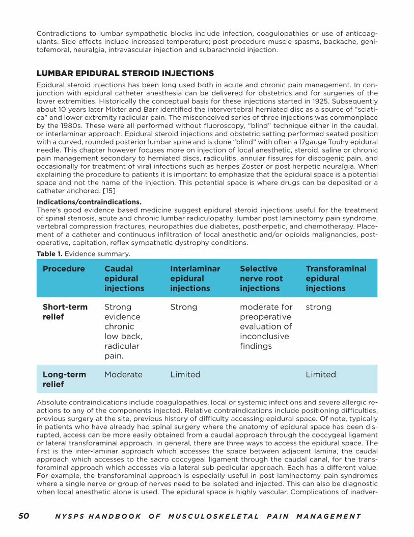
50 N Y S P S H A N D B O O K O F M U S C U L O S K E L E TA L P A I N M A N A G E M E N T
Contradictions to lumbar sympathetic blocks include infection, coagulopathies or use of anticoag-ulants. Side effects include increased temperature; post procedure muscle spasms, backache, geni-tofemoral, neuralgia, intravascular injection and subarachnoid injection.
LUMBAR EPIDURAL STEROID INJECTIONSEpidural steroid injections has been long used both in acute and chronic pain management. In con-junction with epidural catheter anesthesia can be delivered for obstetrics and for surgeries of the lower extremities. Historically the conceptual basis for these injections started in 1925. Subsequently about 10 years later Mixter and Barr identified the intervertebral herniated disc as a source of “sciati-ca” and lower extremity radicular pain. The misconceived series of three injections was commonplace by the 1980s. These were all performed without fluoroscopy, “blind” technique either in the caudal, or interlaminar approach. Epidural steroid injections and obstetric setting performed seated position with a curved, rounded posterior lumbar spine and is done “blind” with often a 17gauge Touhy epidural needle. This chapter however focuses more on injection of local anesthetic, steroid, saline or chronic pain management secondary to herniated discs, radiculitis, annular fissures for discogenic pain, and occasionally for treatment of viral infections such as herpes Zoster or post herpetic neuralgia. When explaining the procedure to patients it is important to emphasize that the epidural space is a potential space and not the name of the injection. This potential space is where drugs can be deposited or a catheter anchored. [15]
Indications/contraindications.There’s good evidence based medicine suggest epidural steroid injections useful for the treatment of spinal stenosis, acute and chronic lumbar radiculopathy, lumbar post laminectomy pain syndrome, vertebral compression fractures, neuropathies due diabetes, postherpetic, and chemotherapy. Place-ment of a catheter and continuous infiltration of local anesthetic and/or opioids malignancies, post-operative, capitation, reflex sympathetic dystrophy conditions.
Table 1. Evidence summary.
Procedure Caudal epidural injections
Interlaminar epidural injections
Selective nerve root injections
Transforaminal epidural injections
Short-term relief
Strong evidence chronic low back, radicular pain.
Strong moderate for preoperative evaluation of inconclusive findings
strong
Long-term relief
Moderate Limited Limited
Absolute contraindications include coagulopathies, local or systemic infections and severe allergic re-actions to any of the components injected. Relative contraindications include positioning difficulties, previous surgery at the site, previous history of difficulty accessing epidural space. Of note, typically in patients who have already had spinal surgery where the anatomy of epidural space has been dis-rupted, access can be more easily obtained from a caudal approach through the coccygeal ligament or lateral transforaminal approach. In general, there are three ways to access the epidural space. The first is the inter-laminar approach which accesses the space between adjacent lamina, the caudal approach which accesses to the sacro coccygeal ligament through the caudal canal, for the trans-foraminal approach which accesses via a lateral sub pedicular approach. Each has a different value. For example, the transforaminal approach is especially useful in post laminectomy pain syndromes where a single nerve or group of nerves need to be isolated and injected. This can also be diagnostic when local anesthetic alone is used. The epidural space is highly vascular. Complications of inadver-

51C H A P T E R 4 . I N T E R V E N T I O N A L P A I N M A N A G E M E N T
tent needle placement include bleeding, inadvertent intrathecal administration of steroid and/or local anesthetic which could possibly result in high spinal blocks, or respiratory depression if opioids are administered intrathecal he, subdural administration because patchy anesthesia of prolonged dura-tion. Spinal puncture with these large epidural needles can result in posterior oral culture headaches with the risk of less than 1%. The caudal approach is a safer technique in the setting of anticoagulation. The transforaminal approach has inherent risks associated with trauma to the dorsal root, intravascu-lar injection of particulate steroids and bleeding, and even disastrous consequences of clotting of the artery of Adamkowiez.
Anatomy/physiology.Anatomically epidural space extends superiorly from the spinal dural layers near the base of the brain or the foramen magnum and inferiorly it continues down to the sacrococcygeal membrane. The lateral borders extend to the vertebral pedicle anteriorly it is bounded by the posterior longitudinal ligament and posteriorly it extends to the vertebral laminar bodies. The space contains connective tissue epi-dural fat veins arteries and lymphatics. It is interesting to note that the epidural veins do not have valves. This is important because abdominal pressure can cause reflex engorgement of the epidural veins. The space also varies in the cervical region where it can be anywhere between 1 to 2 mm down to the widest area and the lumbar space up to 6 mm. The area is widest in the cervical region between C7 and T1, which forms an ideal access point for cervical epidural steroid injections.
Technique.Most epidural steroid injections, whether cervical, thoracic, or lumbar are done in the fluoroscopic room with non-ionic contrast enhancement for documentation epidural space. Steroids that are most often used include long acting steroids Depo-Medrol, triamcinolone, or dexamethasone. After con-sent is obtained the patient is positioned in the prone position and fluoroscopic images are taken of the area. The end plate of vertebral bodies is squared and the spinous process is positioned midline. The interim of her opening is identified and after local anesthesia is administered and epidural needle is advanced into the space traversing the skin connective tissue, supraspinous, inter-spinous, and liga-ment flavum. At this point the needle will be engaged in this tough ligament and further advancement will be done for the loss of resistance syringe either plastic or glass, containing 2 to 3 ML of air, saline, or a combination of air and saline. An abrupt loss of resistance indicates the traversing of the tough connective tissue and entrance into the epidural space. Confirmation is made to ensure absence of blood, cerebrospinal fluid, and then 2 to 3 mL of non-ionic contrast either Isovue, or Omnipaque is injected under live fluoroscopic guidance. Documentation of the spread of the contrast media show-ing often a “soap bubble” appearance where the contrast spreads around the epidural fat, confirms accurate needle tip location. Steroid solution mixed with saline and or a long-acting local anesthetic such as preservative free bupivacaine 0.25% is injected in incremental doses under periodic fluoro-scopic imagery. The caudal approach requires fluoroscopic views initially in the lateral view the sacro coccygeal ligamentous curve. The needle is advanced through the ligament and into the caudal ca-nal, contrast is then again administered confirming cephalad spread lateral view, which follows the contour of the sacrum, and a Christmas tree appearance in the PA view often with contrast enhance-ment of lower sacral roots. The loss of resistance syringe using the caudal approach is typically not required. The transforaminal approach requires a three-quarter “Scottie dog” radiological view and typically a 22gauge blunt bevel needle is advanced just under the pedicle into the foramen. Contrast is injected under direct flight fluoroscopic view in the PA view demonstrating highlighting of the nerve root. Digital subtraction imagery can be helpful to eliminate the inadvertent injection into the blood vessel. Most physicians will choose a non-particulate steroid such as dexamethasone, which may avoid inadvertent particulate injection into the radicular artery.
FACET BLOCKS AND RFFacet blocks including medial branch peri-articular and intra-articular injections are useful in the treat-ment of inflammation, arthritic conditions including trauma to the facet or zygopophyseal or z-joints joints. These are divided into cervical, thoracic and lumbar facets. The angulation of the facets differs in each of these regions based on the required movement of that segment of the spine. Evidence based medicine literature support for diagnostic medial branch blocks show a diagnostic predictabil-ity of 75 to 100% as compared to limited value for diagnostic intra- articular or peri articular blocks. [15]

52 N Y S P S H A N D B O O K O F M U S C U L O S K E L E TA L P A I N M A N A G E M E N T
Indications/contraindications.Spinal pain can because by many different neural sensitive structures including nerves, discs, liga-ments, joints and fascia and muscle. Facets form anchoring points on either side of the posterior as-pect of the disk. Depending on the angulation the facet joints allow motion in the cervical thoracic and lumbar region. Simple degeneration, trauma, infectious processes, or fractures can injure these joints, similar to any other articular surface joints in the body. As the inter-vertebral disc degenerates weight is transmitted laterally onto the joints causing the joints to have increased weight bearing, and they compensate with increased bone growth some of which can protrude into the spinal canal resulting in subsequent bony spinal stenosis. Facet pain has a very different distribution of pain as compared to radicular pain. Facet joint pain is non-dermatomal. Aggravation of cervical facets typically cause pain in the neck occasionally associated with headaches and referred radiation to the shoulder and between the shoulder blades as the lower cervical facets are involved. Facet arthropathy causes local-ized and proximally radiating non-dermatome pattern pain. Similar to other joint arthropathies facet joint pain is often worsened in the morning and is relieved somewhat with activity, heat and NSAIDs. Mechanical motion at the affected joint causes reproduction of pain. This diagnostic specificity is uti-lized to advantage assessing pain, pre-and post facet joint medial branch blocks, and documented. The specificity of this is over 95%.
Anatomy/physiology.Facet joints are formed by the superior and inferior articular facet services of connecting vertebral levels. Like articulating joints anywhere else in the body the spinal facets are considered true joints with in articular surface, synovial fluid, and specific angulation and limitation of motion. The nerve supply of the facets is from two adjacent level medial branches. Posterior primary rami divide into three divisions, lateral intermediate and medial branches. The medial branch hugs close to the superi-or articular process where it connects to the transverse process, and passes in a groove, under a small ligament called the mammillo accessory ligament, that bridge between two small bony prominences called mammary bodies. These are more pronounced in the lumbar region. The medial branch gives innervating fibers to the same level and passes a branch to the level below. This is why in order to en-ervate his single level the medial branch one level higher must also be blocked. In other words, each medial branch provides innervation to half the joint at the same level and half the joint one level be-low. This relationship has an exception at the L5 level where it is the dorsal ramus instead of a medial branch that goes over the SAP over the sacral medial ala.
Technique.Today facet blocks are performed with fluoroscopic guidance. Ultrasound guidance is an emerging potential in the near future. The blind technique has fallen out of favor. Physicians who specialize in interventional techniques of the spine are best suited to perform cervical and thoracic blocks. For me-dial branch blocks, the level above and at the affected joint is targeted due to the dual innervation off of the posterior primary ramus. All are performed in the prone position except for the upper cervical levels, which can be performed in lateral decubitus.
Cervical medial branch blocks: Patient position: prone, cervical pillow, and chest raised with hard pil-low to allow a gentle flexion of the neck. Some physicians use the lateral position, affected side up, for the higher cervical facet levels. Materials: 23 gauge or 22 gauge 3.5” spinal needles. Three ml syringe, five ml syringe, 25, or 27gauge local anesthetic needle, long clamp to identify target under fluoros-copy. Medication: Bupivacaine 0.25%, triamcinolone 40 mg per ml. The ‘waist’ of the articular pillar is identified and the target located at the narrowest part on PA view, and at the center of the parallelo-gram of the facet on a lateral view. The volume injected should be less than ½ ml.
Thoracic medial branch blocks: Patient position: prone. Materials: 23gauge or 22gauge 3.5” spinal nee-dles. Three ml syringe, five ml syringe, 25, or 27gauge local anesthetic needle, long clamp to identify under fluoroscopy. Medication: Bupivacaine 0.25%, triamcinolone 40 mg per MLNeedles are targeted in the PA view to avoid inadvertent needle traversing the lung.
Lumbar medial branch blocks: Patient position: prone. Materials: 23 gauge or 22 gauge 3.5” spinal needles, three ml syringe, five ml syringe, 25, or 27gauge local anesthetic needle, long clamp to iden-tify under fluoroscopy. Medication: Bupivacaine 0.25%, triamcinolone 40 mg per ml. Procedure: As with most blocks of the spine it is important to start with a true AP view. Square off the superior end plate of S1. The L5 medial branch block is typically blocked first. Mark off the sacral ala at the junction with the superior articular process, this is the target location.

53C H A P T E R 4 . I N T E R V E N T I O N A L P A I N M A N A G E M E N T
Advance a 22gauge spinal needle through an anesthetized skin weal and under fluoroscopic guidance advance needle tip to target. For the remaining lumbar levels, return to a true PA position, square off the vertebral body endplates, then rolled the C-arm toward the affected side until a three-quar-ter view, or a “Scottie dog” view was obtained. The target will be the “eye” of the Scottie dog, this is where the superior articular process base meets the transverse process. Touch the needle tip to the bone, document images in both this view and in a PA view. Contrast is typically not necessary. This area is not vascular. Inject a total volume of half mL local anesthetic (lidocaine or Bupivacaine). Steroid can also be injected in this area however the patient must be examined within one-hour post proce-dure to determine effect. [15]
Intra articular Facet Joint blocks: These are injections into the joint itself. The facet joint is seen as between the superior and inferior articular processes in a squared PA view with slight lateral or medial oblique rotation until the joint is visible and this space is viewable. Thoracic and cervical facet joint injections are complex and carry a much higher risk of inadvertent cord injury and should be consid-ered only by pain medicine specialists. The materials and technique is similar with the exception of the target area. Resistance is encountered as the spinal needle traverses the joint. Care is taken not to pass through the anterior aspect of the joint. [15]
Radiofrequency of the medial branches are done using a similar technique. The needle is insulated with and exposed active tip. The needle is often placed and a slight upward angulation in order to al-low greatest contact of the exposed needle tip with the area of anticipated nerve location. This allows for the largest possible lesion. Repeat radiofrequency ablations are usually limited to three times per year. Often the pain relief can be upwards of 10 months, the time it takes for the nerve to regenerate.
Author’s tips: 1. If sedation is used to provide comfort for patient during procedures it is important to avoid the use of any opioids. This can mask a patient’s response two and in accurately placed needle. Instead, use only midazolam or propofol.
2. Curving the tip of your spinal needle to approximately 15° makes course corrections much simpler during the needle advancement.
3. Try using a 25gauge 3.5” spinal needle to avoid skin local anesthesia infiltration.
4. Inject Lidocaine as spinal needle is being withdrawn to avoid any paraspinal muscle spasms.
5. Re-examine the patient 30 minutes post procedure to reproduce facet joint pain, document. Document the improvement.
6. For radiofrequency procedures, document the response to both sensory and motor stimulation and save final needle positions images.
SACROILIAC JOINT BLOCKS AND RFBecause of the complexity of the sacroiliac joint and biomechanics, there has not been any singular diagnostic test to isolate the pain generator. The sacroiliac joint can be responsible for up to 20% back and leg pain. Injuries to the sacroiliac joint are often seen with fall on the buttocks, or in rear-ended motor vehicle accidents causing full movement of the sacroiliac joint at the time of impact. [15]
Table 2. Evidence summary.
Procedure Diagnostic injections
Intra- articular blocks
SIJ neurotomy) Conventional and pulsed RF
Short-term relief Limited Limited
Long-term relief Limited Limited
Diagnostic Predictability Moderately useful

54 N Y S P S H A N D B O O K O F M U S C U L O S K E L E TA L P A I N M A N A G E M E N T
Indications/contraindications.Absolute contraindications include coagulopathies, local or systemic infections and severe allergic re-actions to any of the components injected. Relative contraindications include positioning difficulties, previous surgery at the site, previous history of difficulty accessing the joint. Complications include inadvertent nerve damage to the large sciatic nerve, which travels in close proximity to the inferior portion of the joint. For the initial injection or the therapeutic phase injection should be limited to no more than 4 to 6 per year. Documentation of at least greater than 50% decrease in pain lasting over six weeks is required. Sacroiliac joint radiofrequency ablation typically is done no more than three times per year again assuming the above 50% decrease in pain for four months is documented.
Anatomy/physiology.This joint is a true diarthodial synovial joint with extensive innervation and a comment yet undiag-nosed cause of low back pain in adults. Immobile in males, hormonal changes in females during the third trimester allows the joint some mobility.
Technique.Patient position: prone. Materials: 23 gauge or 22 gauge 3.5” spinal needles. Three ml syringe, five ml syringe, 25, or 27 gauge local anesthetic needle, long clamp to identify target under fluoroscopy. Medication: Bupivacaine 0.25%, triamcinolone 40 mg per ml. Fluoroscopic images are taken and the inferior portion of the joint is identified. Using a cranial caudal and oblique rotation of the C-arm the inferior portion of the joint is identified between two lines. This is sometimes described as overlap-ping the “rivers”. The 22 or 23gauge spinal needle is placed through sterile and anesthetized skin and advanced until bony contact is made at the inferior portion of the joint. The needle is advanced until resistance is encountered in the ligament. Frequent fluoroscopic images are required during bony contact to identify the redirection of the needle tip. Once in position contrast is injected and typically seen traversing upwards into the joint. PA and lateral views are documented. It is important to have the patient able to respond and identify if the larger underlying somatic nervous contacted. This would cause reproduction of radicular pain. Steroid with local anesthetic is injected. Post procedure the patient should be re-examined in the degree of pain relief documented.
Radiofrequency ablation of the sacroiliac joint: After fluoroscopic identification of the joint as de-scribed above, a radiofrequency probe, with a 10 mm exposed tip is advanced into the joint. Sensory stimulation at 50 Hz and motor stimulation at 2 Hz is performed with the patient able to respond and identify the source of pain and to rule out inadvertent sciatic nerve stimulation. Ablation of either 75 or 90°C is carried out for 60 to 90 seconds. Further radiofrequency lesions are carried superiorly along the course of the joint. The L5 medial branch/posterior primary ramus is targeted and the depression of the sacral ala infero lateral to the L5 transverse process.
INTRADISCAL THERAPIESIt has been long recognized that the intervertebral discs carry a rich innervation particularly along the annulus. Disrupted discs and tears tend to have highly sensitized annular fibers. This is often seen either as low back pain or low back and radicular pain. Diagnosis of these conditions, disc pathology, can be difficult with MRI and CAT scans. Provocation discography, which involves injection of contrast material into the nucleus pulposus of discs with the patient able to respond, has proved effective in locating annular tears and sensitive discs. Percutaneous discectomy has been suggested as an alter-native to an open discectomy. These include laser discectomy’s, radiofrequency collation or nucleop-lasty, mechanical and manual or decompression either with an automated device or by use of a long clamp followed by subsequent cauterization of the interior annular fibers have been used. The ratio-nale has been that reduction of internal disk volume decreases tension and pressure on the annular wall and subsequent nerve root reducing pain. [15]

55C H A P T E R 4 . I N T E R V E N T I O N A L P A I N M A N A G E M E N T
Table 4. Evidence summary.
Procedure Lumbar discography
Intra-discal electro thermal therapy (IDET)
Radiofrequency posterior annuloplasty
Short-term relief
Limited Limited
Long-term relief
Indeterminate Indeterminate
Diagnostic Predictability
Strong for diagnos-ing discogenic pain in conjunction with other diagnostic injections
Table 5. Evidence summary.
Procedure Automated percutaneous lumbar discectomy
Percutaneous laser discectomy
DeKompressor Nucleoplasty
Short-term relief
Moderate Moderate Limited Limited
Indications/contraindications.Absolute contraindications include coagulopathies, local or systemic infections and severe allergic re-actions to any of the components injected. Relative contraindications include positioning difficulties, previous surgery at the site, previous history of difficulty accessing the joint. Complications include discitis, a particularly difficult to treat condition often requiring surgical intervention.
Anatomy/physiology.Intervertebral disc procedures are typically limited to the lumbar spine. Occasionally the thoracic spine may be attempted. The key identifying points for disc related procedures in the lumbar spine or identification of the superior articular process and adequate squaring of the vertebral and plates above and below. Cervical disc procedures should be limited to physicians who have extensive exper-tise. The L5/S1 intervertebral disc can be difficult to access in males due to the high iliac crest.
Technique.Patient position: prone, pillow under abdomen to reduce the lumbar lordosis. Full surgical prep and drape. Materials: Three ml syringe, five ml syringe, 25, or 27-gauge local anesthetic needle, long clamp to identify target under fluoroscopy. Preoperative antibiotics are ministered within 30 minutes of the procedure. Postoperative antibiotics are usually not required. For provocation discography, immedi-ate CAT scan post procedure is required. Medication: Bupivacaine 0.25%, triamcinolone 40 mg per ml. Fluoroscopic images are taken and the target disk is squared with and plates above and below. The C-arm is rotated obliquely until the superior articular process transects the disc by approximately 50%. The target point is just anterior to the superior articular process and the midpoint of the disc. To access the disk more posteriorly further oblique rotation may be required. This is particularly the case for posterior herniations. A 16gauge or 14gauge (manufacture provide) is advanced through sterile and anesthetized skin and soft tissues to contact the anterior portion of the superior articular pro-cess. The patient must be awake enough to respond if the traversing route is contacted. Resistance is encountered as the needle is pushed past the annular fibers and into the pulposus. Contrast is often injected into the disk to confirm proper placement of the needle. Images are recorded. The remainder of the procedure is performed based on the manufacturer’s recommendations. Disc material that is removed is sent for pathology for quantification and assessment. Post procedure a small quantity of steroid lidocaine antibiotic mixture is injected into the disk.

56 N Y S P S H A N D B O O K O F M U S C U L O S K E L E TA L P A I N M A N A G E M E N T
VERTEBRAL AUGMENTATIONThe aging population has seen an increase in low back pain secondary to vertebral compression frac-tures. These fractures cause pain at the fracture site at the level of the vertebral body, can cause spinal instability and kyphoscoliotic changes. Vertebroplasty and kyphoplasty are two effective methods for treating these conditions; both involving placing a needle into the vertebral body and injecting radi-opaque bone cement. In the latter procedure the vertebral body is expanded either by a balloon or other expansion device to create a central cavity to accommodate the bone cement. [15]
Table 6. Evidence summary.
Procedure Vertebroplasty Kyphoplasty
Short-term relief Moderate Moderate
Long-term relief Moderate Moderate
Indications/contraindications.Absolute contraindications include coagulopathies, local or systemic infections and severe allergic re-actions to any of the components injected. Relative contraindications include positioning difficulties, previous surgery at the site, previous history of difficulty accessing the spine. Complications include inadvertent leakage of bone cement into the area of the neural foramen or epidural space. This may require subsequent neurosurgical decompression and/or fusion.
Anatomy/physiology.The thoracic and lumbar vertebral bodies are typically treated. The key to this procedure is to identify and center the pedicle of the affected level. In the lumbar spine compression fractures are typically in the L1/L2 levels.
Technique.Patient position: prone with pillow under abdomen to decreased lumbar lordosis Manufacturer provid-ed needle and bone cement mixer. Medication: local anesthetic, preoperative antibiotics. Full surgical prep and drape. Materials: 23 gauge or 22 gauge 3.5” spinal needles. Three ml syringe, five ml syringe, 25, or 27 gauge local anesthetic needle, long clamp to identify target under fluoroscopy. The affected vertebral compression fracture is identified and the endplate squared. The C-arm is open leaked until the pedicle is seen at approximately the 50% midline of the vertebral body. Using the manufacturer provided needle/trocar this is advanced through previously anesthetize skin until bony contact at the midpoint of the pedicle is made. Then using multiple PA and lateral views this is advanced into the pedicle taking care not to fracture it. A manufacturer provided sterile mallet is used to gently tap the trocar into place. Final needle position is confirmed in the PA and lateral views. Manufacturer provided cement mixer and cement is then gently pumped into the collapse vertebral body with continuous first topic confirmation. For kyphoplasty a balloon is advanced and inflated under pressure to create a cavity. The needle is removed by reducing the injected pressure to prevent leaving a bone cement tail. The patient during this procedure is likely sedated with a small amount of opioid however is able to respond to any questions.
IMPLANTABLE THERAPIESThese procedures are of two types. The first involves an electrical current of controlled frequency and amplitude to scramble to pain in the dorsal column of the spine. The second involves drug delivery directly into the intrathecal space for less common conditions the epidural space for continuous drug delivery. Both procedures involve an initial diagnostic phase or trial. Intrathecal therapies can be use-ful in patients with spasticity disorders and malignancies. Intrathecal medication can reduce common side effects associated with large doses of opioids orally. [15]

57C H A P T E R 4 . I N T E R V E N T I O N A L P A I N M A N A G E M E N T
Table 7. Evidence summary.
Procedure Spinal cord stimulation Implantable intrathecal drug administration
Short-term relief
Strong Strong for neuropathic or malignancy pain
Long-term relief
Strong Moderate
Indications/contraindications.Absolute contraindications include coagulopathies, local or systemic infections and severe allergic re-actions to any of the components injected. Relative contraindications include positioning difficulties, previous surgery at the site, previous history of difficulty accessing the space. Complications include inadvertent placement of the electrode lead into the intrathecal space, or inadvertent contact and trauma to the spinal cord. Hematomas in this region can cause profound neurologic effects requiring further surgery.
Anatomy/physiology.The key points to understand in this anatomy are to accurately document and count the lumbar/thoracic levels. Placement of the tip of either the electrode for in the case of spinal cord stim leads or the catheter in the case of intrathecal or epidural drug delivery systems is important. The patient documents the pain relief. 50% or greater decrease in pain and clinical re-examination are required along with a lengthy discussion before the decision is made for permanent electrode or lamitrode placement. Prior to intrathecal pump implantation a trial is performed. Different trials have been tried however the evidence is greatest with continuous intrathecal medication delivery in an in-hospital setting. Single shot epidural opioids as well as continuous epidural opioids are not as predictive.
Technique.Patient position: prone, pillow under abdomen to reduce the lumbar lordosis. Full surgical prep and drape. Materials: Three ml syringe, five ml syringe, 25, or 27-gauge local anesthetic needle, long clamp to identify target under fluoroscopy. Preoperative antibiotics are ministered within 30 minutes of the procedure. Postoperative antibiotics are usually not required. For provocation discography, immedi-ate CAT scan post procedure is required. Medication: local anesthetic. Fluoroscopic images are taken and the target level, the point of entrance for the epidural space is marked. The space is accessed in a much steeper caudal/cephalic approach. This is to ensure that there is no inadvertent puncture of the dura for spinal cord stimulator placement. For spinal cord stimulator’s two electrodes are placed for the trial. The patient is then allowed to map his pain while different frequency combinations are tried. The electrode is adjusted cephalad or caudal accordingly. The final position is then documented flu-oroscopically and secured with sutures to the skin. Sterile dressing is applied and the electrodes con-nected to an external generator. These electrodes are then removed one week later. Continuous an-tibiotics are usually not required. Intrathecal catheters are similarly placed into the intrathecal space. Permanent placement involves a tunneled catheter with a pre-agreed upon pump reservoir position.
EPIDURAL INJECTED STEROIDSIn April 2014, the FDA issued new box learnings on the safety of epidural he injected steroids. This warning is unrelated to the contamination of steroids that was reported previously. This warning was added to recommend practitioners discussed the risks of injected steroids and inform patients of the possibility of vascular occlusion, paraplegic, death. The FDA did not discourage the usage of steroids for epidural injections.
Epidural steroids include triamcinolone, betamethasone, dexamethasone, and methylprednisolone. All of these are considered intermediate the long acting steroids with varying degrees of glucocorti-coid and mineral cord quite effects. The particulate steroids include triamcinolone and methylprednis-olone. Non-particulate includes the dexamethasone and the betamethasone. [16]

58 N Y S P S H A N D B O O K O F M U S C U L O S K E L E TA L P A I N M A N A G E M E N T
Dexamethasone and betamethasone or non-particulate steroid. Dexamethasone has been associated with transient side effects including tingling along the nerve, headache and paralysis. These are all manage conservatively long-lasting effects were noted. [18]
Steroids may be prepared with preservatives or without. It is unclear whether preservatives pose any problems especially since they’re diluted with local anesthetic and saline prior to injection.
Epidural injected steroids have been used safely in the epidural, for over 40 years despite the FDA approval.
Confusion exists among the public and often practitioners about the role of the FDA and its regulation and medication. FDA can issue warnings an additional labeling, however the ultimate judicious use of the medication has awfully able is determined by the physician. Many medications are used in off label use, a notable example is gabapentin which was first introduced as a safe non-hepatic metabolized renal excreted antiepileptic medication. However, it’s predominant use today is in neuropathic pain conditions an off label use.
However, there are risks associated with neuraxial steroid injections which factor operator technique, volume of injectate, route injected, and choice of steroid.
Particulate steroids which include triamcinolone and betamethasone have been suggested as a cause of vascular occlusion in procedures such as transforaminal’s in the cervical thoracic and lumbar spine. Case reports of transient or even permanent paraplegia have also been reported with interlaminar and caudal epidural steroid injections however most of these cases are related to unrecognized arteriove-nous abnormalities causing pre-existing cord compression that was likely exacerbated with simply the volume of injected material. [17]
So in summary the choice of steroids and technique are dependent on the physician however avoiding particulate steroids during transforaminal procedures may be prudent. Whether or not to use a pre-servative free solution is more controversial. And of course, having adequate training in the technique is paramount.

59C H A P T E R 4 . I N T E R V E N T I O N A L P A I N M A N A G E M E N T
REFERENCES
Amrhein, T., & Joshi, A. (2016). Technique for CT Fluoroscopy Guided Lumbar Medical Brach Blocks and Radiofrequency Ablation. American Journal of Radiology(207), 631-634.
Awal, S., & Madabushi, R. (2016). CRPS: Early Lumbar Sympathetic Block is Better Compared to Other Interventions. Pain Physician Journal(19), E363.
Bhatia, A., & Peng, P. (2015). Stellate Ganglion Block Regional Nerve Blocks in Anesthesia and Pain Therapy: Traditional and Ultrasound Guided Techniques.
Bogduk, N. (1997). International Spinal Injection Society Guidelines for the Performance of Spinal Injection Procedures: Part 1: Zygapophyseal Jpint Blocks. Clint Pain(13), 292-297.
Dreyfuss, P. H., Dryer, S. J., & Herring, S. A. (1995). Contempory Concepts in Spine Care: Lumbar zygapophyseal (Facet) Joint Injections. Spine, 2040-2047.
Gerwin, R. (2016). Myofacial Trigger Point Pain Syndromes. Semin Neiro(36), 469-473.
Ghai, A., & Kaushik, T. (2016). Stellate Ganglion Blockade Tchniques and Modalities.
Acta Anaesthesiologica Belg., 67:15.
MacVicar, J., & Borowczk, J. (2013). Lumbar Medical Branch Radiofrequency Neurotomy in New Zealand, Pain Medicine (Vol. 14).
Manchikanti, L., & Boswell, M. V. (2004). Prevalence of Facet Joint Pain in Chronic Spinal Pain of Cervical, Thoracic, and Lumbar Regions. BMC Musculoskeletal Disorder(5(1)), 15.
Ramirez, L., Rugcks, J., & Ortiz, J. (2016). Radiofrequency Neurolysis for Lumbar Pain Using a Variation of the Original Technique. Pain Physician(19), 155-161.
Scott, A. N., & Guo, B. (2009). Triiger Point Injections for Chronic Non Malignant Musculoskeletal Pain: A Systematic Review. American Acadamy of Pain Medicine, 10(1), 54-69.
Slipman, C., & Derby, R. (2008). Interventional Spine in Algorithemic Approach.
Stitz, M. Y., & Sommer, H. M. (1999). Accuracy of Blind Versus Fluoroscopcally Guided Caudal Epidural Injection. Spine(24), 1371-1376.
Weinstein, S. M., Herring, S. A., & Derby, R. (1995). Contemporary Concepts in Spine Care. Epidural Steriod Injections Spine, 1842-1846.
Pain generators of the spine: Kuslich SD, Ulstrom CL, Michael CJ. The tissue origin of low back pain and sciatica: a report of pain response to tissue stimulation during operation on the lumbar spine using local anesthesia. Orthop Clin North Am. 1991;22(2):181-187.
Comparison of the Particle Sizes of Different Steroids and the Effect of Dilution: A Review of the Relative Neurotoxicities of the Steroids. Honorio T. Benzon, M.D.; Teng-Leong Chew, Ph.D.; Robert J. McCarthy, Pharm.D.; Hubert A. Benzon, M.D.; David R. Walega, M.D.
Acute Paraplegia After Lumbar Steroid Injection in Patients With Spinal Dural Arteriovenous Fistulas: Case Reports. https://www.ncbi.nlm.nih.gov/pmc/articles/PMC5108724/#!po=34.2105
Dexamethasone adverse effects during TFESI http://www.painphysicianjournal.com/current/pdf?article=MjMyMg%3D%3D&journal=88.

60 N Y S P S H A N D B O O K O F M U S C U L O S K E L E TA L P A I N M A N A G E M E N T

61C H A P T E R 5 . R E H A B I L I TAT I O N T E C H N I Q U E S
CHAPTER 5. REHABILITATION TECHNIQUES
Bradley Cash MD.
Rehabilitation specialists focus on the body as a whole. Examination of posture, range of motion, motor strength, body mechanics and core strength are essential in determining the pathology of body dysfunction or pain and the function as a whole person in determining the best options for cor-rective action. Pain management is complex and includes various types of conservative options for treatments. The three main categories of conservative management include therapeutic treatment options, pharmacologic management and injection therapies. In this chapter the focus will remain on therapeutic techniques for pain management.
The Rehabilitation of an individual includes both physical and mental reconditioning. The complex components of both somatic and cognitive deficits are the goal of the various techniques to enhance function. The various options include those with purely somatic, some with purely psychological and those with mixed elements. There is never a “correct” option as treatment must be individualized. Learn from experience which methods are the most effective.
PHYSICAL THERAPY TECHNIQUESManipulative TherapyAlso known as Manual Therapy and is done by Massage Therapists, Physical Therapists, Chiropractors, Osteopaths and Occupational Therapists. It encompasses various methods to treat musculoskeletal pain and disability thru different types of manipulation, joint mobilization, “kneading”, massage, gen-tle adjustments and soft tissue mobilization.
There are discreet differences between manipulation, mobilization and massage. Manipulation in-volves the introduction of a rapid rotation, shear or distraction force into an articulation. Manipula-tion is often associated with an audible “popping” caused by the instantaneous breakdown of the gas bubbles that form during a joint cavitation. Mobilization is a slower, more controlled process of articular and myofascial stretching with the intention to improve myofascial and biomechanical elas-ticity. Massage is typical the repetitive rubbing, kneading or stripping of myofascial tissues to improve interstitial fluid dynamics. The key difference in a manipulation versus mobilization is that once the manipulation is initiated the process must undergo full completion.
Manipulative Therapy is done more commonly now by Physical Therapists. Most undergo special training and receive a certificate regarding their abilities to treat patients with manipulation. One of the more common theories is the Maitland technique which has an Australian origin. Historically, it was Chiropractors that were involved in manipulative therapies. The greater forces were required to improve mobility especially in patients that there was profound deficits. A more recent technique which has been even more aggressive performed by Chiropractors and other practitioners is Ma-nipulation under Anesthesia. During this technique the patient is sedated. After sedation a joint or spinal segment is manipulated to undergo its full range of motion. The goal is to enhance range of motion where deficits exist due to soft tissue restrictions. The sedation allows the patient to be in a relaxed state and be fully ranged beyond what pain or spasms may allow in an awake state. This is not fully accepted in the medical world due to the underlying fear that during sedation the normal feedback mechanisms are not functional to allow a patient to be guarded or “stop” a painful range of motion, with the fear of causing soft tissue, tendon or ligament damage. Therefore the benefits of manipulation under anesthesia must clearly outweigh the risks, before deciding to proceed.
FeldenkraisThis is a Somatic Educational System which was designed by Moshe Feldenkrais to reduce pain, improve range of motion, improve physical function and promote general well- being, by improving the patient’s awareness of themselves and by improving movement abilities. Education about the reduction of pain and removal of biomechanical unsound movement habits is part of the process. The education of biomechanics, posture and efficiency of movements of various activities is essential with this method. Without the elimination of inefficient biomechanical processes, the method is not successful.

62 N Y S P S H A N D B O O K O F M U S C U L O S K E L E TA L P A I N M A N A G E M E N T
The approach believes that increasing a person’s proprioceptive and kinesthetic self- awareness can lead to greater function and ease and pleasure of movement. This technique is most similar to the Al-exander Technique which provides more education about movement and not a hands on manipula-tive type of treatment. The practitioner directs attention to habitual movements which are inefficient or strained causing pain, and then teaches new patterns using gentle, slow repeated movements. Slow repetition of the new pattern is a key feature to teaching the proper new biomechanical pro-cess. The movements may be either passive (done by the practitioner) or active (done by the patient)
(Knaster, Mirka 1996) Discovering the Bodies Wisdom: A Comprehensive Guide to more than 50 Mind- Body Process; Bantam pages 232- 8
Rosen Method
The Rosen Method of Rosen Method Bodywork was developed by a Physical Therapist, Marion Rosen as a type of Complimentary or Alternative technique to healing. It is described as a “psychosomatic” of mind over body technique which attempts to integrate the emotional or mental patterns within the body with the physical expression. The goal is to identify or uncover the “unconscious” pattern of physical stress that is held within the body as muscular holding or tension, feeling or behavior. The main theory is that a person protects themselves within the body from past painful experiences by blocking patterns in the mind and separating them from the true self. The most common protective mechanism is through musculoskeletal tension and pain which can be observed by the bodywork practitioners. The patterns that are observed are musculoskeletal tensions, abnormal postures and shortness of breath. The method is practices by non- intrusive touch, verbal interaction and creating an awareness of each breath. This method has the goal to integrate the body, mind, emotions and spirit and “unlock” the unconscious.
(Raso, Jack 1997) “Unnaturalistic Methods” The Expanded Dictionary of Metaphysical Healthcare, Al-ternative Medicine, Paranormal Healing and Related Methods)
Craniosacral Therapy
This is a unique type of alternative therapy with a bodywork of therapeutic touch. The idea is that the practitioner using the minuscule movements of the synarthrodial joints of the cranium with a gentle touch to alter or manipulate the flow of cerebrospinal fluid and aids in “primary respiration”. The practitioner also may apply gentle touch or movements to the lower spine or pelvis around the area of the sacrum. Patients report deep relaxation during treatment and the therapeutic effects are thought to be responsive to endorphins and the endocannabinoid system. Craniosacral thera-py was originated in the 1970’s by a team of Osteopaths after years of research in the field of Oste-opathy regarding the movements between the temporal bones and the parietal bones.
The “primary respiratory mechanism” is theorized to be responsive to the inherent motility in the central nervous system, fluctuation in cerebrospinal fluid, mobility of the intracranial and intraspinal dural membranes, mobility of the cranial bones and involuntary movement between the sacrum and pelvis. Practitioners note that there are small rhythmic movements between the cranial bones that contribute to cerebrospinal spinal fluid and arterial pressures. It is the manipulation between these bones that contribute to the therapeutic effect.
Most craniosacral therapists are Physical Therapist that have taken specialized courses to teach the technique. The clinical studies of effectiveness are somewhat controversial but there is much anec-dotal relief in various case studies.
(Russell J, Rovere A, (2009) “ Craniosacral Therapy” American Cancer Society Complete Guide to Com-plementary and Alternative Cancer therapies, (2nd edition) American Cancer Society pages 187- 9
Trager Approach
This method is also typically done as a subset of Physical Therapy. It is done with extensive amounts of somatic education. It uses both deep physical and mental patterns in order to promote; deep relaxation, increased physical mobility and mental clarity. The approach is named after its founder who was a Psychiatrist by training and used the deep seated psychological relaxation to enhance the physical state of well- being. It was originally used to treat patients with neuromuscular and neu-rological conditions. It was used to enhance function but has not altered either medical or surgical treatments.

63C H A P T E R 5 . R E H A B I L I TAT I O N T E C H N I Q U E S
At the beginning of a treatment session, the practitioner creates a state of meditation. Once in the hyper- relaxed state, the use of gentle touch combined with active and passive fine movements, the body is taught how to mobilize using less effort. Body contact may range from gentle to firm but it is not applied with firm resistance. It uses the body’s natural mobility to enhance function. The idea not to use any strain or resistance is to not induce pain. The approach teaches to body to move more efficiently without inducing pain. The patient is taught how to move with the least amount of effort with reduction of the fear of pain. The teaching is to move with the path of least resistance and the least amount of tension possible. Success in this treatment must involve the patient’s ability to achieve deep relaxation and allow the practitioner to guide them through the series of movements. It typically is done without any other external stimuli
PHYSICAL MODALITIESPhysical Therapists use these types of passive treatments to supplement active treatments such as therapeutic exercise and manual techniques. The idea is to help strength, relax, heal and reduce pain and inflammation with the use of modalities. Modalities alone are not effective but are used to adjunct treatment to Physical Therapy or Chiropractor to prepare a patient for the therapeutic session.
Electrical Stimulation/TENSA series of electrical currents are applied to the body thru electrodes that are strategically placed along a specific muscle, muscle group or along a nerve pathway. The current may be altered through the unit and varies in frequency and intensity. The idea is to relax the muscle and reduce pain via the Gate Theory. By stimulating the neural pathways, the pain fibers are blocked. This helps reduce painful spasms and contributes to the alleviation of pain. A practitioner may use a large unit called an “E- stim” to treat a patient in a therapeutic session combined with moist heat. This is the most common during treatment. A patient may also be given a TENS unit which is a smaller portable unit that can be worn at various intervals throughout the day under the clothing. This also can be adjusted in intensity and frequency for various pain conditions, but is not nearly as strong as the E- stim used in office treatment.
Laser TherapyLow level lasers are used to stimulate cells and enhance functions as opposed to higher level lasers which are used to cut and destroy tissues. It is also known as Cold Laser therapy. There are variable dose wavelengths which are used for treatment of both acute and chronic pain. The treatment is fo-calized toward a specific site of “damaged” tissue and is applied typically for a few minutes. It seems to be most effective in treatment of muscle and soft tissue injuries especially chronic back pain and spasms and less effective in chronic arthritic conditions. It does help in acute flare ups of arthritic conditions for reduction of inflammation. The exact mechanism is unknown but it seems that the low level laser light might react with the mitochondria affecting the electron transport chain to reduce painful spasms and swelling. It also may enhance blood flow to specific areas to promote healing. Laser research continues at a furious pace to find effective treatments for various clinical situations. At this point, it is still considered somewhat experimental and many insurance carriers will not reim-burse the treatment.
UltrasoundThis technique has been used by Therapists since the 1940’s for treatment of painful inflammatory and arthritic conditions. It is accomplished by transmitting sound waves through a round headed or wand like probe with direct contact to the skin through a gel which helps transmit the waves from the metal probe through the skin. Therapeutic ultrasound is in a range of 0.8 to 3.0 MHz
The waves are generated electrically through a vibration of crystals within the probe and transmit-ted through the skin causing a vibration of local tissues and generating heated. Heat variations can be made by using a continuous ultrasound or pulsed ultrasound. One would use a pulsed ultra-sound when excessive heat formation is not desired, such as a newer acute tissue injury.
Ultrasound causes increased tissue relaxation, increased local blood flow and scar tissue break-down. The intensity and density of the ultrasound wave may be adjusted to achieve the desired re-sult. Ultrasound depth varies from 5- 10cm.

64 N Y S P S H A N D B O O K O F M U S C U L O S K E L E TA L P A I N M A N A G E M E N T
There are contraindications to ultrasound which include; malignancy, growth plates in children, local metal implants, local infection, local acute inflammation, vascular abnormalities, and pregnancy and over the eyes, skull, spinal cord in laminectomy site and testicles.
Moist Heat/Cold PacksHeat therapy is also known as Thermal Therapy and is used to treat local pain, spasms and reducing inflammatory conditions. For rehabilitation techniques it is the most common form of pre-treatment to increase extensibility of collagen tissues, decrease joint stiffness, reduce pain, reduce spasms, in-creasing blood flow and reducing soft tissue edema. Moist heat is believed to be more effective in energy transfer than dry heat and is more comfortable for a patient to tolerate, but studies indicate that it is the heat itself that promotes vasodilation and enhanced blood flow to stimulate tissue recovery and enhance soft tissue mobility. It is typical to apply local heat superficially when penetrates up to 5cm used as pre- treatment for a therapeutic technique.
Cold therapy or Cryotherapy is the local use of cold temperatures in medical therapy. This is one of the oldest forms of treatment, dating back 300 years and is used to reduce inflammation and reduce blood flow to locally damaged tissue and additionally reduce pain and spasms which are a direct re-sult of the injury. The goal is to cause vasoconstriction of small blood vessels to decrease cell growth and reproduction, increase cell survival, decrease inflammation, decrease pain and decrease spasms. Oxygen consumption is reduced as well as nerve conduction activity which reduces pain. Also the after effects of vasoconstriction is a reflective vasodilation and healing of injured tissues.
Cryotherapy is used to enhance thermal transfer of energy from damaged tissue to the therapeutic modality. Ice is typically applied for 15- 20 minute intervals.
CHIROPRACTIC TECHNIQUESThis is the most popular form of alternative medicine which focus on mechanical disorders of the mus-culoskeletal system especially the spine. The general health is affected via the nervous system and centered on the spine. Although most Chiropractors focus on the neurological and musculoskeletal systems, many try to incorporate general medical and primary care conditions.
The main treatment technique is manual therapy via spinal manipulation and is focused around the theory of vertebral subluxation which is not only alters spinal mechanics and causes pain, but in-volved in general health. The field was founded in the 1890’s and has been promoted widely. Stud-ies suggest that the most effective techniques yield the best results for lumbar spine disorders The field has always been controversial but more recent evidence based medicine has contributed to its greater acceptance in society
Most of the new age practitioners use a combination of “low velocity” adjustments with therapeutic techniques that Physical Therapy practices with the use of modalities. The more traditional tech-niques involve more “high velocity” adjustment techniques to treat vertebral subluxations. These techniques provide more controversial results with variable medical efficacy. Chiropractors provide significant pain management as part of the rehabilitation process.
OSTEOPATHIC MANIPULATIONA Doctor of Osteopathy receives additional training on how to use the hands to manipulate the body to promote healing. This method of manipulation provides some Chiropractic principles. Osteopathic Manipulative Medicine is a hands on method to apply pressure to the muscles and joints to stretch and provide resistance. It focuses on treating the body as a whole and utilizing the spinal column as the core. Structural problems around the spinal column are the focus of “soft tissue techniques” in which pressure is applied to the muscles close to the spinal cord, or the paraspinal muscles. The “muscle energy technique” asks the patient to flex the muscles in a specific direction while providing resistance to counter the flexion. These are more gentle manipulations than typical Chiropractic mo-bilizations.
The technique is most useful to treat neck and back pain, but contraindicated in conditions of cancer or spinal fusion.

65C H A P T E R 5 . R E H A B I L I TAT I O N T E C H N I Q U E S
CORE STRENGTHENING TECHNIQUESYogaYoga has originated in India as a physical, mental and spiritual practice or discipline. It is a physical method that centers on the spine and core which had first been practiced over 1000 years ago and became popular in the Western world on since 1980. There is a Hindu and Buddhist origin which re-fers to higher levels of concentration to a mind over body experience with the purpose of attaining a goal for the body.
The physical postures of Modern Yoga are designed to alleviate health problems, reduce stress and make the spine supple. Yoga is designed to complete exercise programs to focus on the core in Physical Therapy programs. It is a popular technique used to promote stretching and core strength-ening while promoting mental and spiritual awareness of the body.
PilatesThis is another core strengthening method which is a physical fitness system which was devel-oped in the 20th century. It is a method of controlled movements which improves flexibility, builds strength, develop control and endurance throughout the entire body. It emphasizes alignment, breathing and development of a strong core while improving coordination and balance.
Different exercises can be modified to reflect variable levels of expertise from beginner to advanced levels.
The major principle behind Pilates is the “control” of body movements, exercise and to concentrate on all movements during the exercise and be aware of the muscles that are being isolated. The concentration about the exercise and muscle recruitment is more important than the exercise it-self. The breathing technique is associated with the concentration during exercise. Pilates may also be done as part of a mat exercise or with a reformer. Typically mat exercise uses gravity as resis-tance to strengthen the core and the use of a reformer gives an extra level of strengthening. Pilates is typically used as an extension to Physical Therapy when a program is complete. This is a method of home exercise program.
Core Methods (Barre)This is designed to combine principles of Pilates, Yoga and Ballet classes to provide lean muscle mass with increased tone and strength. It combines weights to isolate muscles in the extremities, floor exercises to work on the core and seated exercise to work specifically on abdominal muscles. It is usually taught in a class setting but may be one on one. It is also an extension of Physical Therapy and may be used after a program is complete as a method of home exercise. The method itself not only works on tone and strengthening but also has an element of cardiovascular exercise as the heart rate increases/decreases during certain activities.
REIKI METHODThis is a Japanese technique for stress reduction and relaxation that promotes healing. The word itself can be broken down as follows “Rei” means Gods Wisdom or the Higher Power and “Ki” means life force energy. So the concept is spiritually or psychologically guided. Alterations in life force energy is directly correlated with health and with sickness.
Reiki treats the person as a whole including body, emotions, mind and spirit creating many beneficial effects that include relaxation, feelings of peace, security and well- being. It is a simple, natural and safe method of healing and self- improvement that everyone can use. The practitioner does this by “laying the hands” on specific areas of the body and creating energy flow. It is a mental technique which is useful to work alongside a physical technique to promote healing. A Reiki Master teaches a student to tap into a supply of “life force energy” and use this to promote healing.
Conscious healing is important to accept as a principle to promote continued healing. A Reiki student learns these ideas of transfer of energy and promotes healing from within. It is used to treat painful syndromes with conscious efforts to heal and promote recovery.

66 N Y S P S H A N D B O O K O F M U S C U L O S K E L E TA L P A I N M A N A G E M E N T
MENTAL IMAGERYMental imagery is an adjunct alternative treatment which helps transform thought into action. The practitioner uses various meditation and soft talking techniques to help the patient envision a bet-ter scenario and offset the negative thoughts associated with pain or disability to focus on more positive thoughts. This is a hands off technique with the theory of mind over matter. The use of mental imagery is a description of the body undergoing a specific movement that it would normally be unable to do. The body must imagine the movement or activity. It is theorized that the brains centers responsible for the movement undergo a physiological phenomenon during the imagina-tion of the movement. With repeated sessions the imagination sense is strong to overcome the inability, to become a real ability.
Overall, Mental imagery is a cognitive task in which a function, a behavior or a performance is re-hearsed mentally, as if a person is actually performing it.
ALEXANDER TECHNIQUEThis is a technique which is frequently used after a therapeutic technique is completed and pain is under control which was first developed in the 1890’s. It is not an alternative type of treatment for acute pain. However it is a method of education which is helpful to prevent recurrence of behaviors which are negative for posture and muscle re- education. It is a type of maintenance education which focuses on body posture and functional movements for everyday activity.
The techniques first goal is to “unlearn” poor posture and movement habits. It is based on the un-derstanding that the balance of tension between the head, neck and back profoundly influences our entire body and the way we move. The application of the technique is thru body awareness and conscious thought. It is used to relearn the “correct way” with the best posture and body move-ments to do everyday activities, i.e. getting out of bed, getting up from a chair without arms, and getting into/out of a car.
The Alexander technique overall is a skill to learn to relieve pain, reduce stress and enhance health and well- being. It teaches movement and posture in the most natural and optimal way for the body, maintaining good posture, and balanced muscle tone and easy breathing. The goal of teaching is to remove harmful body habits from excess tension and strain to manage and reduce stress and strain.
Anyone with poor posture or excess stress or strain in the muscles can benefit from learning the Al-exander Technique.
ROLFINGThis is another alternative form of treatment which utilizes a holistic approach of soft tissue manipu-lation and movement education which organizes the whole body in gravity. The technique is a type of structural integration utilizing techniques of Osteopathy of manual manipulation and craniosacral techniques. Rolfing was founded in the 1950’s and is considered a hands on type of technique. Some massage therapists consider it a type of aggressive deep tissue massage.
The primary goal of Rolfing is to improve the alignment and movement of the body while optimizing the function of the body. The practitioner manipulates the fascia in order to maintain optimal relation-ship in conjunction with the muscles and other soft tissues in relation to gravity. Rolfers use a combi-nation of passive and active soft tissue manipulation for movement retraining. The theory behind it is that fascia become “bound up” and the restrictions of this connective tissue reduces the mobility of all muscles. Many patients that experience sessions of Rolfing may complain that it is painful and there is soreness to follow, however, it is the release of the fascia which enhance movement and cause positive results. There are patients that may have light bruising after treatments. [1]

67C H A P T E R 5 . R E H A B I L I TAT I O N T E C H N I Q U E S
BRACINGProviding supportive bracing to the spinal column may be used to alleviate pain, provide stability and correct deformities. Bracing is done for therapeutic reasons for soft tissues when there is spasms of muscles and strains of ligaments. It limits some mobility and provides relative “rest” to the core tissues used to support the upper body. Long term use of soft or flexible braces can eventually be-come detrimental as it may weaken the core. Braces are doing some of the work of the “core mus-cles” which support the mid- section of the body, which after prolonged time cause core weakness and reliance on the braces for support.
Hard Braces or Lumbar- Sacral Orthoses are used for periods of several weeks to reduce range of mo-tion to promote healing of fractures or any areas of instability. These period are very well defined and based on healing. The use of hard or plastic braces are somewhat uncomfortable for patients to wear for extending periods of time. The rigid support provides significant limitation of range of motion compared to soft braces or binders.
Bracing for spinal deformities also utilizes hard or rigid bracing techniques, however braces typically extend higher or lower than the site of the lesion in order to counter- act forces or apply opposite forces to the area of deformity to perform corrective action. Bracing for deformities typically requires bracing for 23 hours per day for a period of several months.
TRACTION-DECOMPRESSIONSkeletal traction and Spinal Decompression are techniques for relieving pressure on the long bones or spinal column using external forces. The purpose of traction is to regain length or alignment of bones or vertebrae, lessen or eliminate muscle spasms, relieve pressure on spinal nerves and pre-vent deformities or muscle contractures. Chiropractors use a computerized spinal decompression machine designed to custom fit a patient’s spine in series for multiple weeks to relieve pressure from a herniated disc and impingement of a nerve. Physical Therapists use a traditional traction machine in which they can specifically adjust the forces by changing the weight or angle/alignment of the force applied. Cervical and Lumbar traction is typically done as part of a Physical Therapy treated for any patient with disc displacement. It is considered to help over time alleviate the pres-sure on the nerve root. The negative pressure in the disc space promotes reduction of edema and theorized to help draw the disc from impinging the nerve root. Clinical studies suggest no medical efficacy in long term studies, however various case reports demonstrate significant improvement. Traction is contraindicated in cases with acute injury, fracture and spinal instability.
KINESIOTAPINGKinesiotape was developed over 25 years ago in Japan by Dr. Kenzo Kase. The objective is to create a therapeutic tape and taping technique to support joints and muscles without restricting range of motion. It is unlike traditional and standard taping techniques such as athletic taping and strapping, which reduce range of motion, do not support fascia and may actually inhibit the actual healing process of traumatized tissue. Kinesiotape is identical to human skin in both thickness and elasticity, which allows kinesiotape to be worn without binding, constricting or restriction of movement. Kinesio-tape is a non-restrictive latex free taping applied over muscles to reduce pain and inflammation, relax overused tired muscles and support muscles in movement. It is water proof and may be worn 24 hours/day for several days straight, as a patient may shower with it. Kinesiotape facilitates lymphatic drainage by microscopically lifting the skin. The lifting effect causes convolutions in the skin which increase the interstitial space and facilitate reduction of inflammation. Kinesiotaping facilitates the body’s natural healing process while providing support and stability to muscles and joints as well as providing extended soft tissue manipulation to prolong the benefits of manual therapy. Kinesiotaping successfully treats a variety of musculoskeletal, neuromuscular and neurological medical conditions.

68 N Y S P S H A N D B O O K O F M U S C U L O S K E L E TA L P A I N M A N A G E M E N T
MEDICAL MASSAGEMedical Massage is one of the oldest forms of treatment that was first documented more than 5000 years ago by the Chinese. It is done through a thorough evaluation and assessment and ap-ply manual forces with the hands to a specific location of the body. Medical massage is used most often to reduce spasms and improve mobility to soft tissues areas; including, tendons, ligaments, fascia, skin, scar tissue and of course skeletal muscle. There is no specific massage style or method which is accepted as Medical Massage, but rather may be any type of massage that the practi-tioner chooses to use.
The results of Medical Massage are based on outcomes. The patient typically has improved mobili-ty, range of motion and function. Massage may be used for many type of medical conditions, as the manual technique is also known to improve blood flow to the targeted tissue. A Licensed Massage Therapist (LMT) is certified to perform all types of massages on patients. Physical Therapists typically do some manual therapy or medical massage on patients in need. Some of the more known types of massage are Swedish, Shiatsu, Thai and Deep Tissue. Patients generally tolerate all types of massage with the most common adverse event being muscle soreness which feels like strain from overstretch or exercise.
BIOFEEDBACKOverall, Biofeedback is a popular method to gain control over normally involuntary bodily functions such as heart rate and blood pressure. The involuntary responses that are sought to be controlled by Biofeedback are all nervous system responses. Biofeedback is commonly used to prevent or treat conditions of chronic pain, Migraine headaches, incontinence and high blood pressure.
The exact mechanism of action of Biofeedback is widely unknown but the idea of using the mind to improve awareness of what is going on inside the body is a method to improve overall health. A hy-pothesis is that Biofeedback promotes relaxation which may relieved a number of conditions relat-ed to stress. When the body is under stress the heart rate increases, blood pressure rises, muscles increase tension or spasms, respiration increase and perspiration occurs.
During a Biofeedback session sensors or electrodes are attached to the skin or the fingers. The sensors then send a signal to a monitor which in turn displays a sound, light or image which is rep-resentative of the involuntary bodily response which occur involving the heart and blood pressure, skin temperature, sweating or muscle activity. Other more advanced monitoring systems may include EMG and EEG.
Based on these immediate feedback reactions, responses are altered to eliminate the unwanted negative responses. The mind becomes more aware of the involuntary responses and through the use of sensory feedback on the monitor, the conditions are treated. Traditional Biofeedback ses-sions are done in a Therapist office but there are computer programs that can hook up sensors and monitors to keep track of longer term control, from outside in the community.
A biofeedback therapist focuses on relaxation techniques for fine tuning bodily functions. There are several types of relaxation techniques that are employed; deep breathing; alternating muscle relax-ation and tightening; guided imagery (focus on a specific image to enhance relaxation) and Mindful-ness Meditation (focused thoughts to reduce negative emotions). Outcomes are measured when a patient can reduce stress without the use of monitors.
REFLEXOLOGYThis is an alternative form of medicine involving application of pressure to the feet and hands with specific thumb, finger and hand techniques without the use of any lubricants, including oils or lotions. It is based on a system of zones or reflex areas which supposedly reflect different parts of the body via images on the hands or feet with the idea that manipulation or pressure on the area will have an effect of that part of the body and reflected by physical change.

69C H A P T E R 5 . R E H A B I L I TAT I O N T E C H N I Q U E S
According to all the latest medical evidence and research there is no convincing medical evidence to support that Reflexology has any statistical improvement in outcomes for any condition. The be-lief that Reflexologists have is that by manipulating one of the zones on the hand or foot, the bodily response may be influenced thru the Qi. The Qi is the bodily energy or life force that can have an influence on a health condition. They believe that blockage of the energy field can prevent healing and that manipulation can help the flow of energy and influence healing by relieving stress. Accord-ing to the theory, the body is divided into ten equal vertical zones, five on the left and five on the right and there are reflected thru areas on the foot or hand. Another theory is that pressure on the feet send signals to “balance” the nervous system and release the endogenous endorphins that promote healing and relieve stress. It is important to note that since there are no clinical studies that demonstrate clinical efficacy with treatment, a practitioner must not delay diagnostic testing when a patient is getting Reflexology, as there are no proven benefit.
70 N Y S P S H A N D B O O K O F M U S C U L O S K E L E TA L P A I N M A N A G E M E N T
REFERENCES
[1] Rolf, Ida. Reestablishing the Natural Alignment and Structural Integration of Human Body for Vitality and Well Being. P 15

71C H A P T E R 6 . S T E M C E L L S
CHAPTER 6. STEM CELLS
Isaac J. Kreizman MD, Richard A. Gasalberti MD, Aziz Abdurakhimov MD.
Stem cells are undifferentiated cells that can divide thought mitosis and provide specific progenitor cells. The following three criteria commonly used to describe stem cells: (1) Self renewal, (2) differen-tiation into specific progeny and (3) in vivo reconstruction of damaged organ. There three types of stem cells can be considered to regenerate damaged tissue in practice (see Figure 1).
MAPC
ESC
RSC
Figure 1. MAPC-multipotent adult progenitor cells (they have high plasticity, an excellent option for transplantation); RSC-resident stem cells (they are located within related tissue); ESC-embryonic stem cells (they are pluripotent, however, immunogenic and may produce tumors).
EMBRYONIC STEM CELLSESCs are pluripotent stem cells, i.e., cells that can give rise to any fetal or adult cell type. They derived from the inner cell mass of a blastocyst, an early-stage preimplantation embryo. Embryonic stem cells can be found in a number of tissues, including umbilical cord blood. ESCs restricted in use for human trials due to immunogenicity and carcinogenicity.
ADULT STEM CELLSAdult stem cell undergoes symmetrical division to form two identical daughter cells. Newly formed daughter cells can remain on their initial state or differentiate to progenitor cell. Adult stem cells are found throughout the body in many tissues. They have high self-renewal capacity to maintain stem cell pool and undergo differentiation into progenitor cell to generate the entire cell types of the organ from which originate. The surrounding stem cell microenvironment called the stem cell niche provides intrinsic factors and extrinsic cues to regulate the cell quantity, division, self-renewal and differentia-tion.
Adult stem cells are multipotent, i.e., differentiate into multiple cell types with restriction to a given tissue. Wnt, Notch, Sonic Hedgehog are the main regulatory signaling pathways of ASC differentia-tion. The primary role of stem cells is to maintain and repair the tissue in which they are located. Many studies indicating high differentiation potential of adult stem cells. Regenerative medicine uses adult stem cells for therapeutic purposes of genetic and degenerative disorders. Genes encoding for “stem-cell-ness” of embryonic stem cells introduced in vitro into a differentiated somatic cells, which leads to reprogramming of the cell nucleus, giving rise to cells called induced pluripotent stem cells. [1]

72 N Y S P S H A N D B O O K O F M U S C U L O S K E L E TA L P A I N M A N A G E M E N T
Hematopoietic stem cellsHematopoietic stem cells (HSCs) during ontogeny, arise within embryonic yolk sac and aorta-go-nad-mesonephros, then expended in the fetal liver and spleen prior to migration to the bone marrow before birth2. Within the bone HCSs migrate from inner surface toward the center during differentia-tion and give rise to divers lineages of blood cells (see Figure 2).
ISOLATION. Most of HSCs found under normal steady-state conditions in bone marrow however some are able to migrate from bone marrow into the circulation and home to hematopoietic organs in order to drive the process of hematopoiesis. Some of them may migrate to thymus and develop into T-cells in infants. The combinations of SCF, GM-CSF & G-CSF therapy are showed to be most effective for stem cell mobilization. In response to mobilizing agents the number of circulating stem cells can be increased. Hematopoietic stem cells can be harvested from bone marrow and peripheral blood. Monoclonal antibodies against stem cell surface antigens are extensively used for isolation from he-mopoietic tissues. In vitro and in vivo assay systems such as LTBM culture, the pre-CFU assay and CFU blast assay used for testing candidate cells. Isolated HSCs and progenitor stem cells can be used for transplantation and gene therapy.
Figure 2. Hematopoietic stem cell differentiation.
Mesenchymal stem cellsMultipotent stem cells are located on the tissue side of every blood vessel known as pericytes. They commonly found in adipose, bone marrow, circulating blood (few) and joints. MCSs contribute to mature tissues by entering specific linage pathways (see Figure 3).
Figure 3. The mesenchymal stem cell differentiation.

73C H A P T E R 6 . S T E M C E L L S
Due to injury activated MSCs secrete an array of cytokines and chemokines involved with anti-inflam-mation, immunomodulation, anti-apoptosis, proangiogenesis, proliferation and chemoattraction. The key immunomodulatory cytokines include PG2, TGF-β1, HGF, SDF-1, IL-4, IL-6, IL-10. MSCs orchestrate the process of differentiation with differentiated and undifferentiated cells. No risk of genetic disease transmission or rejection.
MSCs are easily can be isolated from a bone marrow aspirates or adipose tissue. Some isolated mono-clonal antibodies (SH2, SH3 and SH4) preferentially bind to cell surface antigens on marrow derived mesenchymal cells, used for MSC isolation [3]. Isolated cells can be used for bone repair, cartilage re-pair, marrow regeneration, plastic surgeries, tendon repair and gene therapy. Several components can affect proliferation of MSCs, such as platelet rich plasma (PRP), hyaluronic acid and protein scaffolds.
Bone marrow derived stem cellsBone marrow derived multipotent stem cells (BM-MSCs) differentiate to chondrogenic phenotype when induced in vitro that can be used as optimal seed cells for cartilage tissue engineering. Cartilage tissue damage in adults incapable for regeneration.
Bone marrow aspirate harvested by bone marrow aspiration from the anterior or posterior iliac crest (approximately 10 ccs aspirated at different sites) and population of cells placed in different me-dia components that include FBS for growth. Aspirated bone marrow concentrate contains MSCs, HSCs and platelets. Under fluoroscopic/ultrasound guidance BM-MSCs combined with growth factors (platelet lysate) can be injected into affected tissue.
Adipose derived stem cellsAdipose tissue contains a subset of progenitor cells exhibiting multipotent plasticity with osteogen-ic, chondrogenic, adipogenic and myogenic differentiation potential. Adipose tissue is an excellent source of autologous stem cells [4]. High amount of multipotent cells have been found within the stro-mal vascular fraction (SVF) of adipose tissue. Adipose tissue composed of adipocytes, erythrocytes, leukocytes, fibroblast and multipotent adipose derived stem cells (ADSCs).
Adipose tissue removed by direct lipectomy or routine liposuction techniques (closed syringe system harvest is used). Acquired SVF grafts can be prepared for injection by gravity separation, by density gradient centrifugation (3-4 min, 1000 gr force) and by commercially available filtration (Figure 4). Combination of AD-SVF and PRP or BMAC improves healing of arthritic joints, degenerated cartilag-inous structures, muscle tears, tendinosis and tendon tears that have failed conservative treatment and radiation necrosis.
In patients with degenerative and musculoskeletal defects AD-SVF combined with 5 ml of PRP (AF-G:PRP ratio 50%) and injected into the affected areas. Adult stem cell injections are always followed by 2-3 monthly PRP injections, that contains a variety of growth factors and secretory proteins that enhance the healing process and recruitment.
Figure 4. Shows centrifuged lipoaspirate tube with layers of cells. Pallet layer contains high concen-tration of endothelial and mesenchymal cells. (by Aziz Abdurakhimov MD.)
Free lipid layer (Upper oil fraction)
Lipid layer (Adipose fat graft)
Serousanguinous layer
Pallet

74 N Y S P S H A N D B O O K O F M U S C U L O S K E L E TA L P A I N M A N A G E M E N T
JOINT INJECTIONSJoint pain caused by osteoarthritis, osteonecrosis and cartilage injuries is treated by injecting a stem cells into the affected tissue, which provides pain relief and regeneration of lesions [5].
Knee osteoarthritis.Intra-articular knee joint injection by AD-MSCs or infrapatellar fat pad-derived MSCs combined with 3 cc of PRP, reduces pain and decreases articular cartilage defect. Injections are guided under ultra-sound control (see Figure 5).
Figure 5. Anterior aspect of the right arthritic knee joint showing percutaneous intra-articular injec-tion. (by Aziz Abdurakhimov MD.)
Meniscal lesions.Percutaneous injections of PRP into and around the meniscus under ultrasound guidance are per-formed for treatment of meniscal lesions. Large meniscal lesions can be injected with combination of PRP and ASCs. 0.5-1 cc of PRP is injected three time times with seven-day interval (see Figure 6).
Figure 6. Anterior aspect of the right knee joint with medial meniscal tear showing percutaneous in-jection of medial meniscus. (by Aziz Abdurakhimov MD.)

75C H A P T E R 6 . S T E M C E L L S
Avascular necrosis of the femoral head. Intra-articular core decompensation under fluoroscopic guidance followed by PRP saturation are used in patients with osteonecrosis grade I or IIA. 10 cc of PRP are delivered into the necrotic area though a previously inserted trocar (see Figure 7).
Figure 7. Anterior aspect of the hip joint showing perforations filled with PRP via previously inserted trocar. (by Aziz Abdurakhimov MD.)
INTERVERTEBRAL DISC INJECTIONSChronic back pain caused by degenerative intervertebral disc disease can be effectively treated by injecting stem cells into the nucleus pulposus. Transplanted cells promote intervertebral disc repair (see Figure 8).
Figure 8. Shows antero-lateral aspect of the eighth and ninth thoracic vertebrae with degenerative intervertebral disc injection (at the top). Normal intervertebral disc after stem cells transplantation (at the bottom). (by Aziz Abdurakhimov MD.)
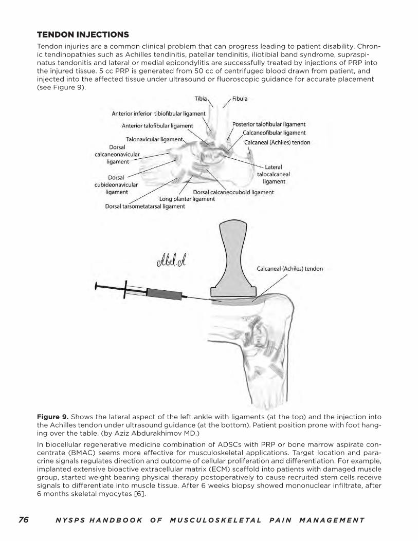
76 N Y S P S H A N D B O O K O F M U S C U L O S K E L E TA L P A I N M A N A G E M E N T
TENDON INJECTIONSTendon injuries are a common clinical problem that can progress leading to patient disability. Chron-ic tendinopathies such as Achilles tendinitis, patellar tendinitis, iliotibial band syndrome, supraspi-natus tendonitis and lateral or medial epicondylitis are successfully treated by injections of PRP into the injured tissue. 5 cc PRP is generated from 50 cc of centrifuged blood drawn from patient, and injected into the affected tissue under ultrasound or fluoroscopic guidance for accurate placement (see Figure 9).
Figure 9. Shows the lateral aspect of the left ankle with ligaments (at the top) and the injection into the Achilles tendon under ultrasound guidance (at the bottom). Patient position prone with foot hang-ing over the table. (by Aziz Abdurakhimov MD.)
In biocellular regenerative medicine combination of ADSCs with PRP or bone marrow aspirate con-centrate (BMAC) seems more effective for musculoskeletal applications. Target location and para-crine signals regulates direction and outcome of cellular proliferation and differentiation. For example, implanted extensive bioactive extracellular matrix (ECM) scaffold into patients with damaged muscle group, started weight bearing physical therapy postoperatively to cause recruited stem cells receive signals to differentiate into muscle tissue. After 6 weeks biopsy showed mononuclear infiltrate, after 6 months skeletal myocytes [6].

77C H A P T E R 6 . S T E M C E L L S
REFERENCES
[1] Vinay K, Abul K. Abbas, Jon C. Aster. (2013). Robbins basic pathology. Elsevier. 60-61
[2] I.L., Weissman. (2000). Translating stem and proginitor cell biology to the clinic. Science.;287: 1442-1446
[3] S.E., Haynesworth. (1992). cell surface antigens on human marrow derived mesenchymal cells are detected by monoclonal antibodies. Bone 13: 69-80
[4] Hung SC, Chen NJ, Hsieh SL, Li H, Ma HL, Lo WH. (2002). Isolation and characterization of size-sieved stem cells from human bone marrow. Stem Cells.;20:249-258.
[5] Rabago D, Mundt M, Zgierska A, Grettie J. (2015). Hypertonic dextrose injection (prolotherapy) for knee osteoarthritis: Long term outcomes.;23: 388-395
[6] Brain Mailey, Ava Hosseni. (2014). Adipose derived stem cells. Springer. 169

78 N Y S P S H A N D B O O K O F M U S C U L O S K E L E TA L P A I N M A N A G E M E N T
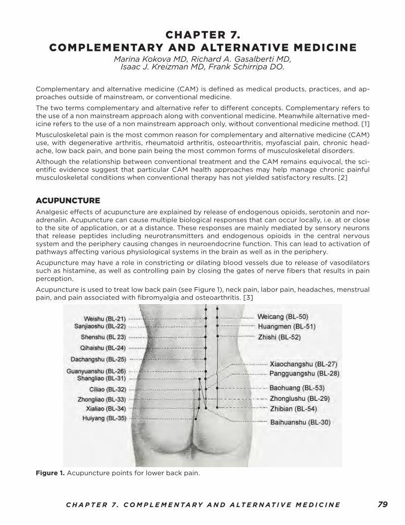
79C H A P T E R 7 . C O M P L E M E N TA R Y A N D A LT E R N AT I V E M E D I C I N E
CHAPTER 7. COMPLEMENTARY AND ALTERNATIVE MEDICINE
Marina Kokova MD, Richard A. Gasalberti MD, Isaac J. Kreizman MD, Frank Schirripa DO.
Complementary and alternative medicine (CAM) is defined as medical products, practices, and ap-proaches outside of mainstream, or conventional medicine.
The two terms complementary and alternative refer to different concepts. Complementary refers to the use of a non mainstream approach along with conventional medicine. Meanwhile alternative med-icine refers to the use of a non mainstream approach only, without conventional medicine method. [1]
Musculoskeletal pain is the most common reason for complementary and alternative medicine (CAM) use, with degenerative arthritis, rheumatoid arthritis, osteoarthritis, myofascial pain, chronic head-ache, low back pain, and bone pain being the most common forms of musculoskeletal disorders.
Although the relationship between conventional treatment and the CAM remains equivocal, the sci-entific evidence suggest that particular CAM health approaches may help manage chronic painful musculoskeletal conditions when conventional therapy has not yielded satisfactory results. [2]
ACUPUNCTUREAnalgesic effects of acupuncture are explained by release of endogenous opioids, serotonin and nor-adrenalin. Acupuncture can cause multiple biological responses that can occur locally, i.e. at or close to the site of application, or at a distance. These responses are mainly mediated by sensory neurons that release peptides including neurotransmitters and endogenous opioids in the central nervous system and the periphery causing changes in neuroendocrine function. This can lead to activation of pathways affecting various physiological systems in the brain as well as in the periphery.
Acupuncture may have a role in constricting or dilating blood vessels due to release of vasodilators such as histamine, as well as controlling pain by closing the gates of nerve fibers that results in pain perception.
Acupuncture is used to treat low back pain (see Figure 1), neck pain, labor pain, headaches, menstrual pain, and pain associated with fibromyalgia and osteoarthritis. [3]
Figure 1. Acupuncture points for lower back pain.

80 N Y S P S H A N D B O O K O F M U S C U L O S K E L E TA L P A I N M A N A G E M E N T
TAI CHITai Chi, being a mind-body exercise therapy, involves integrated dynamic musculoskeletal breathing and meditation training along with slow motion and weight shifting.
Tai chi has three major components:
1. Movement. All of the major muscle groups and joints are needed for the slow, gentle movements in tai chi. Tai chi improves balance, agility, strength, flexibility, stamina, muscle tone, and coordination.
2. Meditation. Research shows that meditation soothes the mind, enhances concentration, reduces anxiety, and lowers blood pressure and heart rate.
3. Deep breathing.
It is typically used to manage chronic back and neck pain and pain associated with arthritis especially among the elderly because of the gentle nature of the exercise and safety. [4][5]
HERBAL PREPARATIONSHerbal medicines are among the most popular forms of complementary treatments. A large propor-tion of these herbal remedies is used for musculoskeletal pain relief. Most of the herbal medicines have an effect on the eicosanoid metabolism, inhibiting one or both of the cyclooxygenase and lipoxygen-ase pathways. The ingredients of phytomedicines may be synergistic or antagonistic.
The most popular herbal analgesics are:
• Cayenne (Capsicum frutescens). Patients with fibromyalgia experience less tenderness and significant increase in grip strength. [6]
• Devil’s claw (Harpagophytum procumbens) preparation produces reduction in pain and increase in mobility in patients with osteoarthritis. [7]
• Phytodolor (a proprietary preparation that contains Populus tremula, Fraxinus excelsior, and Solidago virgaurea [goldenrod]) has demonstrated efficacy for painful arthritic conditions in a number of studies. [8]
• Gamma-linolenic acid (GLA)-containing herbs. Blackcurrant (Ribes nigrum) seeds, borage (Boragio officinalis), evening primrose (Oenothera biennis) oil contain high amounts of GLA, an essential fatty acid that exerts anti-inflammatory activity by interfering with prostaglandin metabolism. These herbs showed objective signs of reduced disease activity and pain relief in paints with osteoarthritis, active synovitis and rheumatoid arthritis. [9]
NUTRITIONAL SUPPLEMENTSAvocado-soybean unsaponifiables (ASU), glucosamine and chondroitin may be effective in treatment of osteoarthritic pain. Fish oil and docosahexaenoic acid both have anti-inflammatory activity through their effects on prostaglandin metabolism and have clinical effect in rheumatoid arthritis [10][11].
• Univestin derived from standardized extracts from the roots of Scutellaria baicalensis and the heartwood of Acacia catechu combined with ginger, rutin (a flavonoid), and bromelain and papain (plant enzymes) combats pain in osteoarthritis. [21]
• Fish oil fatty acids reduce pain and swelling. Fish that are especially rich in the beneficial oils known as omega-3 fatty acids include mackerel, herring, tuna, salmon, cod liver, whale blubber, and seal blubber. Two of the most important omega-3 fatty acids contained in fish oil are eicosapentaenoic acid (EPA) and docosahexaenoic acid (DHA) [22]
• Glucosamine and Chondroitin Sulfate. This combination may provide additional pain relief for some people with knee and hand osteoarthritis. The benefit is usually modest (about 8 to 10 percent improvement) and it works slowly (up to 3 months). [23]
• DMSO & MSM (dimethyl sulfoxide and methylsulfonylmethane). These two sulfur com pounds share similar properties. Their potent anti-inflammatory actions block the pain response in

81C H A P T E R 7 . C O M P L E M E N TA R Y A N D A LT E R N AT I V E M E D I C I N E
nerve fibers. Remarkable feature of these substances is that they can be used orally, intrave nously, or topically. Most patients with arthritis find topical application the most convenient. [24]
• Capsaicin. Capsicum frutescens. Capsaicin works by depleting substance P, a neuropeptide produced by the nerves that carry pain sensation. Skin ointments containing capsaicin have been shown to significantly relieve arthritis pain. The first application often causes a burning sensation; however, as substance-P is depleted, this discomfort goes away. [25]
MASSAGEMassage is one of the most popular CAM methods for neck and back pain [12]. Massage can reduce pain in patients with metastatic bone pain on an immediate, intermediate and long term time frame. It may also relieve pain of wide variety of etiologies: headache pain, postoperative pain, back and leg pain in pregnant women, chronic pain, myalgia, carpal tunnel syndrome, pain from distal radial trauma [13][14]. Massage promotes pain relief through the use of mechanical manipulation of body tissues with rhythmic pressure and stroking [15], this in turn increases blood circulation, reduce inflammation, promote mitochondrial biogenesis in the skeletal muscle, increase vagal activity and decrease cortisol levels [16]. Altogether these factors contribute to reduction of painful sensations.
The most common type and the most widely recognized category of massage is the Swedish mas-sage. The Swedish massage techniques vary from light to vigorous. It involves five styles of strokes: effleurage (sliding or gliding), petrissage (kneading), tapotement (rhythmic tapping), friction (cross fi-ber or with the fibers) and vibration/shaking. By relieving muscle tension Swedish massage has shown significant results in reducing joint stiffness, improving function in patients with osteoarthritis of the knee and significantly reducing back pain. The effectiveness is shown to last for as long as 15 weeks. [26][27]
Figure 2. Massage techniques. • Effleurage: a smooth, gliding stroke used to relax soft tissue. (see Figure 2.) • Petrissage: the squeezing, rolling, or kneading that follows effleurage. (see Figure 2.) • Friction: deep, circular movements that cause layers of tissue to rub against each other, helping to increase blood flow and break down scar tissue. (see Figure 2.) • Tapotement: a short, alternating tap done with cupped hands, fingers, or the edge of the hand [28]

82 N Y S P S H A N D B O O K O F M U S C U L O S K E L E TA L P A I N M A N A G E M E N T
YOGAYoga is a mind-body and exercise practice that combines breath control, meditation, specific stretch-es and body positions to strengthen muscles and obtaining tranquility.
Yoga is thought to relief pain by promoting the pain center in brain to regulate the gate-controlling mechanism located in the spinal cord. It also stimulates secretion of natural painkillers (endorphines) in the body. Breathing exercises used in yoga can also contribute to pain reduction. Because muscles tend to relax during exhalation, lengthening the time of exhalation can help produce relaxation and reduce tension and pain. Trials have found that yoga is most effective in improvement symptoms and function for patients with chronic low back pain. [17]
SPINAL MANIPULATIONSpinal manipulation is localized force directed at specific spinal segments. It is performed by using the hands or a device to apply a controlled force to a joint of the spine and is practiced by chiropractors, osteopathic physicians, allopathic physicians, physiotherapists, and other healthcare professionals to treat a range of primarily musculoskeletal problems [18].
The American Chiropractic Association defines spinal manipulation as a passive manual maneuver during which the three joint complex may be carried beyond the normal voluntary physiological range of movement into the para-physiologic space without exceeding the boundaries of anatomic integrity [19].
The essential characteristic is a low- or high-velocity low-amplitude thrust administered briefly, sud-denly, and carefully and directed at the thoracolumbar and lumbosacral junctions at the end of the normal passive range of movement. It is done to increase the joint’s range of movement. This method is mainly used for lower back pain management [20].

83C H A P T E R 7 . C O M P L E M E N TA R Y A N D A LT E R N AT I V E M E D I C I N E
REFERENCES
[1] U.S. Department of Health and Human Services, National Institutes of Health, National Center for Complementary and Alternative Medicine. Complementary, alternative, or integrative health. August 4, 2015.
[2] Furlan A, Yazdi F, Tsertsvadze A, et al. Complementary and alternative therapies for back Pain II. Evidence report/technology assessment No. 194; 2010.
[3] Allais, L. Cochrane Database of Systematic Reviews, January 2009.
[4] Jacobson, B. H., Chen, H. C., Cashel, C. & Guerrero, L. The effect of Tai Chi Chuan training on balance, kinesthetic sense, and strength.
[5] Vitetta, L., Anton, B., Cortizo, F. & Sali, A. Mind-body medicine: stress and its impact on overall health and longevity. Ann N Y Acad Sci. 1057, 492–505 (2005).
[6] McCarthy DJ, Csuka M, McCarthy G, Trotter D. Treatment of pain due to fibromyalgia with topical capsaicin: a pilot study. Semin Arthritis Rheum. 1994;23(suppl 3):41-47.
[7] Lecomte A, Costa JP. Harpagophytum dans Parthrose Etudes en double insu contre placebo. 37 2 Le Magazine. 1992;15:27-30.
[8] Ernst E, Chrubasik S. Phyto-anti-inflammatories: a systematic review of randomized, placebo-controlled, double blind trials. Rheum Dis Clin North Am. 2000;1:13-27.. Chen EW, Fu AS, Chan KM, Tsang WW. The effects of Tai Chi on the balance control of elderly persons with visual impairment: a randomised clinical trial. Age Ageing. 2012;41(2):254-9.
[9] Levanthal LJ, Boyce EG, Zurier RB. Treatment of rheumatoid arthritis with gammalinolenic acid. Ann Intern Med. 1993;119:867-873.
[10] Pavelka K, Gatterova J, Olejavero M, Machacek S, Glacovelli G, Rovati LC. Glucosamine sulfate use and delay of progression of knee osteoarthritis: a 3 year, randomized, placebo-controlled, double-blind study. Arch Intern Med. 2002;162:2113-2123.
[11] McCarthy GM, Kenny D. Dietary fish oil and rheumatic diseases. Semin Arthritis Rheum. 1992;21:368-375.
[12] Wolsko PM, Eisenberg DM, Davis RB, Kessler R, Phillips RS. Patterns and perceptions of care for treatment of back and neck pain: results of a national survey. Spine (Phila PA 1976). 2003;28(3):292-297.
[13] Nixon, M., Teschendorff, J., Finney, J., Karnilowicz, W. Expanding the nursing repertoire: the effect of massage on post-operative pain. Aust J Adv Nurs. 1997. 14(3), 21-6.
[14] Hughes, D., Ladas, E., Rooney, D., Kelly, K. Massage therapy as a supportive care intervention for children with cancer, Oncol Nurs Forum; 2008.
[15] Cafarelli E, Flint F. The role of massage in preparation for and recovery from exercise: an overview. Sports Med. 1992;14(1):1-9.
[16]Justin D. Crane1, Daniel I. Ogborn2, Colleen Cupido1, Simon Melov3, Alan Hubbard4, Jacqueline M. Bourgeois5 and Mark A. Tarnopolsky. http://stm.sciencemag.org/content/4/119/119ra13?ijkey=68811eea24ebde099eb64889ebb6252bbb1a7e64&keytype2=tf_ipsecshay.
[20] Pubmed. Evaluation of the effectiveness and efficacy of Iyengar yoga therapy on chronic low back pain. Williams K, Abildso C, Steinberg L, Doyle E, Epstein B, Smith D, Hobbs G, Gross R, Kelley G, Cooper L Spine (Phila Pa 1976). 2009;34(19):2066.

84 N Y S P S H A N D B O O K O F M U S C U L O S K E L E TA L P A I N M A N A G E M E N T
[21] http://whitakerwellness.com/health-concerns/chronic-pain-treatment/chronic-back-pain-treatment/
[22] http://www.webmd.com/vitamins-supplements/ingredientmono-993-fish%20oil.aspx?activeingredientid=993
[23] American Academy of Orthopaedic Surgeons (February 2013), “Five Things Physicians and Patients Should Question”, Choosing Wisely: an initiative of the ABIM Foundation, American Academy of Orthopaedic Surgeons, retrieved 19 May 2013
[24] R.J. Herschler, “Dietary and pharmaceutical uses of methylsulfonylmethane and compositions comprising it”, U.S. Patent 4,514,421. April 30, 1985. Accessed 2011-03-12.
[25] http://whitakerwellness.com/health-concerns/chronic-pain-treatment/chronic-back-pain-treatment/
[26] Braun, Mary Beth. Introduction to Massage Therapy (Third Edition). Lippincott Williams & Wilkins. p. 16.
[27] “Swedish Massage”. Massagereister.com
[28] http://www.webmd.com/balance/guide/massage-therapy-styles-and-health-benefits#2-3
[17] U.S. Department of Health and Human Services, National Institutes of Health, National Center for Complementary and Alternative Medicine. Complementary, alternative, or integrative health: http://nccam.nih.gov/health/whatiscam. August 4, 2015.
[18] American Chiropractic Association. Policy Statement on Spinal Manipulation. February 2003. Available at: http://www.acatoday.com/content_css.cfm?CID=1083. Accessed April 16, 2007.
[19] Ernst E. Prevalence of use of complementary/alternative medicine: a systematic review. Bull World Health Organ. 2000;78:252-257

85C H A P T E R 8 . E M E R G I N G A N D N O V E L T H E R A P I E S
CHAPTER 8 . EMERGING AND NOVEL THERAPIES
Robert Duarte MD, Todd Schlifstein DO.
Over the past decade there has been a tremendous interest over new and innovative medical ther-apies created to manage musculoskeletal pain. These new therapeutic modalities, with a focus on regenerative medicine, include but not limited to the prolotherapy, human stem cells, platelet rich plasma therapy, and use of hormones such as the growth hormone oxytocin.
Regenerative medicine in pain management, where the body regenerates or rebuilds itself, is relative-ly new and rapidly evolving area.
Prolotherapy also known as proliferation therapy is a treatment that employs an injection of an irritant solution, most commonly hyperosmolar dextrose, into a joint space, ligament or tendon insertion in an effort to relieve pain [2]. More recently, Platelet Rich Plasma (PRP) and autologous adult stem cells have been used. These products will be discussed later. The term, prolotherapy, originally described by Dr. George S. Hackett, from the word “proli” meaning offspring and proliferate - to produce new cells in rapid succession. The mechanism of prolotherapy is still under investigation and remains un-clear.
However, it is thought to work by stimulating the body’s natural healing mechanisms to lay down new tissue in the weakened area. In 2007 Cochrane review found, when used alone, prolotherapy ineffective in treating chronic low back pain. When used in conjunction with manipulation, exercise and other interventions, prolotherapy showed it may improve chronic low back pain and disability. In 2016, a systematic review of dextrose prolotherapy for chronic musculoskeletal pain supported the use of dextrose prolotherapy for the treatment of tendinopathies, knee and finger joint OA and spinal / pelvic pain due to ligament dysfunction. Efficacy in acute pain as first line therapy, and in myofascial pain could not be determined from the literature. Despite some anecdotal evidence and limited well designed research, prolotherapy remains an alternative treatment and, therefore, not accepted by most insurance companies including medicare. [5]
Patients with chronic low back pain, tendonitis, osteoarthritis, and sports related injuries can be con-sidered for prolotherapy [2]. Generally, patients receive prolotherapy treatment every two to 6 weeks for up to 6 months. The patient should be made aware that the injection may result in a temporary increase in pain with mild stiffness and pain. Stem cells have attracted much of the attention in the field of regenerative medicine.
Autologous and recombinant products and technology are at the center of emerging and novel ther-apies. There is ongoing research into bone, ligament, cartilage, and tendon healing. This research has generated the development of products that stimulate growth factors and secretory proteins. These growth factors and proteins help to maximize the healing of tissues and to expedite the healing pro-cess. Unfortunately, many of these new technologies and techniques are not well studied.
Currently, there are several technologies being used today in Pain Medicine. Some of these products presently used include the following. Platelet-Rich Plasma (PRP) is one of the most commonly used products. It has been widely used for many different problems and there are many different ways of producing the PRP. The use of Stem cells has been growing rapidly and its potential role in so many different physical conditions.
There are different types of stem cells. Mesenchymal Stem Cells (MSCs), Bone Marrow Derived MSCs (BMSCSs), and Adipose-Dervied MSCs (AMSCs) Regonkine is a tradename of a autologous product used to help reduce inflammation by Interlukin Factors.
Platelet Rich plasma is an Autologous product first used back in the 1970s to help general surgery and dental procedures help the healing process. PRP assists the healing process by increasing the concen-trations of autologous growth factors and secretory proteins factors. It has been well studied well in maxillofacial surgery and general surgery. More recently, orthopedic use has increased tremendously as well as research into orthopedic uses of PRP. The belief is that the PRP enhances healing process , bringing healing and growth factors to an area of need and to an area that may not have blood supply. PRP has been used in orthopedic surgery, sports medicine, wound care, and pain management since

86 N Y S P S H A N D B O O K O F M U S C U L O S K E L E TA L P A I N M A N A G E M E N T
the 1990’s. The science behind PRP explains its role in the healing process of ligaments, and tendons. Platelets contain numerous proteins, cytokines, and other bioactive factors that initiate and regulate wound healing. Normal Platelet counts in blood 150 to 350,000/L . Plasma is the fluid portion of blood. PRP concentrate is made from the patient’s own blood. After the blood is centrifuged, it sepa-rates into serum (top coat), the platelets and white blood cells (“buffy coat”), and the red blood cells ( bottom layer). PRP has platelet concentrations 1,000,000/L in 5 mL of plasma is associated with enhanced wound healing. Most PRP preparations have between 3 to 5 times the normal growth factor concentrations. Platelets synthesize and release more than 1,100 biologically active proteins. There are many bioactive factors in PRP. Cytokines in PRP include Transforming growth factor (TGF), Platelet derived growth factor (PDGF), Insulin like growth factor (IGF-I,IGF-II), Fibroblast growth factor (FGF), Vascular endothelial growth factor (VEGF). Wound healing is a complex process. Exogenous deliv-ery of a single growth factor has limited benefit. Non-growth factors contained with dense granules in platelets also play a role- serotonin, histamine, dopamine, calcium, and adenosine. There are three stages of healing inflammation, proliferation and remodeling. . The Clot formation also lays a role as forms “scaffolding” or framework for the healing process. [4][9]
Presently, orthopedic uses of PRP include chronic tendinopathies, acute ligamentous injuries, osteoar-thritis, muscle injuries and intra-operative use. These indications are also the most frequently studied uses, and are areas of need in the field of orthopedic and pain medicine.
Chronic or refractory later epicondylitis is one of the most well studied etiologies. Most studies show-ing favorable results. Studies have focused on Refractory Cases of conservative treatment. MRI or Ultrasound to confirm Extensor Carpi radialis brevis tendinopathy. Strengthen progression post injec-tion. Return to activities over 6 to 8 weeks. Return to Sports with FROM and no pain and no tender-ness. This protocol has shown good results in several studies.
Achilles tendonitis or tendinopathy.Tendon injuries occur from hypovascularity, microtrauma, tendon degeneration, weakness. PRP is suppose to improve vascularity and promote healing of the tendon. Tendons have no direct blood-flow, therefore directly injecting the tendon to deliver healing and growth factors seems logical. There are many smaller studies showing good efficacy. These studies had Varying pathology including tendonitis, tendonosis , fibrosis, and partial tearing. [1]
Plantar fasciitis Patients who failed conservative treatment has been studied. A common problem that is often slow to improve. Injection of PRP into the medial plantar fascia under guidance of ultrasound has been used and studied. Guidance is ned to ensure proper placement of the PRP. Pain and swelling does occur. Inflammation from the injection activates the platelets to release their healing and growth factors. Post injection, immediate weight bearing and ice for the first 48 hours to allow inflammation to activate the process. Return to activities in 6 to 8 weeks. No study looked at torn fascia on fascitis. An area that needs to be investigated.
Chronic patella tendinopathy or jumper’s knee.Current treatment is rest from activities and physical therapy. This problem is often slow to improve and returns often on return to activity. Tendon changes are found to be angiofibroblastic hyperplasia. PRP is thought to be an adjunct to accelerate healing process and to bring healing and growth factors to an area of minimal bloodflow.
Patella tendinopathy has no prospective,large randomized trials. Many studies that are retrospective, many case studies, histological animal studies also have been done. Consider in patients who have failed conservative treatment for 3 months. Clinical findings are supported by MRI and/or ultrasound findings. No NSAIDs 1 week before an injection and for 3 to 4 weeks post injection. [6]
Acute ligamentous injuries in athletes and getting them back to play quickly. Medical collateral liga-ment injuries are being looked at for PRP. One study looked at professional soccer players with grade II MCL injuries. One injection, within 72 hours post injury. Return to play time shortened by 27%. This significant reduction in healing time demonstrates the potential benefit in ligamentous injuries and in acute injuries. Further research needs to be done in these areas. It maybe difficult to obtain an MRI and get a PRP injection within 72 hours in the non-professional athlete population.
The Goal of PRP is to give a high concentration of platelet growth factors to enhance thehealing pro-cess. PRP may be advantageous in sports medicine. Little data aside from many case series to support its use. Widespread use in maxillofacial surgery, plastic surgery and orthopedic surgery continues.

87C H A P T E R 8 . E M E R G I N G A N D N O V E L T H E R A P I E S
Stem cells are characterized by their ability to renew themselves through cell division and differentiate into a diverse range of specialized cell types (also see Chapter 6). There are many sources for stem cells. Human embryos contain pluripotent stem cells. Induced pluripotent stem cells are generated by taking cells, for example the skin, and then injecting a small number of genes into the cells which con-verts them to stem cells. A concern with this source of stem cells is the introduction of an oncogene. Most uses today of stem cells utilize adult stem cells. [3]
Sources of adult stem cells include bone marrow, and adipose tissue. Most adult stem cells are multi-potent, the cells can differentiate into some cells types but not all cell types.
Mesenchymal stem cells (MSCs ) are multi-potent progenitor cells. MSC’s can differentiate into osteo-cytes, adipocytes, and chondrocytes. Autologous stem cells are easily harvested. MSCs inhibit CD+4, CD+8, and Natural Killer Cells. They secrete cytokines including PGE-2, GM-CSF, IL-1RA, IL-7, IL-8, IL-10 and IL-11. MSCs are adult stem cells and do not have the unlimited capacity for self renewal.
The most commonly studies and used MSCs for cartilage repair is Bone Marrow MSCs (BMSCs). It is estimated that about 1000 more times MSCs can be obtained from each gram of adipose tissue when compared to bone marrow.
There have been promising results from injecting BMSCs in mild to moderate osteoarthritis. No se-rious adverse events have occurred. Most studies are lacking in significant numbers and long term follow up of patients . Many more promising studies have come out recently. [12]
Adipose derived MSCs often harvest the cells, they do not culture them but the cells are derived from adipose cells, then isolated by centrifuging. These cells are not confirmed stem cells. Some of the studies with AMSCs have been done with PRP. These studies have been effective in reducing pain and cartilage regeneration. These studies lack MRI follow up to determine cartilage regrowth, limited number of patients and no long term follow up. [11]
Determining the optimal dose of MSCs was looked at directly in Jo et al in 2014. Low dose 1.0 x 10^7 , medium dose, 5.0 x10^7 and high dose 1.0 x 10^8. Size of the cartilage defect decreased in size in the High dose MSCs study. It is still to be determined what is the optimal dose, for what diagnosis and size of defect.
The previously mentioned techniques have shown promising results. Better results have been shown in most studies with the high dose MSCs, but the optimal dose has yet to be determined. Single dose treatments have been effective but many studies show several treatments maybe necessary. The ulti-mate solution has yet to be determined.
Hormone based therapy.Recently oxytocin has emerged as a new and innovative hormone based therapy that has been found to target specific pain receptors to induce analgesia. Historically the polypeptide hormone oxytocin has played a major role in parturition and milk let down reflex. More recently oxytocin serves in a range of psychosocial and psychophysiological phenomenon including the reward system and memory pro-cessing. Research during the 1980 and 1990’s discovered the role of oxytocin in both neural and be-havioral processing. At the time, clinical application was geared towards a wide array of psychiatric disease, most notably drug addiction.
Introduction of oxytocin into the body has been efficacious via intrathecal insertion. This neuropep-tide hormone involves both central and peripheral physiological properties. Centrally, oxytocin serves as a neuromodulator in both social behavior and physiological stress response. Furthermore, oxytocin acts as a neurotransmitter and acts on the amygdala activating the hypothalamic-pituitary axis to de-crease the stress hormone cortisol and attenuating the influence of stress and anxiety on nociceptive signaling Peripherally, oxytocin modulates the inflammatory response and can improve healing.
Recent studies released by Trigemnina found intranasal oxytocin to be efficacious in the treatment of chronic migraine. In a single dose, placebo controlled, double blind study on 40 people who suffered from chronic migraine, it was found that 42% of patients at 2 hrs and 55% at 4 hrs.hrs. had reduced pain compared to 11% and 28% for placebo.
There have been studies showing that acute and chronic low back pain in humans produced changes of oxytocin levels in the plasma and CSF fluid. In a study conducted by Yang to study the effects of oxytocin on low back was studied through intrathecal injection of oxytocin into rats. Studies conclud-ed that oxytocin had a dose related analgesic effects at low levels.

88 N Y S P S H A N D B O O K O F M U S C U L O S K E L E TA L P A I N M A N A G E M E N T
REFERENCES
[1] Barboni B, Russo V, Curini V, et al. Achilles tendon regeneration can be improved by amniotic epithelial cells allotransplantation. Cell Transplant. 2012:[Epub ahead of print].
[2] Dagenais S, Yelland MJ, DelMar C, Schoene ML. Prolotherapy injections for low back pain. Cochrane Back and Neck Group 2007
[3] Díaz-Prado S, Muiños-López E, Hermida-Gómez T, et al. Human amniotic membrane as an alternative source of stem cells for regenerative medicine. Differentiation. 2011;81(3):162-171.
[4] Giusti I, Rughetti A, D’Ascenzo S, et al. Identification of an optimal concentration of platelet gel for promoting angiogenesis in human endothelial cells. Transfusion. 2009;49(4):771-778.
[5] Hauser RA, Lackner JB, Steilen-Matias D, Harris DK. A systematic review of dextrose prolotherapy for chronic musculoskeletal pain. Clinical Medicine Insights Arthritis and musculoskeletal disorders. 2016:9:139-159
[6] Hyun I. Allowing innovative stem cell-based therapies outside of clinical trials: Ethical and policy challenges. J Law Med Ethics. 2010:38(2):277-85.
[7] Masuda K, Lotz JC. New challenges for intervertebral disc treatment using regenerative medicine. Tissue Eng Part B Rev. 2010;16(1):147-58.
[8] McJunkin T, Lynch P, Deer TR, Anderson J, Desai R. Regenerative medicine in pain management. Pain Medicine News Spec. 2012:35-38.
[9] Nguyen R, Borg-Stein J, McInnis K. Applications of platelet-rich plasma in musculoskeletal and sports medicine: A evidence-based approach. PM&R. 2011:3(3):226-250.
[10] Ohno T, Kaneda H, Nagai Y, Fukushima M. Regenerative medicine in critical limb ischemia. J Atheroscler Thromb. 2012;19(10)883-9.
[11] Van Osch GJ, Brittberg M, Dennis JE, Bastiaansen-Jenniskens YM, Erben RG, Knottinen YT, Luyten FP. Cartilage repair: Past and future—lesson for regenerative medicine. J Cell Mol Med. 2009;13(5):792-810.
[12] Vora A, Borg-Stein J, Nguyen RT. Regenerative injection therapy for osteoarthritis: Fundamental concepts and evidence-based review. PM R. 2012;4(5 Suppl):S104-9

89C H A P T E R 9 . T E L E M E D I C I N E
CHAPTER 9. TELEMEDICINECarlisle St. Martin, MD.
If a picture is worth a thousand words then the value of a live, moving image is priceless. The ability to have true meaningful exchanges with patients via telemedicine will allow for higher standards of care when coupled with laboratory data, radiologic imaging and other high-tech diagnostic techniques. Telemedicine has immediate applications across primary, specialty, and critical care sub-specialties, and should be promoted and implemented to the fullest that current available technology can allow.
Introduction: Early AdoptersOf course, one of the earliest adopters of telemedicine was radiology. These early adopters used tele-medicine to transmit radiological images and their interpretations between radiologists and ordering physicians. Radiologists, who could be located on the other side of the globe, read these images in real time as our daily routines continued. In many cases, by the time we got back to work the next morning, the previous day’s scans could be read by the time our morning routines begin.
For physicians in an emergency setting, this turnaround time is invaluable to crisis situations, which require reading and interpretations of imaging stat and often beyond the purview of regular busi-ness hours. It is difficult enough to provide radiological services beyond regular business hours for most imaging centers, let alone get the images, be they CT, MR, or x-ray, be read and interpreted. A patient pursuing imaging late at night in New York, would benefit from a teleradiologist operating in California, for by the time said patient’s physician is ready to reassess the patient the next morning, fully interpreted scans will have been transmitted across the country, and read securely with a report generated, transcribed and returned.
However within teleradiology these services can usually be seen as being unidirectional, in that it is the sending of static images and then the transmission of either a fax or telephone communication images – sometimes utilizing a DICOM viewer. Interface between the physician and the patient is kept to a minimum and the telemed technology at play here is purely functional. Since its earliest days telemedicine has evolved to include numerous other specialties and subspecialties where diagnostic practice and meaningful patient-physician interactions can occur.
Setting the StageIn most major American hospitals expanding access to quality medical care is seen as a top priori-ty. Access to specialists should be highlighted for the economic benefits telemedicine can bring to achieve this goal. In the current setting, specialists shuttle from one center to another, and larger pa-tient populations remain hard to reach. This inefficient system can be remedied through centralizing services via an integrated telemedicine platform. Through centralization, sharing specialist resources on demand becomes an efficient and increasingly viable solution. This technique has been utilized across many major North American hospital groups such as NYU, Cornell, and the Cleveland Clinic.
When looking at the general populace, special attention should be paid to beneficiaries of recent healthcare laws such as those looking to rectify low access to qualified medical care. Telemedicine can allow equal access regardless of terrain, weather conditions, climate, or even socio-economic status. This is especially important to rural areas of the United States. Rural states – both wealthy and less so – could benefit from telemed programs. Benefits can be measured in the amount of specialties available in a given rural health clinic, as well as in the quality of care.
A major medical system can establish a connected health program to ensure the same standards of care are met in surrounding areas where there are barriers to access. In New York, for example, qual-ity of care standards from New York City-based hospitals such as Columbia-Presbyterian, NYU or Mt. Sinai can be utilized in a telemedicine program to help ailing communities in upstate New York. This scenario has been in place in a select group of nursing homes in upstate New York since 2010. We have provided the nursing homes with on-demand emergency consultations during evening hours when

90 N Y S P S H A N D B O O K O F M U S C U L O S K E L E TA L P A I N M A N A G E M E N T
on-staff physicians are not available. Since its inception, this telemedicine platform has expanded to create installations at 17 nursing homes, giving effective emergency access to patients in critical care situations 24/7. Given telemedicine’s nature, we are now able to truly maximize our resources because team members, whether they are located in Tokyo or New York, can offer support 24/7.
Telemedicine services don’t simply expand healthcare, they also provide a platform for innovation. With telemedicine programs, providers can provide more personalized healthcare, tailored to the individual patient’s needs. We can get the services that are needed to those who need it most with-in a critical time period and always with a fresh team available regardless of the time of day. These consultative teams are innovative because they can be tailor-made to fit the recipient’s profile. As in the nursing home example above, our telemedicine team consists of both emergency medicine and neurology specialists who can address the high number of traumatic and neurological ailments of the geriatric population.
Telemedicine platforms can also be tailored to other niches. Emergency Medical Care NYC is a 24-hour urgent care facility located in the heart of Times Square. The facility’s team members are able to service many of the hotels in Midtown allowing for tourists and travelers to be seen and receive full diagnostic work-ups, including CT scans, in the event of a travel emergency. These rapid workups can be performed with patients being seen within 10 minutes of arrival. While they are waiting, patients are able to access telemedicine consultations from various specialists while their cases are reviewed.
Building on the theme of travel medicine, in our Japanese International Clinic, we cater to a niche clientele of Japanese tourists who use overseas travel insurance while visiting New York. In case of a travel emergency, physicians and staff – many of whom speak Japanese – can see patients. However, upon return to Japan, a patient follow-up may be required to assess the patient’s status. Telemedicine allowed us to be able to see these patients in Japan and instruct them on follow up care. We are also able to get additional history through coordinating with their primary doctors in their home country.
Diagnostic Technique & Basic Functions 1. Consultations: Assuming the person who requested the consultation is another physician then the consult can be performed from the requesting physician’s office to the consulting specialist over a dis-tance whereby the requesting provider is able to demonstrate portions of the exam when requested by the consultant.
2. Diagnostic strategies: Additional testing and consults can be arranged remotely preventing the patient from having to make multiple trips; this also allows for multiple consultations to be performed simultaneously. This is a rare occurrence as multi-disciplinary input while the patient is present usually only takes place within the teaching program or grand rounds.
Legal Framework and Best PracticesAlthough initial interest was for the implementation within rural communities, where accessibility was a question, it was later understood that even urban communities could benefit from telemedicine. In an urban setting, access was not only constrained by distance but also by the sparse availability of specialists. To travel a short distance within a major American city can sometimes take a very long time – during rush hour, it is not uncommon for a 1.5-mile car trip from the United Nations to Emergen-cy Medical Care to take as long as 45 minutes. Scheduling can also be an issue; many specialist prac-tices are fully booked weeks in advance. After all, within urban agglomerations there is a significantly higher demand for all services. Hence in January 2016 New York became a parity state allowing for the reimbursement of telemedicine services on par with face-to-face encounters of the same E/M level.
This support at the state-level has been a boon to telemedicine providers and has started a trend to-wards wider acceptance of telemedicine as a valuable alternative to office encounters. Similarly, there has been broad support from the U.S. Department of Health and Human Services as well as interest from the Centers for Medicare and Medicaid for similar laws. This trend of increasing local and state government support will allow telemedicine to be utilized across multiple and diverse environments. Within the highest levels of government this support may bring about national parity and acceptance of telemedicine for American healthcare participants. Future utilization is being discussed with other groups such as the New York National Guard, who have expressed interest in our platforms, as well as

91C H A P T E R 9 . T E L E M E D I C I N E
the Canadian Mounted Police and Japan Self Defense Forces. Field applications are just one of numer-ous applications where telemedicine is seen as beneficial at this time.
In a humanitarian context, the benefits of telemedicine to bring healthcare to rural and locations in need are innumerable. As we have seen before, traditional barriers to healthcare access, such as cli-mate or topography, can be bypassed with technological advances. In 2017, a trial study was undertak-en to examine the application of telemedicine services in a more unique locale. In the rural mountain villages of northern Thailand, areas with meager medical resources, we established a telemedicine platform, which we left in the care of GONGOVA at Ban Mai Huay Hia village (in the Chom Thong District of Chiang Mai). One of the doctors we were able to collaborate with – whose grandmother we were treating using our telemedicine platform following a subdural hematoma – successfully demon-strated both the need and practicality of a telemedicine program in the humanitarian context. The patient was followed in Bangkok for more than three months, during which time several MRI’s and CT’s were performed and successfully reviewed via telemedicine. The patient, whose care was being managed in New York and Bangkok, recovered fully without any sequelae.
With the labor and service costs associated with quality care being successfully distributed be-tween more economical and efficient sectors, the decentralized nature of the telemedicine system has allowed us to not only stretch our resources, but also to expand our medical knowledge as well. Non-medically trained persons – or persons with only minimal experience (or people not trained to U.S. standards) – are able to access high-level consultative care while in the field via telemedicine. This allows for the person who is traveling without a medical expert to have access to expert opinions. In case of an emergency or in a rural area where there is virtually no chance of trained physicians be-coming available, this type of knowledge and consultative care can be lifesaving.
The FutureLooking forward, the technology and applications used in telemedicine are constantly evolving. We have pioneered the use of connected tools such as Bluetooth-enabled autoscopes, stethoscopes, heart monitors, and other diagnostic tools to aid us in this task.
Support & AcknowledgmentsThere are many doctors whose support of this effort in its early years is most notable. My friend and colleague Dr. Richard A. Gasalberti provided a forum to discuss telemedicine issues. Dr. Steve Okhra-vi, because of his faith in this system, gave us a permanent home to establish multiple centers. In the early years of this technology, Dr. Fink allowed me to demonstrate the many ways that telemedicine could be used – his time and interest were a great source of inspiration. I must also acknowledge the young doctors who supported me during those early years – many of these young researchers are still helping us today.

92 N Y S P S H A N D B O O K O F M U S C U L O S K E L E TA L P A I N M A N A G E M E N T
REFERENCES
[1] “What is Telemedicine?”. Washington, D.C.: American Telemedicine.
[2] Berman, Matthew; Fenaughty, Andrea (June 2005). “Technology and managed care: patient benefits of telemedicine in a rural health care network”. Health Economics. Wiley. 14 (6): 559–573. doi:10.1002/hec.952. PMID 15497196.
[3] “Telemedicine sanction in Idaho clouds doctor’s future”. Spokesman.com. Retrieved 7 March 2016.
[4] Hiers, Mary. “Everything You Need to Know About Telemedicine”. Retrieved 10 July 2014.

93C H A P T E R 1 0 . T H E R O L E O F R A D I AT I O N I N P A I N M A N A G E M E N T
CHAPTER 10. THE ROLE OF RADIATION
IN PAIN MANAGEMENTDavid Gasalberti MD, Richard A. Gasalberti MD, Isaac J. Kreizman MD.
Introduction. Radiotherapy has been a cornerstone of pain management since the discovery of x-rays over a century ago. Advances in the delivery of radiation over the decades allowed for more penetrat-ing x-rays at higher energies. As a consequence, the treatment of painful metastatic bone and visceral lesions with less skin toxicity became a reality. The majority of patients treated with palliative radiation have advanced or metastatic cancers and consequently have a poor overall prognosis. The goal of care in this setting is to provide an acceptable quality of life for the patient by maximizing pain control while minimizing toxicity. While radiotherapy remains an effective means of controlling pain, a survey of hospice care providers conducted by Lutz et al. 2007 demonstrates that only 10% of providers con-sidered Radiation Oncologists as part of a palliative care team despite the fact that 40% of patients receive radiation at some point during treatment. This chapter will review appropriate indications for palliative radiation therapy focusing primarily on pain management in the oncologic setting.
BONE METASTASISBackground. Over 100,000 people with cancer will develop bone metastasis annually. The most com-mon primary sites are breast, prostate, and lung cancer. The axial skeleton is the most common area of spread with the lumbar spine being the most common site. In the appendicular skeleton, the femur is the most common site. Bony metastatic disease if left untreated can lead to pathologic fractures, spinal cord compression, sleep disturbances and an overall decrease in quality of life. The mechanism of bone pain is unclear and several theories have emerged including periosteal stretching, tumor in-duced nerve injury, or osteolysis caused by the activation of osteoclasts. Overall, radiation response rates are between 60-70% and patients report an improved or stable quality of life. [4]
Workup. Patients usually present with symptoms that are largely dependent upon the site of metas-tasis. For instance, a patient complaining of groin or hip pain with a history of prostate cancer could have a femoral metastasis. Palpation over the area in question or bearing weight on the affected limb may elicit pain, but this may not always be the case. Although a thorough medical history can help clinch the diagnosis, a complete musculoskeletal and neurologic exam is essential especially when ruling out benign etiologies such as muscle spasm, herniated disks, advanced osteoarthritis, frac-ture from trauma, etc. If patients with a history of malignancy report sudden paralysis, parasthesias, difficulty walking, bowel incontinence, or bladder incontinence then it is prudent to suspect a spinal cord compression or cauda equina syndrome which will require urgent evaluation and treatment with radiation and surgery if confirmed. Imaging is an essential part of this workup and there are several modalities that can detect and localize metastatic lesions. The goal in this case is to select an imaging test that is both sensitive, specific, and cost effective. Plain radiographs are easy to obtain and eco-nomical, but suffer from a lack of sensitivity. A substantial amount of bone loss (30-50%) must occur before it can be detected with plain radiography thus hampering its sensitivity (see Fig. 1A).
Computed tomography (CT) scans are far more sensitive but speed and cost are sacrificed compared to radiography (see Fig. 1B). However, the clinical yield from a CT scan is far greater than a plain radio-graph. They can provide information not only on location and extent but also the degree of cortical in-volvement, which is helpful in risk stratifying patients for impending pathologic fracture and the need for prophylactic fixation. In addition, they help guide clinicians if a biopsy is required to determine the origin of the primary tumor. Unfortunately, CT imaging cannot distinguish between metastatic disease and more benign processes such as osteoporosis. Magnetic resonance imaging is the most sensitive test available for detecting metastatic disease and is also the most costly and time consuming of the three imaging modalities mentioned. Sensitivity is reported to be 90-100%. MRI is particularly useful at detecting neurovascular involvement with tumors. This is of the utmost importance when ruling out a spinal cord compression. In addition, an MRI can help distinguish between pathologic fractures from osteoporosis or malignancy (see Fig.1C).

94 N Y S P S H A N D B O O K O F M U S C U L O S K E L E TA L P A I N M A N A G E M E N T
Fig 1A-C. Various Imaging modalities for demonstrating bone metastasis in bony pelvis. A.) Plain Radiograph. B.) Computed Tomography. C.) Magnetic Resonance Imaging
Medical Management. Patients with bone metastasis will inevitably experience pain related to these lesions. Medical therapy for pain control and the systematic escalation of therapy is well illustrated in the World Health Organization analgesic ladder. This ladder consists of three steps which start with the use of non-opioid analgesics such as NSAIDS to potent analgesics such as oxycodone or fentanyl (see Fig. 2). While patients report sustained pain relief with this approach, the side effects of opiate medications such as constipation, nausea, and mood changes can limit pain control.
Fig 2. WHO’s Pain Relief Ladder
Surgical Intervention. The location of bone metastasis can pose a risk for pathologic fracture espe-cially in weight bearing areas such as the femur or lower spine which can have profound functional im-plications. Identifying and intervening on these patients before a fracture occurs is far more effective and technically less challenging than delaying until a fracture is encountered. In order to identify these patients, the Mirels scoring system was developed and takes into account location, pain severity, cor-tical involvement, and radiographic appearance (see Table. 1). A twelve point scoring system ranging from 4-12 is then devised. A score of 8 is correlated with a 15% risk of fracture. Orthopedic evaluation is generally warranted above a score of 8. Two to three weeks post-operatively, the patients are typi-cally taken for radiation of the same area that was stabilized to prevent tumor seeding and recurrence.
A B C

95C H A P T E R 1 0 . T H E R O L E O F R A D I AT I O N I N P A I N M A N A G E M E N T
SCORE
1 MILD UPPER EXTREMITY <1/3 BLASTIC
3 SEVERE PERITROCHANTERIC >2/3 LYTIC
2 MODERATE LOWER EXTREMITY 1/3 - 2/3 MIXED
PAIN LOCATION CORTICAL
INVOLVEMENT RADIOGRAPHIC APPEARANCE
Table 1. The Mirels Scoring Criteria for risk stratifying impending pathologic fracture. there are four categories given a score of 1-3. which are added together. The scores range from 4-12. Scores >8 should seek orthopedic evaluation.
Radiotherapy. Radiation is an integral part of pain management, prevention of pathologic frac-ture, and local tumor control. The treatment and planning in most cases is fairly straightforward. In non-emergent situations, patients undergo a CT simulation over the areas to be treated. The data from these scans will be used to direct the radiation beams avoiding vital structures that may cause unwanted symptoms such as diarrhea, nausea, or vomiting in the case of lumbar vertebral radiation (see Fig. 3). Treatments are delivered through a linear accelerator which generates x-rays and can be projected and modified as a field onto the patient. There are several doses and fractionation schemes utilized for treatment (see Table. 2). Several early studies of various fractionation schemes have been explored in various randomized prospective trials. The important point is that all of these trials show no difference in pain control. The Dutch Bone Metastasis trial evaluated single fraction treatment with 8Gy versus 24Gy in 6 fractions. The overall and complete pain response rates were not statistically different. However, the risk of pathologic fracture (2% vs 4%) was higher in the short course arm and the risk of requiring retreatment was also higher in the single fraction arm (7% vs 25%). Perez et al 2013 enumerate the following conclusions based on thirty years of research into palliation for bone metastasis. They consist of the following:
1. Single fraction treatments of 8Gy provide similar pain relief when compared to more protracted regimens (e.g. 26Gy in 6 fractions or 30Gy in 10 fractions).
2. Retreatment rates are higher with shorter course treatments
3. Patients have better response rates if they are treated at lower pain levels rather than waiting until the pain is unbearable.
4. There is no known dose response for palliative bone metastasis.
With this in mind, the standard in the United States remains 30Gy in 10 fractions for the majority of cases. Concerns about the need for retreatment and the risk of pathologic fracture are often cited by Radiation Oncologists as the most compelling reasons for a longer course of treatment. Single fraction doses could be considered in cases where the anticipated life expectancy is shorter than the average time to retreatment, difficulty in obtaining timely medical transport, or patient preference. Single fraction treatments remain controversial in the United States.
Targeting painful bone metastasis with external beam radiotherapy is quite beneficial for patients and improves overall quality of life. Response rates from palliative radiation are much higher than with chemotherapy [3]. It is a cost effective tool in palliation without the side effects of opiates.

96 N Y S P S H A N D B O O K O F M U S C U L O S K E L E TA L P A I N M A N A G E M E N T
Figure 3. Radiation treatment plan for the Lumbar spine.
Dose (Gy)
30 10
8 1
30 15
15 3
20 5
40 15
Number of Fractions
Table 2. Dose fractionation schemes for bone metastasis
VISCERAL METASTASISBackground. The most common areas of visceral metastasis include the brain and the lung and for the most part they do not cause pain. However, in some cases, a primary tumor will metastasize to an or-gan such as the liver or adrenal glands which can cause abdominal pain, early satiety, distention, and obstruction. In addition, recurrent pelvic malignancies such as cervical cancer, endometrial cancer, or rectal cancer may also produce pelvic pain and uncontrollable bleeding. Radiation therapy plays an essential role in both instances.
Liver Metastasis. The liver is a common site of metastasis from breast, lung, and colorectal cancers. Common symptoms include abdominal pain in the right upper quadrant, nausea, weight loss, early satiety, jaundice, and fever. Even in the metastatic setting, patients with limited disease in the liver may be eligible for curative resection. This is particularly true of colorectal cancers. In those cases, it is important to realize that while palliation is important these patients can still be cured despite the extent of disease. However, most patients will present with multiple liver lesions and large disease burden that cannot be cured. In addition to radiotherapy, these patients benefit from systemic che-motherapy as well as interventional procedures such as hepatic arterial chemo-embolization. Typical-ly, palliative radiation is done in circumstances where the patient has large tumor burden and other medical co-morbidities. The limiting factor is Radiation Induced Liver Damage (RILD) which can lead to liver failure and may hasten death. Baseline liver function tests would be advisable prior to starting treatment. In many cases, patients have multiple lesions and require whole liver irradiation. Multiple fractionation schemes exist including 30Gy in 15 fractions, 20Gy in 10 fractions, or 21Gy in 3 fractions [3]. All of these regimens are roughly equivalent with 55% of patients reporting improvement in symp-toms.
Selective Internal Radiotherapy (SIRT) is an interventional procedure which introduces Yttrium-90 attached to small resin microspheres into the hepatic arterial system. This would be considered in
97C H A P T E R 1 0 . T H E R O L E O F R A D I AT I O N I N P A I N M A N A G E M E N T
patients with diffuse metastasis but otherwise good functional status. Hendlisz et al 2010 conducted a phase III trial with 5FU +/- Y-90 embolization demonstrating the effectiveness of this treatment in chemo-refractory patients with liver metastasis. [5]
Adrenal Metastasis. While an uncommon site of metastasis overall, adrenal metastasis are usually en-countered in lung cancer patients. While there may be a role for surgery in those with limited disease burden, most patients are appropriate for palliative radiation. The data on adrenal metastasis is sparse and follow up is limited due to survival being on the order of months. Radiation dose is limited by acute bowel toxicity resulting in nausea. No late effects were seen but this is likely due to the limited life expectancy of the patients. The most common regimen is 30Gy in 10 fractions.
Splenic Metastasis. Tumor spread to the spleen is quite uncommon and more likely to be associated with hematologic malignancies. These patients will typically complain of left upper quadrant pain, early satiety, and abdominal distension. Fortunately, the spleen is quite radiosensitive and responds to doses far smaller than what is employed at other organ sites. Baseline blood counts are advisable in this scenario as patients may quickly develop anemia, thrombocytopenia, and lymphopenia. Doses are typically under 5Gy and delivered twice weekly with a CBC measured after each treatment.
Pelvic Recurrences. Pelvic malignancies are notorious for causing a significant amount of pain as well as bleeding and obstruction. Most common pelvic recurrences include colorectal cancers, endometri-al cancers, and cervical cancers. A multimodality approach is usually employed in these patients as it increases the probability of successful palliation of symptoms. For recurrent colorectal cancers, the patient may benefit from a diverting colostomy in the case of obstruction. They may also benefit from chemotherapy and radiation for painful bulky disease in the pelvis. Several studies examining rectal cancer patients that are surgically inoperable given 5-fluorouracil accompanied with radiation appear to have a high success of pain relief. This approach is limited to patients that have not received prior pelvic irradiation. Patients that have received prior radiation to the pelvis usually treated with surgery and/or chemotherapy. There are some instances in which radiation can be employed. This usually requires a review of the prior radiation plan and treatment to a far smaller volume than originally treated. A more conformal treatment modality such as interstitial brachytherapy may be employed. Brachytherapy is a superior modality when targeting a relatively small volume. The doses of radiation are required for palliation in this area are usually around 50Gy with conventional fractionation using external beam. If brachytherapy is utilized, then doses of 10-15Gy delivered in 1 fraction or 5Gy twice per day to a total dose of 30-40Gy which results in long lasting pain relief. If brachytherapy is not an option, a faster but more protracted regimen called the “QUAD-shot” may be employed. It consists of a 3.7Gy BID regimen over a 2 day period followed by a 3-6 week treatment break. This regimen is repeated for 2 additional cycles for a total of 44.4Gy [2]. Seventy-Five percent of the patients that completed the course had remission or stable disease. Those unable to tolerate or continue with a full course of treatment still have a >50% chance of disease remission. Common side effects of all of these treatments include nausea, diarrhea, and dysuria. Brachytherapy carries the additional risk of bowel perforation, infection, and bleeding.

98 N Y S P S H A N D B O O K O F M U S C U L O S K E L E TA L P A I N M A N A G E M E N T
REFERENCES
1. Spanos W, Guse C, Perez C, et al. Phase II study of multiple daily fractionations in the palliation of advanced pelvic malignancies: preliminary report of RTOG 8502. Int J Radiat Oncol Biol Phys. 1989;17:659–661.
2. Lutz, S. T., Chow, E. L., Hartsell, W. F. and Konski, A. A. (2007), A review of hypofractionated palliative radiotherapy. Cancer, 109: 1462–1470. doi:10.1002/cncr.22555
3. Edward C. Halperin MD, Luther W. Brady MD, Carlos A. Perez MD, David E. Wazer MD. Perez & Brady’s Principles and Practice of Radiation Oncology (Perez and Bradys Principles and Practice of Radiation Oncology) Sixth Edition. 2013. Lippincott Williams and Wilkins. Philadelphia, PA.
4. Caissie, A. et al. 2012. Assessment of Health-related Quality of Life with the European Organization for Research and Treatment of Cancer QLQ-C15-PAL after Palliative Radiotherapy of Bone Metastases. Clinical Oncology , Volume 24 , Issue 2 , 125 - 133.
5. Hendlisz A1, Van den Eynde M, Peeters M, Maleux G, Lambert B, Vannoote J, De Keukeleire K, Verslype C, Defreyne L, Van Cutsem E, Delatte P, Delaunoit T, Personeni N, Paesmans M, Van Laethem JL, Flamen P. Phase III trial comparing protracted intravenous fluorouracil infusion alone or with yttrium-90 resin microspheres radioembolization for liver-limited metastatic colorectal cancer refractory to standard chemotherapy. J Clin Oncol. 2010 Aug 10;28(23):3687-94. Epub 2010 Jun 21.
6. Steenland E1, Leer JW, van Houwelingen H, Post WJ, van den Hout WB, Kievit J, de Haes H, Martijn H, Oei B, Vonk E, van der Steen-Banasik E, Wiggenraad RG, Hoogenhout J, Wárlám-Rodenhuis C, van Tienhoven G, Wanders R, Pomp J, van Reijn M, van Mierlo I, Rutten E. The effect of a single fraction compared to multiple fractions on painful bone metastases: a global analysis of the Dutch Bone Metastasis Study. Radiother Oncol. 1999 Aug;52(2):101-9.

99C H A P T E R 1 1 . G U I D E L I N E S A N D N Y L A W
CHAPTER 11 . GUIDELINES AND NY LAW
Lawrence F. Kobak, DPM, JD,(Diplomate American Academy of Pain Management)
I. What Are Clinical Practice Guidelines?A Clinical Practice Guideline is a document that defines a standard of diagnosing, care and treatment that is generally accepted and presumed followed by a group of health care providers. Radiologists might have a Clinical Practice Guideline for diagnosing and treating breast cancers; so might oncol-ogists. PMR physicians and Orthopedists might have their own Clinical Practice Guidelines for treat-ment of various chronic conditions. Sometimes, organizations might have joint guidelines. Some orga-nizations, such as the New York State Pain Society, could generate guidelines that are multi-specialty in nature. A government agency such as the CDC or Workers’ Compensation often generates Clinical Practice Guidelines.
The key is if the guideline is based upon reliable standards and measures. The guideline should pres-ent a systematic approach to the subject at hand. It should be based upon reliable research and stud-ies. It should draw upon physicians and healthcare providers who are knowledgeable and experienced in the topic at hand. The inherent value of a Clinical Practice Guideline is the willingness of a physician population to adopt the guideline into their daily clinical practice. While a guideline might become part of a law or adopted by an organization, in the end, the guideline is only as good as its quality.
By quality, we mean applicability in a useful way to the patient. If the guideline presents a paradigm for treatment, can a practitioner reasonably follow it? Does the guideline call upon you to perform reasonable actions? Is the bar set too high before a definitive diagnosis can be made? Often, this can only be determined by taking the guidelines out for a test drive. In other words, good guidelines must work in the real world. Impractical guidelines will soon be ignored by the practitioner.
Clinical Practice Guidelines may be used to either defend or prosecute a physician administratively, as with the Office of Professional Medical Conduct, concerning a physician’s license. Since the “rules of evidence” are not adopted at administrative hearings, the administrative law judge has wide latitude as to whether or not the guidelines can be admitted as evidence. Administrative hearings include the Office of Professional Medical Conduct and hospital hearings. The physician has a better chance of getting them admitted into evidence if your attorney can make a showing that the guidelines are accepted by the medical community.
Clinical Practice Guidelines also play a potential role in medical malpractice suits in New York. Mal-practice suits are civil in nature. As such, the “rules of evidence” apply. By definition, Clinical Practice Guidelines are hearsay. Hearsay is any statement made by somebody else, to prove the truth of the matter being talked about. An example of hearsay is when an expert witness testifies that Dr. Jones’ injection of 1cc of 1% lidocaine into the trigger point was efficacious or indicated as per the Clinical Practice Guidelines of the “ABCD Society”. Can the judge allow this hearsay into evidence? At the risk of sounding like an attorney, “maybe”. There are several hearsay exceptions. The trick is Alter-native Dispute Resolution, Professional Discipline, Governmental Investigations, Litigation and Arbi-tration, Contracts and Business Transactions, White Collar Crime, Regulatory Compliance, Practice Formation, Mergers and Acquisitions, Asset Protection and Estate Planning, Medical Financial Audits.
The highest court in New York State is known as the Court of Appeals, not the Supreme Court. For-tunately, the Court of Appeals actually considered this matter in 2006. There was a medical malprac-tice case involving an anesthesiologist. As part of that anesthesiologist’s testimony, he testified to following a flow chart or paradigm in deciding to allow the surgery to proceed without the pa-tient having a prior cardiac evaluation. The judge allowed the flow chart into evidence. The Court of Appeals agreed. The witness, the treating physician, could use the guidelines or paradigm as evidence because he was not using it to prove the truthfulness of the guideline, but just to inform the Court as to the process he took in formulating his medical opinion prior to the patient’s surgery. That might seem to be a distinction without a difference, but there it is! A guideline may be admitted into evidence under the proper conditions. The Court also held that as long as the treating physician partially (not totally) relied on the guideline in his medical decision making, the guideline was allowed

100 N Y S P S H A N D B O O K O F M U S C U L O S K E L E TA L P A I N M A N A G E M E N T
into evidence. Not only that, but the attorney can ask all sorts of questions to the witness about the guidelines, such as who formulated it, who it is endorsed by, and who else uses it. If the guideline is endorsed by a reliable entity and widely used, it will carry more weight with a jury. Hinlicky v Dreyfuss, 6 NY3d 636 Slip Op (2006). It is generally inadmissible for expert, non-treating witnesses to refer-ence Clinical Practice Guidelines. When the defense attorney is cross-examining the plaintiff’s expert witness, the defense attorney may ask if the plaintiff’s expert considers a Clinical Practice Guideline as authoritative. In the rare instance when the expert admits that he/she considers it authoritative, it may be admissible in a state court. In federal courts, if the judge rules that the guideline in question was established as authoritative, the guidelines may be admitted into evidence. Most, but not all, medical malpractice cases in New York are in state courts.
The National Practitioner Clearinghouse, part of the Federal Government’s AHRQ (Agency for Health-care Research and Quality) is the website for thousands of Clinical Practice Guidelines that meet some pretty stringent federal standards for quality. Frankly, the authors of guidelines that make the cut on this website have jumped through many hoops.
To demonstrate the maze of guidelines that are out there, I typed in the search terms “chronic pain” on the National Practitioner Clearinghouse website. 454 different guidelines appeared! What is useful, and what is not, often involves common sense. If you practice in New York, the Chronic Pain Dis-order Medical Treatment Guidelines from the Colorado Division of Workers’ Compensation will not be as helpful to you as compared to the New York version. The ACR Appropriateness Criteria Chronic Foot Pain will apply more to a radiologist than a similar guideline written for a physical therapist.
Clinical Practical Guidelines are hardly new. They have been around for over 20 years. Of course, guidelines must be updated periodically to keep up with new treatment regimens and scientific dis-coveries. Depending on the subject, updates might have to be more frequent. For example, guidelines concerning the Zika virus might have to be updated more often as it is currently more thoroughly studied, than perhaps the efficacy and dosage of Tetracycline.
One of the big raps on Clinical Practice Guidelines is that it promotes cookbook medicine. If properly written, a good guideline will leave room for clinical judgment. A well-written guideline will promote better and more consistent medical decisions.
One might ask what happens if you decide not to follow a generally accepted Clinical Practice Guide-line. The short answer is, you better have a good reason for departing from the guideline and you should definitely explain it in that patient’s medical record!
II. CDC Practice Guideline for Prescribing Opioids for Chronic PainIn March of 2016, the Center for Disease Control (CDC) issued national Clinical Practice Guidelines “on prescribing opioids for chronic pain, for patients who are at least 18 years old, outside of active cancer treatment, palliative care and end-of-life care.” (CDC Guideline for Prescribing Opioids for Chronic Pain, United States, 2016). This guideline is a potential game-changer for the primary care physician who treats this type of chronic pain. The CDC stated that it felt that the guideline was necessitated by primary care physicians’ concern with misusing opioid pain medication in patients with chronic pain, defined as pain more than 3 months. Due to concern of addiction and over-dosage, these physicians desired authoritative guidance in opioid prescribing for chronic pain in non-cancer patients. Many organizations, states and federal agencies, such as the American Pain Society, the American Academy of Pain Medicine, the US Department of Veterans Affairs, have developed their own guide-lines for prescribing opioids. The CDC guideline purports to include the most up-to-date scientific evidence along with a systematic review of the evidence, review by experts in this area. The guide-line is targeted for use by family physicians and internists who treat patients for chronic pain.
The guideline states: “In summary, evidence on long-term opioid therapy for chronic pain outside of end-of-care remains limited, with insufficient evidence to determine long-term versus no opioid ther-apy, though evidence suggests risk for serious harms that appears to be dose-dependent.” It also states that several non-opioid therapies have been found effective in treating chronic pain, such as exercise therapy, behavioral modification techniques, mixing exercise with psychological approach-es, acetaminophen, NSAIDS COX-2 inhibitors, some anticonvulsants and some antidepressants, and for short-term relief of chronic pain, an epidural injection.

101C H A P T E R 1 1 . G U I D E L I N E S A N D N Y L A W
The guideline then goes on to deal with 12 issues dealing with determining when to initiate or continue the use of opioids for chronic pain. Each will be discussed after being quoted, with its implications on your practice:
1. Nonpharmacological therapy and nonopioid pharmacological therapy are preferred for chronic pain. Clinicians should consider opioid therapy only if expected benefits for both pain and function are anticipated to outweigh risks to the patient. If opioids are used, they should be combined with nonpharmacologic therapy and nonopioid pharmacologic therapy, as appropriate.
Your chart should indicate, in detail, how you are employing these specific recommendations, includ-ing specific reference to how you want the patient to functionally improve. As an example-the goal to walk 10 blocks instead of 1 block. The chart note should state how the patient is progressing or not in this regard. Do not just mention a goal and ignore it later on. What non-opioid therapies have been tried? By whom? How long? Drill down as to detail.
2. Before starting opioid therapy for chronic pain, clinicians should establish treatment goals for all patients, including realistic goals for pain and function and should consider how opioid therapy will be discontinued if benefits do not outweigh risks. Clinicians should continue opioid therapy only if there is clinically meaningful improvement in pain and function that outweigh risks to patient safety.
The “takeaway” from this recommendation, is that your medical chart should list specific realistic goals for each patient. General, lofty goals such as “walk better” are not sufficient. Quantify this in blocks walked. “Improved ability to lift” should be quantified perhaps by the weight of an object to be lifted when the patient is on opioid therapy as opposed to prior to the institution of such treatment. Quantify the pain as to type of pain; what precipitates it and in what ways it is ameliorated by the opi-oid therapy. Again, generalizations as to “increase function or activities in daily living,” will not suffice. If it is found that your patient is not obtaining sufficient benefits that outweigh the risks, an actual plan to wean them off the opioids should be included in your chart. If a patient is at heightened risk to take opioids due to some pathology, that should be noted in the chart and what you, the physician, is doing to monitor this.
3. Before starting and periodically during opioid therapy, clinicians should discuss with patient known risks and realistic benefits of opioid therapy and patient and clinician responsibilities for managing therapy. This recommendation states that there should be informed consent prior to and while the patient is under opioid therapy. Remember, informed consent is not just a signed consent form; it is a process of educating the patient. It is highly recommended that this process be memorialized in your chart. When you educate your patient, which should be noted in your medical record; and not just the first time, but every time the patient is educated. If a pamphlet or handout is given to a patient, note with specificity the handout given to the patient.
4. When starting opioid therapy for chronic pain, clinicians should prescribe immediate-release opi-oids instead of extended-release/long acting opioids. The key here is that it was found that lon-ger-acting opioids appear to entail greater dosages than when shorter-acting alternatives were given on a p.r.n. basis. If you have a legitimate reason for starting therapy with an extended-release opioid, your record should spell out the reason why this is so.
5. When opioids are started, clinicians should prescribe the lowest effective dosage. Clinicians should use caution when prescribing opioids at any dosage, should carefully reassess evidence of individual benefits and risks when considering increasing dosage to 50 or more morphine milligram equivalents (MME, and should avoid increasing dosage to 90 or more per day or carefully justify a decision to ti-trate dosage to 90 or more MME/day.
While preparing this guideline, the CDC did not find that high doses of opioids for chronic pain was efficacious. It did find that high doses of opioids potentially compromised patient safety. Most prac-titioners are already titrating their patients’ doses when using opioids. However, it is of the utmost importance to clearly chart the titrating process and letting the reader of your chart into your thought process concerning how you arrived at the dosage you are currently prescribing; if that dosage might be increasing or decreasing, and why. Pain and function are not the only consideration; is the patient at increased risk to become addicted? Was the patient addicted to opioids before? Does the patient have a history of addictive behavior? Does the patient have some type of pathology or genetic predis-

102 N Y S P S H A N D B O O K O F M U S C U L O S K E L E TA L P A I N M A N A G E M E N T
position to having undesirable side-effects if dosage is too high? Your patient care should deal with all of these potential issues. Your chart should reflect that in fact, you did deal with these issues.
6. Long-term opioid use often begins with treatment of acute pain. When opioids are used for acute pain, clinicians should prescribe the lowest effective dose of immediate-release opioids and should prescribe no greater quantity than needed for the expected duration of pain severe enough to require opioids. Three days or less will often be sufficient; more than seven days will rarely be needed.
The guideline now steps into the area of prescribing opioids in acute situations. Although this area is not the primary focus, this recommendation should not be ignored. We get back to medical justifica-tion of what dose is prescribed and how long the opioid is given. Your record should justify your pre-scription. Your record should pay particular attention to acute situations where you want to prescribe for more than seven days.
7. Clinicians should evaluate benefits and harms with patients within 1 to 4 weeks of starting opioid therapy for chronic pain or of dose escalation. Clinicians should evaluate benefits and harms of con-tinued therapy with patients every 3 months, or more frequently. If benefits do not outweigh harms of continued opioid therapy, clinicians should optimize other therapies and work with patients to taper and discontinue opioids.
It is very obvious that lower doses of opioids, less pills, alternate therapies, such as NSAIDS, physical therapy, biofeedback, are preferred. Forewarned is forearmed! Document what you are doing to de-crease the dosage of opioids. If you are trying physical therapy, note it. Then, note the progress or lack of same. Note if there is patient compliance or not. You not only have to ask, but you have to chart that you asked and note the answer.
8. Before starting and periodically during continuation of opioid therapy, clinicians should evaluate risk factors for opioid-related harms. Clinicians should incorporate into the management plan, strate-gies to mitigate risk, including considering offering naloxone when factors that increase risk for opioid overdose, such as history of overdose, history of substance use disorder, higher opioid dosages or concurrent benzodiazepine use, are present.
Does your patient abuse alcohol? Is your patient currently on a benzodiazepine? This should be noted in detail and factored in to your treatment plan. Consider prescribing naloxone for the appropriate patient. Please, note all prescriptions and patient recommendations in your chart.
9. Clinicians should review the patient’s history of controlled substance prescriptions using state pre-scription drug monitoring program data (PDMP) to determine whether the patient is receiving opioid dosages or dangerous combinations that put him or her at high risk for overdose. Clinicians should re-view PDMP data when starting opioid therapy for chronic pain and periodically during opioid therapy or chronic pain, ranging from every prescription to every 3 months.
In New York, there is the ISTOP program. Obtain the PDMP data as suggested in suggestion 9 for each chronic pain patient taking opioids. Make sure that your records reflect that you checked ISTOP and what was found. It would not hurt to place a copy of that patient’s PDMP data in your chart.
10. When prescribing opioids for chronic pain, clinicians should use urine testing before starting opi-oid therapy and consider urine drug testing at least annually to assess for prescribed medication as well as other controlled prescription drugs and illicit drugs.
This might seem obvious, but do not forget to place the urine drug test results in the patient’s chart. Initial them and date it, if your EMR allows. The results should also be incorporated in the body of your progress notes. For example, if the results show patient compliance, the note should state that. If there is any irregularity, such as the patient testing positive for cocaine, the record should state that too. Then, the record must state what action you took as a result of that. That might vary from a warn-ing for the first violation to stopping the patient’s opioid prescription for any additional violations.
11. Clinicians should avoid prescribing opioid pain medication and benzodiazepines concurrently whenever possible.
As prescribing opioids and benzodiazepines together places the patient at greater risk for respiratory depression, you should have a very good reason for the medical necessity to combine the two. Use an alternate treatment regime instead and let your record show why.

103C H A P T E R 1 1 . G U I D E L I N E S A N D N Y L A W
12. Clinicians should offer or arrange evidence-based treatment (usually medication-assisted treat-ment with buprenorphine or methadone in combination with behavioral therapies) for patients with opioid use disorder.
You yourself need not provide this treatment; you can refer such patients out for treatment for opioid use disorder. However, your chart should reflect what is occurring and what is recommended, and what you are doing for this patient.
In conclusion, your chart should reflect, in detail, how you are complying with all of the relevant guide-line recommendations. Administrative agencies, such as the Office of Professional Medical Conduct might find this CDC Guideline authoritative. That will mean that if you did not follow the guideline’s recommendations, there will be a presumption of substandard patient care. Only a carefully worded and detailed medical record will explain why you might have deviated from the guideline recommen-dations. In any case, a complete and accurate medical record is the single best thing that the primary care provider can do to protect him or herself from the various agencies, insurance companies, mal-practice attorneys and criminal prosecutors. If utilized appropriately, clinical guidelines will provide a shield for you and your treatment of your patients.
Kern Augustine Conroy & Schoppmann, P.C., Attorneys to Health Professionals, DrLaw.com, is solely devoted to the representation and defense of physicians and other health care professionals.
Mr. Kobak may be contacted at 1-800-445-0954 or via email at [email protected].

w w w . N Y P a i n S o c i e t y . o r g
New York Headquartered
Clinical Toxicology • AcutisDiagnostics.com • (516) 253-2171
Test us.
Advancing the Science of Pharmacogenomics www.PremierDNA.com • (855) 531-5400