height estimation using 2 -...
TRANSCRIPT


HEIGHT ESTIMATION USING 2ND
& 3RD
METACARPAL BONE AMONG ADULT
MALAY POPULATION ATTENDING
HOSPITAL UNIVERSITI SAINS
MALAYSIA (HUSM)
BY:
DR SITI MAYZURA BT ABDUL RASID
Dissertation Submitted in Partial Fulfillment of the
Requirement for the Degree of Master of Medicine
(RADIOLOGY)
UNIVERSITI SAINS MALAYSIA 2015

To my dear husband,Shahrul Zaman B. Ab
Ghani,
Thank you for the understanding,patience and
support which enable me to complete this
dissertation.
My parents,Mr Abdul Rasid Taib and Mrs
Mahnie Daud,
Thank you for the full support and always be
there when needed

i
ACKNOWLEDGEMENTS
Alhamdulillah, thanks to Allah SWT, The Almighty for giving me the
strength and good health to complete this dissertation. My heartiest appreciation to
my supervisor, Dr Nik Munirah Bt Nik Mahdi (Lecturer, Radiologist and Head of
Department of Radiology, School of Medical Sciences, University Sains Malaysia,
KubangKerian, Kelantan) for their guidance, supervision and advice.
Deepest gratitude to the following individuals for the support of this
dissertation project. Their patience, help and encouragement make this
dissertation a success.
All lecturers/Radiologists, Department of Radiology, School of Medical
Sciences, Universiti Sains Malaysia, Kubang Kerian, Kelantan.

ii
TABLE OF CONTENTS
Page
ACKNOWLEDGEMENTS
i
TABLES OF CONTENTS
ii
LIST OF TABLES
vi
LIST OF FIGURES
vii
ABBREVIATIONS
x
ABSTRAK
xi
ABSTRACT
xiii
CHAPTER ONE:INTRODUCTION
1
CHAPTER TWO:LITERATURE REVIEW 3
2.1 OVERVIEW 3
2.2 ANATOMY AND DEVELOPMENT
5
2.2.1 EMBRYOLOGY OF UPPER LIMB 5
2.2.2 LEFT HAND
8
2.2.3 METACARPAL BONE
8
2.2.4 GROSS ANATOMY OF THE LEFT 2nd METACARPAL BONE
10
2.2.5GROSS ANATOMY OF THE LEFT 3rd METACARPAL BONE 12
2.2.6 RADIOLOGICAL ANATOMY OF METACARPAL BONE 14
2.3HISTORY OF STATURE ESTIMATION 15
2.3.1 OVERVIEW
15
2.3.2IMPORTANCE OF REGRESSION EQUATION 15
2.3.3EFFECT OF AGEING ON STATURE
18
iii
2.3.4ESTIMATION OF STATURE FROM VARIOUS PART OF HUMAN BODY
19
2.3.5 ESTIMATION OF STATURE FROM METACARPAL BONE
22
CHAPTER THREE: OBJECTIVES
24
3.1 GENERAL OBJECTIVES 24
3.2 SPECIFIC OBJECTIVES 24
3.3 RESEARCH QUESTION 3.4 HYPOTHESIS
25
25
CHAPTER FOUR:METHODOLOGY
26
4.1 STUDY DESIGN 26
4.2 POPULATION AND SAMPLE
26
4.2.1Reference population 26
4.2.2 Sample population 26
4.2.3 Inclusion criteria 27
4.2.4 Exclusion criteria
27
4.2.5 Selection of study sample 4.2.6 Sampling method 4.2.7 Informed consent
27
28
28
4.3 SAMPLE SIZE CALCULATION
29
4.4 RESEARCH TOOLS
32
4.5 LEFT HAND RADIOGRAPH POSTERIOR ANTERIOR (PA) VIEW 32
4.5.1 Patient and part position
33
4.5.2 Protocol and processing
33

iv
4.5.3 Acceptability criteria of left hand radiograph (PA) view
35
4.6 MEASUREMENT OF METACARPAL BONE
38
4.6.1 Measurement of 2nd metacarpal bone length
38
4.6.2 Measurement of 2nd metacarpal bone width
38
4.6.3 Measurement of 3rd metacarpal bone length
41
4.6.4 Measurement of 3rd metacarpal bone width
41
4.7 VALIDATION OF TECHNIQUE MEASUREMENT
44
4.8MEASUREMENT OF HEIGHT
45
4.9 DATA COLLECTION
49
4.10 STATISTICAL ANALYSIS 50
4.11 DEFINITIONS
4.11.1 Malay
4.11.2 Stature vs height
4.11.3 Sexual dimorphism
56
56
57
58
CHAPTER FIVE: RESULTS
59
5.1 DEMOGRAPHY 59
5.2 COMPARISON OF STATURE-METACARPAL BONE PARAMETERS
(LENGTH AND WIDTH) OF LEFT HAND BETWEEN MALE AND
FEMALE
5.2.1 Mean of height
5.2.2 Metacarpal bone parameter
5.2.2.1 Second metacarpal bone length
5.2.2.2 Second metacarpal bone width
5.2.2.3 Third metacarpal bone length
5.2.2.4 Third metacarpal bone width
61
61
64
64
67
69
71
1.1 5.3CORRELATION BETWEEN HEIGHT AND METACARPAL BONE
1.2 PARAMETER
75
v
1.3 5.4 PREDICTION OF STATURE FROM THE PARAMETERS
(LENGTH AND WIDTH) OF LEFT METACARPAL BONES BASED
ON EQUATION FORMULA
5.4.1 Simple linear regression equation 5.4.2 Multiple linear regression equation
84
84
87
CHAPTER SIX: DISCUSSION
91
6.1DEMOGRAPHY 91
6.2MEAN OF HEIGHT
92
6.3 MEAN OF METACARPAL BONE PARAMETER
94
6.4CORRELATION BETWEEN METACARPAL BONE PARAMETERS AND STATURE
96
6.5STATURE PREDICTION USING METACARPAL BONE PARAMETERS
98
6.5.1 Linear regression
98
6.6.2 Multiple linear regression 100
CHAPTER SEVEN: CONCLUSION
102
CHAPTER EIGHT: LIMITATION
104
CHAPTER NINE:RECOMMENDATION
105
REFERENCES
106
APPENDIX
111

vi
LIST OF TABLES Page
Table 4.1 Metacarpal bone parameter of left hand from previous
study
29
Table 4.2 Acceptability criteria for left hand radiograph 37
Table 4.3 Interclass correlation between researcher and radiologist 45
Table 4.4 Strength Of Correlation Interpretation 50
Table 5.1 Height and metacarpal bone parameters comparison between female and male
74
Table 5.2 The correlation betweenstature and metacarpalbone(2nd and 3rd) parameter
75
Table 5.3 The regression equations for stature and SEE by simple linear regression
86
Table 5.4 Multiple linear analysis and regression equations for stature for both genders.
88
Table 5.5
Final model of regression equations for stature estimation and SEE by multiple linear regression analysis
90
vii
LIST OF FIGURES
Page
Figure 2.1
Scanning micrographs shows the development of the upper limb
6
Figure 2.2
Gross anatomy of bones of the left hand 9
Figure 2.3
Gross anatomy of the left 2nd metacarpal bone 11
Figure 2.4
Gross anatomy of the left 3rd metacarpal bone 13
Figure 2.5 Left hand radiograph (posterior anterior view) 14
Figure 4.1 Patient part and position 34
Figure 4.2
Normal left hand radiograph (PA) projection 36
Figure 4.3
Measurement of 2nd metacarpal bone length 39
Figure4.4
The measurement of 2nd metacarpal bone width 40
Figure 4.5 Measurement of 3rd metacarpal bone length 42
Figure 4.6 Measurement of 3rd metacarpal bone width 43
Figure 4.7 The Frankfort plane 47
Figure 4.8 Measuring height with stadiometer 48
Figure 5.1
Study sample by gender 60
Figure 5.2
Total subject height 61
Figure 5.3
Male subject height 62
Figure 5.4
Female subject height 63
Figure 5.5
Box and Whisker Plots mean parameter of second metacarpal bone length for male subjects
65
Figure 5.6 Box and Whisker Plots mean parameter of second metacarpal bone length for female subjects
66
Figure 5.7 Box and Whisker Plots mean parameter of second metacarpal bone width for male subjects
67
Figure 5.8 Box and Whisker Plots mean parameter of second metacarpal bone width for female subjects
68
Figure 5.9 Box and Whisker Plots mean parameter of third metacarpal bone length for male subjects
69

viii
Figure 5.10 1.4 Box and Whisker Plots mean parameter of third metacarpal bone length for female subjects
70
Figure 5.11 Box and Whisker Plots mean parameter of third metacarpal bone width for male subjects
71
Figure 5.12 Box and Whisker Plots mean parameter of third metacarpal bone width for female subjects
72
Figure 5.13 The scatter plot of correlation between second metacarpal bone length and height for male subjects
76
Figure 5.14 The scatter plot of correlation between second metacarpal bone length and height for female subjects
77
Figure 5.15 The scatter plot of correlation between third metacarpal bone length and height among male subjects
78
Figure 5.16 The scatter plot of correlation between third metacarpal bone length and height among female subjects
79
Figure 5.17 The scatter plot of correlation between second metacarpal bone width and height among male subjects
80
Figure 5.18 The scatter plot of correlation between third metacarpal bone width and height among male subjects
81
Figure 5.19 The scatter plot correlation between second metacarpal bone width and height for female subjects
82
Figure 5.20 The scatter plot correlation between third metacarpal bone width and height for female subjects.
83

ix
ABBREVIATIONS AND TERMS
HUSM-Hospital Universiti Sains Malaysia
PACS - Picture Archiving and Communications System
DICOM- Digital Imaging and Communications in Medicine
PA - Posterior Anterior
MDCT - Multi-Detector Computed Tomography
MCB - Metacarpal bone
SEE - Standard Error of Estimation

ABSTRAK

x
ANGGARAN KETINGGIAN BERDASARKAN TULANG METAKARPAL
KE-2 DAN KE-3 DI KALANGAN POPULASI MELAYU DEWASA DI
HOSPITAL UNIVERSITI SAINS MALAYSIA (HUSM)
Latarbelakang:
Anggaran ketinggian individu merupakan parameter penting dalam
pemeriksaan forensik. Masalah yang selalu dihadapi berkenaan kes-kes
bencana besar atau keganasan di mana angota tubuh badan manusia
terbahagi kepada beberapa bahagian. Banyak kajian telah dilakukan untuk
menentukan ketinggian daripada tulang panjang. Hubungan antara tulang
tertentu dan perkadaran boleh digunakan untuk membantu proses
identifikasi sekiranya rangka yang lengkap tidak diperolehi daripada tempat
kejadian.
Secara keseluruhananya, tidak banyak kajian yang dijalankan
mengenai anggaran ketinggian di kalangan rakyat Malaysia. Sehingga kini,
tiada kajian yang diterbitkan di kalangan populasi Melayu Kelantan
menggunakan tulang metakarpal. Oleh itu, dalam kajian ini usaha telah
dilakukan untuk mencari hubungan antara ketinggian dan ukuran tulang
metakarpal kedua dan ketiga di kalangan populasi orang dewasa yang hadir
ke Hospital Universiti Sains Malaysia (HUSM).
Objektif:
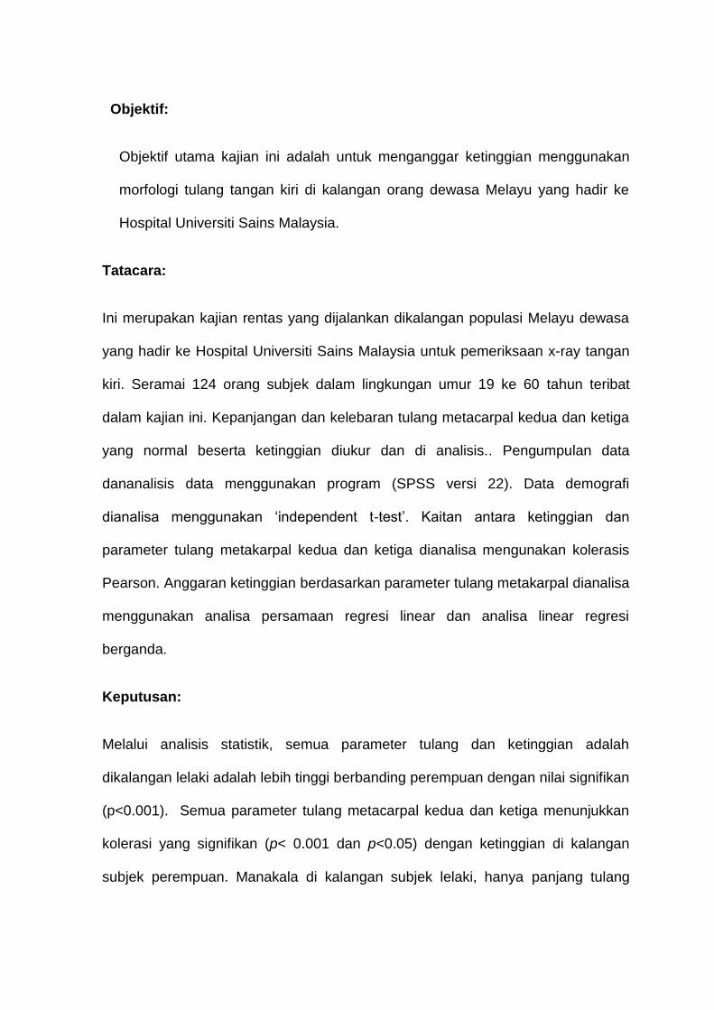
Objektif utama kajian ini adalah untuk menganggar ketinggian menggunakan
morfologi tulang tangan kiri di kalangan orang dewasa Melayu yang hadir ke
Hospital Universiti Sains Malaysia.
Tatacara:
Ini merupakan kajian rentas yang dijalankan dikalangan populasi Melayu dewasa
yang hadir ke Hospital Universiti Sains Malaysia untuk pemeriksaan x-ray tangan
kiri. Seramai 124 orang subjek dalam lingkungan umur 19 ke 60 tahun teribat
dalam kajian ini. Kepanjangan dan kelebaran tulang metacarpal kedua dan ketiga
yang normal beserta ketinggian diukur dan di analisis.. Pengumpulan data
dananalisis data menggunakan program (SPSS versi 22). Data demografi
dianalisa menggunakan ‘independent t-test’. Kaitan antara ketinggian dan
parameter tulang metakarpal kedua dan ketiga dianalisa mengunakan kolerasis
Pearson. Anggaran ketinggian berdasarkan parameter tulang metakarpal dianalisa
menggunakan analisa persamaan regresi linear dan analisa linear regresi
berganda.
Keputusan:
Melalui analisis statistik, semua parameter tulang dan ketinggian adalah
dikalangan lelaki adalah lebih tinggi berbanding perempuan dengan nilai signifikan
(p<0.001). Semua parameter tulang metacarpal kedua dan ketiga menunjukkan
kolerasi yang signifikan (p< 0.001 dan p<0.05) dengan ketinggian di kalangan
subjek perempuan. Manakala di kalangan subjek lelaki, hanya panjang tulang
metacarpal kedua dan ketiga menunjukkan nilaisignifikan dengan ketinggian
(p<0.001). Analisa regresi linier menghasilkan ralat piawaian anggaran dari ±3.94
cm ke ±4.97 cm untuk subjek lelaki dan ±3.92 cm ke ±4.55 cm untuk subjek
perempuan. Daripada analisa regresi linear, empat persamaanregresi terhasil
untuk setiap kumpulan jantina. Analisa linier berganda pula menghasilkan nilai
ralat piawaian anggaran lebih kecil daripada parameter kepanjangan tulang kedua
bagi subjek lelaki(±3.94 cm). Untuk subjek perempuan, analisa linear berganda
menghasilkan nilai ralat piawaian yang terkecil(±3.78 cm) daripada kepanjangan
dan kelebaran tulang metacarpal kedua. Oleh yang demikian, satu persamaan
regressi berganda dihasilkan bagi setiap jantina berdasarkan parameter tulang
metacarpal kedua.
Kesimpulan:
Secara kesimpulannya, ketinggian di kalangan populasi dewasa Melayu Kelantan
boleh ditentukan menggunakan tulang metacarpal kedua iaitu kepanjangan tulang
bagi subjek lelaki dan bagi perempuan kepanjangan dan kelebaran di kalangan
populasi dewasa Melayu di Kelantan.

ABSTRACT

xiii
HEIGHT ESTIMATION USING 2ND& 3RD METACARPAL BONE
AMONG ADULT MALAY POPULATION ATTENDING
HOSPITAL UNIVERSITI SAINS MALAYSIA (HUSM)
Background:
Estimation of individual’s stature is an important parameter in forensic
examination. Problems encountered in cases of mass disaster or assaults
where the body was dismembered. Many studies have been encountered to
determine stature by taking measurement of the long bones. The
relationship between specific bone and proportion can be used to help in the
process for identification in the absence of complete skeleton from a crime
scene.
In general, there is a scarcity of literature regarding the estimation of
stature (height) among Malaysia population. Up to date there was no
published study regarding height estimation among Malay population in
Kelantan using metacarpal bone parameters. The regression formulae are
population specific. Therefore, the aim of the present study was to set a
general formula of stature estimation for both adult males and females using
anthropometrical measurements of second and third metacarpal bones
using AP hand radiograph among Malay adults attending Hospital Universiti
Sains Malaysia.
xiv
Objectives:
The general objective of this study was to estimate stature from the
morphology of left hand radiograph among adult Malay population attending
Hospital Universiti Sains Malaysia.
Patients and methods:
This was a cross-sectional study among adult Malay Kelantan population
attended HUSM for posterior anterior (PA) view radiograph of left hand. A
total of 124 subjects with age ranged from 19 to 60 years old were included.
The length and width of the normal 2nd and 3rd metacarpal bones together
with their heights were measured.Data entry and analysis were performed
usingStatistical Package for Social Sciences (SPSS version 22) software
programme. The descriptive demographic data were measured using
independent t-test. The correlation between stature and parameters of
metacarpal bones were performed using Pearson Correlation. The
prediction of stature from the parameters of metacarpal bone were using
simple linear regression and multiple (stepwise) regression analysis.
xv
Results:
Statistical analysis revealed that parameters formale subjects were higher than
the female subjects (p <0.001). There was significant correlation between all the
parameters of second and third metacarpal bones in female subjects (p value
<0.001 and p value 0.05). For male subjects, significant correlation noted
between stature and the length of second and third metacarpal bone (p value
<0.001).
Using simple linear regression analysis, the standard error of estimate (SEE)
ranged from ±3.94cm to ±4.97cm for males and ±3.92cm to ±4.55cm for
females. From the simple linear regression analysis, four regression equations
were established for each gender. The multiple linear regression analysis
revealed smallest SEE from the second metacarpal bone length for male (SEE
±3.94). For female subject the multiple linear regression analysis revealed
smallest SEE revealed from the second metacarpal bone length and width (SEE
±3.78 cm). Therefore, one multiple regression equation was established for
each gender using the second metacarpal bone parameter.
Conclusion:
It was concluded that stature can be determined successfully using 2nd
metacarpal bone parameters which were the length for male and both length
and width for female among adult Malay population in Kelantan.

CHAPTER 1
INTRODUCTION

1
1.0 INTRODUCTION
With the increasing frequency of mass disasters and fatal assaults, the
identification of isolated extremities and their parts is the ultimate goal in the
investigation for identity of victims. The process generally begins with formulation
of a biological profile (osteobiography); specifically, estimation of sex, age,
ethnicity and stature.
Forensic anthropologists while dealing with skeletal remains have very little
choice to use anatomical method for stature reconstruction due to non-availability
of complete skeleton from a scene of crime in most of the cases. Thus, they have
no choice to use a relatively less precise method of stature reconstruction, i.e. the
mathematical method. It is the method for calculating the height by considering the
mathematical regression coefficients obtained from the measurements of many
bones of the body.
A formula for one population does not necessarily yield reliable results for
another due to inherent population variations that may be attributed to genetic and
environmental factors as climate, nutrition and lifestyle. Thus, separate regression
formulae should be developed in order to determine stature for each population
group.

2
In general, there is a scarcity of literature regarding the estimation of stature
(height) among Malaysia population. Furthermore, there is no study for the height
estimation from metacarpal bones’ dimensions performed among Malaysian
especially Malay population in Kelantan. The regression formulae are population
specific. Therefore, the aim of the present study was to set a general formula of
stature estimation for both adults male and female using anthropometrical
measurements of second and third metacarpal bones using AP hand radiograph
among Malay adults attending Hospital Universiti Sains Malaysia.

CHAPTER 2
LITERATURE REVIEW

3
2.0LITERATURE REVIEW
2.1 OVERVIEW
Forensic radiology was first introduced since 1895’s and since then it
becomes more popular. In forensic radiology, the personal identification,
age estimation and establishing a cause of death is possible. Various
radiological methods had been used including plain radiograph, dental and
fluoroscopy. Nowadays multidetector computed tomography (MDCT) has
become more popular in forensic work (Saunders et al., 2011).
It is well known that difficult to encounter full body parts or skeletal
remains in forensic and anthropological investigations especially when the
skeletal remains are fragmented or incomplete. Therefore, the degree of
expertise is required to perform the analyses(Pretorius et al., 2006).
Many research works have been done on the metacarpal
morphology for populations originating from Western countries. It is
accepted that studies of stature estimation based on an isolated body part
should be population specific(Pelin et al., 2010).The size and morphological
shape of metacarpal bones may vary among ethnicity. The reasons for
these differences are genetic and environmental factors such as dietary
pattern and life style.

4
There are a few available study on metacarpal bone morphology among
Asian population, the nearest among Singaporean population (Basir, 2008).
However, no documented study about the morphology of metacarpal bone
among Malaysian population has been done.

5
2.2 ANATOMY& DEVELOPMENT
2.2.1 EMBRYOLOGY OF THE UPPER LIMB
The limbs develop from somatopleuric mesenchyme in the lateral
body wall. At a specific site along the main body axis, the regions of the
somatopleuric mesenchyme proliferate extensively to give rise to limb buds
as viewed at postaxial border (Fig 2.1 A). These buds are the first visible
signs of limb development and are rimmed by a longitudinal ridge of high
columnar epithelial cells, the apical ectodermal ridge viewed from the
postaxial border (Fig2.1B). From the limb buds, (in dorsolateral view) the
shoulder and elbow region can be discerned. More distally, a hand plate
also has formed (Fig 2.1C). Subsequently, from the hand plate the digital
ray will be formed in each digit and the edges of the plate becoming
notched (Fig 2.1D). The notch is separated for the formation of fingers and
proliferation begins at the distal end of each digitto form nail bed (Fig 2.1E).
The nail development continues as each of the fingers has the tactile pad
distally (Fig 2.1 F).
The early limb bud contains a mixed population of mesenchymal
cells consist of somatopleuric mesenchyme, that gives rise to the
connective tissues of the limb, including cartilage, bone, tendon and loose
connective tissue, paraxial mesenchyme from the somites gives rise to the
myogenic cells of the muscles, and angiogenic mesenchyme produces an
extensive vascular network in the early limb bud.
6
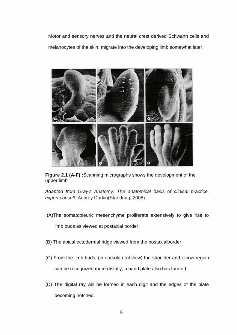
Motor and sensory nerves and the neural crest derived Schwann cells and
melanocytes of the skin, migrate into the developing limb somewhat later.
Figure 2.1 (A-F) :Scanning micrographs shows the development of the upper limb
Adapted from Gray's Anatomy: The anatomical basis of clinical practice,
expert consult. Aubrey Durkin(Standring, 2008)
(A)The somatopleuric mesenchyme proliferate extensively to give rise to
limb buds as viewed at postaxial border.
(B) The apical ectodermal ridge viewed from the postaxialborder
(C) From the limb buds, (in dorsolateral view) the shoulder and elbow region
can be recognized more distally, a hand plate also has formed.
(D) The digital ray will be formed in each digit and the edges of the plate
becoming notched.

7
(E) The notch is separated for the formation of fingers and proliferation
begins at the distal end of each digit to form nail bed.
(F)The nail development continues as the each of the finger has the tactile
pad distally.

8
2.2.2 LEFT HAND
Each hand consists of 27 bones, 8 form the wrist, and 19 form the
palm. The hand bones consist of the carpal bones, metacarpals and
phalanges. At birth, only the shafts of the metacarpals and phalanges are
present.
2.2.3. METACARPAL BONE
The metacarpals consist of five cylindrical bones in each hand. They
articulate with the carpal bones proximally and distally with the proximal row
of phalanges. They are numbered from the lateral or thumb to the medial
side as shown in (Fig 2.2), the anatomy of bone of the left hand.
A metacarpal consists of a shaft and two extremities. The base
articulates with the carpals and they also articulate on the side with the
adjacent metacarpal bones (except for the thumb). The shafts are slightly
concave on the palm surface and are triangular in shape.
The first metacarpal is the shortest and thickest of the metacarpals.
The second is the longest metacarpal. The third, fourth and fifth
metacarpals decrease in size. The articular surfaces on the heads of the
metatarsals are restricted laterally and are well marked anterior-posteriorly.
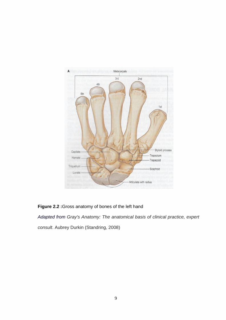
9
Figure 2.2 :Gross anatomy of bones of the left hand Adapted from Gray's Anatomy: The anatomical basis of clinical practice, expert
consult. Aubrey Durkin (Standring, 2008)

10
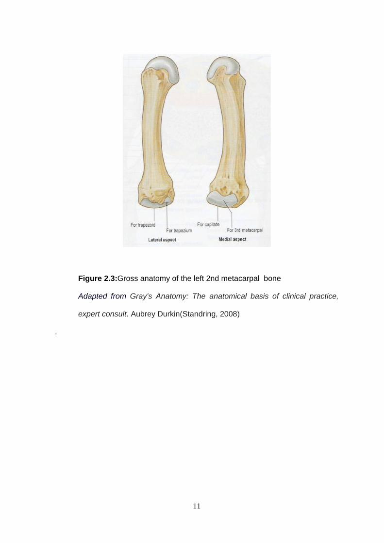
2.2.4GROSS ANATOMY OF THE LEFT 2nd METACARPAL BONE
The second metacarpal bone is the longest of the four remaining
bones; it, however, exceeds the third by a very slight difference. It is further
distinguished from the third by the diminutive size of the articular facet on
the radial side of its carpal extremity. Figure 2.3shows the gross anatomy
and Figure 2.5 shows the radiological anatomy of the left second
metacarpal bone.
Apart from being the longest, its base is also the largest. Its base is
prolonged upward and medial ward, forming a prominent ridge. It presents
four articular facets: three on the upper surface and one on the ulnar side.
Of the facets on the upper surface the intermediate is the largest and is
concave from side to side, convex from before backward for articulation
with the lesser multangular; the lateral is small, flat and oval for articulation
with the greater multangular; the medial, on the summit of the ridge, is long
and narrow for articulation with the capitate. The facet on the ulnar side
articulates with the third metacarpal.
11
Figure 2.3:Gross anatomy of the left 2nd metacarpal bone Adapted from Gray's Anatomy: The anatomical basis of clinical practice,
expert consult. Aubrey Durkin(Standring, 2008)
.
12
2.2.5 GROSS ANATOMY OF THE LEFT 3rd METACARPAL BONE
The third metacarpal bone, though shorter than the second, is
manifestly thicker and stronger. It has a short styloid process, projecting
proximally form the radial side of dorsal surface. Its base with a convex
anterior facet for articulation with the capitate. A strip-like facet, constricted
centrally articulates with the base of second metacarpal bone on the lateral
part and fourth metacarpal bone on the medial side. Figure 2.4 shows the
gross anatomy and Figure 2.5 shows the radiological anatomy of the left
3rdmetacarpal bone.

13
Figure 2.4 :Gross anatomy of the left 3rd metacarpal bone Adapted from Gray's Anatomy: The anatomical basis of clinical practice,
expert consult. Aubrey Durkin(Standring, 2008)

14
2.2.6 RADIOLOGICAL ANATOMY OF METACARPAL BONE
Figure 2.5: Left hand radiograph (posterior anterior view)
15
2.3HISTORY OF STATURE ESTIMATION
2.3.1 OVERVIEW
Various methods have developed in the last few centuries for stature
estimation either the anatomical or mathematical. The mathematical
method make use of one or more bone of length to estimate the stature of
individual.
The first study is by Rollet 1888, assesed the correlation between stature
and long bone lengths. He measured the length of radius, ulna, humerus,
femur, fibula and tibia and table of stature estimation.
Then, (Pearson, 1899a), used the data to creat regression formulae
for estimating stature.The advantage of mathematical method is, a single
bone can be used to estimate stature. The bone length is substituted into
regression equation. The outcome of the equation calculated gives the total
skeletal height of the living stature. However, the disadvantages is that,
different formula is required for a different population, for each different
bone and also saperately for each sex (Sanli et al., 2005).
2.3.2 IMPORTANCE OF REGRESSION EQUATION
Stature determination is one of the earliest values needed in
identifying human. In the 20th century with more mass disaster happen from
homicides activities to war victims and mass accidental disaster, most of
the victim’s body parts are capitulated. In forensic sciences and medicine
16
the stature of an individual can be determined from anatomical
measurement or mathematical method (Bhasin et al., 2002).
The anatomical method was noted more reliable in the presence of the
complete individual body (Bidmos, 2005; Pearson, 1899b). However, in the
absences of the completed human body or skeleton, the mathematical
method was preferred.
The mathematical method was created to determine the regression
equation and estimating stature of an individual or person (Pearson,
1899a).When a full skeletal or complete human body were presented, the
role of mathematical method in estimation height becomes second. The
role of mathematical method in estimating stature in Asian country was
pioneered by India(Badkur et al., 2011).
Malay race had occupied the portion of adjacent island in Southeast
Asia (SEA), Sumatera, coast of Borneo as well as islands in between these
area and particularly peninsular Malaysia. Malays were traced by the
anthropologist as originated from north-western part of Yunnan in China.
The earlier Malay noted as proto-Malay were people probably from coastal
Borneo who lengthened to Sumatera as consequence of trading and ocean-
going activities. Malay today known as modern Malay of Peninsular
Malaysia and coast of the Malay Archipelago were results of mixture of
other different races such as modern India, Thai, Arab and Chinese. As for
Kelantan, a state in the region of Peninsular Malaysia has 95.7 % of Malay
population(Negara, 2012).
17
Due to specific genetically distribution among Modern Malay, a
specific regression analysis must be performed for specific estimation
stature formula. This make the estimation of stature from human skeletal
remains is a major challenge in every country(Menezes et al., 2009).The
limb length to stature proportions differ between human populations(Krishan
and Sharma, 2007)
Other than that, previous study noted that specific gender and population
need specific equation for estimation that derive from specific simple liner
regression or multiple linear regression analysis(Bidmos and Asala, 2003).
Many studies have been carried out to estimate stature by taking
measurements from radiographic materials. There are different methods in
obtaining metacarpal measurements, generally categorized into invasive
and non-invasive. Plain radiograph is anon-invasive, practical and more
accessible mode of measurement, and delivers the minimum radiation dose
to subjects(MOU et al., 2011). These provide the suitability tool for
metacarpal measurement in this study.
18
2.3.3 EFFECT OF AGIENG ON STATURE
It is generally accepted that stature declines with age. According to
Giles and Hutchinson (1991), it is a reasonable assumption that
populations, particularly those in the United States, will begins to decrease
in stature starting in their midforties. This minimal decrease begins for
males at about 1mm/year and for female at about 1.25mm/year. These
factors must be accounted when estimating in an older individual, generally
including over the age of 45 years old (Giles and Klepinger, 1988).
Galloway found that height reduces average by 0.16 cm per years after
age 45 (Galloway, 1988). She suggested correction of the maximum height
-0.16 cm (age 45), incorporated into the stature estimation equation when
analysing older individuals. Trotter stated that stature decrease from age 30
by 0.06 cm per year (Trotter and Gleser, 1977).
19
2.3.4ESTIMATION OF STATURE FROM VARIOUS PARTS OF HUMAN
BODY
Estimation of stature in victim is a one of the major concern in
Forensic Investigation and anthropology. Every part of the human body is
unique in its determination ration to the height. It is amazing to discover that
every part of the body is different in its own way for a similar body
component in other person. There is also relationship between each part of
the body and the whole body.
Nothing represents this truth more than the relationship that various
parts of the body have to stature and gender identity on a person. Many
authors have studied estimation from mutilated body parts can be done
based on ratio of the body part concerned in relation to entire body.
Kate and Majumdar 1976, sucessfully estimated stature from the
length of femur and humerus by regression method and outometry is an
Indian sample. A study by Boldsen in 1984 statistically evaluated the
prediction of stature from length of the long bones in different European
population. A study done among Indian population done measuring 12
anthropometric parameters on ulna and multi-linear regression equation
were computed for stature estimation(Badkur and Nath, 1990).
A study done in Japan, proved that stature estimation can actually
be derived not only form the long bones but also from the somatometry of
the skull(Chiba and Terazawa, 1998). Theys carried out a study to
20
detemine the stature from the somatometry ofthe skullamong 124 Japase
cadavers.
The regression equations calculated for both sexes with SEE (standard
error of estimation) (S.E= 6.59cm) and (S.E= 6.96) . These are noted to be
larger than other parts of the body. However, usefull in cases where
indentification is required by means of only the skull is available.
In the year of 2002, (D.Radoinova et al., 2002) performed a study for
stature estimation among the Bulgarian population. In this study, the
statures and lengths of humerus, tibia and fibula were measured in 416
forensic cases.
The stature of a person can be determined by employing the software
system of multivariate regression statistic to analyze data from the hand. A
study performed in China involving 52 adult males whereby they measure
the upper limb. Measurement of hand length, hand width, length of palm,
width of palm, length of thumb, index finger, middle finger, ring finger and
little finger, which , after analysis has turned out to have applicable value to
determine person’s height (Xian-yue, 2004).
The study has proved that predicting the stature from the length of
the limb long bones taking into account sex- and age-related changes.
Mubarak Bidmos(2005) reported that adult stature estimation can be
measured by calcaneus measurement on cadaver with the standard error of
estimation around 4 to 5 cm. The stature estimation can be calculated from
the individual bone measurement or using combination of variables
(Bidmos, 2006).
21
The relationship between humerus, radius, ulna, femur, tibia, fibula
and clavicle with the stature estimation have been a topic of research for
decades (Nath and Badkur, 2002; Oberoi et al., 2006).
Among the North Indian population, a study to determine the relationship
between stature and various dimensions of hands and feet was performed.
The hand length, breadth, foot length and foot breadth were taken
independently right and left for each 246 subjects. The linear and multiple
regression equations for stature estimation were calculated and found to be
positive and statistically significant. The highest correlation coefficient noted
between stature and foot length with lowest SEE (standard error of
estimation). These provides the reliability and accuracy in estimating stature
of unknown individual among the North Indian Population (Krishan and
Sharma, 2007).
In the year of 2010, a study to evaluate the correlation between
stature of an individual with six parameters; hand length, hand-width, foot-
length, foot-width, forearm length and knee-to ankle length among healthy
individual in local population of Mumbai was performed. From the sample of
300 medical students, it was found that all six parameters showed
correlation with stature. However, the best was forearm-length (r=0.6)
followed by foot-length (r= 0.6). The mathematical formulae for estimating
stature were performed through basic linear regression for stature
estimation in the population of Mumbai (Chikhalkar et al., 2010).
22
2.3.5ESTIMATION OF STATURE FROM METACARPAL BONE
Formula for the estimation of stature from metacarpal bone length
are presented by (Meadows and Jantz, 1992). Developed regression
equation from two samples of metacarpals specimens. In this study, two
samples of metacarpal specimens were employed in the analysis: one of
212 individuals from the Terry Collection, and one of 55 modern males, all
of whom had measured statures. The midline length was taken on each
metacarpal bone. The stature was regressed on the basis of the metacarpal
length to derive equations. Comparisons between the Terry Collection male
and the modern sample showed the latter to have longer metacarpals and
greater statures. In spite of the differences noted, the Terry equations
perform acceptably on modern individuals and performance was slightly
better for whites than for blacks.
A study conducted in 372 radiographs of hand from 186 adults was
performed to find correlation with body height. The length and mid-width of
proximal, middle and distal phalanges and metacarpal bones were
measured. It was reported, that the body-height index of metacarpal bones
and phalanges provide definite assistance in judgment of the height and
gender of a person in forensic medicine. was gender difference among
them (SUN and CHEN, 2005)
In the year of 2006, a study to estimate the stature from x-rays of
metacarpal bone was performed among Turkish population (Sağir, 2006).
It is a study of 100 female and male patient’s hand radiograph. From the
study, five regression models has been set up base on 5 metacrapal
23
bones.The best regression model noted in this study, to estimate stature
from x-rays of metacarpals were using the measurement of 2nd and 3rd
metacarpals bone.
Not only this, another study performed in Japan results in similar
findings. (MOU et al., 2011)study the association between the metacarpal
and height of population in Gaomi City. This study involving a total of 280
men and 331 women respondents had their metacarpal bone measured
and correlate with their height. From this study, the best correlation noted
between the second metacarpal length and height followed by the third
metacarpal bone length.
Another study performed among 157 Egyptians adults using multi-
detector computed tomography (MDCT) of the left handto measure the 2nd
and 3rd metacarpal bone length and width. Their stature were determined.
Statistical analysis revealed that all variables are significant for female. As
for the male population, the Pearson’s correlation found to be significant
between the stature and second metacarpal width, third metacarpal bone
length and width. Linear regression equations were calculated with
standard error (SEE) 4.53cm to 4.71 for male and 5.45cm to 5.87cm for
female. Not only that,the multiple regression equations were also calculated
resulting into the best model for both gender (Zaher et al., 2011)

CHAPTER 3
OBJECTIVE
24
3.0 OBJECTIVES
3.1 GENERAL OBJECTIVE
To estimate stature from the morphology of left hand radiograph among
adult Malay population attending Hospital Universiti Sains Malaysia.
3.2 SPECIFIC OBJECTIVES
1. To compare the stature-metacarpal bone parameters (length and width) of
left hand between female and male.
2. To determine the correlation between stature and the parameters (length
and width) of left metacarpal bones.
3. To predict stature from the parameters (length and width) of left metacarpal
bones based on equation formula.