hindrance of the myosin power stroke posed by the .../67531/metadc3688/m2/1/high... · hydrolyzes...
TRANSCRIPT

APPROVED:
Douglas Root, Major Professor Kent D. Chapman, Committee Member Rebecca Dickstein, Committee Member Arthur J. Goven, Chair of the Department of
Biological Sciences Sandra L. Terrell, Dean of the Robert B.
Toulouse School of Graduate Studies
HINDRANCE OF THE MYOSIN POWER STROKE POSED BY THE PROXIMITY TO
THE TROPONIN COMPLEX IDENTIFIED USING A NOVEL LRET
FLUORESCENT NANOCIRCUIT
Pilar G. Coffee Castro-Zena
Thesis Prepared for the Degree of:
MASTER OF SCIENCE
UNIVERSITY OF NORTH TEXAS
May 2007
Coffee Castro-Zena, Pilar G. Hindrance of the Myosin Power Stroke Posed by
the Proximity to the Troponin Complex Identified Using a Novel LRET Fluorescent
Nanocircuit. Master of Science (Biochemistry), May 2007, 49 pp., 1 table, 12
illustrations, references, 53 titles.
A novel luminescence resonance energy transfer (LRET) nanocircuit assay
involving a donor and two acceptors in tandem was developed to study the dynamic
interaction of skeletal muscle contraction proteins.
The donor transmits energy relayed to the acceptors distinguishing myosin
subfragment-1 (S1) lever arm orientations. The last acceptor allows the detection of
S1’s bound near or in between troponin complexes on the thin filament. Additionally,
calcium related changes between troponin T and myosin were detected.
Based on this data, the troponin complex situated every 7 actin monomers,
hinders adjacently bound myosins to complete their power stroke; whereas myosins
bound in between troponin complexes undergo complete power strokes.

ii
Copyright 2007
by
Pilar G. Coffee Castro-Zena

iii
ACKNOWLEDGEMENTS
I wish to acknowledge Dr. Douglas Root for his enlightened mentoring and
continuous support. Additionally, special thanks to Dipesh Patel for providing Cy7-
troponin T.
This work was supported by grants from the National Institutes of Health and the
National Institute of Arthritis and Musculoskeletal and Skin Diseases.

iv
TABLE OF CONTENTS
Page
ACKNOWLEDGEMENTS .............................................................................................iii LIST OF TABLES.........................................................................................................vii LIST OF ILLUSTRATIONS.......................................................................................... viii LIST OF ABBREVIATIONS...........................................................................................ix Chapter
1. INTRODUCTION ..................................................................................... 1 1.1 Acto-Myosin Components and Muscle Contraction 1.2 The Lever Arm Model and Myosin Subfragment-1 1.3 Transition State Analogs of ATP Hydrolysis by Myosin 1.4 Objectives
2. EXPERIMENTAL MATERIALS AND METHODS .................................. 12
2.1 Preparation of Myosin Subfragment-1 by Papain Treatment and Endogenous RLC Heat Extraction
2.2 DTNB Purification and Terbium Chelate Conjugation of RLC 2.3 CS124-Tb-RLC Exchange to S1 and Labeling Ratio by Terbium
Titration 2.4 Tropomyosin-α Purification and Cy7 Labeling 2.5 Nanocircuit Assay in Solution 2.6 Nanocircuit LRET Measurements 2.7 Data Analysis: Single and Double Exponential Fitting of Decay
Curves 3. EXPERIMENTAL DESIGN .................................................................... 22
3.1 Nanocircuit Components 3.2 Theory of Luminescence Resonance Energy Transfer (LRET)
4. RESULTS .............................................................................................. 26
4.1 FRET Strategy: No Cross Contamination Between Donor and Last Acceptor

v
4.2 Nanocircuit LRET Distances Compared to X-ray Diffraction and Other FRET Measurements of the Myosin Lever Arm to the Nucleotide Binding Site
4.3 Calcium Induced Thin Filament Structural Changes Detected by the Nanocircuit.
4.4 Post-Power Stroke Lever Arm Conformations Detected on Myosins Bound Close to Cy7-Tropomysin α and Far Away From the Troponin Complex. Pre-Power Stroke Conformations Detected on Myosins Bound Close to Cy7-Troponin T
5. DISCUSSION ........................................................................................ 35
5.1 Steric Hindrance on Lever Arm Rotation Model: Myosins bound Proximally to the Troponin Complex have a Lever Arm biased towards Pre-Power Stroke State whereas Myosins bound in between the Troponin Complex have Lever Arms in Post-Power Stroke States
5.2 What Value Does a Prevention of a Power Stroke near a Troponin Complex Have?
5.3 Transition State Analogs Aluminum and Beryllium Fluoride behave similarly causing Pre-Power Stroke Conformations when observed with Troponin T and Post-Power Stroke Conformations when observed from Tropomyosin
5.4 Cooperativity of Activation 5.5 Calcium Induced Thin Filament Structural Changes Detected by
Nanocircuit 5.6 Other Possible Applications of the Fluorescent Nanocircuit 5.7 Conclusions
REFERENCES............................................................................................................ 45

vi
LIST OF TABLES
Page
1. Fluorescent nanocircuit analysis of lever arm position ..................................... 30
vii
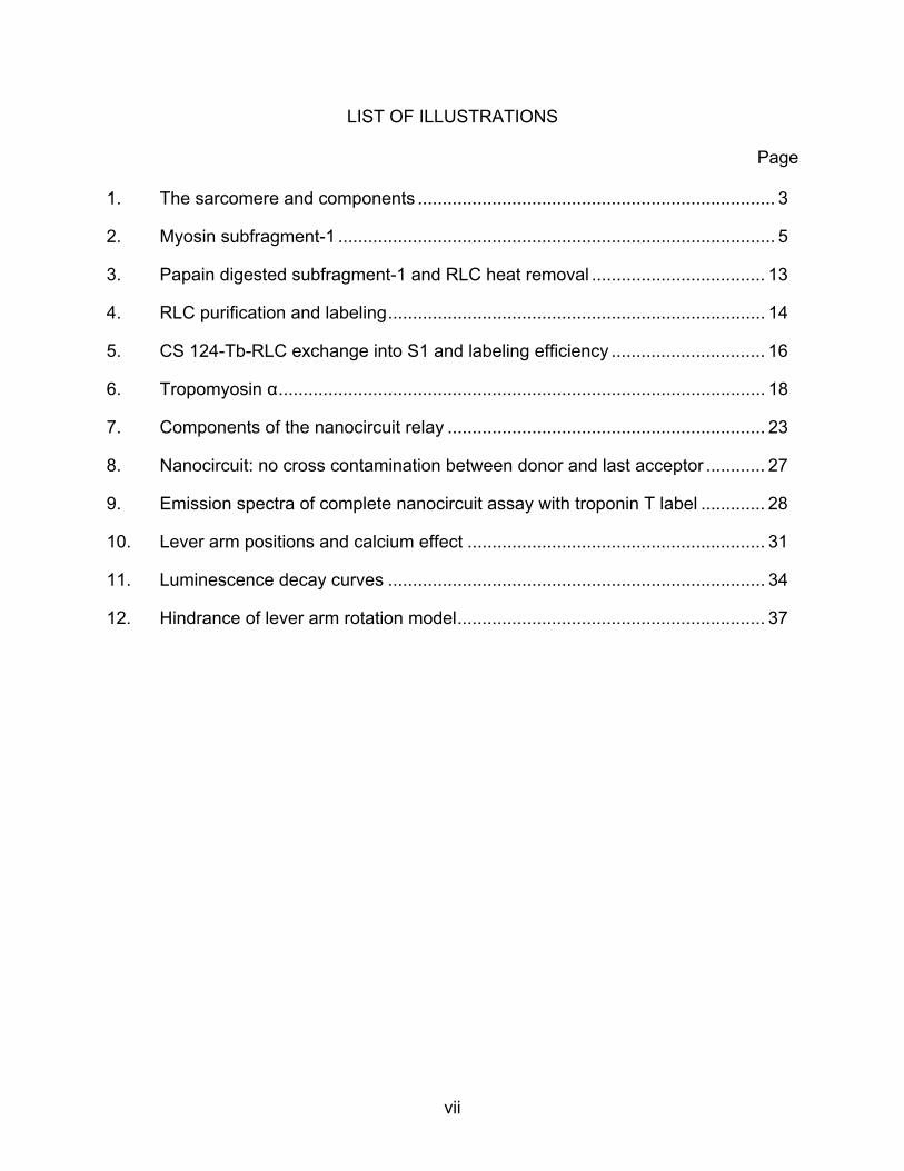
LIST OF ILLUSTRATIONS
Page 1. The sarcomere and components ........................................................................ 3
2. Myosin subfragment-1 ........................................................................................ 5
3. Papain digested subfragment-1 and RLC heat removal ................................... 13
4. RLC purification and labeling............................................................................ 14
5. CS 124-Tb-RLC exchange into S1 and labeling efficiency ............................... 16
6. Tropomyosin α.................................................................................................. 18
7. Components of the nanocircuit relay ................................................................ 23
8. Nanocircuit: no cross contamination between donor and last acceptor ............ 27
9. Emission spectra of complete nanocircuit assay with troponin T label ............. 28
10. Lever arm positions and calcium effect ............................................................ 31
11. Luminescence decay curves ............................................................................ 34
12. Hindrance of lever arm rotation model.............................................................. 37
viii
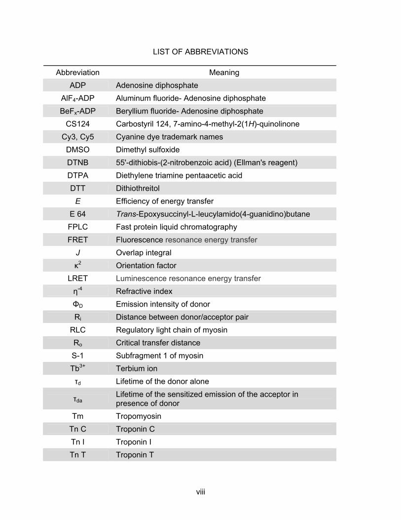
LIST OF ABBREVIATIONS
Abbreviation Meaning ADP Adenosine diphosphate
AlF4-ADP Aluminum fluoride- Adenosine diphosphate BeFx-ADP Beryllium fluoride- Adenosine diphosphate
CS124 Carbostyril 124, 7-amino-4-methyl-2(1H)-quinolinone Cy3, Cy5 Cyanine dye trademark names DMSO Dimethyl sulfoxide DTNB 55'-dithiobis-(2-nitrobenzoic acid) (Ellman's reagent) DTPA Diethylene triamine pentaacetic acid DTT Dithiothreitol
E Efficiency of energy transfer E 64 Trans-Epoxysuccinyl-L-leucylamido(4-guanidino)butane FPLC Fast protein liquid chromatography FRET Fluorescence resonance energy transfer
J Overlap integral κ2 Orientation factor
LRET Luminescence resonance energy transfer η-4 Refractive index ΦD Emission intensity of donor Ri Distance between donor/acceptor pair
RLC Regulatory light chain of myosin Ro Critical transfer distance S-1 Subfragment 1 of myosin Tb3+ Terbium ion τd Lifetime of the donor alone
τda Lifetime of the sensitized emission of the acceptor in presence of donor
Tm Tropomyosin Tn C Troponin C Tn I Troponin I Tn T Troponin T

1
CHAPTER 1
INTRODUCTION
1.1 Acto-Myosin Components and Muscle Contraction
Myosins comprise a large superfamily of motor proteins that couple movement
along actin filaments with the hydrolysis of ATP. There are about 20 classes of myosins
classified as such on the basis of amino acid sequence differences in their ATP-
hydrolyzing motor domains with additional differences among classes found in the
structure of their tail domains. Myosins II in particular are studied for their role in muscle
contraction. The myosin II molecule is comprised of two heavy chains and two light
chains. The globular motor domain of each heavy chain catalyzes the hydrolysis of ATP
and interacts with the actin thin filament. Two light chains, the essential light chain and
the regulatory light chain wrap around the neck region of each heavy chain. The tail
domains of myosin II heavy chains contain heptads repeat sequences involved in
dimerization in which several tails interact to form a rod-like α-helical coiled coil
responsible for the formation of the thick filaments.
Skeletal muscle is also referred to as striated muscle due to the consistent,
highly ordered interdigitating arrangement of the thin and thick myofilaments into
sarcomeres (Fig.1A). The sarcomere is the fundamental, functional unit of skeletal
muscle. The thin filaments consist predominantly of G-actin, a 42 KDa protein. G-actin
monomers polymerize end to end in the sarcomere to form filamentous F- actin, and
two F-actin strands coil around each other in a double helical array in a manner
analogous to two pearl necklaces wrapped together. Tropomyosin, a rope-like
regulatory protein coils around F-actin covering actin active binding sites for myosin. At

2
regular intervals along the thin filament is the other regulatory protein troponin.
Troponin is composed of 3 polypeptide chains TnT, TnI and TnC. These polypeptides,
together with tropomyosin assemble on the polymerized actin filament at a ratio of 1 Tn
complex: 1 Tm dimer: 7 actins (Fig. 1B). Skeletal and cardiac muscle contraction is
controlled by intracellular Ca 2+, where troponin is the Ca 2+ receptive protein that plays
the key role in regulating the sliding action between the two myofilaments. During
muscle contraction, the filaments slide resulting in the interactions between the myosin
cross-bridges with the actin thin filaments where the myosin head, binds to actin,
hydrolyzes ATP and converts this chemical energy into mechanical energy for force
transmission along the filaments that results in force production.
The myosin S1 fragment (subfragment-1) is the structure that forms the cross-
bridges from the myosin thick filament to the actin thin filaments during muscle
contraction and it is obtained by the proteolytic treatment of myosin with cysteine
proteinase papain (Fig. 2A). The x-ray crystal structure of chicken skeletal S1 fragment
was solved in 1993 (Rayment et al., 1993a),
1.2 The Lever Arm Model and Myosin Subfragment-1
Fig. 2B illustrates subfragment 1 to be composed of both a catalytic site (where
the actin binding and ATP hydrolyzing domains are) and the light chain binding domain
or lever arm, an approximately 9 nm in length α-helix stabilized by two calmodulin-like
light chains, the regulatory light chain (RLC) and the essential light chain (ELC). This
last domain or lever arm is believed to swing while the actomyosin interface remains
rigid (Cooke, 1986; Rayment et al., 1993a; Holmes, 1997). In this theory, the myosin II

3
Fig 1. The sarcomere and components: A) The sarcomere is the basic unit of contraction, defined by two Z-lines. In the A band both filaments overlap, the I band is composed of only thin filament proteins. B) Inset from A: calcium binds to the troponin complex causing a conformational rearrangement of tropomyosin that makes possible the myosin-actin interaction. Actin stimulates ATPase activity in the globular myosin head that results in the production of force along the actin filaments.
1A)
Actin
Troponin Tropomyosi
Troponin C
Myosin head
Troponin T
1B)
Z line
Actin
Myosin head
I band
A band

4
heads latch onto a nearby actin thin filament and the light chain binding domain flips in a
lever-like motion propelling the actin filament. The myosin head then lets go of the actin,
the neck cocks back, and the head reattaches at a new point on the thin filament,
starting the process again. In this manner, the lever arm of myosin is the structure that
amplifies the subtle changes occurring at the actin binding and ATP hydrolyzing
domains of myosin and converts them into the great molecular displacement called the
“power stroke.” This power stroke is the step during the contraction cycle that pulls the
actin filaments towards the center of the muscle sarcomere; its molecular mechanisms
have not completely been elucidated nor have those of the effects of regulatory proteins
troponin and tropomyosin in the lever arm conformations of nearby S1 heads.
To this date, “tail-wagging” or “rotating lever arm” models are being proposed
based on the many structurally diverse crystallographic structural data accumulated in
these past 10 years. Shorter Dictyostelium myosin heavy chain fragments had been
crystallized in the presence of different bound nucleotides and transition state analogs
(Fisher et al., 1995; Smith and Rayment, 1996; Gulick et al., 1997). Although these
constructs were too short to carry the light chains, large-scale structural changes had
been observed near the carboxy-ends of certain transition state structures where the
lever arm would emerge. Up to this point, the structural data had identified two different
states of myosin differing not only in the orientation of the lever arm where the most
dramatic conformational changes occur but also in two more subtle changing regions of
the motor domain: the 50 kDa cleft and the converter region. The first crystal structure
of chicken skeletal muscle S1 without nucleotide and no actin was considered to
correspond to the post-power stroke or rigor state (Rayment et al., 1993a).

5
2A) 2B) Fig 2. Myosin subfragment-1: A) Treatment of myosin with papain generates two S-1 fragments and a rod. B) X-ray crystal structure of chicken skeletal S-1 fragment (Rayment et al., 1993a), the catalytic domain composed of the actin binding and ATP hydrolysis domains and the lever arm with one regulatory light chain (light green) and one essential light chain (light yellow) wrapped around.
S1 Rod
Papain treatment
ATP hydrolysis domain
Regulatory Light chain
Lever arm
Actin binding domain

6
The truncated expressed Dictyostelium motor domains lacking the lever arm and
complexed to various nucleotide analogs were modeled in two conflicting ways; if the
lever arm was present, in a “primed” transition state (lever arm in the up position) or a
post-power stroke state (down position lever arm). In 1999 a scallop S1 unmodified
structure complexed to MgADP with an intact lever arm revealed a third conformation of
the lever arm situated almost parallel to the actin filament axis which has been
interpreted as a detached ATP state occurring between the rigor and transition states
(Houdusse et al., 1999). The following year, the same group reported two more
structural states for a total of three different conformational states of the intact scallop
S1 that depend on the specific nucleotide or nucleotide analog to which it is bound:
nucleotide-free motor with post-power stroke lever arm (near rigor state), transition state
conformation: lever arm primed in pre-power stroke state and lastly ADP detached
state. The first two conformations correspond to the span of the entire power stroke of
the actomyosin cycle (Houdusse et al., 2000).
1.3 Transition State Analogs of ATP Hydrolysis by Myosin
Myosin hydrolyzes ATP to produce the energy required for muscle contraction.
The elucidation of the crystal structure of the S1 fragment of myosin by x-ray diffraction
showed that myosin is a back door enzyme, one in which the ATP substrate binds in a
site of the S1 molecule that is opposite to the binding site of the catalyst actin (Rayment
et al, 1993a; Fig. 2B). In the absence of ATP, the myosin S1 cross bridges bind tightly
to the actin filaments. In the presence of ATP, binding to ATP causes a rapid
dissociation of the actomyosin complex, therefore the crossbridges bind to either ATP or
7
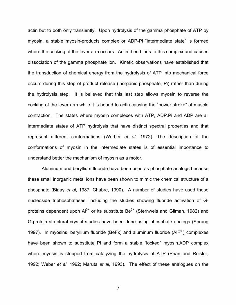
actin but to both only transiently. Upon hydrolysis of the gamma phosphate of ATP by
myosin, a stable myosin-products complex or ADP-Pi “intermediate state” is formed
where the cocking of the lever arm occurs. Actin then binds to this complex and causes
dissociation of the gamma phosphate ion. Kinetic observations have established that
the transduction of chemical energy from the hydrolysis of ATP into mechanical force
occurs during this step of product release (inorganic phosphate, Pi) rather than during
the hydrolysis step. It is believed that this last step allows myosin to reverse the
cocking of the lever arm while it is bound to actin causing the “power stroke” of muscle
contraction. The states where myosin complexes with ATP, ADP.Pi and ADP are all
intermediate states of ATP hydrolysis that have distinct spectral properties and that
represent different conformations (Werber et al, 1972). The description of the
conformations of myosin in the intermediate states is of essential importance to
understand better the mechanism of myosin as a motor.
Aluminum and beryllium fluoride have been used as phosphate analogs because
these small inorganic metal ions have been shown to mimic the chemical structure of a
phosphate (Bigay et al, 1987; Chabre, 1990). A number of studies have used these
nucleoside triphosphatases, including the studies showing fluoride activation of G-
proteins dependent upon Al3+ or its substitute Be2+ (Sternweis and Gilman, 1982) and
G-protein structural crystal studies have been done using phosphate analogs (Sprang
1997). In myosins, beryllium fluoride (BeFx) and aluminum fluoride (AlF4-) complexes
have been shown to substitute Pi and form a stable “locked” myosin.ADP complex
where myosin is stopped from catalyzing the hydrolysis of ATP (Phan and Reisler,
1992; Weber et al, 1992; Maruta et al, 1993). The effect of these analogues on the

8
crystallographic structures of scallop myosins, dictyostelium myosins as well as smooth
muscle myosins has also been documented. Recombinant truncated Dictyostelium S1
has been complexed with ADP.BeFx and ADP.AlF4- (Fisher et al, 1995) as well as ADP-
vanadate (Smith and Rayment, 1996); and the crystal structures have been solved.
These studies concluded that the S1-ADP.BeFx mimics the “pre-hydrolyzed” state
Myosin.ATP, whereas the S1-ADP.AlF4- (as well as the ADP.vanadate) behaves more
like a “true transition state” mimicking the Myosin.ADP.Pi intermediate state. The
results from the smooth myosin S1 crystal structure however, indicate that the
ADP.BeFx and ADP.AlF4- are identical and closely resemble the Dictyostelium
ADP.AlF4- and Dictyostelium ADP.Vanadate (Dominguez et al, 1998). Further
biochemical studies (Peyser et al, 1997; Phan et al, 1997 and Ajtai et al, 1999) support
the conclusions from the dictyostelium results: S1.ADP.BeFx resembles Myosin.ATP
whereas S1.ADP.ALF4- (as well as S1.ADP.vanadate) resembles the Myosin.ADP.Pi
intermediate state. Therefore, not only does the assigned transition state varies with
the type of metal fluoride ion but it has been proposed that it varies also with the ionic
strength particularly in the S1.ADP.BeFx complex (Peyser et al, 2001), and the
presence of other contractile proteins (Phan and Reisler, 1993; Nikolaeva et al, 1994;
Kraft et al, 2005 ).
In the present study, transition state analogs aluminum and beryllium fluoride
were used in an attempt to characterize the transient weak binding states of ADP-Pi of
the ATPase cycle. In the light of previous studies, assigning a particular kinetic state to
any of these analogs has proven difficult due to their properties being dependent on the
assay conditions and the presence of other proteins of the sarcomere. When used in
9
the nanocircuit in solution, both metallofluorides have exhibited no difference among
themselves. However, when used in the nanocircuit in conjunction with the labeled
troponin T they allowed the identification of pre-power stroke conformations and post-
power stroke conformations were identified when used in conjunction with labeled
tropomyosin. The goal of the spectroscopic nanocircuit is to not only identify different
conformations of the lever arm by averaging the signals from a large number of
molecules but to exploit the signal of the last acceptor of the nanocircuit relay to identify
the most critical conformations to a given transition kinetic step using the analogs.
1.4 Objectives
The different possible orientations of the light chain binding domain mapped by x-
ray crystallographic methods need to be corroborated by studies done in solution and in
the presence of the other regulatory components of muscle contraction to establish their
dynamic interaction. Fluorescence spectroscopy is a technique that can resolve
distances of subnanometer length and together with crystallography can provide
additional detailed information for the understanding of the molecular mechanisms of
contraction and motility. As opposed to x-ray crystallography, fluorescence
spectroscopic methods allow the measurement of conformational changes observed in
dynamic form and closer to physiological conditions. The different kinetic states of
myosin reflect different stages in the ATPase cycle and great effort has been put into
correlating these stages to the available structural data. Additionally, spectroscopic
data, by virtue of being collected in solution, has shown that it is a distribution of myosin
structures that are present at any given time so that a particular kinetic state may

10
correspond to several structures. This novel fluorescent nanocircuit assay was
developed with the objective of studying the molecular dynamic interaction of the
proteins involved in skeletal muscle contraction in solution using fluorescence
resonance energy transfer (FRET) and luminescent lanthanides as energy transfer
donors. In solution, the nanocircuit makes possible the measurement of the kinetic
states of myosin through the averaging of a number of molecules, while the three relay
system ensures that only the molecules bound to the filament and bound closest to any
of the two labels in the thin filament: either Cy7-Troponin T or Cy7-Tropomyosin α, are
averaged in the readings. The purpose is to identify more clearly which conformations
are more critical to the weak binding state by looking at the changes in position of the
RLC relative to the nucleotide binding site of molecules bound proximate to two different
parameter proteins in the thin filament in the presence and absence of ADP and
nucleotide analogs. Furthermore, the nanocircuit provides a way to assess the location
of regulatory protein, troponin T, on the thin filament relative to myosin during the
actomyosin interaction in the presence and absence of calcium. The present work is
first in its kind to label in solution three different sites of the actomyosin cycle as a relay
of energy successfully.
Measurement of distances to establish lever arm movement using energy
transfer between two sites: the light chain and catalytic domain before and after the
power stroke have been done in the past. (Shi et al, 2000; Xu and Root, 1998 and 2000,
Smyczynski and Kasprzak, 1997). Additionally, the determination of the crystal
structures of the troponin complex (Takeda et al. 2003. and Vinogradova et al. 2005)
has further advanced the knowledge of the regulation of muscle contraction at the

11
molecular level but still much of the interactions of all the actomyosin components
remain to be elucidated.
This novel nanocircuit assay, allows measurements in three different sites as a relay
of energy transfer that involve myosin as well as the muscle regulatory proteins of the
thin filament. Being the first time that a third parameter is used to study conformations
of the lever arm, this nanocircuit is a unique assay. For it not to break, the terbium-
labeled RLC in the lever arm, the Cy5-ADP in the nucleotide binding site and a
proximate troponin T or tropomyosin α must be no farther than their Ro Forster distance
for energy transfer to occur. In that case, no time-resolved fluorescence signal would be
observed from Cy7, the last site of the relay.
The hypothesis tested in this study is the hindrance posed by the periodically
situated troponin complexes to myosins that when binding adjacent to the Tn complex
situated every 7 actin monomers along a thin filament, are unable to complete their
power stroke. Furthermore, this study tests the lack of hindrance measured in the lever
arm conformations of myosins bound in between the Tn complexes.

12
CHAPTER 2
EXPERIMENTAL MATERIALS AND METHODS
2.1 Preparation of Myosin Subfragment-1 by Papain Treatment and Endogenous RLC Heat Extraction
Myosin subfragment 1 (S-1) was prepared by papain digestion of rabbit skeletal
myosin. Myosin (stored flash-frozen) in 0.5 M KCL, 10mM imidazole, pH 7.0 was
centrifuged at 100,000 x g (47,000 rpm) in a TLA 100.3 rotor (Beckman Optima ™ TL-
100 ultracentrifuge) for 1 hour at 4oC to remove aggregates. The cleared supernatant
was dialyzed against 1 L of 0.1 M KCl, 10mM imidazole, pH 7.0 for 15 hours (overnight)
at 10oC, lowering the salt concentration causes the myosins to form filaments. The next
day, MgCl2 was added to a final concentration of 2 mM prior to digestion. Fresh papain
was added to a final concentration of 0.2 mg/mL to the protein solution and incubation
was done for 7 minutes at 25oC or room temperature in a shaker. The proteolytic
reaction was stopped by the addition of 50 ul of E-64 (from SIGMA) per ml of myosin
solution. The products were dialyzed 15 hours (overnight) against 1 L of 0.04 M KCl, 10
mM imidazole, pH 7.0. The next day ultracentrifugation at 100,000 x g (47,000 rpm in a
TLA 100.3 rotor) for 1 hour at 4oC yields S1 in the supernatant (Fig. 3A). The S1
preparation (containing the endogenous Regulatory Light Chain (RLC) was run through
a first Toyopearl® 55-F gel filtration column to buffer exchange the protein to 50 mM
Kpr, 20 mM EDTA, 10 mM Kpi, 0.5 mM DTT, pH 7.0 (Fig. 3A). The eluted S1 in K
propionate buffer was heated at 30oC for 15 minutes to extract the endogenous RLC.
Separation of the stripped RLC was accomplished by a second Toyopearl® 55-F gel
filtration step after heat removal (Fig. 3B). Fig. 3C shows the 12% SDS-PAGE of S1
devoid of RLC with a molecular weight of about 95 kDa as compared to broad-range

13
prestained SDS-PAGE standards from Bio-Rad.
3A)
3B) 3C)
208-115-80-
S1 My
Fig 3. Papain digested subfragment-1 and RLC heat removal. Gel filtration chromatography after papain digestion (3A). Gel filtration after heat removal of regulatory light chain (3B). SDS-PAGE of S1 devoid of RLC at expected MW of 90 kDa (3C).
2.2 DTNB Purification and Terbium Chelate Conjugation of RLC
RLC was dissociated from rabbit skeletal muscle myosin with DTNB (Wagner,
1982) and purified by β-mercaptoethanol reduction followed by Superdex™-75 gel
20-
1 st Toyopearl
-0.02
0
0.02
0.04
0.06
0.08
0.1
0.12
0 5 10 15 20 25 30
fractions
AU28
0
After heat removal: 2nd Toyopearl
-0.05
0
0.05
0.1
0.15
0.2
0 2 4 6 8 10 12 14 16 18 20 22 24 26 28 30
Fractions
AU
280
S1 (with endogenous RLC
S1 Endogenous RLC
MW S1 Std (no RLC)

14
Dyalisis, membrane concentration and Desalting of RLC labeled with Cs124
00.10.20.30.40.50.60.70.8
250 300 350 400
Wavelength
Abso
rban
ce
after concentration + dilution after Dyalisis
DTNB Purification of RLC: Superdex 75
-0.5
0
0.5
1
1.5
2
0 7 14 21 28 35 42 49 56 63 70
fractions
AU
280
4A) 4B)
4C) Fig. 4. RLC purification and labeling. Dissociation of RLC achieved by treatment of myosin with sulfhydryl reactant DTNB followed by BME reduction and separation by Superdex-75 gel filtration chromatography (4A). 15% SDS-PAGE gel indicating the separation of high purity RLC (fracs 25-31) from higher molecular weight myosin heavy chain and thin filament regulatory proteins (fracs 17-22) and from excess DTNB (fracs 34-55)(4B). Absorption spectra of removal of excess CS124-DTPA terbium chelate from labeled RLC after desalting and dialysis (4C). filtration chromatography to remove excess Ellman’s reagent (Fig. 4A). RLC of high
purity was obtained as shown by a 15% SDS-PAGE of the eluting fractions 25-31 from
the FPLC and removal of excess DTNB reagent in fractions 34-58 (Fig. 4B).
RLC
DTNB
20kDa RLC
280 337
after dialysis

15
The purified rabbit skeletal RLC was labeled with a 1,500 x molar excess of an
equimolar mixture of carbostyril-124 and DTPA in DMSO to conjugate terbium with
DTPA/ CS124. DTPA is a dianhydride reagent that attaches to carbostyril via one
anhydride, the other one interacts with the amine groups of lysine residues of RLC. The
great excess of free reagents were dialyzed over a weekend and subjected to several
rounds of buffer exchange by concentration and dilution using a Millipore Centricon®
centrifugal filter device with a membrane of a molecular weight cut off of 5,000; 4 times
smaller than the RLC protein’s molecular weight (Fig. 4C).
2.3 CS 124-Tb-RLC Exchange to S1 and Labeling Ratio by Terbium Titration
Cs-124 labeled RLC was added at molar excess to S1 to be exchanged back
with overnight dialysis in 1 mM MgCl2, 0.1 M KCl and 10 mM imidazole, pH 7 overnight.
The next day, excess free labeled RLC was separated by Superdex™ 75 gel filtration
(Fig 5A) and protein was concentrated using Centricon® concentrators to 2 uM (about
0.25 mg/ml) as read on a HP UV-Vis spectrophotometer (extinction coefficient for S1 is
0.75 ml mg-1cm-1)
Terbium titration was carried on an SLM-Aminco Bowman II luminescence
spectrometer (Spectronic Instruments, Rochester, NY) with dual xenon arc and
flashlamp light sources. Titration was carried by incubating the CS 124-RLC-S1 with
increasing amounts of terbium chloride solution. Excitation was set at 337 nm with
emission collected at 547, the intensity of emission was plotted against the
concentration of terbium chloride, indicating a 100% labeling ratio (Fig. 5B)

16
5A) 5B) Fig 5. CS 124-Tb-RLC exchange into S1 and labeling efficiency. FPLC Superdex-75 separation of residual CS 124-Tb-RLC after exchange to S1 previously devoided of endogenous regulatory light chain (5A). Labeling ratio of 1 to 1 or 100 % labeling efficiency calculated by terbium titration of CS 124-Tb-RLC-S1 as estimated by following the luminescence enhancement and saturation upon titration of 0.9 uM S1 with 0.9 uM terbium chloride solution (5B). 2.4 Tropomyosin-α Purification and Cy7 Labeling
Tropomyosin was extracted from acetone powder from rabbit skeletal back
Superdex-75
-0.01
0
0.01
0.02
0.03
0.04
0.05
0.06
1 3 5 7 9 11 13 15 17 19 21 23 25 27 29
Fractions
AU
280
S1 with incorporated CS 124-RLC
Excess CS 124-RLC
Terbium Titration of 0.9 uM Cs124-RLC labeled S1
0
1
2
3
4
5
6
0 0.5 1 1.5
[Terbium]
Fluo
resc
ence
uM
Rel
ativ
e In
tens
ity

17
muscle mince in 1.0 M KCl, 0.5 mM DTT at pH 7 according to a modified protocol of
Bailey and further purified as α,α- and α,β-tropomyosin by hydroxyapatite
chromatography as the method described by Cummins and Perry (Colowick, 1982).
The proteins were eluted by a linear gradient of 1-200 mM phosphate buffer to separate
the two subfractions: α (higher electrophoretic mobility) and β subunits (Fig. 6). The α
subunit contains a single cysteine residue, whereas the β subunit has two cysteine
residues per 33 kDa. The single cysteine containing Tm-α was picked for labeling.
Fractions containing only α,α-tropomyosin were pooled to a final concentration of 4
mg/ml and were denatured by dialysis in 6 M urea and 10 mM imidazole pH 7. Labeling
was carried at denaturing conditions of 6 M urea with Cy7-maleimide with the purpose
of labeling the single Cys 190 residue in the rabbit Tm-α. Cys 190 is involved in
disulphide crosslinking of the polypeptide chains of a dimer and it is highly conserved in
the α tropomyosins in human, pig, rat and rabbit (Perry, 2001) . The labeled protein
was flash frozen for storage until needed.
The troponin subunits were purified after the tropomyosin extraction from rabbit
skeletal muscle in denaturing conditions 6 M urea with FPLC chromatography in a
DEAE-Sephadex A-50 column as described in Colowick (Colowick, 1982), with an
additional chromatography step with Sephadex G50/75 for the removal of lower
molecular weight contaminants in the troponin I subunit isolation.
The Cy7 labeled troponin T subunit used is a naturally occurring isoform from
skeletal avian breast muscle that has calcium binding properties and a single cysteine
used for the attachment of different fluorescent probes (Jin and Root, 2000; Zhang et
al., 2004)

18
Fig 6. Tropomyosin α. Tm α molecule 7Å resolution crystal structure from porcine ventricles crystallized by Whitby and Philips in 2000, showing Cys 190 highly conserved residue in pig, human, rat and rabbit (6A). 15% SDS-PAGE of hydroxyapatite separation of tropomyosins α (higher electrophoretic mobility) and β from rabbit skeletal muscle (6B)
βα
6A)
6B)
Cys-190
N
C

19
2.5 Nanocircuit Assay in Solution
The acto-S1-Tropomyosin-Troponin complex experiments were performed in 0.1
M KCl, 2 mM MgCl 2, and 10 mM imidazole, pH 7.0. The concentrations in the assay of
labeled S1 were 0.5-0.8 uM, while the molar ratios of S1 to actin assayed were 1:1 and
1:7 in order to analyze two conditions: saturating or 1 S1 per 1 actin molar ratio and at 1
S1 per 7 actin molar ratio. Since TnT repeats at every 7 actin monomers, the idea is to
measure if crowding of S1 heads affects lever arm conformations in the saturated
versus the non saturated conditions. No significant effect of actin saturation with
myosin S1 was clearly observed, therefore for detection efficiency purposes, saturating
conditions were used. Actin was reconstituted by dialysis in assay buffer and
assembled with 1/7 ratio of renatured tropomyosin, troponin I, C, and Cy7 troponinT.
The complex is added to the system (case I, Fig. 7). The same ratios apply to the thin
filament reconstitution involving the Cy7 label at tropomyosin α (case II, Fig. 7). Other
conditions are measured with no nucleotide, with CY5-ADP at final equimolar amounts
to S1, with transition analogs beryllium and aluminum fluoride (at 0.1 mM final
concentration) and + and – calcium removed by the addition of EGTA at also 0.1 mM
final concentration of CaCl2.
All contractile protein components were mixed in a 100 ul total volume reaction in
an eppendorf UVette® without CY5-ATP for a “donor” only measurement of emission
and decays at 540 nm, 690 nm and 780 nm in the appropriate buffer. First acceptor
CY5-ADP labeling the nucleotide binding pocket was added as CY5-ATP for the
nanocircuit to occur and appropriate signals were read after a 5 minute incubation (to
allow ATP hydrolysis) at 540 nm, 690 nm and 780 nm as well.
20
Papain, DTPA, beryllium, aluminum chloride, sodium fluoride, and terbium
chloride hexahydrate were obtained from Sigma Aldrich. Cy5 and Cy7 from Amersham-
Pharmacia Biotech.
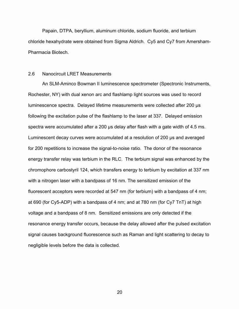
2.6 Nanocircuit LRET Measurements
An SLM-Aminco Bowman II luminescence spectrometer (Spectronic Instruments,
Rochester, NY) with dual xenon arc and flashlamp light sources was used to record
luminescence spectra. Delayed lifetime measurements were collected after 200 µs
following the excitation pulse of the flashlamp to the laser at 337. Delayed emission
spectra were accumulated after a 200 µs delay after flash with a gate width of 4.5 ms.
Luminescent decay curves were accumulated at a resolution of 200 µs and averaged
for 200 repetitions to increase the signal-to-noise ratio. The donor of the resonance
energy transfer relay was terbium in the RLC. The terbium signal was enhanced by the
chromophore carbostyril 124, which transfers energy to terbium by excitation at 337 nm
with a nitrogen laser with a bandpass of 16 nm. The sensitized emission of the
fluorescent acceptors were recorded at 547 nm (for terbium) with a bandpass of 4 nm;
at 690 (for Cy5-ADP) with a bandpass of 4 nm; and at 780 nm (for Cy7 TnT) at high
voltage and a bandpass of 8 nm. Sensitized emissions are only detected if the
resonance energy transfer occurs, because the delay allowed after the pulsed excitation
signal causes background fluorescence such as Raman and light scattering to decay to
negligible levels before the data is collected.

21
2.7 Data Analysis: Single and Double Exponential Fitting of Decay Curves
Emission scans with donor only and in the presence of acceptors were
subtracted of a baseline, and instrument correction was performed to compensate for
the lack of sensitivity of the photomultiplier after wavelength 600 nm. Final curves were
smoothed to 15 points and exported to excel for plotting as shown in Fig. 8. The
nanocircuit is broken if any of the three components is farther apart than their Ro
distance for energy transfer to occur, so the decay of signal from the terminal acceptor
reports on the conformation of myosin S1 bound near troponin (or tropomyosin) while
the decays of previous fluors of the circuit report on the average conformations of all S1
regardless of their position (Fig. 9).
The lifetimes of the decay curves were calculated by MacCurveFit Software
(version 1.3.3 by Kevin Raner). Lifetimes at 547 nm were fitted single and double
exponentially to account for the molecules that are bound to actin and the ones
unbound as illustrated in Fig. 9. The efficiency of energy transfer (E) and distance (R)
between donor and acceptors were determined by comparing lifetime measurements of
the donor without the acceptor present and the sensitized emission of the acceptor in
the presence of the donor per equations 1, 2, and 3 (section 1.6).
22
CHAPTER 3
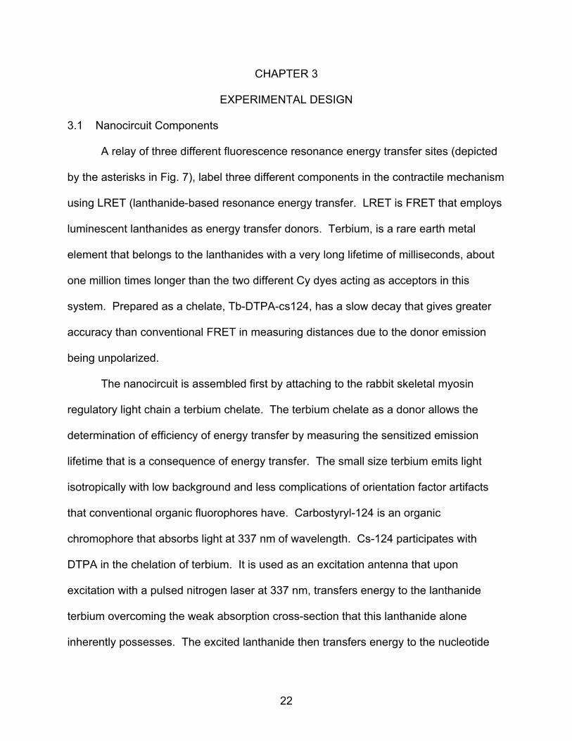
EXPERIMENTAL DESIGN 3.1 Nanocircuit Components
A relay of three different fluorescence resonance energy transfer sites (depicted
by the asterisks in Fig. 7), label three different components in the contractile mechanism
using LRET (lanthanide-based resonance energy transfer. LRET is FRET that employs
luminescent lanthanides as energy transfer donors. Terbium, is a rare earth metal
element that belongs to the lanthanides with a very long lifetime of milliseconds, about
one million times longer than the two different Cy dyes acting as acceptors in this
system. Prepared as a chelate, Tb-DTPA-cs124, has a slow decay that gives greater
accuracy than conventional FRET in measuring distances due to the donor emission
being unpolarized.
The nanocircuit is assembled first by attaching to the rabbit skeletal myosin
regulatory light chain a terbium chelate. The terbium chelate as a donor allows the
determination of efficiency of energy transfer by measuring the sensitized emission
lifetime that is a consequence of energy transfer. The small size terbium emits light
isotropically with low background and less complications of orientation factor artifacts
that conventional organic fluorophores have. Carbostyryl-124 is an organic
chromophore that absorbs light at 337 nm of wavelength. Cs-124 participates with
DTPA in the chelation of terbium. It is used as an excitation antenna that upon
excitation with a pulsed nitrogen laser at 337 nm, transfers energy to the lanthanide
terbium overcoming the weak absorption cross-section that this lanthanide alone
inherently possesses. The excited lanthanide then transfers energy to the nucleotide

23
Cy7-Troponin T (CASE I)
Terbium chelate on RLC Cy5-ADP on myosin or (donor) (1st acceptor) Cy7-Tropomyosin (CASE II)
(2nd acceptor)
Fig 7. Components of the nanocircuit relay. Donor, 1st acceptor and two possibilities for a 2nd acceptor. Excitation of the CS-124 antenna with a pulsed laser excites the terbium chelate on the lever arm which transfers energy to its acceptor, Cy5-ADP on labeling the nucleotide binding site of myosin, which in turn relays the energy to any of the two last acceptor on the thin filament: Cy7-troponin (case I) or Cy7-tropomyosin α (case II)
Cy7-Troponin T
Cy5-ADP
Terbium chelate: DTPA -cs124-Tb-RLC
Pulsed nitrogen laser
C
N O
CO-2
N
N
CO-2
CO-
C
O
NH
N
O
CH3
Tb3+
Cy7-Tropomyosi
-
Terbium chelate: Cs 124-DTPA-
-
Cy5-ADP
Pulsed nitrogen laser
C
N
OCO-
2
N
N
CO-2
CO-
C
O NH
N
O
CH
Tb3
Troponin
CASE I CASE II

24
binding site which is the second relay site and first acceptor of energy labeled by Cy5-
ADP. This second site in turn transfers energy to the third and last relay site: the Cy7
attached to a unique single cysteine in the alternative splicing isoform of Troponin T or
to Cys 190 in the Tropomyosin α subunit in the thin filament. Both cases are illustrated
with asterisks in Fig. 7.
3.2 Theory of Luminescence Resonance Energy Transfer (LRET)
The efficiency (E) of energy transfer between the donor lanthanide chelate in the
RLC and the acceptor 1 (Cy5-ADP) or acceptor 2 (Cy7-TnT) was determined by
comparing the lifetime measurements of the donor without the acceptor present and the
“sensitized” emission of the acceptor in the presence of the donor by using the
equation:
E = 1 - τda/τd (Eq. 1) Where τda is the apparent lifetime of the sensitized emission of the acceptor in the
presence of the donor since the delayed acceptor fluorescence should be due only to
the energy transfer from the donor and τd is the lifetime of the donor alone.
Furthermore, the efficiency of LRET (E) for the distance (Ri) between donor and
acceptor was calculated from the equation:
E = Σi (R6
o/R6i) / (1 + Σi (R6
o/R6i) (Eq. 2)
Where Ri is the actual separation distance between donor-acceptor pair and Ro is the
critical transfer distance that corresponds to a distance of 50% energy transfer
efficiency and calculated as follows:

25
Ro = 8.79 x 10⁻¹¹ J κ2η-4φD)1/6 nm (Eq. 3) Where J is the calculated overlap integrals for the acceptor with the donor; κ2 is the
dynamic average value of the orientation factor, 2/3; the isotropic emission of terbium
will limit the orientation factor uncertainty of at the most, 10% (Selvin and Hearst, 1994)
From equation 2, efficiency of energy transfer “E”, it is observed that FRET
diminishes as the sixth power of the distance between the donor and the acceptor;
therefore, no FRET occurs for distances greater than 2 Ro. Since Ro is on the order of
10 to 70 Angstroms, by performing FRET measurements it is possible to distinguish
proteins that are merely nearby in the same vicinity from those proteins that are
interacting with each other. Ro depends on the relative orientation and spectral
properties of the two dyes and the donor quantum yield. The calculated Ro for the
terbium-Cy5 pair is 5.1 nm.

26
CHAPTER 4
RESULTS
4.1 FRET Strategy: No Cross Contamination Between Donor and Last Acceptor
In this study, the RET (resonance energy transfer) strategy was to pick three
appropriate labeling probes: a terbium chelate as the donor and two different organic
dyes as acceptors of well separated emission spectra. The energy transfer circuit
breaks if any of the three components is farther than Ro critical transfer distance. As a
donor, the terbium chelate provides great advantages such as a millisecond lifetime
following the excitation of the nitrogen excitation pulse, a high quantum yield and lack of
polarization. In this manner, the emission that is recorded from the acceptors due to
energy transfer (the sensitized emission) gets measured with no background
contamination and the determination of distances is highly accurate due to the no
polarity characteristic of the terbium chelate. Fig. 8A shows that after adding ADP to
the assay with donor and last acceptor, no conformational changes are reported at 780
nm unless the middle acceptor Cy5-ADP is added as shown in Fig. 8B. Additionally Fig.
8B illustrates that when the donor and last acceptor are in the assay without Cy5-ADP,
no energy transfer due to cross-contamination is observed between the RLC (donor)
and the last acceptor tropomyosin α until Cy5-ADP is added when time-resolved
fluorescent emission changes are observed at both 690 and 780. Consequently, Cy7
signal at 780 nm only reports RET of myosins bound to actin and adjacent to labeled
tropomyosin.

27
8A) Broken nanocircuit using ADP:
8B) Nanocircuit emission using Cy5-ADP:
Fig 8. Nanocircuit: no cross contamination between donor and last acceptor. Nanocircuit with second acceptor Cy7-tropomyosin α. In the presence of unlabeled ADP, no energy transfer is observed between the RLC (donor) and the last acceptor tropomyosin-α showing no cross-contamination between these two labeling sites representing a “broken” energy transfer circuit (8A). Donor only and nanocircuit occurring after the addition of Cy5-ADP (first acceptor), causing time-resolved fluorescent emission changes at both 690 and 780. Consequently, Cy7 signal at 780 nm only reports RET of myosins bound to actin and adjacent to labeled tropomyosin.
0
0.2
0.4
0.6
0.8
1
1.2
1.4
1.6
450 550 650 750
wavelength
Nor
mal
ized
Inte
nsity
Donor onlyADPEGTA
Wavelength
Nor
mal
ized
Inte
nsity
-0.02
0.18
0.38
0.58
0.78
0.98
1.18
450 550 650 750
wavelength
Nor
mal
ized
Inte
nsity
Donor onlyCy5-ADPEGTA
Wavelength
Nor
mal
ized
Inte
nsity

28
Fig. 9 shows the complete emission scans of the assay performed with second
acceptor Cy7-TnT at different conditions: S1 molecules bound to Cy5-ADP, or CY5-
ADP.AlF4 and in the presence of calcium chloride and absence of calcium by chelation
with EGTA.
Fig. 9: Emission spectra of complete nanocircuit assay with troponin T label. The complete emission spectra of a nanocircuit assay with Cy7-TnT as second acceptor. Donor is Tb chelate-RLC (donor only in blue), first acceptor Cy5-ADP (magenta) or Cy5-ADP.AlF4 (yellow) and second acceptor Cy7-TnT. Assay conditions were performed in the presence of Ca 2+; until removal by the addition of EGTA (yellow)
4.2 Nanocircuit LRET Distances Compared to X-ray Diffraction and Other FRET Measurements of the Myosin Lever Arm to the Nucleotide Binding Site
Table 1 compares two distances a short (pre-power stroke) and a longer one
(post-power stroke) calculated from the nanocircuit measurements and shown in bold.
When measured from the sensitized emission of Cy5-ADP to regulated actin in the
00.20.4
0.60.8
11.2
450 550 650 750wavelength
Nor
mal
ized
Inte
nsity
Donor only Cy5-ADPCy5ADP.AlF4 EGTA
Wavelength
Nor
mal
ized
Inte
nsity
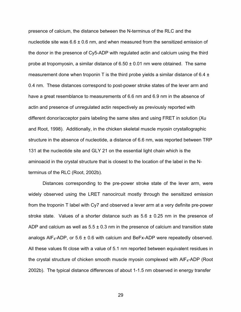
29
presence of calcium, the distance between the N-terminus of the RLC and the
nucleotide site was 6.6 ± 0.6 nm, and when measured from the sensitized emission of
the donor in the presence of Cy5-ADP with regulated actin and calcium using the third
probe at tropomyosin, a similar distance of 6.50 ± 0.01 nm were obtained. The same
measurement done when troponin T is the third probe yields a similar distance of 6.4 ±
0.4 nm. These distances correspond to post-power stroke states of the lever arm and
have a great resemblance to measurements of 6.6 nm and 6.9 nm in the absence of
actin and presence of unregulated actin respectively as previously reported with
different donor/acceptor pairs labeling the same sites and using FRET in solution (Xu
and Root, 1998). Additionally, in the chicken skeletal muscle myosin crystallographic
structure in the absence of nucleotide, a distance of 6.6 nm, was reported between TRP
131 at the nucleotide site and GLY 21 on the essential light chain which is the
aminoacid in the crystal structure that is closest to the location of the label in the N-
terminus of the RLC (Root, 2002b).
Distances corresponding to the pre-power stroke state of the lever arm, were
widely observed using the LRET nanocircuit mostly through the sensitized emission
from the troponin T label with Cy7 and observed a lever arm at a very definite pre-power
stroke state. Values of a shorter distance such as 5.6 ± 0.25 nm in the presence of
ADP and calcium as well as 5.5 ± 0.3 nm in the presence of calcium and transition state
analogs AlF4-ADP, or 5.6 ± 0.6 with calcium and BeFx-ADP were repeatedly observed.
All these values fit close with a value of 5.1 nm reported between equivalent residues in
the crystal structure of chicken smooth muscle myosin complexed with AlF4-ADP (Root
2002b). The typical distance differences of about 1-1.5 nm observed in energy transfer

30
Table 1: Fluorescent nanocircuit analysis of lever arm position: comparison of conformations of myosins bound to thin filaments proximate to TnT versus tropomyosin. Compares two distances a short (pre-power stroke) and a longer one (post-power stroke) calculated from the nanocircuit measurements in bold between S1 myosins bound proximate to the troponin complex or in between troponin complexes along tropomyosin.
Sample Nucleotide Wave- length (nm)
Exponentials in fit
2nd accept: TroponinT E R(nm)
2nd accept: Tropomyosin E R(nm)
Acto-S1+Ca2+ ADP 547 Single 0.22±0.07 6.4±0.4 0.19±0.00 6.50±0.01 Acto-S1+Ca2+ ADP 547 Multiplea 0.46±0.14 5.4±0.5 N/A N/A
Acto-S1+Ca2+ ADP 690 Single 0.19±0.1 6.6±0.6 --- --- Acto-S1+Ca2+ ADP 780 Single 0.40±0.05 5.6±0.2 0.27±0.03 6.02±0.17 Acto-S1+Ca2+ ADP-
BEFx 547 Single 0.16±0.11 6.8±0.7 0.21±0.00 6.37±0.02
Acto-S1+Ca2+ ADP-BEFx
547 Multiplea 0.61±0.2 4.8±0.6 N/A N/A
Acto-S1+Ca2+ ADP-AlF4 547 Single 0.13±0.05 7.1±0.4 0.15±0.02 6.79±0.15 Acto-S1+Ca2+ ADP-AlF4 547 Multiplea 0.73±0.2 4.2±0.6 N/A N/A Acto-S1+Ca2+ ADP-
BEFx 780 Single 0.38±0.17 5.6±0.6 0.25±0.04 6.13±0.19
Acto-S1+Ca2 ADP-AlF4 780 Single 0.41±0.09 5.5±0.3 0.23±0.06 6.24±0.34 Acto-S1+EGTA
ADP-BEFx
547 Single 0.12±0.07 7.1±0.6 0.24±0.01 6.20±0.04
Acto-S1+EGTA
ADP-BEFx
547 Multiplea 0.38±0.1 5.7±0.1 N/A N/A
Acto-S1+EGTA
ADP-AlF4 547 Single 0.12±0.04 7.2±0.6 0.17±0.01 6.62±0.08
Acto-S1+EGTA
ADP-AlF4 547 Multiplea 0.8±0.1 4.1±0.1 N/A N/A
Acto-S1+EGTA
ADP-BEFx
780 Single 0.37±0.2 5.7±0.7 0.30±0.07 5.89±0.34 Acto-S1+EGTA
ADP-AlF4 780 Single 0.41±0.16 5.5±0.6 0.23±0.02 6.24±0.12
aIndicates multiple exponential fits performed by constraining additional components to the fits to both the 547 and 690 decays, so that only the fastest decaying component was extracted. By multiple fitting, the unbound donors signal is discarded and the shorter, fastest component corresponds to the bound molecules only. No multiple fitting was required for the tropomyosin data.
measurements (between bent and extended lever arm, Fig. 10) are corroborated by the
chicken crystal structure differences of 6.6 nm – 5.1 nm = 1.5 nm and the scallop
muscle myosin crystal structure differences of 1.4 nm (Root 2002b).

31
Pre-power stroketransition state analogs
Post-power stroke
B
Fig. 10: Lever arm positions and calcium effect. Lever arm positions detected by nanocircuit illustrated using TnT labeled by Cy7 as second acceptor: bent lever arm or pre-power stroke and extended lever arm or post-power stroke. Also illustrated unbound molecules that will be detected by 547 nm emissions and not by 780 emissions, explaining the single and double exponential fitting of decays obtained at 547 nm. Extended versus bent S1 conformations estimated to be 1-1.5 nm in difference (A). Calcium induced thin filament structural changes detected by nanocircuit. RET between Cy5-ADP and Cy7-troponin T detects calcium induced changes between S1 and troponin T (B)
A

32
4.3 Calcium Induced Thin Filament Structural Changes Detected by Nanocircuit
Table 1 measurements indicate that the impact of calcium level is small on the
RLC to nucleotide site distance. When using the nanocircuit with troponin T as the
second acceptor, the measured separation distance of N terminus RLC to nucleotide
binding site is 5.6 ± 0.6 nm and 5.5 ± 0.3 nm in the presence of calcium and transition
state analogs beryllium as well as aluminum fluoride respectively, changing only to 5.7 ±
0.7 nm and 5.5 ± 0.6 nm (beryllium and aluminum fluoride respectively) upon addition of
calcium-chelator EGTA at 1 mM concentration. The emission spectra collected (Fig.
10B), however, indicates that calcium does have an impact, indicated by a pronounced
increase in the ratio of Cy7-troponin T sensitized emission relative to Cy5-ADP emission
upon the addition of 1mM EGTA in the presence of aluminum or beryllium fluoride. The
increased energy transfer observed between the Cy5-ADP and Cy7-troponin T sites
could be due to a conformational change or orientation changes between the two
dipoles when calcium is bound. Additionally, these results show that there are still
bound S1 molecules after the removal of calcium which may presumably be weak
binding interactions.
4.4 Post-Power Stroke Lever Arm Conformations Detected on Myosins Bound Close to Cy7-Tropomysin α and Far Away From the Troponin Complex. Pre-power Stroke Conformations Detected on Myosins Bound Close to Cy7-Troponin T
Table 1 summarizes the distances measured using the nanocircuit on the lever
arm positions of S1 molecules bound close to the troponin complex and bound along
and between troponin complexes. Measurements of the nanocircuit using as second
acceptor troponin T, indicate that the location of the binding of S1 relative to troponin T

33
influences the position of the lever arm. Depending on the proximity of S1 to troponinT,
the sensitized emissions of Cy5-ADP and Cy7-TnT should differ. Table 1 shows that
the calculated distance from the RLC to Cy5-ADP is 6.4 nm ± 0.4 nm but when this
distance is measured through the Cy7-TnT sensitized decay under identical conditions,
the distance drops to 5.6 ± 0.2 nm. These measurements suggest that when S1 binds
near troponin T, the lever arm orients preferentially towards a pre-power stroke
conformation, presumably due to steric hindrances; rather than in the more typical case,
when it binds further away from troponin.
Measurements of the nanocircuit using as second acceptor tropomyosin labeled
at the single Cys 190 of tropomyosin α with CY7, indicate a lever arm rotation of S1
molecules bound near tropomyosin and in between troponin complexes of distances
that correspond to predominantly post-power stroke conformations in support to the
present hypothesized model. Post-power stroke conformations are observed
regardless of the presence of transition state analogues in S1’s bound along and in
between troponin complexes.

34
Fig. 11: Luminescence decay curves: Decay curves following excitation of CS124 in RLC with Last Acceptor Cy7-TnT. Measured in a luminescence spectrometer after excitation at 337 nm and estimated by single exponential curve fitting at 547, 690 and 780 nm. Lifetime of the donor changes as read at different emissions to isolate specific conformations of S1 lever arms along the thin filament. At emission 547 nm unbound and bound S1’s detected. At emission 780 nm, only S1’s bound close to troponin T are isolated.
Tb-RLC-S1 only Tb-RLC-S1+Cy5-ADP at 547 Tb-RLC-S1+Cy5-ADP at 690 Tb-RLC-S1+Cy5-ADP at 780
y
0 0.001 0.002 0.003 0.004 0.005
-0.5
0
0.5
1
1.5
2
2.5
3
3.5
4
4.5
5
5.5
Nor
mal
ized
lum
ines
cenc
e
Time (S)

35
CHAPTER 5
DISCUSSION
5.1 Steric Hindrance on Lever Arm Rotation Model: Myosins Bound Proximally to the Troponin Complex have a Lever Arm Biased towards Pre-Power Stroke State whereas Myosins Bound in between the Troponin Complex have Lever Arms in Post-Power Stroke States
The distances observed using the nanocircuit on the lever arm positions of S1
molecules bound close to the troponin complex indicate that the location of the binding
of S1 relative to troponin T influences the position of the lever arm. This pre-power
stroke positioning has been observed not only in the presence of the transition state
analogs but in the presence of Cy5ADP alone. S1 binds near troponin T with lever arm
positions preferentially towards a pre-power stroke conformation than when it binds
further away from troponin, in between troponin complexes and near tropomyosin in
which case the lever arm of S1 molecules situate preferentially at post-power stroke
conformations. Measurements from the nanocircuit corroborate the data calculations
done by triangulation measurements using FRET and computational docking that yield
an atomic model of S1 bound to one side of the troponin complex with less steric
hindrance when the lever arm is in a pre-power stroke orientation (Patel, et al., 2005).
Fig. 12 illustrates in schematic form this concept that based on the present study and
the docking experiments, it is suggested that preferential different lever arm
conformations are presumably due to steric hindrances.
The molecular docking experiments using LRET measurements were performed
with various constrains from different sites of the myosin head to the same avian breast
troponin T isoform as the one used in the present study (Patel, et al., 2005). The
troponin T isoform, has calcium binding properties that were discovered during its

36
characterization and a single cysteine (used for the attachment of probes) situated very
close to the interface between the T, I and C subunits (Root and Jin, 2000). The
nanocircuit results indicate the first of a two way effect that occurs during actomyosin
interaction. First, the troponin T position in the thin filament affects the binding of S1 to
a preferentially pre-power stroke state. Secondly, the triangulation measurements
indicate that troponin influences the ability of S1 molecules to bind to specific locations
along the thin filament because these RET measurements situate the troponin complex
near loop 3 in the nearest myosin S1 bound to actin, therefore, modeling based on
these measurements suggest myosin binds to one side of the troponin complex .
Furthermore, molecular modeling docking indicates that the myosin must be in the pre-
power stroke state to bind this close to troponin on actin (Patel, et al., 2005 and Fig. 12).
Models of the actomyosin interface have been built using the x-ray structures of
individual proteins and electron micrographs of actin filaments decorated by S1
(Rayment et al., 1993b, Schroeder et al., 1993), dynamic docking of actomyosin has
also been observed with resonance energy transfer (Root et al, 2002), as well as
computational docking using molecular mechanics (Root, 2002a) to further define the
important residues responsible for the acto-S1 interaction. Loop 3 of S1 has been
defined as a secondary actin binding site of myosin, and has been shown in skeletal
striated muscle myosins, in particular, to cross-link and therefore interact with actin via
loop 3 (Van Dijk, 1999). The results correspond to all the above mentioned models of
the actomyosin interface, and taking them all into consideration, the troponin label is
predicted to be in very close proximity to loop 3 (Patel et al., 2005).

37
Fig. 12: Hindrance on lever arm rotation model. Myosins bound proximally to the troponin complex have a lever arm biased towards pre-power stroke state. Scheme based on the atomic model of troponin docking to actoS1 with less steric hindrance when the lever arm of myosin is in the pre-power stroke position.
5.2 What Value Does a Prevention of a Power Stroke near a Troponin Complex
Have?
Interactions with or near loop 3 of myosin S1 with troponin T hint to the
importance of this region for the regulation of the S1-actin contacts in skeletal muscle
and cardiac systems, both of which are regulated via the troponin-tropomyosin system.
A peptide antibody against the loop 3 region of skeletal muscle has been shown to
inhibit the sliding velocity of actin filaments on in vitro motility assays (Blotnick et al.,
Cy5ADP
Terbium chelate: DTPA-cs124-RLC
Pulsed nitrogen laser
Tn T
C
N O CO-
2
N
N
CO-2
CO-
CO
N
NO
CH
Tb3+

38
1995). Only the motor domains containing the loop 3 of skeletal muscle myosin have
been shown to cross-link and therefore interact with actin, this interaction was not
observed in smooth muscle or recombinant constructs of the slime mold Dictyostelium
myosin II motor (Van Dijk et al, 1999). Additionally, only recombinant chimeric
constructs of Dictyostelium myosin II motor domain where the loop 3 region had been
substituted by the one of skeletal myosin showed significant actin cross-linking (Van
Dijk et al, 1999). The lever arm region of S1 confers great flexibility to myosin and is
primarily responsible for the changes in the sarcomere stretch that produces the sliding
of thick and thin filaments. The S1 lever arm attaches to the thick filament backbone via
subfragment-2 (S2); both regions are the heavy meromyosin product of trypsin digest.
Although not as flexible as the lever arm, a second region of flexibility has been
observed at the hinge connecting the S2 region and the light meromyosin
(Gundapaneni et al, 2005). Furthermore, regulatory components spaced every 7 actin
monomers in the form of the troponin complex may guide the binding positions of S1
molecules in the vicinity. The troponin complex preventing close S1 molecules from
undergoing a power stroke could confer a limit to the sliding of thick filaments allowing
enough flexibility (performed by the majority of S1 molecules that undergo a full power
stroke) but ensuring the preservation of the elastic component to stop and recoil back.
The loop 3 region of the actin-S1 interface, of close contact with troponin T, has been
shown to have a modulating effect in the interaction with actin. The troponin complex
may contribute to the modulating effect of nearby S1 molecules by inhibiting their power
stroke and sequestering the lever arms in pre-power stroke positions. Experiments
cross-linking of S1 to actin done in the presence of Mg.ADP.ALF4- have shown that the
39
area of loop 3 involved in the actomyosin interface is smaller than in the absence of
nucleotide (Martin et al., 2006), this observation corroborates why the troponin complex
can fit better closer to S1 molecules that have a pre-power stroke positioned lever arm
in the actomyosin interphase. A few periodically situated S1 molecules with lever arms
unable to undergo a complete power stroke may create a limit or stop mechanism for
further shortening. This mechanism can aid in the function of the giant protein titin that
spans as a molecular scaffold of the sarcomere further serving as a molecular spring.
Titin extends from the Z-line of the sarcomere to the M-band. Titin, the largest single
polypeptide found in nature, provides binding sites for numerous proteins thus its
importance for the assembly of the sarcomere. Several proteins important for the
stability of the sarcomeric structure are found in the Z-line as well as in the M-band of
the sarcomere. In the Z-line, titin binds to the thin filament system via the protein alpha-
actinin. Therefore, both titin and the troponin complex components in the muscle fibers
may cooperatively influence the recoiling after extension (due to contraction) with
troponin behaving as an elastic limit while titin provides passive tension for optimal
interaction of myofilaments and sarcomere length. Regulation of sarcomere length has
been shown to be important for isometric tension, too long and too short lengths
generate low force whereas intermediate lengths are optimal for tension and force
generation in muscle. Experiments conducted in 1966 defined well this relationship
based on the optimal overlapping of myosin and actin filaments to generate tension. If a
sarcomere of length 3.65 μm (myosin filaments of 1.65 μm and actin filaments of 2 μm
in length) is highly stretched to its limit, there would be no overlap between
interdigitating actin and myosin filaments. Additionally, it has been observed that
40
sarcomere shortening below 2 μm results in great filament overlap but no tension
generation, at this point, the bare zone of the myosin backbones devoided of S1
crossbridges does not allow cross-bridge connections to be made (Gordon et al.,
1966a; 1966b). Troponin T as well as the other subunits of the troponin complex are
important and may also have effects in tension development. Experiments of partial
extractions of the Ca2+ binding troponin C subunit in mammalian skinned muscle fibers,
from which the sarcolemma has been removed, have evidenced the molecular
cooperativity that exists between the other thin filament regulatory proteins. These
experiments have shown that partial extraction of troponin C still results in the
development of Ca2+ insensitive active tension evidencing the cooperativity of the other
functional subunits of the thin filament (Moss et al., 1986; Metzger and Moss, 1991). In
a similar manner, it would be expected that partial extraction of troponin T in muscle
fibers, would produce less uniform sarcomere lengths with shortening in support of the
role of troponin T in influencing further shortening of the sarcomere by affecting the
power stroke of nearby S1 bound cross-bridges.
5.3 Transition State Analogs Aluminum and Beryllium Fluoride behave similarly causing Pre-Power Stroke Conformations when Observed with Troponin T and Post-Power Stroke Conformations when Observed from Tropomyosin
Transition state analogs aluminum and beryllium fluoride were used in order to
characterize and to isolate weak binding states or ADP-Pi of the ATPase cycle. As
observed in previous studies, assigning a particular kinetic state to any of these analogs
has proven difficult due to their properties being dependent on the assay conditions and
the presence of other proteins in the

41
sarcomere. In this present case, the results have been very consistent. When used in
the nanocircuit in solution and in conjunction with the labeled troponin T, they allowed
the identification of pre- power stroke conformations and no difference was observed in
the effects of both metallofluorides, these results are in concordance with the
crystallographic results obtained with both analogs with smooth myosin S1 where no
difference was observed in the use of either of them (Dominguez et al., 1998). The use
of transition state analogs in the tropomyosin labeled assay was not able to induce pre-
power stroke conformations even after the reduction of calcium in bound molecules,
reaffirming the selection of post-power stroke structures of most S1 molecules bound
along tropomyosin. Not being able to force a pre-powerstroke conformation is not
unusual since it has been observed that the presence of other contractile proteins may
affect the state in which the lever arm is isolated using the analogs (Phan and Reisler,
1993). The present results show the greater number of S1 molecules that can undergo
their complete power stroke while few S1 molecules situated with their loop 3 region
close to troponin staying in the short lived pre-power stroke state.
5.4 Cooperativity of Activation
Calcium regulates contraction in striated muscle. As already mentioned, the thin
filament is composed of actin arranged helically with regulatory proteins tropomyosin
binding as a dimer to 7 actin monomers in the thin filament helix and the proteins of the
troponin complex binding every 7 actin monomers as well. In the absence of calcium,
troponin I binds to actin and to tropomyosin via troponin T. Troponin I also interacts with
troponin C, the subunit that upon binding with Ca2+ causes the detachment of TnI to

42
actin allowing tropomyosin to move over the surface of the thin filament to expose weak
and strong myosin binding sites and allowing the interaction of myosin S1 heads to
actin. This interaction, allows the power stroke to occur causing contraction when thin
filaments slide over thick filaments. This model of steric regulation has been observed
consistently with electron micrographs and cryoelectron microscopy and helical image
reconstruction of F-actin-troponin-tropomyosin complexes in the low Ca2+ and high Ca2+
states (Holmes, 1995; Xu, 1999). The nanocircuit results indicate that the prevention of
a complete power stroke near the periodically occurring troponin complex still allows the
recruiting of other S1 molecules for the subsequent cooperative activation of the thin
filament, as complete power strokes have been extracted at wavelength 690 nm
corresponding to S1 molecules bound but not necessarily close to the troponin complex.
These results corroborate further studies done in skinned skeletal muscle fibers that
had shown that complete activation of the thin filament by bound myosin cross-bridges
requires not only the presence of calcium but also strong-binding myosin cross-bridges
to actin in a cooperatively increasing manner (Lehrer, 1994; Gordon et al., 2000)
5.5 Calcium Induced Thin Filament Structural Changes Detected by Nanocircuit.
The impact of calcium level in the lever arm position of the RLC to nucleotide site
distance has been relatively small, in the presence of analogs and even upon addition
of calcium-chelator EGTA at 1 mM concentration. The emission spectra collected (Fig.
10B), however, indicates that calcium does have an impact, indicated by a pronounced
increase in the ratio of Cy7-troponin T sensitized emission relative to Cy5-ADP emission
upon the addition of 1mM EGTA in the presence of aluminum or beryllium fluoride.

43
Increased energy transfer between the nucleotide site and troponin T in response to
calcium provides additional information about the dynamics of the thin filaments. More
may be observed in actual myofibrils where all thin filament proteins are present.
Additionally, Figure 10B illustrates the displacement of the troponin complex relative to
myosin reported by the changes in the sensitized emission in response to calcium
quenching. A likely explanation of the increased energy transfer observed between the
Cy5-ADP and Cy7-troponin T sites could be due to orientation changes between the
two dipoles when calcium is bound. Aluminum fluoride has been shown in the past to
increase the α-helicity and tyrosine fluorescence of Ca2+ binding troponin C (Phan and
Reisler, 1993); the avian troponin T labeled with Cy7, naturally binds calcium and may
be affected some by the phosphate analogs without necessarily affecting distances
between the fluorophores.
5.6 Other Possible Applications of the Fluorescent Nanocircuit
The fluorescent nanocircuit can be applied to different systems to study biological
questions involving different molecular ternary complexes. So far, triple FRET
cascades have been used in synthetic dendrimers, single stranded DNA and RNA
polymerase/DNA complexes using organic fluorophores (Serin et al., 2002; Tong et al.,
2001; Lee et al., 2006). However, the use of a three FRET relay system combined with
lanthanides for greater precision, can be extended to the study of the interactions of
triple stranded DNA, protein folding and to answer more complex interactions such as
dynamic structural changes in solution involving proteins and protein/DNA complexes.
Most significantly, the exploitation of the great time-resolution provided by the mixture of

44
slow lanthanide decays (ms) with fast conventional decays (ns) enables the
construction of nanocircuits that can report structural changes in the rate-limiting (slow
decaying) steps of the relay only in the presence of the terminal acceptor. Even in the
presence of a small fraction of lanthanide donor and/or first acceptor (CY5) relative to
the terminal acceptor (CY7), it is possible to precisely quantify the energy transfer in this
first step (lanthanide to CY5) only when the terminal acceptor (CY7) is in close
proximity. This degree of discrimination is has only been reported with the time-resolved
probes as used in the unique fluorescent nanocircuit described herein.
5.7 Conclusions
The goals of a newly developed spectroscopic nanocircuit were successfully
accomplished. Not only did it allow the identification of different conformations of the
lever arm by signal averaging from a large number of molecules, but it also permitted
exploiting the signal of the last acceptor of the nanocircuit relay to identify the most
critical conformations to a given transition kinetic step using phosphate analogs and in
proximity to two different regulatory proteins of the thin filament: troponin and
tropomyosin. The skeletal myosin lever arm exists in various orientations prior to the
association of myosin to actin. Upon initial binding events with actin, pre-power stroke
orientations are selected on S1 molecules bound close to the troponin complex the
same ones that cannot complete their power stroke. S1 molecules bound along
tropomyosin and in between troponin complexes on the other hand, are driven towards
post-power stroke conformations upon phosphate release.

45
REFERENCES Bauer, C. B., Holden, H. M., Thoden, J. B., Smith, R., and Rayment, I. (2000). X-ray
structures of the Apo and Mg·ATP-bound states of Dictyostelium discoideum myosin motor domain. Journal of Biological Chemistry, 275, 38494-38499
Bigay, J., Deterre, P., Pfister, C., and Chabre, M. (1987). Fluoride complexes of aluminum or beryllium act on G-proteins as reversible bound analogues of the gamma phosphate of GTP. EMBO Journal, 6, 2907-2913
Blotnick, E., Miller, C., Groschel-Stewart, U., and Muhlrad, A. (1995). Immunochemical probing of the functional role of the 238-246 and 567-574 sequences of myosin heavy chain. European Journal of Biochemistry, 232, 235-240
Chabre, M. (1990). Aluminofluoride and beryllofluoride complexes: new phosphate analogs in enzymology. Trends in Biochemical Sciences, 15, 6-10
Colowick, S. and Kaplan, N. (1982). Structural and contractile proteins. The contractile apparatus and the cytoskeleton. Methods in Enzymology, 85, 234-259
Cooke, R. (1986). The mechanisms of muscle contraction. Critical Reviews in Biochemistry, 21, 53-118
Dominguez, R., Freyzon, Y., Tybus, K.M., and Cohen, C. (1998). Crystal structure of a vertebrate smooth muscle myosin motor domain and its complex with the essential light chain: visualization of the pre–power stroke state. Cell, 94, 559-571
Ebashi, S. and Endo, M. (1968). Calcium ion and muscle contraction. Progress in Biophysics and Molecular Biology, 18, 123–183
Ebashi, S., Endo, M. and Otsuki, I. (1969). Control of muscle contraction. Quarterly Review of Biophysics, 2, 351–384
Fisher, A. J., Smith, C. A., Thoden, J. B., Smith, R., Sutoh, K., Holden, H. M. and Rayment, I. (1995). X-ray structures of the myosin motor domain of Dictyostelium discoideum complexed with MgADPBeFx and MgADPAlF4. Biochemistry, 34, 8960-8972
Getz, Elise Burmeister, Cooke, Roger and Selvin Paul R. (1998). Luminescence resonance energy transfer measurements in myosin. Biophysical Journal, 74, 2451-2458
Gordon, A.M., Huxley, A.F., and Julian, F.J. (1966). Tension development in highly stretched vertebrate muscle fibres. Journal of Physiology, 184, 143-169

46
Gordon, A.M., Huxley, A.F., and Julian, F.J. (1966). The variation in isometric tension with sarcomere length in vertebrate muscle fibres. Journal of Physiology, 184, 170-192
Gordon, A.M., Homsher, E., and Regnier, M. (2000). Regulation of contraction in striated muscle. Physiological Reviews, 80, 853-924
Gulick, A. M., Bauer, C. B., Thoden, J. B., Pate, E., Yount, R. G., and Rayment, I. (2000). X- ray structures of the Dictyostelium discoideum myosin motor domain with six non-nucleotide analogs. Journal of Biological Chemistry, 275, 398-408
Gundapaneni, D., Xu, J., and Root, D.D. (2004). High flexibility of the actomyosin crossbridge resides in the skeletal myosin subfragment-2 as demonstrated by a new single molecule assay. Journal of Structural Biology, 149, 117-126
Holmes, K.C. (1995). The actomyosin interaction and its control by tropomyosin. Biophysical Journal, 68, 2-7
Holmes, K. C. (1997). The swinging lever-arm hypothesis of muscle contraction. Current Biology, 7, 112-118
Houdusse, A., Kalabokis, V.N., Himmel, D., Szent-Györgyi, A.G., and Cohen, C. (1999). Atomic structure of scallop myosin subfragment S1 complexed with MgADP: A novel conformation of the myosin head. Cell, 97, 459-470
Houdusse, A., Szent-Györgyi, A.G., and Cohen, C. (2000). Three conformational states of scallop myosin S1. Proceedings of the National Academy of Sciences USA, 97, 11238-11243
Jin, J.P., and Root, D.D. (2000). Modulation of troponin T molecular conformation and flexibility by metal ion binding to the NH2-terminal variable region. Biochemistry, 39, 11702-11713
Kraft, T., Mählmann, E., Mattei, T., and Brenner B. (2005). Initiation of the power stroke in muscle: Insights from the phosphate analog AlF4. Proceedings of the National Academy of Sciences USA, 102, 13861–13866
Lee, N.K., Kapanidis, A.N., Koh, H.R., Korlann, Y., Ho, S.O., Kim, Y., Gassman, N., Kim, S.K., Weiss, S. (2006). Three-color alternating-laser excitation of single molecules: Monitoring multiple interactions and distances. Biophysical Journal, Oct 13; (E-pub ahead of print)
Lehrer, S.S. (1994). The regulatory switch of the muscle thin filament: Ca2+ or myosin heads. Journal of Muscle Research and Cell Motility, 15, 232-236
Martin, B., Karczewska, E., and Plitszka, B. (2006). Effect of nucleotide on interaction of the 567-578 segment of myosin heavy chain with actin. Biochimica et Biophysica Acta, 1764, 217-222

47
Maruta, S., Henry, G.D., Sykes, B.D., and Ikebe, M. (1993). Formation of the stable myosin-ADP-aluminum fluoride and myosin-ADP-beryllium fluoride complexes and their analysis using 19F NMR. Journal of Biological Chemistry, 268, 7093-7100
Metzger, J.M., and Moss, R.L. (1991). Kinetics of a Ca2+-sensitive cross-bridge state transition in skeletal muscle fibers. Journal of General Physiology, 98, 233-248
Moss, R.L., Allen, J.D., and Greaser, M.L. (1986). Effects of partial extraction of troponin complex upon the tension-pCa relation in rabbit skeletal muscle. Journal of General Physiology, 87, 761-774
Nikolaeva O.P, Dedova I.V, Khvorova I.S, and Levitsky D.I. (1994). Interaction of F-actin with phosphate analogues studied by differential scanning calorimetry. FEBS Letters, 351, 15-18.
Patel, D., Coffee, P., and Root, D.D. (2005). The structural relationship of muscle myosin to thin filament regulatory proteins. Gordon Summer Research Conference Abstract
Perry, S. (2001). Vertebrate tropomyosin: Distribution, properties and function. Journal of Muscle Research and Cell Motility, 22, 5-49
Peyser, Y.M., Ajtai, K, Burghardt, T.P. and Muhlrad, A. (2001). Effect of ionic strength on the conformation of myosin subfragment 1-nucleotide complexes. Biophysical Journal, 81, 1101–1114
Phan, B., and Reisler, E. (1992). Inhibition of myosin ATPase by beryllium fluoride. Biochemistry, 31, 4787-4793
Phan, B., and Reisler, E. (1993). Aluminum fluoride interactions with troponin C. Biophysical Journal, 65, 2511-2516
Rayment, I; Rypniewski, W.R; Schmidt-Bäse, K., Smith, R., Tomchick, D. R., Benning, M. M., Winkelmann, D. A., Wesenberg, G., and Holden, H. M. (1993a). Three-dimensional structure of myosin subfragment-1: A molecular motor. Science, 261, 50-58
Rayment, I., Holden H.M., Whittaker, M., Yohn, C.B., Lorenz, M., Holmes, K.C., and Milligan, R.A. (1993b). Structure of the actin-myosin complex and its implications for muscle contraction. Science, 261, 58-65
Root, D.D. (2002a). A computational comparison of the atomic models of the actomyosin interface. Cell Biochemistry and Biophysics, 37, 97-110
Root, D.D. (2002b). The dance of actin and myosin. A structural perspective. Cell Biochemistry and Biophysics, 37, 111-139

48
Schroeder, R.R, Manstein, D.J., Jahn, W., Holden, H., Rayment, I., Holmes K.C., and Spudich J.A. (1993). Three-dimensional atomic model of F-actin decorated with Dictyostelium myosin S1. Nature, 364, 171-174
Serin, J.M., Brousmiche, J.M., and Frechet, J. (2002). Cascade energy transfer in a conformationally mobile multichromophoric dendrimer. Chemical Communications, 22, 2605-2607
Smith, C.A. and Rayment, I. (1996). X-ray structure of the magnesium(II).ADP.vanadate complex of the Dictyostelium discoideum myosin motor domain to 1.9 Å resolution. Biochemistry, 35, 5404-5417
Smyczynski, C. and Kasprzak, A. A. (1997). Effect on nucleotides and actin on the orientation of the light chain-binding domain in myosin subfragment 1. Biochemistry, 36(43), 13201-13207
Sprang, SR. (1997). G protein mechanisms: insights from structural analysis. Annual Review of Biochemistry 66, 639-678
Sternweis, P.C., and Gilman, A.G. (1982). Aluminum: A requirement for activation of the regulatory component of adenylate cyclase by fluoride. Proceedings of the National Academy of Sciences USA, 79, 4888–4891
Takeda, S., Yamashita, A., Maeda, K., and Maéda Y. (2003). Structure of the core domain of human cardiac troponin in the Ca 2+ saturated form. Nature 424, 35–41
Tong, A.K., Jockusch, S., Li, Z., Zhu, H.-R., Akins, D.L., Turro, N.J., and Ju, J. (2001). Triple fluorescence energy transfer in covalently trichromophore-labeled DNA. Journal of the American Chemical Society, 123, 12923-12924
Van Dijk, J., Furch, M., LaFont, C., Manstein, D., and Chaussepied, P. (1999). Functional characterization of the secondary actin binding site of myosin II. Biochemistry, 38, 15078-15085
Vinogradova, M., Stone, D., Malanina, G., Karatzaferi , C., Cooke , R., Mendelson, R. and Fletterick, R. (2005). Ca2+-regulated structural changes in troponin. Proceedings of the National Academy of Sciences USA, 102, 5038–5043
Werber, M.M., Peyser, Y.M., and Muhlrad, A. (1992). Characterization of stable beryllium fluoride, aluminum fluoride, and vanadate containing myosin subfragment 1-nucleotide complexes. Biochemistry, 31, 7190-7197
Xu, C., Craig, R., Tobacman, L., Horowitz, R., and Lehman, W. (1999). Tropomyosin positions in regulated thin filaments revealed by cryoelectron microscopy. Biophysical Journal, 77, 985-992

49
Xu, J and Root, D.D. (1998). Domain motion between the regulatory light chain and the nucleotide site in skeletal myosin. Journal of Structural Biology 123, 150-161
Xu, J and Root, D.D. (2000). Conformational selection during weak binding at the actin and myosin interface. Biophysical Journal, 79, 1498-1510
Zhang, Z., Jin, J.P., Root, D.D. (2004). Binding of calcium ions to an avian flight muscle troponin T. Biochemistry, 43, 2645-2655.