histone deacetylase 3 is required for efficient t cell
TRANSCRIPT

Histone Deacetylase 3 Is Required for Efficient T Cell Development
Kristy R. Stengel,a Yue Zhao,a Nicholas J. Klus,b Jonathan F. Kaiser,a Laura E. Gordy,c Sebastian Joyce,c,e Scott W. Hiebert,a,d
Alyssa R. Summersb
Department of Biochemistry, Vanderbilt University School of Medicine, Nashville, Tennessee, USAa; Department of Biology, The University of the South: Sewanee,Sewanee, Tennessee, USAb; Department of Pathology, Microbiology and Immunology, Vanderbilt University School of Medicine, Nashville, Tennessee, USAc; Vanderbilt-Ingram Cancer Center, Vanderbilt University School of Medicine, Nashville, Tennessee, USAd; Veterans Administration Tennessee Valley Healthcare System, Nashville,Tennessee, USAe
Hdac3 is a key target for Hdac inhibitors that are efficacious in cutaneous T cell lymphoma. Moreover, the regulation of chroma-tin structure is critical as thymocytes transition from an immature cell with open chromatin to a mature T cell with tightly con-densed chromatin. To define the phenotypes controlled by Hdac3 during T cell development, we conditionally deleted Hdac3using the Lck-Cre transgene. This strategy inactivated Hdac3 in the double-negative stages of thymocyte development and causeda significant impairment at the CD8 immature single-positive (ISP) stage and the CD4/CD8 double-positive stage, with few ma-ture CD4� or CD8� single-positive cells being produced. When Hdac3�/� mice were crossed with Bcl-xL-, Bcl2-, or TCR�-ex-pressing transgenic mice, a modest level of complementation was found. However, when the null mice were crossed with miceexpressing a fully rearranged T cell receptor �� transgene, normal levels of CD4 single-positive cells were produced. Thus,Hdac3 is required for the efficient transit from double-negative stage 4 through positive selection.
Histone deacetylases (HDACs) are a heterogeneous group of 18mammalian enzymes that remove acyl groups from the side
chain of lysine (1). The class I enzymes (HDAC1, -2, -3, and -8) arehomologous to the Saccharomyces cerevisiae yeast Rpd3 and targetmultiple lysine residues in the N-terminal tails of histone H3 andH4 as well as deacetylate nonhistone targets. These enzymes act inconcert with a corepressor (mSin3A or mSin3B for HDAC1 andHDAC2, NCOR1 or NCOR2 for HDAC3) that stimulates theiractivity (2–5). Inositol phosphates are key cofactors of HDACs 1to 3 that bridge the deacetylase and the corepressor to allow reg-ulation by inositol phosphate signaling and sensing of the meta-bolic state of the cell (6, 7). Several of the class II HDACs are poordeacetylases on histone substrates, but HDAC4, HDAC5, andHDAC7 can recruit HDAC3 to deacetylate targets (8–10). Theclass III enzymes are sirtuins 1 to 7 and are homologues of theyeast Sir2. These are NAD�-dependent enzymes that sensechanges in NAD levels to respond to changes in cellular metabo-lism (11–13). HDAC11 is the only member of the 4th class ofdeacetylase and modulates immunity (14, 15).
These enzymes have been the target of wide-ranging searchesfor small molecules that can rewrite the epigenetic code for use innumerous diseases, including neurological diseases, inflamma-tory disorders, and cancer (1). Broad-spectrum HDAC inhibitors(vorinostat and rhomidepsin) have gained FDA approval for theireffectiveness against cutaneous T cell lymphoma (16). These smallmolecules inhibit multiple class 1 and/or class 2 HDACs but haveserious side effects, which has stimulated the development ofmore selective inhibitors (17). A key to the development of betterdrugs is the identification of the HDAC(s) that mediates the actionof the approved compounds as well as the HDAC(s) responsiblefor the side effects.
Genetic approaches are being applied to dissect the physiolog-ical roles of individual HDACs in tissues that are affected by thesedrugs and are changing the way that we view individual HDACs.For example, while Hdac1 and Hdac2 can heterodimerize andsubstitute for one another, deletion of Hdac1 was embryonic le-thal at embryonic day 9.5, whereas Hdac2�/� mice showed cardiac
defects shortly after birth (18–20). Heart-specific double deletionof Hdac1/2 caused more dramatic cardiac phenotypes, includingarrhythmia and severe ventricular dilation (18). Hematopoiesis isanother key target tissue, as HDAC inhibitors not only act in T celllymphoma but also show promise in myeloid leukemia and B celllymphoma. While germ line deletion of Hdac3 caused early em-bryonic lethality (21, 22), hematopoietic stem cell deletion ofHdac3 caused a dramatic loss of B cells and T cells and defects instem cell self-renewal. The stem cell defect appeared to be due todefects in DNA replication, while the loss of lymphopoiesis wastraced to a loss of the lymphoid-primed multipotent progenitorcells (LMPPs) (23). In contrast, double deletion of Hdac1 andHdac2 in hematopoietic stem cells caused megakaryocyte apopto-sis and thrombocytopenia, which are also observed in patientstreated with HDAC inhibitors (19, 20, 24).
Developing T cells are an ideal model system in which to dissectthe roles of individual HDACs in normal physiology. Stepwiseremoval of the 4 alleles of Hdac1 and Hdac2 in thymocytes yieldeda gradient of Hdac activity, and removal of all four alleles caused ablock in early thymic development at double-negative stage 3(DN3) (25, 26). However, as the amount of Hdac1/2 activity de-creased, the mice developed T cell lymphomas, with a particularlyhigh incidence being noted in Hdac1�/�/Hdac2�/� mice. In contrast,the double-knockout mice did not develop tumors, indicating that alow level of Hdac1/2 activity is required for tumorigenesis. In addi-tion, the tumors that developed in Hdac1�/�/Hdac2�/� mice re-
Received 16 July 2015 Accepted 19 August 2015
Accepted manuscript posted online 31 August 2015
Citation Stengel KR, Zhao Y, Klus NJ, Kaiser JF, Gordy LE, Joyce S, Hiebert SW,Summers AR. 2015. Histone deacetylase 3 is required for efficient T celldevelopment. Mol Cell Biol 35:3854 –3865. doi:10.1128/MCB.00706-15.
Address correspondence to Scott W. Hiebert, [email protected], orAlyssa R. Summers, [email protected].
Copyright © 2015, American Society for Microbiology. All Rights Reserved.
3854 mcb.asm.org November 2015 Volume 35 Number 22Molecular and Cellular Biology

mained sensitive to Hdac inhibitors, providing a genetic rationalefor the use of these compounds in a synthetic lethal strategy (25,26).
Here, we have examined the physiological roles of Hdac3 bydeletion of this gene during early thymocyte development usingLck-Cre. Analysis of the developmental markers CD4 and CD8showed impaired maturation of double-negative cells, an increasein immature single-positive (ISP) CD8� cells, and impaired mat-uration of double-positive (DP) cells, causing a dramatic decreasein single-positive (SP) CD4� and CD8� cells. Gene expressionanalysis pinpointed T cell receptor (TCR) signaling to be a possi-ble mechanism and transgenic mouse expression of a combinedTCR�� transgene to provide a high level of complementation ofthymocyte development.
MATERIALS AND METHODSMouse strains. Mice were maintained following Vanderbilt UniversityMedical Center guidelines and in accordance with an Institutional AnimalCare and Use Committee-approved protocol. All mice analyzed were be-tween 3 and 6 weeks of age unless otherwise noted. All experimentalstrains were in a C57BL/6 mouse genetic background. ROSA26-Lox-Stop-Lox-GFP, p56lck proximal promoter Cre (Lck-Cre), OT-II, TCR�, Bcl2,and Bcl-xL mice were obtained from The Jackson Laboratory, while con-ditional Hdac3-knockout mice were generated in the lab of S. W. Hiebert(21).
Flow cytometry. Single-cell suspensions were formed from thymustissue that had been passed through a 70-�m-pore-size filter with ice-coldphosphate-buffered saline (PBS). Red blood cells were subsequently lysedwith erythrocyte lysis buffer (buffer EL; Qiagen). Cells were then stainedwith fluorescence-labeled antibodies for 30 min in PBS containing 0.5%fetal bovine serum (FBS) at 4°C. All thymocytes were first gated throughgreen fluorescent protein (GFP) to select for cells expressing Cre and thenevaluated using lineage markers, as indicated below. Fluorochrome-con-jugated antibodies to the following were used: from eBioscience, antibod-ies to CD4 (RM4-5 antibody), CD8 (53-6.7 antibody), CD44 (IM7 anti-body), CD25 (PC61.5 antibody), TCR� (H57-597 antibody), CD147(RL73 antibody), CD5 (53-7.3 antibody), and CD69 (H1.2F3 antibody),and from BD Biosciences, antibody to CD3 (145-2C11 antibody). Analy-sis was performed with a 3-laser BD LSRII or BD Fortessa flow cytometer.The acquired data were analyzed using FlowJo software (TreeStar).
Microarray and real-time quantitative PCR. Thymocyte single-cellsuspensions were sorted with a BD FACSAria cell sorter, with the firstgating being on GFP-positive cells and then anti-CD4 and anti-CD8markers being used to identify double-positive cells. Total RNA was pu-rified from the sorted thymocytes with a PerfectPure RNA extraction kit(5 Prime, Inc., Gaithersburg, MD). RNA was pooled from 2 wild-type(WT) and 2 Hdac3�/� animals for a total of 3 biological replicates from 6mice each. RNA was prepared and hybridized to an Affymetrix GeneChipmouse gene 1.0 ST array for analysis in the Vanderbilt Functional Genom-ics Shared Resource. Data were analyzed using GeneSpring (Agilent Tech-nologies) and Panther classification system software. Gene ontology anal-yses of the microarray data were performed using the web-based tool kitWebGeStalt. P values were calculated using a hypergeometric test (raw Pvalues) and adjusted by multiple testing (adjusted P values). Enrichedcategories identified using different databases are presented in the figures.Quantitative reverse transcription-PCR (qRT-PCR) was performed usingSybr green and real-time PCR.
Western blot analysis. Where noted, thymocytes were sorted by GFPstatus prior to lysis in radioimmunoprecipitation assay (RIPA) buffercontaining protease inhibitors. Cleared lysates were resolved by SDS-PAGE. Specific proteins were detected with the antibodies to the follow-ing: from Cell Signaling Technology, phosphorylated extracellular signal-regulated kinases (ERKs) 1 and 2 with a Thr residue at position 202 andTyr residue at position 204, extracellular signal-regulated kinases 1 and 2,
histone H3, and histone H4; from Abcam, Hdac3, histone H3 with atrimethylated lysine 9 (H3K9me3), histone 4 with an acetylated lysine 12(H4K12ac), H4K5ac, and tubulin; and from Upstate Biotechnology,H3K9ac and H4K16ac.
T cell activation assay. Thymocytes were isolated from WT andHdac3�/� mice, resuspended in 5 ml PBS containing 0.5% fetal bovineserum, and rested on ice for 1 h. The cells were then filtered through a70-�m-pore-size cell strainer and resuspended in ice-cold PBS at a densityof 5 � 106 cells per ml. Cells were stimulated with anti-mouse CD3 anti-body (10 �g/ml) that had been pre-cross-linked with goat anti-hamsterimmunoglobulin antibody (10 �g/ml) for 2, 5, 20, and 45 min at 37°C.Untreated cells served as controls. Cells were collected by centrifugation,and whole-cell lysates were made using RIPA buffer containing proteaseand phosphatase inhibitors prior to Western blotting.
RNA-Seq analysis. Single-cell thymocyte suspensions were sorted forDP stage 1 (DP1; GFP positive [GFP�] CD4� CD8� CD5lo TCR�lo) andDP2 (GFP� CD4� CD8� CD5hi TCR�-intermediate [TCR�int]) popula-tions. Total RNA was extracted with the TRIzol reagent (Ambion) andprovided to the Vanderbilt Vantage Core Shared Resource for libraryconstruction and sequencing. Briefly, samples were depleted of rRNAusing a RiboGone mammalian rRNA removal kit (Clontech). Librarieswere created with a SMARTer stranded mRNA deep-sequencing (RNA-Seq) kit (Clontech) and sequenced with an Illumina HiSeq 2500 se-quencer on an SR-50 apparatus run using a Rapid (v2) kit (Illumina) andaiming for 28 million reads per sample. Preprocessed reads were alignedto the mouse transcriptome (version mm10; downloaded from the UCSCGenome Browser) using the TopHat program, and differential gene ex-pression was determined using the Cuffdiff program as previously de-scribed (27).
Accession number. The gene expression data for these studies can befound under GEO accession number GSE72917.
RESULTSHdac3 is essential for efficient development of functional Tcells. To examine the role of Hdac3 in T cell development, wecrossed our Hdac3 floxed mice with Lck-Cre transgenic mice. TheLck promoter is expressed during the double-negative stages of Tcell development, such that Hdac3 is inactivated relatively early inthymocyte development. We additionally crossed our mice withROSA26-Lox-Stop-Lox-GFP transgenic mice to assess the Cre-me-diated recombination efficiency in our model (28), such thatgreen fluorescent protein-positive (GFP�) cells expressed Cre andthus simultaneously had an Hdac3 deletion. This approach al-lowed us the additional control of assessing GFP-negative (GFP�)cells and GFP� cells with the Hdac3 deletion in the same mouse.Hdac3 inactivation was variable, as detected using fluorescence-activated cell sorting (FACS) for GFP status (Fig. 1A), and thelevels of GFP closely correlated with the level of Hdac3 inactiva-tion, as determined by Western blot analysis (Fig. 1B). SortedGFP� cells showed that the loss of Hdac3 resulted in globalchanges in the levels of histone acetylation by Western blot anal-ysis, including increased levels of H4K5, H4K12, and H4K16 acet-ylation (Fig. 1C). Assessment of Lck-Hdac3�/� mice with 85% ormore GFP� thymocytes showed normal numbers of thymocytes(Fig. 1D and E) and a normal thymus architecture (data notshown).
Next, we used flow cytometry with anti-CD4 and anti-CD8 toassess thymocyte development into the two major classes of Tcells. Hdac3-null thymocytes appeared to accumulate at the CD4/CD8 double-positive stage, with decreased progression to single-positive CD4� and CD8� cells. The loss of CD4� cells was themost dramatic, with 80% of CD4� cells being lost, while CD8�
cells were reduced by about 40 to 60% (Fig. 1D and data not
T Cell Deletion of Hdac3 Impairs Positive Selection
November 2015 Volume 35 Number 22 mcb.asm.org 3855Molecular and Cellular Biology

Stengel et al.
3856 mcb.asm.org November 2015 Volume 35 Number 22Molecular and Cellular Biology

shown). Consistent with the majority of thymocytes accumulatingat the DP stage of development, FACS analysis of splenocytesshowed a dramatic reduction in the number of T cells that wereGFP� and either CD4� or CD8� (Fig. 1E and F), which is consis-tent with the effects observed on inducible NK T cells after CD4�
Cre-mediated deletion of Hdac3 (29). While there were fewerHdac3�/� T cells in the spleen, the architecture of the spleen inLck-Hdac3�/� mice was relatively normal (data not shown),which was likely due to the preponderance of cells that escapedCre-mediated Hdac3 deletion and were wild type, indicating thatHdac3 is required for efficient T cell development.
Lck-Cre-dependent cleavage is typically thought to occur inCD4/CD8 double-negative stage 2 (DN2; CD25�/CD44�) todouble-negative stage 4 (DN4; CD25�/CD44�), and small num-bers of GFP� cells were first detected in DN2, but they continuedto accumulate through DN3 (CD25�/CD44�) and into DN4(data not shown) (30, 31). When we gated on GFP� cells, wenoted 50% more CD25� cells compared to the number in thecontrols (Fig. 2A). TCR� expression commences from DN3 (31–34), and when the DN3 and DN4 populations of GFP� cells werepermeabilized prior to further stratification using intracellular ex-pression of TCR�, roughly a third of the Hdac3�/� DN4 cellslacked expression of TCR� (Fig. 2B), suggesting a defect in thetransition from DN4 to the double-positive stage.
The accumulation of CD8 single-positive cells in the thymus(Fig. 1E and F) but a lack of CD8 single-positive cells in the spleen(Fig. 1E and F) suggested that the CD8� thymocytes were imma-ture cells that were inefficiently moving toward the double-posi-tive stages. Therefore, we used cell surface expression of CD147and CD3 to enumerate immature CD8� cells. In Hdac3�/� mice,there was a 2- to 4-fold increase CD8� CD147� CD3� immaturesingle-positive (ISP) thymocytes (Fig. 2C), indicating impaireddevelopment from DN4 cells through the CD8 single-positivestage prior to the CD4/CD8 double-positive stage.
The loss of Hdac3 affects gene expression. Given the linksbetween Hdac3 function and transcriptional control and the ap-parent block of thymocyte maturation at the CD4/CD8 double-positive stage, we analyzed global gene expression of FACS GFP�
CD4� CD8� thymocytes to further probe how the loss of Hdac3might affect development. Microarray analysis showed augmentedexpression of genes involved in T cell differentiation, but many ofthese genes were turned off rather than induced by the loss ofHdac3. Indeed, the number of genes showing a loss of expressionoutnumbered the number of genes showing an induction of ex-pression by nearly 2 to 1. A group of critical regulatory genesnormally induced upon positive selection (Id2, Id3, Myc, Egr1, andEgr2) (35), as well as genes required for T cell functions (e.g., Gfi1)and cell cycle progression, was downregulated in Hdac3-null DPcells (Fig. 3A). The downregulation of Myc, a target of TCR sig-
naling that controls metabolism, and many genes that contributeto cell cycle control, such as E2F5 and cyclin D2 (Fig. 3B), sug-gested that these cells were less robust than control cells. There wasalso the deregulation of several key transcriptional regulators, in-cluding Id2 and Id3, as noted above (which would allow unfet-tered E-protein functions), Gfi1, Lmo4, and the Ikaros familymember Helios (Ikzf2), all of which could disrupt T cell develop-ment (36).
Additionally, there was a decrease in the levels of expression ofgenes normally upregulated in the CD4 lineage. Perhaps mostimportantly for cells in the early DP stages of T cell development,when positive selection is set to begin or is taking place, there werenumerous signaling genes affected by the loss of Hdac3 (Fig. 3Aand B). Interleukin-7 (IL-7) is a key cytokine for lymphoid prolif-eration (37, 38), and the level of expression of the gene for itsreceptor (IL7ra) was 2.3-fold lower in the Hdac3-deficient thymo-cytes (Fig. 3A). Likewise, the level of expression of Jak2 was 1.5-fold lower and that of CD44 was 1.6-fold lower in the absence ofHdac3 (Fig. 3A). Conversely, the dual-specificity phosphataseDusp5, as well as Vav3 and Il2ra (CD25), was upregulated (Fig.3A). Dusp5 is a negative regulator of ERK signaling that was re-cently found to be suppressed by class 1 HDACs in muscle cells(39). The level of Dusp5 mRNA was roughly 2- to 3-fold higher inHdac3�/� thymocytes in both microarray and qRT-PCR analyses,but Dusp5 protein levels were only modestly affected (data notshown).
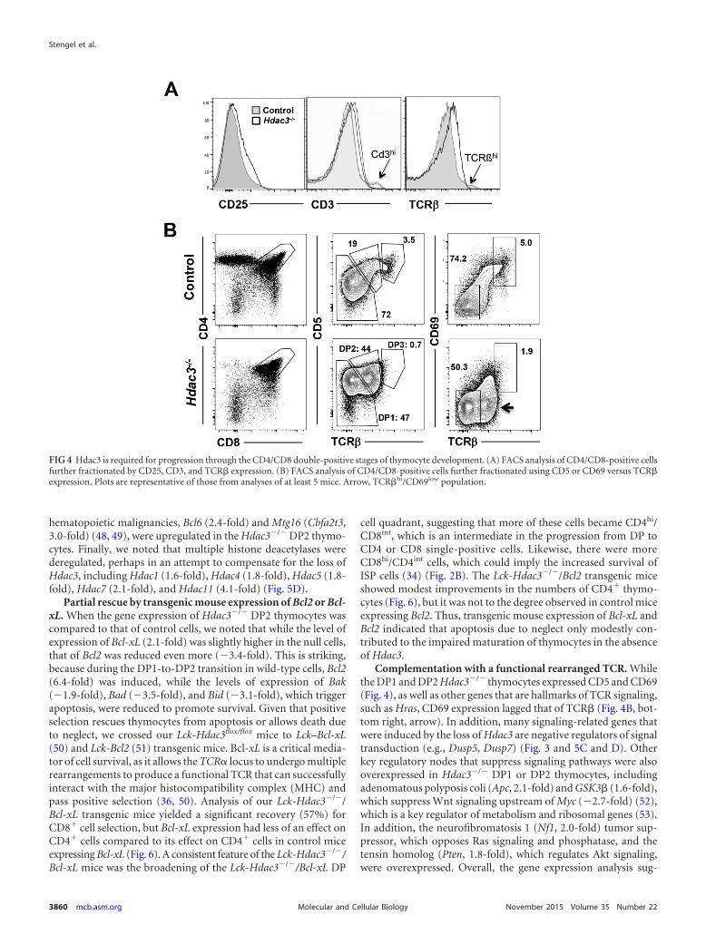
Hdac3 is required for efficient positive selection. The amountof CD25 was higher in the double-negative stage 3 (DN3) popu-lation (Fig. 2), and the amount of CD25 was higher at the mRNAlevel in double-positive cells (Fig. 3A). Therefore, we used flowcytometry to further scrutinize the CD4/CD8 double-positive thy-mocytes in Hdac3�/� mice. As predicted by the mRNA expressionanalysis, Hdac3�/� double-positive cells expressed higher levels ofCD25 on the cell surface (Fig. 4A). While we noted a significantpopulation of CD4/CD8 double-negative cells lacking the intra-cellular expression of TCR� (Fig. 2), the majority of DP cells ex-pressed somewhat more TCR� on the cell surface, although therewas a reduction in the small number of cells expressing the highestlevels of TCR� (Fig. 4A). Likewise, CD3 was expressed at signifi-cantly higher levels in the Hdac3-deficient thymocytes (Fig. 4A),but there was a reduction in the number of CD3hi thymocytes (Fig.4A) (40, 41).
Given the lack of the CD3hi and TCR�hi populations, we usedcell surface expression of CD5 and CD69 versus that of TCR� toexamine the maturation of double-positive cells. In Hdac3�/�
thymocytes there was a skewing toward CD5hi TCR�int and sig-nificantly fewer CD5low TCR�low cells in the null mice (double-positive stage 1 [DP1]) (Fig. 4B). Further, in addition to a reduc-tion in DP1 cell numbers, the median level of CD5 expression was
FIG 1 Loss of Hdac3 affects histone acetylation and thymocyte development. (A) FACS analysis of thymocytes from control mice (no GFP expression) andLck-Hdac3�/� mice (GFP�). Shown is a tracing for a control mouse that lacks GFP expression and two Lck-Hdac3flox/� mice in that 60% of the thymocytes areGFP� and thus have the Hdac3 deletion. (B) Western blot of Hdac3 expression in total thymocytes with various degrees of GFP expression. Hdac3 knockout isvariable, as indicated by GFP expression. �/�, Lck-Hdac3�/� mice; F/�, Lck-Hdac3flox/� mice. (C) Western blot of histone modification on thymocytes of eitherWT-Lck-Hdac3�/� or Lck-Hdac3flox/� mice sorted using GFP expression. (D) FACS analysis using anti-CD4 and anti-CD8. Unless otherwise noted, plots arerepresentative of those for at least 5 mice. The numbers indicate the percentages of cells analyzed. (E) Bar graphs showing the percentage of GFP-positive T cellsfrom thymic and splenic subpopulations in control or Hdac3�/� mice. Student’s t test was used to evaluate each pair, and the differences in CD4� and CD8� cellsfrom the spleen were significant (P � 0.05), whereas only the difference for CD4� cells from the thymus were statistically significant at this level. KO, knockout.(F) FACS analysis of T cells containing CD4 and CD8 versus GFP from the thymus (Thy) and spleen (Spl) showing that Hdac3�/� thymocytes do not persist inthe spleen. Plots are representative of those from analyses of at least 5 mice.
T Cell Deletion of Hdac3 Impairs Positive Selection
November 2015 Volume 35 Number 22 mcb.asm.org 3857Molecular and Cellular Biology

noticeably higher in the Hdac3�/� DP1 population. At the sametime, there was a drop in the number of CD5hi TCR�Hi (DP3) cellsin the Hdac3�/� mice, suggesting that these cells were impaired intheir development to DP3 and on to the single-positive stages.Likewise, CD69 is a marker of TCR signaling in cells undergoingpositive selection that is subsequently suppressed as thymocytesmature (42), to allow emigration from the thymus (reviewed inreference 43). Hdac3�/� double-positive cells expressing high lev-els of TCR� failed to coexpress CD69 (Fig. 4B, right, arrow), andthe cells completing positive selection that were TCR�hi CD69hi
failed to accumulate in the absence of Hdac3 (Fig. 4B), suggestingthat Hdac3 contributes to positive selection.
Inactivation of Hdac3 alters the gene expression program inthymocytes undergoing positive selection. Given the defects that
we observed in DP1 and DP2 of thymocyte development, we fur-ther subdivided the CD4/CD8 double-positive populations ana-lyzed earlier (Fig. 3) to define how inactivation of Hdac3 affectsgene expression. The DP1 and DP2 populations were isolatedfrom wild-type and Hdac3�/� mice and mRNA deep-sequencing(RNA-Seq) analysis was performed. During the transition of wild-type DP1 cells to DP2, just over 600 genes were induced, whilenearly 3 times this number were downregulated (35) (Fig. 5A).Central among the induced genes were the gene for the IL-7 re-ceptor (nearly 40-fold increase), which promotes survival (44);the genes for CD69 (14-fold) and CD53 (40-fold); as well as Bcl2(6-fold), which also impairs apoptosis to promote survival (45).Further, there was an induction of a transcriptional program thatincludes key regulators of T cell development, such as the E-pro-
FIG 2 Hdac3 is required for progression through early thymocyte development. (A) FACS analysis of GFP� double-negative (CD4�/CD8�) thymocytes usingCD44 and CD25 to delineate DN1 to DN4. The numbers in the lower corners of the gates indicate the percentages of cells analyzed. (B) FACS analysis ofintracellular TCR� expression by DN3 and DN4 cells. (C) (Left) FACS analysis of total thymocytes using CD4 and CD8 markers, with the results for CD8� cellsbeing replotted using CD147 and CD3 expression. (Right) Bar graph presenting the composite results for 6 mice.
Stengel et al.
3858 mcb.asm.org November 2015 Volume 35 Number 22Molecular and Cellular Biology

tein inhibitor of differentiation (Id2, 6-fold), Helios (Ikzf2, 4.7-fold), the NFB component RelB (4-fold), the Ets family memberEtv5 (4-fold), and the homeodomain protein Hhex (3.7-fold).Conversely, the cell cycle machinery was suppressed in DP2 cells,as the levels of expression of cyclin E (�5.7-fold), cyclin A2 (�3.5-fold), cyclin B2 (�4.6-fold), Cdk1 (�3.8-fold), E2F1 (�3.5-fold),E2F2 (�4.6-fold), E2F8 (�4.7-fold), and Myc (�2-fold) were alldecreased. While the level of expression of Rorc (RORt), a keycontributor to positive selection (46), decreased, as expected, dur-ing this transition (35), the drop was relatively small (�1.7-fold).Conversely, Nur77 (Nr4a1) was induced (7.9-fold) and its relatedfamily member Nor1 (Nr4a3) was dramatically induced (35-fold).Induction of these genes is consistent with these nuclear hormonereceptor family members being robustly activated by TCR signal-ing in DN2 cells in preparation for negative selection (47).
When the DP1 cells lacking Hdac3 were compared to wild-typeDP1 cells, over 1,000 genes were significantly induced, with someof the largest changes in expression occurring among key tran-scriptional regulators, such as retinoic acid receptor alpha (Rara,2.2-fold) and Mef2a, Mef2b, and Mef2c (Fig. 5A). While RORt(Rorc) contributes to positive selection (46), Rorb was inducednearly 3-fold, with the expression of Rorc was unchanged. Similarto our prior analysis (Fig. 3), CD25 (Il2ra) was overexpressed inboth DP1 and DP2 in Hdac3�/� thymocytes (Fig. 5B and C). TheHdac3�/� DP2 thymocytes also expressed 3.7-fold more Rorb,and while the level of expression of Rorc was higher, it was more inline with this gene not being slightly downregulated during theDP1-to-DP2 transition (1.8-fold). It is also notable that transcrip-tion factors and corepressors that recruit Hdac3 and that auto-regulate their own expression, including two genes associated with
FIG 3 Gene expression analysis of WT and Hdac3�/� (Null) CD4/CD8-positive thymocytes. (A) Heat map showing the log2 values of the relative fluorescenceintensity of 1,800 genes affected by the loss of Hdac3. (Right) Gene ontology groupings of genes and the fold change in gene expression. (B) Bar graph showingthe pathways affected by the inactivation of Hdac3. P values were calculated using a hypergeometric test (raw P value) and adjusted by multiple testing (adjustedP values [adjP]).
T Cell Deletion of Hdac3 Impairs Positive Selection
November 2015 Volume 35 Number 22 mcb.asm.org 3859Molecular and Cellular Biology
hematopoietic malignancies, Bcl6 (2.4-fold) and Mtg16 (Cbfa2t3,3.0-fold) (48, 49), were upregulated in the Hdac3�/� DP2 thymo-cytes. Finally, we noted that multiple histone deacetylases werederegulated, perhaps in an attempt to compensate for the loss ofHdac3, including Hdac1 (1.6-fold), Hdac4 (1.8-fold), Hdac5 (1.8-fold), Hdac7 (2.1-fold), and Hdac11 (4.1-fold) (Fig. 5D).
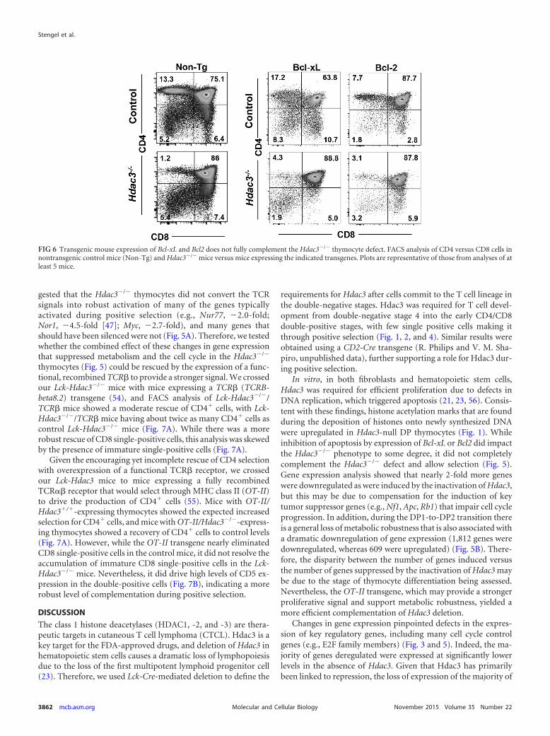
Partial rescue by transgenic mouse expression of Bcl2 or Bcl-xL. When the gene expression of Hdac3�/� DP2 thymocytes wascompared to that of control cells, we noted that while the level ofexpression of Bcl-xL (2.1-fold) was slightly higher in the null cells,that of Bcl2 was reduced even more (�3.4-fold). This is striking,because during the DP1-to-DP2 transition in wild-type cells, Bcl2(6.4-fold) was induced, while the levels of expression of Bak(�1.9-fold), Bad (�3.5-fold), and Bid (�3.1-fold), which triggerapoptosis, were reduced to promote survival. Given that positiveselection rescues thymocytes from apoptosis or allows death dueto neglect, we crossed our Lck-Hdac3flox/flox mice to Lck–Bcl-xL(50) and Lck-Bcl2 (51) transgenic mice. Bcl-xL is a critical media-tor of cell survival, as it allows the TCR� locus to undergo multiplerearrangements to produce a functional TCR that can successfullyinteract with the major histocompatibility complex (MHC) andpass positive selection (36, 50). Analysis of our Lck-Hdac3�/�/Bcl-xL transgenic mice yielded a significant recovery (57%) forCD8� cell selection, but Bcl-xL expression had less of an effect onCD4� cells compared to its effect on CD4� cells in control miceexpressing Bcl-xL (Fig. 6). A consistent feature of the Lck-Hdac3�/�/Bcl-xL mice was the broadening of the Lck-Hdac3�/�/Bcl-xL DP
cell quadrant, suggesting that more of these cells became CD4hi/CD8int, which is an intermediate in the progression from DP toCD4 or CD8 single-positive cells. Likewise, there were moreCD8hi/CD4int cells, which could imply the increased survival ofISP cells (34) (Fig. 2B). The Lck-Hdac3�/�/Bcl2 transgenic miceshowed modest improvements in the numbers of CD4� thymo-cytes (Fig. 6), but it was not to the degree observed in control miceexpressing Bcl2. Thus, transgenic mouse expression of Bcl-xL andBcl2 indicated that apoptosis due to neglect only modestly con-tributed to the impaired maturation of thymocytes in the absenceof Hdac3.
Complementation with a functional rearranged TCR. Whilethe DP1 and DP2 Hdac3�/� thymocytes expressed CD5 and CD69(Fig. 4), as well as other genes that are hallmarks of TCR signaling,such as Hras, CD69 expression lagged that of TCR� (Fig. 4B, bot-tom right, arrow). In addition, many signaling-related genes thatwere induced by the loss of Hdac3 are negative regulators of signaltransduction (e.g., Dusp5, Dusp7) (Fig. 3 and 5C and D). Otherkey regulatory nodes that suppress signaling pathways were alsooverexpressed in Hdac3�/� DP1 or DP2 thymocytes, includingadenomatous polyposis coli (Apc, 2.1-fold) and GSK3� (1.6-fold),which suppress Wnt signaling upstream of Myc (�2.7-fold) (52),which is a key regulator of metabolism and ribosomal genes (53).In addition, the neurofibromatosis 1 (Nf1, 2.0-fold) tumor sup-pressor, which opposes Ras signaling and phosphatase, and thetensin homolog (Pten, 1.8-fold), which regulates Akt signaling,were overexpressed. Overall, the gene expression analysis sug-
FIG 4 Hdac3 is required for progression through the CD4/CD8 double-positive stages of thymocyte development. (A) FACS analysis of CD4/CD8-positive cellsfurther fractionated by CD25, CD3, and TCR� expression. (B) FACS analysis of CD4/CD8-positive cells further fractionated using CD5 or CD69 versus TCR�expression. Plots are representative of those from analyses of at least 5 mice. Arrow, TCR�hi/CD69low population.
Stengel et al.
3860 mcb.asm.org November 2015 Volume 35 Number 22Molecular and Cellular Biology

FIG 5 RNA-Seq analysis of DP1 and DP2 thymocytes. (A) The heat map depicts the changes in gene expression between WT and Hdac3�/� DP1 or DP2thymocytes that were FACS prior to RNA-Seq analysis. (B) Venn diagrams show the number of genes that were upregulated or downregulated between controlDP1 and DP2 cells (blue) and then the number of genes whose expression changed upon deletion of Hdac3 (pink). (C) KEGG analysis of genes dysregulated inHdac3�/� DP1 and DP2 thymocytes. MAPK, mitogen-activated protein kinase; ER, endoplasmic reticulum. (D) Heat maps of four groups of genes from theKEGG analysis in panel C. P values were calculated using a hypergeometric test (raw P value) and adjusted by multiple testing (adjusted P values).
T Cell Deletion of Hdac3 Impairs Positive Selection
November 2015 Volume 35 Number 22 mcb.asm.org 3861Molecular and Cellular Biology
gested that the Hdac3�/� thymocytes did not convert the TCRsignals into robust activation of many of the genes typicallyactivated during positive selection (e.g., Nur77, �2.0-fold;Nor1, �4.5-fold [47]; Myc, �2.7-fold), and many genes thatshould have been silenced were not (Fig. 5A). Therefore, we testedwhether the combined effect of these changes in gene expressionthat suppressed metabolism and the cell cycle in the Hdac3�/�
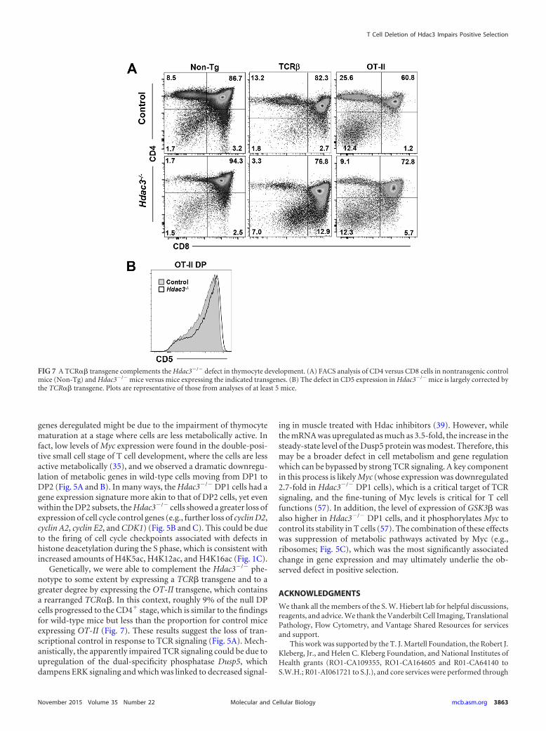
thymocytes (Fig. 5) could be rescued by the expression of a func-tional, recombined TCR� to provide a stronger signal. We crossedour Lck-Hdac3�/� mice with mice expressing a TCR� (TCRB-beta8.2) transgene (54), and FACS analysis of Lck-Hdac3�/�/TCR� mice showed a moderate rescue of CD4� cells, with Lck-Hdac3�/�/TCR� mice having about twice as many CD4� cells ascontrol Lck-Hdac3�/� mice (Fig. 7A). While there was a morerobust rescue of CD8 single-positive cells, this analysis was skewedby the presence of immature single-positive cells (Fig. 7A).
Given the encouraging yet incomplete rescue of CD4 selectionwith overexpression of a functional TCR� receptor, we crossedour Lck-Hdac3 mice to mice expressing a fully recombinedTCR�� receptor that would select through MHC class II (OT-II)to drive the production of CD4� cells (55). Mice with OT-II/Hdac3�/�-expressing thymocytes showed the expected increasedselection for CD4� cells, and mice with OT-II/Hdac3�/�-express-ing thymocytes showed a recovery of CD4� cells to control levels(Fig. 7A). However, while the OT-II transgene nearly eliminatedCD8 single-positive cells in the control mice, it did not resolve theaccumulation of immature CD8 single-positive cells in the Lck-Hdac3�/� mice. Nevertheless, it did drive high levels of CD5 ex-pression in the double-positive cells (Fig. 7B), indicating a morerobust level of complementation during positive selection.
DISCUSSION
The class 1 histone deacetylases (HDAC1, -2, and -3) are thera-peutic targets in cutaneous T cell lymphoma (CTCL). Hdac3 is akey target for the FDA-approved drugs, and deletion of Hdac3 inhematopoietic stem cells causes a dramatic loss of lymphopoiesisdue to the loss of the first multipotent lymphoid progenitor cell(23). Therefore, we used Lck-Cre-mediated deletion to define the
requirements for Hdac3 after cells commit to the T cell lineage inthe double-negative stages. Hdac3 was required for T cell devel-opment from double-negative stage 4 into the early CD4/CD8double-positive stages, with few single positive cells making itthrough positive selection (Fig. 1, 2, and 4). Similar results wereobtained using a CD2-Cre transgene (R. Philips and V. M. Sha-piro, unpublished data), further supporting a role for Hdac3 dur-ing positive selection.
In vitro, in both fibroblasts and hematopoietic stem cells,Hdac3 was required for efficient proliferation due to defects inDNA replication, which triggered apoptosis (21, 23, 56). Consis-tent with these findings, histone acetylation marks that are foundduring the deposition of histones onto newly synthesized DNAwere upregulated in Hdac3-null DP thymocytes (Fig. 1). Whileinhibition of apoptosis by expression of Bcl-xL or Bcl2 did impactthe Hdac3�/� phenotype to some degree, it did not completelycomplement the Hdac3�/� defect and allow selection (Fig. 5).Gene expression analysis showed that nearly 2-fold more geneswere downregulated as were induced by the inactivation of Hdac3,but this may be due to compensation for the induction of keytumor suppressor genes (e.g., Nf1, Apc, Rb1) that impair cell cycleprogression. In addition, during the DP1-to-DP2 transition thereis a general loss of metabolic robustness that is also associated witha dramatic downregulation of gene expression (1,812 genes weredownregulated, whereas 609 were upregulated) (Fig. 5B). There-fore, the disparity between the number of genes induced versusthe number of genes suppressed by the inactivation of Hdac3 maybe due to the stage of thymocyte differentiation being assessed.Nevertheless, the OT-II transgene, which may provide a strongerproliferative signal and support metabolic robustness, yielded amore efficient complementation of Hdac3 deletion.
Changes in gene expression pinpointed defects in the expres-sion of key regulatory genes, including many cell cycle controlgenes (e.g., E2F family members) (Fig. 3 and 5). Indeed, the ma-jority of genes deregulated were expressed at significantly lowerlevels in the absence of Hdac3. Given that Hdac3 has primarilybeen linked to repression, the loss of expression of the majority of
FIG 6 Transgenic mouse expression of Bcl-xL and Bcl2 does not fully complement the Hdac3�/� thymocyte defect. FACS analysis of CD4 versus CD8 cells innontransgenic control mice (Non-Tg) and Hdac3�/� mice versus mice expressing the indicated transgenes. Plots are representative of those from analyses of atleast 5 mice.
Stengel et al.
3862 mcb.asm.org November 2015 Volume 35 Number 22Molecular and Cellular Biology
genes deregulated might be due to the impairment of thymocytematuration at a stage where cells are less metabolically active. Infact, low levels of Myc expression were found in the double-posi-tive small cell stage of T cell development, where the cells are lessactive metabolically (35), and we observed a dramatic downregu-lation of metabolic genes in wild-type cells moving from DP1 toDP2 (Fig. 5A and B). In many ways, the Hdac3�/� DP1 cells had agene expression signature more akin to that of DP2 cells, yet evenwithin the DP2 subsets, the Hdac3�/� cells showed a greater loss ofexpression of cell cycle control genes (e.g., further loss of cyclin D2,cyclin A2, cyclin E2, and CDK1) (Fig. 5B and C). This could be dueto the firing of cell cycle checkpoints associated with defects inhistone deacetylation during the S phase, which is consistent withincreased amounts of H4K5ac, H4K12ac, and H4K16ac (Fig. 1C).
Genetically, we were able to complement the Hdac3�/� phe-notype to some extent by expressing a TCR� transgene and to agreater degree by expressing the OT-II transgene, which containsa rearranged TCR��. In this context, roughly 9% of the null DPcells progressed to the CD4� stage, which is similar to the findingsfor wild-type mice but less than the proportion for control miceexpressing OT-II (Fig. 7). These results suggest the loss of tran-scriptional control in response to TCR signaling (Fig. 5A). Mech-anistically, the apparently impaired TCR signaling could be due toupregulation of the dual-specificity phosphatase Dusp5, whichdampens ERK signaling and which was linked to decreased signal-
ing in muscle treated with Hdac inhibitors (39). However, whilethe mRNA was upregulated as much as 3.5-fold, the increase in thesteady-state level of the Dusp5 protein was modest. Therefore, thismay be a broader defect in cell metabolism and gene regulationwhich can be bypassed by strong TCR signaling. A key componentin this process is likely Myc (whose expression was downregulated2.7-fold in Hdac3�/� DP1 cells), which is a critical target of TCRsignaling, and the fine-tuning of Myc levels is critical for T cellfunctions (57). In addition, the level of expression of GSK3� wasalso higher in Hdac3�/� DP1 cells, and it phosphorylates Myc tocontrol its stability in T cells (57). The combination of these effectswas suppression of metabolic pathways activated by Myc (e.g.,ribosomes; Fig. 5C), which was the most significantly associatedchange in gene expression and may ultimately underlie the ob-served defect in positive selection.
ACKNOWLEDGMENTS
We thank all the members of the S. W. Hiebert lab for helpful discussions,reagents, and advice. We thank the Vanderbilt Cell Imaging, TranslationalPathology, Flow Cytometry, and Vantage Shared Resources for servicesand support.
This work was supported by the T. J. Martell Foundation, the Robert J.Kleberg, Jr., and Helen C. Kleberg Foundation, and National Institutes ofHealth grants (RO1-CA109355, RO1-CA164605 and R01-CA64140 toS.W.H.; R01-AI061721 to S.J.), and core services were performed through
FIG 7 A TCR�� transgene complements the Hdac3�/� defect in thymocyte development. (A) FACS analysis of CD4 versus CD8 cells in nontransgenic controlmice (Non-Tg) and Hdac3�/� mice versus mice expressing the indicated transgenes. (B) The defect in CD5 expression in Hdac3�/� mice is largely corrected bythe TCR�� transgene. Plots are representative of those from analyses of at least 5 mice.
T Cell Deletion of Hdac3 Impairs Positive Selection
November 2015 Volume 35 Number 22 mcb.asm.org 3863Molecular and Cellular Biology

a Vanderbilt digestive disease research grant (NIDDK P30DK58404) anda Vanderbilt-Ingram Cancer Center support grant (NCI P30CA68485).A.R.S. was supported by a fellowship from the NIH (5F32HL090259),K.R.S. was supported by grant 5 T32 CA009582-26 from the NCI and apostdoctoral fellowship (PF-13-303-01-DMC) from the American Can-cer Society, and L.E.G. was supported by training grant HL069765. Theproject described was also supported by the National Center for ResearchResources (grant UL1 RR024975-01) and is now supported by the Na-tional Center for Advancing Translational Sciences (grant 2 UL1TR000445-06).
The content is solely the responsibility of the authors and does notnecessarily represent the official views of the NIH.
We declare that no conflict of interest exists.
REFERENCES1. Seto E, Yoshida M. 2014. Erasers of histone acetylation: the histone
deacetylase enzymes. Cold Spring Harb Perspect Biol 6:a018713. http://dx.doi.org/10.1101/cshperspect.a018713.
2. Hassig CA, Fleischer TC, Billin AN, Schreiber SL, Ayer DE. 1997. Histonedeacetylase activity is required for full transcriptional repression by mSin3A.Cell 89:341–347. http://dx.doi.org/10.1016/S0092-8674(00)80214-7.
3. Laherty CD, Yang WM, Sun JM, Davie JR, Seto E, Eisenman RN. 1997.Histone deacetylases associated with the mSin3 corepressor mediate madtranscriptional repression. Cell 89:349 –356. http://dx.doi.org/10.1016/S0092-8674(00)80215-9.
4. Li H, Leo C, Schroen DJ, Chen JD. 1997. Characterization of receptorinteraction and transcriptional repression by the corepressor SMRT. MolEndocrinol 11:2025–2037. http://dx.doi.org/10.1210/mend.11.13.0028.
5. Yang WM, Yao YL, Sun JM, Davie JR, Seto E. 1997. Isolation andcharacterization of cDNAs corresponding to an additional member of thehuman histone deacetylase gene family. J Biol Chem 272:28001–28007.http://dx.doi.org/10.1074/jbc.272.44.28001.
6. Watson PJ, Fairall L, Santos GM, Schwabe JW. 2012. Structure ofHDAC3 bound to co-repressor and inositol tetraphosphate. Nature 481:335–340. http://dx.doi.org/10.1038/nature10728.
7. Millard CJ, Watson PJ, Celardo I, Gordiyenko Y, Cowley SM, RobinsonCV, Fairall L, Schwabe JW. 2013. Class I HDACs share a commonmechanism of regulation by inositol phosphates. Mol Cell 51:57– 67. http://dx.doi.org/10.1016/j.molcel.2013.05.020.
8. Grozinger CM, Hassig CA, Schreiber SL. 1999. Three proteins define aclass of human histone deacetylases related to yeast Hda1p. Proc NatlAcad Sci U S A 96:4868 – 4873. http://dx.doi.org/10.1073/pnas.96.9.4868.
9. Fischle W, Dequiedt F, Hendzel MJ, Guenther MG, Lazar MA,Voelter W, Verdin E. 2002. Enzymatic activity associated with class IIHDACs is dependent on a multiprotein complex containing HDAC3and SMRT/N-CoR. Mol Cell 9:45–57. http://dx.doi.org/10.1016/S1097-2765(01)00429-4.
10. Petrie K, Guidez F, Howell L, Healy L, Waxman S, Greaves M,Zelent A. 2003. The histone deacetylase 9 gene encodes multiple pro-tein isoforms. J Biol Chem 278:16059 –16072. http://dx.doi.org/10.1074/jbc.M212935200.
11. Chang HC, Guarente L. 2014. SIRT1 and other sirtuins in metabolism.Trends Endocrinol Metab 25:138 –145. http://dx.doi.org/10.1016/j.tem.2013.12.001.
12. Fiorino E, Giudici M, Ferrari A, Mitro N, Caruso D, De Fabiani E,Crestani M. 2014. The sirtuin class of histone deacetylases: regulation androles in lipid metabolism. IUBMB Life 66:89 –99. http://dx.doi.org/10.1002/iub.1246.
13. Roth M, Chen WY. 2014. Sorting out functions of sirtuins in cancer.Oncogene 33:1609 –1620. http://dx.doi.org/10.1038/onc.2013.120.
14. Gao L, Cueto MA, Asselbergs F, Atadja P. 2002. Cloning and functionalcharacterization of HDAC11, a novel member of the human histonedeacetylase family. J Biol Chem 277:25748 –25755. http://dx.doi.org/10.1074/jbc.M111871200.
15. Villagra A, Cheng F, Wang HW, Suarez I, Glozak M, Maurin M,Nguyen D, Wright KL, Atadja PW, Bhalla K, Pinilla-Ibarz J, Seto E,Sotomayor EM. 2009. The histone deacetylase HDAC11 regulates theexpression of interleukin 10 and immune tolerance. Nat Immunol 10:92–100. http://dx.doi.org/10.1038/ni.1673.
16. Stengel KR, Hiebert SW. 2014. Class I HDACs affect DNA replication,
repair, and chromatin structure: implications for cancer therapy. AntioxidRedox Signal 23:51– 65. http://dx.doi.org/10.1089/ars.2014.5915.
17. Wells CE, Bhaskara S, Stengel KR, Zhao Y, Sirbu B, Chagot B, CortezD, Khabele D, Chazin WJ, Cooper A, Jacques V, Rusche J, Eischen CM,McGirt LY, Hiebert SW. 2013. Inhibition of histone deacetylase 3 causesreplication stress in cutaneous T cell lymphoma. PLoS One 8:e68915. http://dx.doi.org/10.1371/journal.pone.0068915.
18. Montgomery RL, Davis CA, Potthoff MJ, Haberland M, Fielitz J, Qi X,Hill JA, Richardson JA, Olson EN. 2007. Histone deacetylases 1 and 2redundantly regulate cardiac morphogenesis, growth, and contractility.Genes Dev 21:1790 –1802. http://dx.doi.org/10.1101/gad.1563807.
19. Wilting RH, Yanover E, Heideman MR, Jacobs H, Horner J, van derTorre J, DePinho RA, Dannenberg JH. 2010. Overlapping functions ofHdac1 and Hdac2 in cell cycle regulation and haematopoiesis. EMBO J29:2586 –2597. http://dx.doi.org/10.1038/emboj.2010.136.
20. Yamaguchi T, Cubizolles F, Zhang Y, Reichert N, Kohler H, Seiser C,Matthias P. 2010. Histone deacetylases 1 and 2 act in concert to promotethe G1-to-S progression. Genes Dev 24:455– 469. http://dx.doi.org/10.1101/gad.552310.
21. Bhaskara S, Chyla BJ, Amann JM, Knutson SK, Cortez D, Sun ZW,Hiebert SW. 2008. Deletion of histone deacetylase 3 reveals critical rolesin S phase progression and DNA damage control. Mol Cell 30:61–72. http://dx.doi.org/10.1016/j.molcel.2008.02.030.
22. Montgomery RL, Potthoff MJ, Haberland M, Qi X, Matsuzaki S,Humphries KM, Richardson JA, Bassel-Duby R, Olson EN. 2008. Main-tenance of cardiac energy metabolism by histone deacetylase 3 in mice. JClin Invest 118:3588 –3597. http://dx.doi.org/10.1172/JCI35847.
23. Summers AR, Fischer MA, Stengel KR, Zhao Y, Kaiser JF, Wells CE,Hunt A, Bhaskara S, Luzwick JW, Sampathi S, Chen X, Thompson MA,Cortez D, Hiebert SW. 2013. HDAC3 is essential for DNA replication inhematopoietic progenitor cells. J Clin Invest 123:3112–3123. http://dx.doi.org/10.1172/JCI60806.
24. Bishton MJ, Harrison SJ, Martin BP, McLaughlin N, James C, JosefssonEC, Henley KJ, Kile BT, Prince HM, Johnstone RW. 2011. Decipheringthe molecular and biologic processes that mediate histone deacetylase in-hibitor-induced thrombocytopenia. Blood 117:3658 –3668. http://dx.doi.org/10.1182/blood-2010-11-318055.
25. Heideman MR, Wilting RH, Yanover E, Velds A, de Jong J, KerkhovenRM, Jacobs H, Wessels LF, Dannenberg JH. 2013. Dosage-dependenttumor suppression by histone deacetylases 1 and 2 through regulation ofc-Myc collaborating genes and p53 function. Blood 121:2038 –2050. http://dx.doi.org/10.1182/blood-2012-08-450916.
26. Santoro F, Botrugno OA, Dal Zuffo R, Pallavicini I, Matthews GM,Cluse L, Barozzi I, Senese S, Fornasari L, Moretti S, Altucci L, PelicciPG, Chiocca S, Johnstone RW, Minucci S. 2013. A dual role for Hdac1:oncosuppressor in tumorigenesis, oncogene in tumor maintenance.Blood 121:3459 –3468. http://dx.doi.org/10.1182/blood-2012-10-461988.
27. Trapnell C, Roberts A, Goff L, Pertea G, Kim D, Kelley DR, PimentelH, Salzberg SL, Rinn JL, Pachter L. 2012. Differential gene and transcriptexpression analysis of RNA-seq experiments with TopHat and Cufflinks.Nat Protoc 7:562–578. http://dx.doi.org/10.1038/nprot.2012.016.
28. Mao X, Fujiwara Y, Chapdelaine A, Yang H, Orkin SH. 2001. Activationof EGFP expression by Cre-mediated excision in a new ROSA26 reportermouse strain. Blood 97:324 –326. http://dx.doi.org/10.1182/blood.V97.1.324.
29. Thapa P, Das J, McWilliams D, Shapiro M, Sundsbak R, Nelson-HolteM, Tangen S, Anderson J, Desiderio S, Hiebert S, Sant’angelo DB,Shapiro VS. 2013. The transcriptional repressor NKAP is required for thedevelopment of iNKT cells. Nat Commun 4:1582. http://dx.doi.org/10.1038/ncomms2580.
30. Allen JM, Forbush KA, Perlmutter RM. 1992. Functional dissection ofthe lck proximal promoter. Mol Cell Biol 12:2758 –2768.
31. Godfrey DI, Kennedy J, Suda T, Zlotnik A. 1993. A developmentalpathway involving four phenotypically and functionally distinct subsets ofCD3� CD4� CD8� triple-negative adult mouse thymocytes defined byCD44 and CD25 expression. J Immunol 150:4244 – 4252.
32. Godfrey DI, Kennedy J, Mombaerts P, Tonegawa S, Zlotnik A. 1994.Onset of TCR-beta gene rearrangement and role of TCR-beta expressionduring CD3� CD4� CD8� thymocyte differentiation. J Immunol 152:4783– 4792.
33. Zeng L, Dalheimer SL, Yankee TM. 2007. Gads�/� mice reveal func-tionally distinct subsets of TCRbeta� CD4� CD8� double-negative
Stengel et al.
3864 mcb.asm.org November 2015 Volume 35 Number 22Molecular and Cellular Biology

thymocytes. J Immunol 179:1013–1021. http://dx.doi.org/10.4049/jimmunol.179.2.1013.
34. Xiong J, Armato MA, Yankee TM. 2011. Immature single-positive CD8�
thymocytes represent the transition from Notch-dependent to Notch-independent T-cell development. Int Immunol 23:55– 64. http://dx.doi.org/10.1093/intimm/dxq457.
35. Mingueneau M, Kreslavsky T, Gray D, Heng T, Cruse R, Ericson J,Bendall S, Spitzer MH, Nolan GP, Kobayashi K, von Boehmer H,Mathis D, Benoist C, Best AJ, Knell J, Goldrath A, Jojic V, Koller D,Shay T, Regev A, Cohen N, Brennan P, Brenner M, Kim F, Rao TN,Wagers A, Heng T, Ericson J, Rothamel K, Ortiz-Lopez A, Mathis D,Benoist C, Bezman NA, Sun JC, Min-Oo G, Kim CC, Lanier LL, MillerJ, Brown B, Merad M, Gautier EL, Jakubzick C, Randolph GJ, MonachP, Blair DA, Dustin ML, Shinton SA, Hardy RR, Laidlaw D, Collins J,et al. 2013. The transcriptional landscape of alphabeta T cell differentia-tion. Nat Immunol 14:619 – 632. http://dx.doi.org/10.1038/ni.2590.
36. Wang R, Xie H, Huang Z, Ma J, Fang X, Ding Y, Sun Z. 2011.Transcription factor network regulating CD(�)CD8(�) thymocytesurvival. Crit Rev Immunol 31:447– 458. http://dx.doi.org/10.1615/CritRevImmunol.v31.i6.10.
37. Sudo T, Nishikawa S, Ohno N, Akiyama N, Tamakoshi M, Yoshida H,Nishikawa S. 1993. Expression and function of the interleukin 7 receptorin murine lymphocytes. Proc Natl Acad Sci U S A 90:9125–9129. http://dx.doi.org/10.1073/pnas.90.19.9125.
38. Trigueros C, Hozumi K, Silva-Santos B, Bruno L, Hayday AC, OwenMJ, Pennington DJ. 2003. Pre-TCR signaling regulates IL-7 receptoralpha expression promoting thymocyte survival at the transition from thedouble-negative to double-positive stage. Eur J Immunol 33:1968 –1977.http://dx.doi.org/10.1002/eji.200323831.
39. Ferguson BS, Harrison BC, Jeong MY, Reid BG, Wempe MF, WagnerFF, Holson EB, McKinsey TA. 2013. Signal-dependent repression ofDUSP5 by class I HDACs controls nuclear ERK activity and cardiomyo-cyte hypertrophy. Proc Natl Acad Sci U S A 110:9806 –9811. http://dx.doi.org/10.1073/pnas.1301509110.
40. Tarakhovsky A, Kanner SB, Hombach J, Ledbetter JA, Muller W,Killeen N, Rajewsky K. 1995. A role for CD5 in TCR-mediated signaltransduction and thymocyte selection. Science 269:535–537. http://dx.doi.org/10.1126/science.7542801.
41. Yang Y, Contag CH, Felsher D, Shachaf CM, Cao Y, Herzenberg LA,Herzenberg LA, Tung JW. 2004. The E47 transcription factor negativelyregulates CD5 expression during thymocyte development. Proc Natl AcadSci U S A 101:3898 –3902. http://dx.doi.org/10.1073/pnas.0308764101.
42. Yamashita I, Nagata T, Tada T, Nakayama T. 1993. CD69 cell surfaceexpression identifies developing thymocytes which audition for T cell an-tigen receptor-mediated positive selection. Int Immunol 5:1139 –1150.http://dx.doi.org/10.1093/intimm/5.9.1139.
43. Weinreich MA, Hogquist KA. 2008. Thymic emigration: when and howT cells leave home. J Immunol 181:2265–2270. http://dx.doi.org/10.4049/jimmunol.181.4.2265.
44. Murray R, Suda T, Wrighton N, Lee F, Zlotnik A. 1989. IL-7 is a growthand maintenance factor for mature and immature thymocyte subsets. IntImmunol 1:526 –531. http://dx.doi.org/10.1093/intimm/1.5.526.
45. Linette GP, Grusby MJ, Hedrick SM, Hansen TH, Glimcher LH,
Korsmeyer SJ. 1994. Bcl-2 is upregulated at the CD4� CD8� stageduring positive selection and promotes thymocyte differentiation atseveral control points. Immunity 1:197–205. http://dx.doi.org/10.1016/1074-7613(94)90098-1.
46. Sun Z, Unutmaz D, Zou YR, Sunshine MJ, Pierani A, Brenner-MortonS, Mebius RE, Littman DR. 2000. Requirement for RORgamma in thy-mocyte survival and lymphoid organ development. Science 288:2369 –2373. http://dx.doi.org/10.1126/science.288.5475.2369.
47. Cheng LE, Chan FK, Cado D, Winoto A. 1997. Functional redundancyof the Nur77 and Nor-1 orphan steroid receptors in T-cell apoptosis.EMBO J 16:1865–1875. http://dx.doi.org/10.1093/emboj/16.8.1865.
48. Ci W, Polo JM, Cerchietti L, Shaknovich R, Wang L, Yang SN, Ye K,Farinha P, Horsman DE, Gascoyne RD, Elemento O, Melnick A. 2009.The BCL6 transcriptional program features repression of multiple onco-genes in primary B cells and is deregulated in DLBCL. Blood 113:5536 –5548. http://dx.doi.org/10.1182/blood-2008-12-193037.
49. Fujiwara T, Lee HY, Sanalkumar R, Bresnick EH. 2010. Buildingmultifunctionality into a complex containing master regulators of he-matopoiesis. Proc Natl Acad Sci U S A 107:20429 –20434. http://dx.doi.org/10.1073/pnas.1007804107.
50. Chao DT, Linette GP, Boise LH, White LS, Thompson CB, KorsmeyerSJ. 1995. Bcl-XL and Bcl-2 repress a common pathway of cell death. J ExpMed 182:821– 828. http://dx.doi.org/10.1084/jem.182.3.821.
51. Linette GP, Hess JL, Sentman CL, Korsmeyer SJ. 1995. Peripheral T-celllymphoma in lckpr-bcl-2 transgenic mice. Blood 86:1255–1260.
52. He TC, Sparks AB, Rago C, Hermeking H, Zawel L, da Costa LT, MorinPJ, Vogelstein B, Kinzler KW. 1998. Identification of c-MYC as a targetof the APC pathway. Science 281:1509 –1512. http://dx.doi.org/10.1126/science.281.5382.1509.
53. van Riggelen J, Yetil A, Felsher DW. 2010. MYC as a regulator ofribosome biogenesis and protein synthesis. Nat Rev Cancer 10:301–309.http://dx.doi.org/10.1038/nrc2819.
54. Uematsu Y, Ryser S, Dembic Z, Borgulya P, Krimpenfort P, Berns A,von Boehmer H, Steinmetz M. 1988. In transgenic mice the intro-duced functional T cell receptor beta gene prevents expression of en-dogenous beta genes. Cell 52:831– 841. http://dx.doi.org/10.1016/0092-8674(88)90425-4.
55. Barnden MJ, Allison J, Heath WR, Carbone FR. 1998. Defective TCRexpression in transgenic mice constructed using cDNA-based alpha- andbeta-chain genes under the control of heterologous regulatory elements.Immunol Cell Biol 76:34 – 40. http://dx.doi.org/10.1046/j.1440-1711.1998.00709.x.
56. Bhaskara S, Knutson SK, Jiang G, Chandrasekharan MB, Wilson AJ,Zheng S, Yenamandra A, Locke K, Yuan JL, Bonine-Summers AR,Wells CE, Kaiser JF, Washington MK, Zhao Z, Wagner FF, Sun ZW,Xia F, Holson EB, Khabele D, Hiebert SW. 2010. Hdac3 is essential forthe maintenance of chromatin structure and genome stability. Cancer Cell18:436 – 447. http://dx.doi.org/10.1016/j.ccr.2010.10.022.
57. Preston GC, Sinclair LV, Kaskar A, Hukelmann JL, Navarro MN,Ferrero I, MacDonald HR, Cowling VH, Cantrell DA. 2015. Single celltuning of Myc expression by antigen receptor signal strength and inter-leukin-2 in T lymphocytes. EMBO J 34:2008 –2024. http://dx.doi.org/10.15252/embj.201490252.
T Cell Deletion of Hdac3 Impairs Positive Selection
November 2015 Volume 35 Number 22 mcb.asm.org 3865Molecular and Cellular Biology