histopathologic evaluation of the subcutaneous tissue...
TRANSCRIPT
64
Dental Research Journal (Vol. 2, No. 2, Winter 2006)
Histopathologic Evaluation of the Subcutaneous Tissue Response to Three Endodontic Sealers
M. Aminozarbian MD*, E. Bahmani MD**; M. Feizianfard MD***
ABSTRACT Introduction: The materials used for obturation of root canal system may be extruded through apical foramen into the periapical tissue. Therefore, biocompatibility of these materials is very important. The purpose of this study was to evaluate the in vivo biocompatibility of three conventional endodontic sealers: AH26, Roth 801, and ZOE after their subcutaneous implantation in rats.
Methods: Twenty-two mature male Albino rats, weighting from 250 to 500grs were used. Each animal received four polyethylene implants; three implants were containing test sealers and one was empty tube as negative control. The animals were sacrificed at third day and third month after implantation and the implants were dissected with 2cm of surrounding tissue margins. Then, tissue reactions to the test materials were evaluated histologically and quantitatively by a pathologist under light microscope, after histologic processing and staining with H-E. The obtained results were analyzed statistically by Mann-Whitney test.
Results: After 3 days, tissue reaction to ZOE was more acute than AH26, Roth 801, and control group. But after 3 months, no significant difference was observed among these three sealers and among those and control group.
Discussion: According to this study, all of the sealers cause inflammatory reactions immediately after contact with tissue, but the intensity of these responses decrease with time. The acute responses of third day changed to chronic, proliferative, healing processes in third month.
Key words: Endodontic Sealers, Tissue reaction (response), Subcutaneous Implantation.
[Dental Research Journal (Vol. 2, No. 2, Winter 2006)]
Introduction The materials used for obturation of root canal system such as sealers, maybe extruded through apical foramen into the periapical tissue. Therefore, the biocompatibility of these materials is very important1.Currently, there are three recommended tests for the biological evaluation and acceptance of endodontic materials: a primary test or cell culture test which provides a general profile of toxicity for the material, a secondary test or material implantation test which evaluates its local toxicity in experimental animals a usage
test in which the material is used in the endodontic treatment of teeth in experimental animals 2.In cell culture tests, several studies have been performed and all have found different degrees of cytotoxicity for various sealers 3,
4, 5. Bouillaguet et al (2004) evaluated cytotoxicity of several sealers at cell culture and reported that the cytotoxicity of sealers increases with time from 24h to 1 week and most sealers are potentially, cytotoxic specially when are mixed freshly 6. Huang et al (2002) demonstrated that AH26 exhibite
*Assistant Professor, Department of Endodontics, Faculty of Dentistry, Medical University of Isfahan, Iran. **Assistant Professor, Department of Endodontics, Faculty of Dentistry, Medical University of Ahvaz, Iran. ***Endodontist, Department of Endodontics, Faculty of Dentistry. Medical University of Isfahan, Iran.
65 Histopathologic Evaluation of….
Dental Research Journal (Vol. 2, No. 2, Winter 2006)
not only in vitro dose dependent cytotoxicity but also genotoxicity7.Azar et al (2000) mentioned that cytotoxicity of ZOE was detectable as early as 1hr after mixing and remained at a high level until 5 week. AH26, however, induced early cytotoxic effects that lasted for 1 week, followed by a substantial reduction in cytotoxicity 8.In usage tests, Bernath and Szabo (2003), after filling the root canals of monkeys, found that all sealers cause inflammatory response and reported that if root filling by Apexit and Grossman’s sealers confine to the canal system, it would not cause inflammation. But similar situations, with endomethasone and AH26, cause mild lymphocytic- plasmocytic infiltration in some cases 9.The reports about biocompatibility of sealers are different. Thus in present study the biocompatibility of three conventional sealers such as AH26, Roth 801, and ZOE are evaluated by subcutaneous implantation (secondary test) in rats.
Materials and methods Twenty-two mature male Albino rats, weighting from 250 to 500gr, were used in this study. Every 11 rats were selected as one group; first group for third day evaluation and second group for third month evaluation. The animals were anesthetized with an intraperitoneal injection of a combination of 50mg/kg of ketamine hydrochloride and 0.5mg/kg of Rompun 2%. The dorsum was shaved at 4 points, two points at anterior (right and left) and two points at posterior (right and left). After disinfection of these areas with 10% povidone iodine, local anesthesia was performed with 2% Lidocaine, containing 1/80000 epinephrine. Then 4 incisions were made in the dorsal skin and 4 subcutaneous
pockets were carefully prepared to a depth of approximately 15mm. The polyethylene tubes, 7mm in length with an inner diameter of 1.7mm were sterilized with ethylen oxide gas and then were filled with the freshly mixed sealers prepared in accordance with the manufactuerers’ instruction. Care was taken to fill the tubes without voids 10.The sealers used in this study were AH26 (Dentsply, Detrey), Roth 801 (AriaDent, Packed by ASIA CHEMI Teb co., IRAN), and ZOE (Kemdent – BP). Three tubes with each sealer and one empty tube (as negative control) were placed carefully in the four pockets of each animal to avoid spilling the material into the tissue. The incisions were closed with silk sutures. For infection control, the Chloramphenicole spray was used on surgical site. The information about each animal was recorded at a data sheet. After periods of 3 days and / or 3 months, the rats were sacrificed by overdosing with intracardiac injection of Ketamine hydrochloride and Rompun. The test tubes and approximately 2cm of the surrounding tissue margin were dissected and placed into 10% formalin. The specimens were processed to be embedded in paraffin after 48h ours of fixation. The blocks were oriented parallel to the long axis of the tubes and serial sections were cut to a thickness of 6 µ msetting of the microtome at the histology laboratory of Medical University of Isfahan. The sections were mounted on glass slides and were stained with Hematoxylin and Eosin. Then the histopathologic evaluation of the specimens was performed by a pathologist who was blind to the specimens under the light microscope. Some criteria was considered for histopathologic evaluation and a tissue response table was designed with 6 scores, according to these criteria (Table 1).
66
Dental Research Journal (Vol. 2, No. 2, Winter 2006)
Table 1: Tissue response table
In this table, minimum reaction was seen in score 3 (negative control). From score 3 to 6, the severity of responses increased and they became more acute. From score 3 to 1, the severity of responses decreased and they became more chronic. Each specimen got a score according to tissue response table and was recorded. Then the data was statistically analyzed by Mann-Whitney and Willcoxon tests.
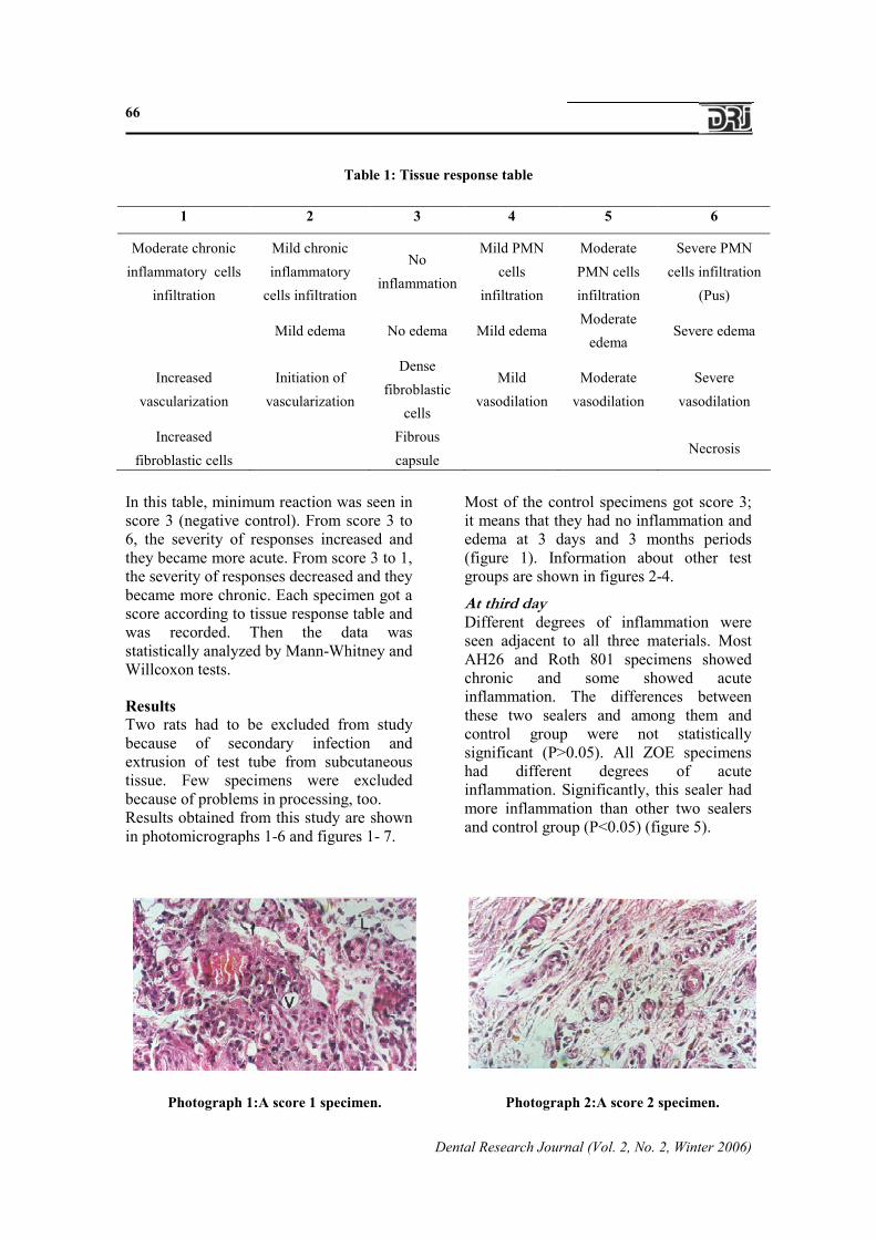
Results Two rats had to be excluded from study because of secondary infection and extrusion of test tube from subcutaneous tissue. Few specimens were excluded because of problems in processing, too. Results obtained from this study are shown in photomicrographs 1-6 and figures 1- 7.
Most of the control specimens got score 3; it means that they had no inflammation and edema at 3 days and 3 months periods (figure 1). Information about other test groups are shown in figures 2-4.
At third day Different degrees of inflammation were seen adjacent to all three materials. Most AH26 and Roth 801 specimens showed chronic and some showed acute inflammation. The differences between these two sealers and among them and control group were not statistically significant (P>0.05). All ZOE specimens had different degrees of acute inflammation. Significantly, this sealer had more inflammation than other two sealers and control group (P<0.05) (figure 5).
Photograph 1:A score 1 specimen. Photograph 2:A score 2 specimen.
1 2 3 4 5 6
Moderate chronic inflammatory cells
infiltration
Mild chronic inflammatory
cells infiltration
Noinflammation
Mild PMN cells
infiltration
Moderate PMN cells infiltration
Severe PMN cells infiltration
(Pus)
Mild edema No edema Mild edema Moderate
edema Severe edema
Increased vascularization
Initiation of vascularization
Dense fibroblastic
cells
Mild vasodilation
Moderate vasodilation
Severe vasodilation
Increased fibroblastic cells
Fibrous capsule
Necrosis