histopathologic observations on early oral implant failures
TRANSCRIPT
COPYRIGHT © 2000 BY QUINTESSENCE PUBLISHING CO, INC. PRINTING
OF THIS DOCUMENT IS RESTRICTED TO PERSONAL USE ONLY. NO PART OF
THIS ARTICLE MAY BE REPRODUCED OR TRANSMITTED IN ANY FORM WITH-OUT WRITTEN PERMISSION FROM THE PUBLISHER.
798 Volume 14, Number 6, 1999
Implants are commonly and successfully used asbone-anchoring elements in oral prosthetic
rehabilitation. A firm and direct anchorage of theimplant to surrounding bone, a condition definedas “osseointegration,”1 is presumably the mostimportant factor to explain the reported long-term clinical success of oral implants. Despitehigh success rates,2–5 failures do occur. Therefore,to further optimize the outcome, etiologies and
factors associated with implant failure should beelucidated. Conceivably, such knowledge isneeded for developing adequate treatment andprevention strategies.
The epidemiology and factors associated withoral implant failures have recently been reviewed.6,7
Implant losses can arbitrarily be divided into early(failure to achieve osseointegration) and late (failureto maintain the established osseointegration)losses.6 One way to discriminate between early andlate losses is to include all failures occurring beforeprosthesis placement in the early group and thoseoccurring after functional loading in the late group,if implants are not immediately loaded. Obviously,this subdivision has limitations, as do all classifica-tions, since it remains difficult to clinically deter-mine to what extent an implant is actually osseoin-tegrated. Limitations of this classification may beparticularly evident for implants that are found tobe clinically stable at abutment connection, butwhich become mobile before placement of thedefinitive prosthesis.8
***Researcher, Institute of Anatomy and Cell Biology, GöteborgUniversity, Göteborg, Sweden.
***Professor, Institute of Anatomy and Cell Biology, GöteborgUniversity, Göteborg, Sweden.
***Professor, The Brånemark Clinic, Public Dental Health andFaculty of Odontology, Göteborg University, Göteborg, Swe-den.
Reprint requests: Dr Marco Esposito, Institute of Anatomy andCell Biology, Göteborg University, P.O. Box 420, SE-405 30Göteborg, Sweden. Fax: +46-(0)31 7733308. E-mail:[email protected]
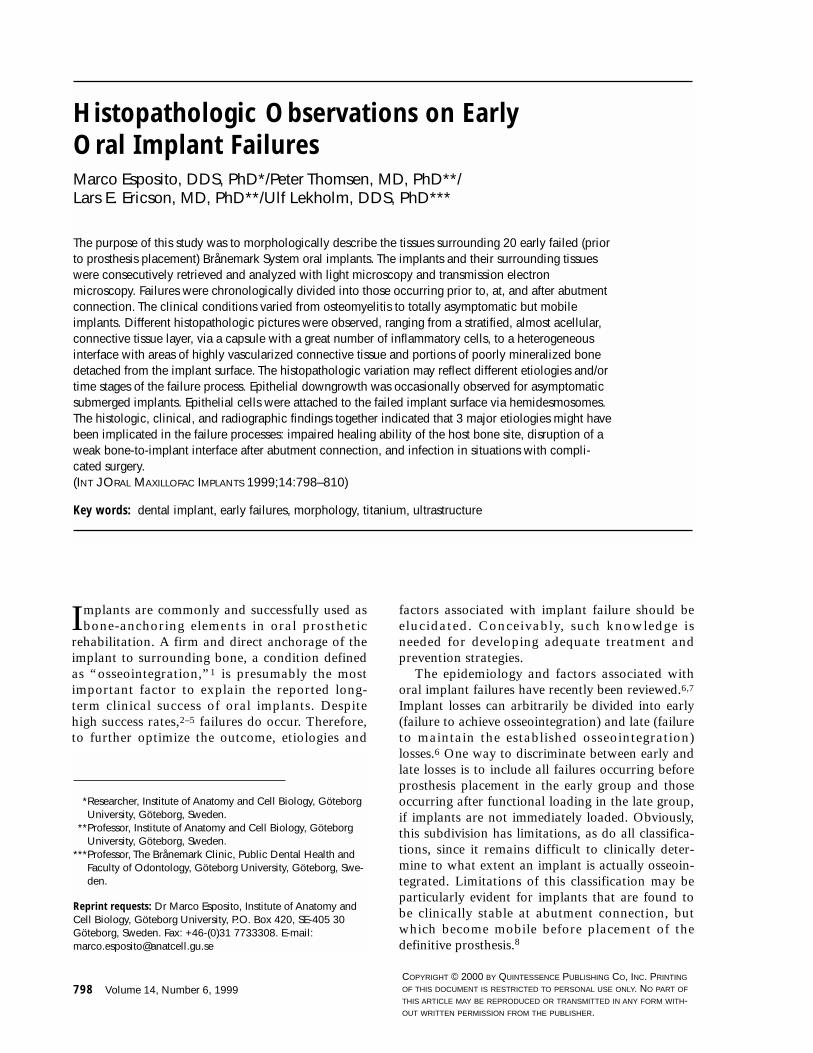
Histopathologic Observations on Early Oral Implant FailuresMarco Esposito, DDS, PhD*/Peter Thomsen, MD, PhD**/Lars E. Ericson, MD, PhD**/Ulf Lekholm, DDS, PhD***
The purpose of this study was to morphologically describe the tissues surrounding 20 early failed (priorto prosthesis placement) Brånemark System oral implants. The implants and their surrounding tissueswere consecutively retrieved and analyzed with light microscopy and transmission electronmicroscopy. Failures were chronologically divided into those occurring prior to, at, and after abutmentconnection. The clinical conditions varied from osteomyelitis to totally asymptomatic but mobileimplants. Different histopathologic pictures were observed, ranging from a stratified, almost acellular,connective tissue layer, via a capsule with a great number of inflammatory cells, to a heterogeneousinterface with areas of highly vascularized connective tissue and portions of poorly mineralized bonedetached from the implant surface. The histopathologic variation may reflect different etiologies and/ortime stages of the failure process. Epithelial downgrowth was occasionally observed for asymptomaticsubmerged implants. Epithelial cells were attached to the failed implant surface via hemidesmosomes.The histologic, clinical, and radiographic findings together indicated that 3 major etiologies might havebeen implicated in the failure processes: impaired healing ability of the host bone site, disruption of aweak bone-to-implant interface after abutment connection, and infection in situations with compli-cated surgery. (INT J ORAL MAXILLOFAC IMPLANTS 1999;14:798–810)
Key words: dental implant, early failures, morphology, titanium, ultrastructure

COPYRIGHT © 2000 BY QUINTESSENCE PUBLISHING CO, INC. PRINTING
OF THIS DOCUMENT IS RESTRICTED TO PERSONAL USE ONLY. NO PART OF
THIS ARTICLE MAY BE REPRODUCED OR TRANSMITTED IN ANY FORM WITH-OUT WRITTEN PERMISSION FROM THE PUBLISHER.
The International Journal of Oral & Maxillofacial Implants 799
Esposito et al
It is believed that excessive surgical trauma,impaired healing, bacterial contamination, andpremature overloading may be the most commoncauses of implant failure at an early stage afterimplant placement. Excessive occlusal stress inconjunction with host characteristics and bacterial-induced marginal bone loss (peri-implantitis) seemto be the major etiologic factors for late losses(apart from mechanical breakdown).7
The first histologic description of early implantfailure was provided by Brånemark et al9 in ani-mals 3 decades ago. This investigation was fol-lowed by a detailed ultrastructural description oftissues surrounding early failures of 1-stage bladeimplants, both in monkeys and humans.10 Unfor-tunately, no description of the clinical picture wasgiven. Thereafter, only sporadic studies, mainly inthe form of single case reports, have been pub-lished regarding this matter.11–18 In 2 recent arti-cles,17,18 early failures were attributed to overheat-ing of the surgical site. However, since the authorsstated that bacteria were observed in all their spec-imens, infection could have been an alternativecause. As judged by the scientific literature, nofirm evidence of the mechanisms for early failureshas been reported. However, the epidemiology ofearly losses has been described in relation to differ-ent implant systems, anatomic locations, and otherfactors.6,8,19–23
Once implant failures are recognized as beingdisease-related, a comprehensive view of the prob-lem, as with any other medical condition, might beobtained. Therefore, it is useful to analyze the epi-demiology (prevalence of the disease and variousinterrelated factors), the histopathology (the grossand microscopic changes of the disease), thepathogenesis (the disease history), and the patho-physiology (the mechanisms of the disease) ofimplant losses and complications.
The objective of the present study was to ana-lyze the histomorphology of tissues surroundingearly failures of oral implants in relation to theclinical history and radiographic findings so as toacquire additional information about possible fail-ure mechanisms.
Materials and Methods
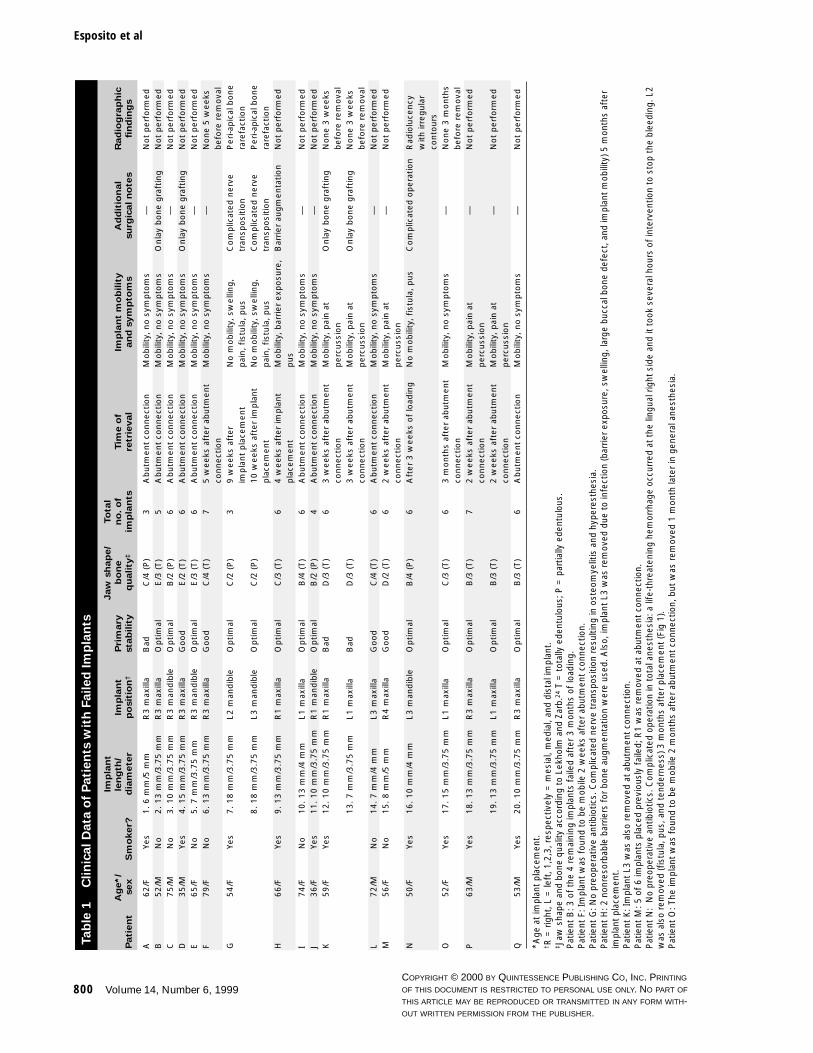
Clinical Data. Twenty failed, commercially puretitanium implants (Brånemark System, Nobel Bio-care AB, Göteborg, Sweden), consecutively removedbefore prosthesis placement (early losses) from 17patients (11 females and 6 males) treated at theBrånemark Clinic, Göteborg University, Göteborg,Sweden, were included in this study (Table 1). One
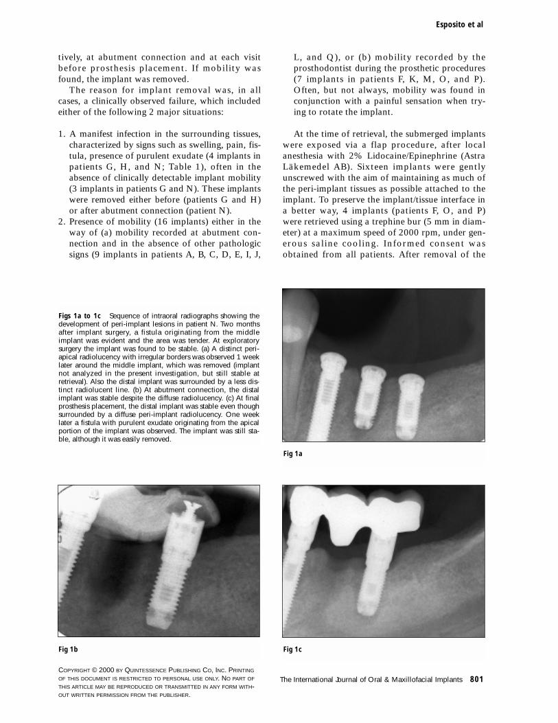
implant was removed 3 weeks after loading and itmay be argued that it should be considered as a lateloss. However, radiographic signs of failure wereevident 2 months after placement (Fig 1).
The following information was recorded atretrieval: additional postoperative drugs (type,dosage, and duration) and diseases; symptoms andtheir duration; peri-mucosal conditions (visualsigns); manual implant stability assessment; andany relevant additional finding. The remaininginformation presented in Table 1 was obtained ret-rospectively by searching the patient’s history andincluded the following: medical and oral anamne-sis including smoking habits; implant operationnotes (jaw bone resorption anatomy and bonequality at the implant site,24 vascularization,preparation of the implant site, primary implantstability, and number, length, and type of implantsplaced); pre- and post-medication; and any addi-tional relevant information.
Failed implants had various lengths (from 6 to 18mm; 5 implants ≤ 8 mm) as well as diameters (from3.75 to 5 mm; 2 were 5 mm wide and 3 were 4 mmwide) and were placed in different arch shapes andbone qualities (6 in extremely resorbed jaws [TypeD and E] and 3 implants in soft bone [Type 4]).24
The majority of losses (14) occurred in totally eden-tulous patients and in maxillae (14). Four of theretrieved implants were placed in grafted bone of 3patients (patients B, D, and K; Table 1).
All implants had been placed following a 2-stagesurgical technique.25 About 1 hour before implantsurgery, patients had routinely been administered 2g of phenoxymethylpenicillin (Kåvepenin, AstraLäkemedel AB, Södertälje, Sweden) orally. Postop-eratively, the same antibiotic treatment was contin-ued for 10 days (2 g + 2 g daily). In patients withpenicillin allergy, 1 g erythromycin was adminis-tered at that time (Ery-Max, Astra Läkemedel AB,Södertälje, Sweden, or Abboticin, Abbot Skandi-navia AB, Kista, Sweden), 1 hour preoperatively,and 1 g + 1 g daily for 10 days. However, patientG, allergic to penicillin, specifically asked forVibramycin, which was not available at the clinic.Patient N, also allergic to penicillin, was operatedon under total anesthesia and received 0.2 g oftetracycline intravenously, 11⁄2 hours after inductionof the general anesthesia. Both patients G and Nreceived postoperatively 100 mg of tetracycline(Vibramycin, Pfizer AB, Täby, Sweden) twice a dayfor 10 days. Therefore, no preoperative antibioticswere administered to those patients.
All implants were checked for stability by man-ual manipulation and/or rotation of each implantwith a pair of forceps or a screwdriver, respec-
COPYRIGHT © 2000 BY QUINTESSENCE PUBLISHING CO, INC. PRINTING
OF THIS DOCUMENT IS RESTRICTED TO PERSONAL USE ONLY. NO PART OF
THIS ARTICLE MAY BE REPRODUCED OR TRANSMITTED IN ANY FORM WITH-OUT WRITTEN PERMISSION FROM THE PUBLISHER.
800 Volume 14, Number 6, 1999
Esposito et al
Ta
ble
1C
lin
ica
l D
ata
of
Pa
tie
nts
wit
h F
ail
ed
Im
pla
nts
Imp
lan
tJaw
sh
ap
e/
To
tal
Ag
e*/
len
gth
/Im
pla
nt
Pri
mary
bo
ne
no
. o
fTim
e o
fIm
pla
nt
mo
bilit
yA
dd
itio
nal
Rad
iog
rap
hic
Pati
en
tsex
Sm
oker?
dia
mete
rp
osit
ion
†sta
bilit
yq
uality
‡im
pla
nts
retr
ieval
an
d s
ym
pto
ms
su
rgic
al n
ote
sfi
nd
ing
s
A62/F
Yes
1. 6 m
m/5
mm
R3 m
axill
aB
adC
/4 (P
)3
Abutm
ent
connect
ion
Mobili
ty, no s
ympto
ms
—N
ot
perf
orm
ed
B52/M
No
2. 13 m
m/3
.75 m
mR
3 m
axill
aO
ptim
alE
/3 (T)
5A
butm
ent
connect
ion
Mobili
ty, no s
ympto
ms
Onla
y bone g
raft
ing
Not
perf
orm
ed
C75/M
No
3. 10 m
m/3
.75 m
mR
3 m
andib
leO
ptim
alB
/2 (P
)6
Abutm
ent
connect
ion
Mobili
ty, no s
ympto
ms
—N
ot
perf
orm
ed
D35/M
Yes
4. 15 m
m/3
.75 m
mR
3 m
axill
aG
ood
E/2
(T)
6A
butm
ent
connect
ion
Mobili
ty, no s
ympto
ms
Onla
y bone g
raft
ing
Not
perf
orm
ed
E65/F
No
5. 7 m
m/3
.75 m
mR
3 m
andib
leO
ptim
alE
/3 (T)
6A
butm
ent
connect
ion
Mobili
ty, no s
ympto
ms
—N
ot
perf
orm
ed
F79/F
No
6. 13 m
m/3
.75 m
mR
3 m
axill
aG
ood
C/4
(T)
75 w
eeks
aft
er
abutm
ent
Mobili
ty, no s
ympto
ms
—N
one 5
weeks
co
nnect
ion
befo
re r
em
ova
lG
54/F
Yes
7. 18 m
m/3
.75 m
mL2 m
andib
leO
ptim
alC
/2 (P
)3
9 w
eeks
aft
er
No m
obili
ty, sw
elli
ng,
Com
plic
ated n
erv
e
Peri-a
pic
al b
one
impla
nt
pla
cem
ent
pai
n, fist
ula
, pus
tran
sposi
tion
rare
fact
ion
8. 18 m
m/3
.75 m
mL3 m
andib
leO
ptim
alC
/2 (P
)10 w
eeks
aft
er
impla
nt
No m
obili
ty, sw
elli
ng,
Com
plic
ated n
erv
e
Peri-a
pic
al b
one
pla
cem
ent
pai
n, fist
ula
, pus
tran
sposi
tion
rare
fact
ion
H66/F
Yes
9. 13 m
m/3
.75 m
mR
1 m
axill
aO
ptim
alC
/3 (T)
64 w
eeks
aft
er
impla
nt
Mobili
ty, bar
rier
exp
osu
re,
Bar
rier
augm
enta
tion
Not
perf
orm
ed
pla
cem
ent
pus
I74/F
No
10. 13 m
m/4
mm
L1 m
axill
aO
ptim
alB
/4 (T)
6A
butm
ent
connect
ion
Mobili
ty, no s
ympto
ms
—N
ot
perf
orm
ed
J36/F
Yes
11. 10 m
m/3
.75 m
mR
1 m
andib
leO
ptim
alB
/2 (P
)4
Abutm
ent
connect
ion
Mobili
ty, no s
ympto
ms
—N
ot
perf
orm
ed
K59/F
Yes
12. 10 m
m/3
.75 m
mR
1 m
axill
aB
adD
/3 (T)
63 w
eeks
aft
er
abutm
ent
Mobili
ty, pai
n a
t O
nla
y bone g
raft
ing
None 3
weeks
co
nnect
ion
perc
uss
ion
befo
re r
em
ova
l13. 7 m
m/3
.75 m
mL1 m
axill
aB
adD
/3 (T)
3 w
eeks
aft
er
abutm
ent
Mobili
ty, pai
n a
t O
nla
y bone g
raft
ing
None 3
weeks
co
nnect
ion
perc
uss
ion
befo
re r
em
ova
lL
72/M
No
14. 7 m
m/4
mm
L3 m
axill
aG
ood
C/4
(T)
6A
butm
ent
connect
ion
Mobili
ty, no s
ympto
ms
—N
ot
perf
orm
ed
M56/F
No
15. 8 m
m/5
mm
R4 m
axill
aG
ood
D/2
(T)
62 w
eeks
aft
er
abutm
ent
Mobili
ty, pai
n a
t —
Not
perf
orm
ed
connect
ion
perc
uss
ion
N50/F
Yes
16. 10 m
m/4
mm
L3 m
andib
leO
ptim
alB
/4 (P
)6
Aft
er
3 w
eeks
of
load
ing
No m
obili
ty, fist
ula
, pus
Com
plic
ated o
pera
tion
Rad
iolu
cency
w
ith ir
regula
r co
nto
urs
O52/F
Yes
17. 15 m
m/3
.75 m
mL1 m
axill
aO
ptim
alC
/3 (T)
63 m
onth
s af
ter
abutm
ent
Mobili
ty, no s
ympto
ms
—N
one 3
month
s co
nnect
ion
befo
re r
em
ova
lP
63/M
Yes
18. 13 m
m/3
.75 m
mR
3 m
axill
aO
ptim
alB
/3 (T)
72 w
eeks
aft
er
abutm
ent
Mobili
ty, pai
n a
t —
Not
perf
orm
ed
connect
ion
perc
uss
ion
19. 13 m
m/3
.75 m
mL1 m
axill
aO
ptim
alB
/3 (T)
2 w
eeks
aft
er
abutm
ent
Mobili
ty, pai
n a
t —
Not
perf
orm
ed
connect
ion
perc
uss
ion
Q53/M
Yes
20. 10 m
m/3
.75 m
mR
3 m
axill
aO
ptim
alB
/3 (T)
6A
butm
ent
connect
ion
Mobili
ty, no s
ympto
ms
—N
ot
perf
orm
ed
*Age
at
impl
ant
plac
emen
t.† R
= r
ight
, L =
left
, 1,2
,3, r
espe
ctiv
ely
= m
esia
l, m
edia
l, an
d di
stal
impl
ant.
‡ Jaw
sha
pe a
nd b
one
qual
ity a
ccor
ding
to
Lekh
olm
and
Zar
b.24
T =
tot
ally
ede
ntul
ous;
P =
par
tially
ede
ntul
ous.
P
atie
nt B
: 3 o
f th
e 4
rem
aini
ng im
plan
ts f
aile
d af
ter
3 m
onth
s of
load
ing.
Pat
ient
F: I
mpl
ant
was
fou
nd t
o be
mob
ile 2
wee
ks a
fter
abu
tmen
t co
nnec
tion.
Pat
ient
G: N
o pr
eope
rativ
e an
tibio
tics.
Com
plic
ated
ner
ve t
rans
posi
tion
resu
lting
in o
steo
mye
litis
and
hyp
eres
thes
ia.
Pat
ient
H: 2
non
reso
rbab
le b
arrie
rs f
or b
one
augm
enta
tion
wer
e us
ed. A
lso,
impl
ant
L3 w
as r
emov
ed d
ue t
o in
fect
ion
(bar
rier
expo
sure
, sw
ellin
g, la
rge
bucc
al b
one
defe
ct, a
nd im
plan
t m
obili
ty) 5
mon
ths
afte
rim
plan
t pl
acem
ent.
Pat
ient
K: I
mpl
ant
L3 w
as a
lso
rem
oved
at
abut
men
t co
nnec
tion.
Pat
ient
M: 5
of
6 im
plan
ts p
lace
d pr
evio
usly
fai
led;
R1
was
rem
oved
at
abut
men
t co
nnec
tion.
Pat
ient
N:
No
preo
pera
tive
antib
iotic
s. C
ompl
icat
ed o
pera
tion
in t
otal
ane
sthe
sia:
a li
fe-t
hrea
teni
ng h
emor
rhag
e oc
curr
ed a
t th
e lin
gual
rig
ht s
ide
and
it to
ok s
ever
al h
ours
of
inte
rven
tion
to s
top
the
blee
ding
. L2
was
als
o re
mov
ed (f
istu
la, p
us, a
nd t
ende
rnes
s) 3
mon
ths
afte
r pl
acem
ent
(Fig
1).
Pat
ient
O: T
he im
plan
t w
as f
ound
to
be m
obile
2 m
onth
s af
ter
abut
men
t co
nnec
tion,
but
was
rem
oved
1 m
onth
late
r in
gen
eral
ane
sthe
sia.
COPYRIGHT © 2000 BY QUINTESSENCE PUBLISHING CO, INC. PRINTING
OF THIS DOCUMENT IS RESTRICTED TO PERSONAL USE ONLY. NO PART OF
THIS ARTICLE MAY BE REPRODUCED OR TRANSMITTED IN ANY FORM WITH-OUT WRITTEN PERMISSION FROM THE PUBLISHER.
The International Journal of Oral & Maxillofacial Implants 801
Esposito et al
tively, at abutment connection and at each visitbefore prosthesis placement. If mobility wasfound, the implant was removed.
The reason for implant removal was, in allcases, a clinically observed failure, which includedeither of the following 2 major situations:
1. A manifest infection in the surrounding tissues,characterized by signs such as swelling, pain, fis-tula, presence of purulent exudate (4 implants inpatients G, H, and N; Table 1), often in theabsence of clinically detectable implant mobility(3 implants in patients G and N). These implantswere removed either before (patients G and H)or after abutment connection (patient N).
2. Presence of mobility (16 implants) either in theway of (a) mobility recorded at abutment con-nection and in the absence of other pathologicsigns (9 implants in patients A, B, C, D, E, I, J,
L, and Q), or (b) mobility recorded by theprosthodontist during the prosthetic procedures(7 implants in patients F, K, M, O, and P).Often, but not always, mobility was found inconjunction with a painful sensation when try-ing to rotate the implant.
At the time of retrieval, the submerged implantswere exposed via a flap procedure, after localanesthesia with 2% Lidocaine/Epinephrine (AstraLäkemedel AB). Sixteen implants were gentlyunscrewed with the aim of maintaining as much ofthe peri-implant tissues as possible attached to theimplant. To preserve the implant/tissue interface ina better way, 4 implants (patients F, O, and P)were retrieved using a trephine bur (5 mm in diam-eter) at a maximum speed of 2000 rpm, under gen-erous saline cooling. Informed consent wasobtained from all patients. After removal of the
Figs 1a to 1c Sequence of intraoral radiographs showing thedevelopment of peri-implant lesions in patient N. Two monthsafter implant surgery, a fistula originating from the middleimplant was evident and the area was tender. At exploratorysurgery the implant was found to be stable. (a) A distinct peri-apical radiolucency with irregular borders was observed 1 weeklater around the middle implant, which was removed (implantnot analyzed in the present investigation, but still stable atretrieval). Also the distal implant was surrounded by a less dis-tinct radiolucent line. (b) At abutment connection, the distalimplant was stable despite the diffuse radiolucency. (c) At finalprosthesis placement, the distal implant was stable even thoughsurrounded by a diffuse peri-implant radiolucency. One weeklater a fistula with purulent exudate originating from the apicalportion of the implant was observed. The implant was still sta-ble, although it was easily removed.
Fig 1a
Fig 1b Fig 1c

COPYRIGHT © 2000 BY QUINTESSENCE PUBLISHING CO, INC. PRINTING
OF THIS DOCUMENT IS RESTRICTED TO PERSONAL USE ONLY. NO PART OF
THIS ARTICLE MAY BE REPRODUCED OR TRANSMITTED IN ANY FORM WITH-OUT WRITTEN PERMISSION FROM THE PUBLISHER.
802 Volume 14, Number 6, 1999
Esposito et al
failed implants, the sites were carefully curettedand closed via a flap procedure. All implant sites,with the exception of those of patient G, healeduneventfully. In patient G, the implant sites wereaffected by osteomyelitis, a condition that requiredseveral months of antibiotic treatment and surgicaldebridement to heal completely.
The retrieved implants were chronologicallydivided into 3 groups: implants that failed beforeabutment connection (group 1 including 3implants), failures identified at abutment connec-tion (group 2 including 9 implants), and losses thatoccurred after abutment connection, but before thedefinitive placement of the prosthesis (group 3including 8 implants).
As a rule, no radiographs were taken ofimplants removed up to the time of abutment con-nection, unless the surgeon, in the presence of clin-ical signs and symptoms, decided otherwise(patients G and N). Intraoral radiographs wereroutinely taken at abutment connection to verifyproper abutment seating. Obtained radiographswere also inspected with regard to density andarchitecture of the bone around the implant.
Tissue Sample Preparation. Sixteen of theretrieved implants, together with their surroundingtissues, were immediately immersed in a 2.5% glu-taraldehyde in 0.01 mol/L sodium cacodylate fixa-tive solution (pH 7.4). Three other implants(patients A, C, and D) were fixed in a 4% bufferedformalin solution (pH 7.4), whereas 1 specimen(patient Q) was immediately frozen and processedfor immunohistochemistry, as previously described.26
The specimens were kept in a fixative solution forbetween 48 hours and 2 weeks before being post-fixed for 1 hour in 2% osmium tetroxide, dehy-drated in increasing concentrations of ethanol toabsolute ethanol, and, finally, embedded in plasticresin (LR White, The London Resin Co Ltd, Hamp-shire, England).
The embedded specimens were divided longitu-dinally into 2 parts by sawing. One half was subse-quently cut horizontally in 2 and 3 pieces, depend-ing on the implant length, and processed with anelectropolishing technique27 to obtain sectionswith an intact implant-tissue interface for lightmicroscopy (LM) and transmission electronmicroscopy (TEM). In brief, by an electrochemicalprocedure the bulk part of the implant was dis-solved, while a thin intact oxide layer was left indirect contact with the tissue. The specimens weremounted in a sample holder and immersed in anelectrolyte containing 5% perchloric acid, 60%methanol, and 35% n-buthanol and cooled to–30˚C. The specimen served as an anode and was
surrounded by a platinum cathode. The electropol-ishing was performed at 24 V for 2 to 4 hours.Afterward, the specimens were carefully rinsed intap water and reembedded in plastic resin (Agar100, Agar Aids, Stansted, Essex, England).
Longitudinal thin sections (1 µm) were cut forLM using a microtome (Microm HM 350, CarlZeiss AB, Stockholm, Sweden) with glass knivesand stained with 1% Azur II and 1% methyleneblue in 1% disodium tetraborate. Selected areaswere cut for TEM using an ultramicrotome (Ultra-cut, Reichert-Jung, Vienna, Austria) with diamondknives and contrasted with uranyl acetate and leadcitrate. The remaining half of selected implant-containing specimens were ground sectioned(down to 10 to 20 µm thickness)28 and stainedwith 1% toluidine blue or subjected to the fracturetechnique,29 sectioned, and stained as the electro-polished specimens.
Sections for LM were examined and pho-tographed in a Nikon Microphot FXA microscope(Bergström Instrument AB, Göteborg, Sweden).Histometric data were obtained using LeitzMicrovid equipment mounted to a Leitz Metallux3 microscope (Leica Microsystems AB, Sollentuna,Sweden) connected to a personal computer. Trans-mission electron microscopy was performed in aZeiss CEM 902 (Carl Zeiss AB) on sections 70 to80 nm thick.
Results
Clinical and Radiographic Observations. A detaileddescription of the clinical data of patients withfailed implants is presented in Table 1. From intra-oral radiographs taken either to investigate the ori-gin of some clinical symptoms (3 implants inpatients G and N) or to ascertain whether the abut-ment was properly seated (4 implants in patients F,K, O), 2 distinctly different radiographic picturesemerged. Where symptoms of infection presented(patients G and N), a diffuse radiolucency withirregular borders surrounding a large portion of theimplant was present (Fig 1). In contrast, implantsthat were asymptomatic and clinically stable atabutment connection, but which were loose a fewweeks later, before prosthesis placement, seemed tohave normal bone tissue in close contact with theimplant surface. An exception was the implantfrom patient O, in which a very thin, radiolucentperi-implant space seemed to surround the entireimplant at abutment connection. However, thisobservation was made retrospectively.
Histopathologic Observations (LM and TEM).For 6 implants, inadequate amounts of tissues
COPYRIGHT © 2000 BY QUINTESSENCE PUBLISHING CO, INC. PRINTING
OF THIS DOCUMENT IS RESTRICTED TO PERSONAL USE ONLY. NO PART OF
THIS ARTICLE MAY BE REPRODUCED OR TRANSMITTED IN ANY FORM WITH-OUT WRITTEN PERMISSION FROM THE PUBLISHER.
The International Journal of Oral & Maxillofacial Implants 803
Esposito et al
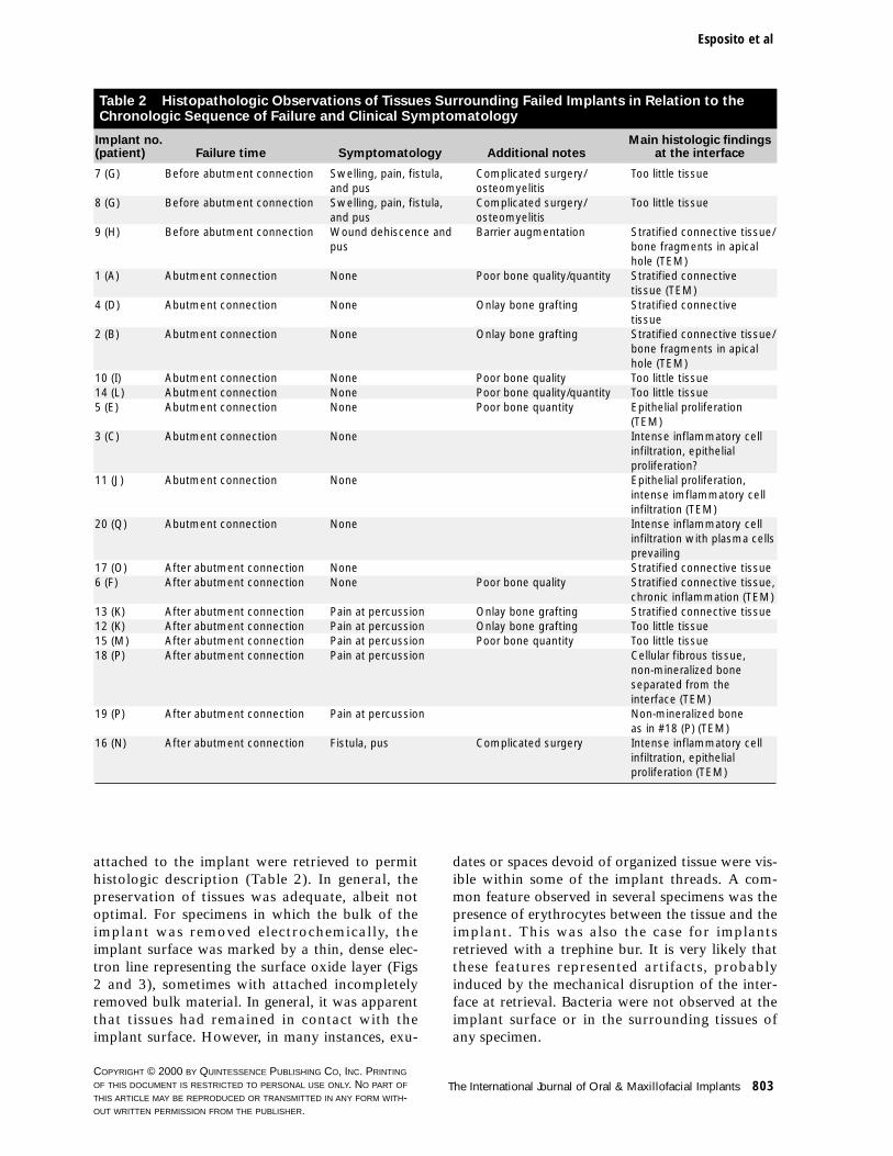
attached to the implant were retrieved to permithistologic description (Table 2). In general, thepreservation of tissues was adequate, albeit notoptimal. For specimens in which the bulk of theimplant was removed electrochemically, theimplant surface was marked by a thin, dense elec-tron line representing the surface oxide layer (Figs2 and 3), sometimes with attached incompletelyremoved bulk material. In general, it was apparentthat tissues had remained in contact with theimplant surface. However, in many instances, exu-
dates or spaces devoid of organized tissue were vis-ible within some of the implant threads. A com-mon feature observed in several specimens was thepresence of erythrocytes between the tissue and theimplant. This was also the case for implantsretrieved with a trephine bur. It is very likely thatthese features represented artifacts, probablyinduced by the mechanical disruption of the inter-face at retrieval. Bacteria were not observed at theimplant surface or in the surrounding tissues ofany specimen.
Table 2 Histopathologic Observations of Tissues Surrounding Failed Implants in Relation to theChronologic Sequence of Failure and Clinical Symptomatology
Implant no. Main histologic findings(patient) Failure time Symptomatology Additional notes at the interface
7 (G) Before abutment connection Swelling, pain, fistula, Complicated surgery/ Too little tissueand pus osteomyelitis
8 (G) Before abutment connection Swelling, pain, fistula, Complicated surgery/ Too little tissueand pus osteomyelitis
9 (H) Before abutment connection Wound dehiscence and Barrier augmentation Stratified connective tissue/pus bone fragments in apical
hole (TEM)1 (A) Abutment connection None Poor bone quality/quantity Stratified connective
tissue (TEM)4 (D) Abutment connection None Onlay bone grafting Stratified connective
tissue2 (B) Abutment connection None Onlay bone grafting Stratified connective tissue/
bone fragments in apical hole (TEM)
10 (I) Abutment connection None Poor bone quality Too little tissue14 (L) Abutment connection None Poor bone quality/quantity Too little tissue5 (E) Abutment connection None Poor bone quantity Epithelial proliferation
(TEM)3 (C) Abutment connection None Intense inflammatory cell
infiltration, epithelial proliferation?
11 (J) Abutment connection None Epithelial proliferation, intense imflammatory cell infiltration (TEM)
20 (Q) Abutment connection None Intense inflammatory cell infiltration with plasma cells prevailing
17 (O) After abutment connection None Stratified connective tissue6 (F) After abutment connection None Poor bone quality Stratified connective tissue,
chronic inflammation (TEM)13 (K) After abutment connection Pain at percussion Onlay bone grafting Stratified connective tissue12 (K) After abutment connection Pain at percussion Onlay bone grafting Too little tissue15 (M) After abutment connection Pain at percussion Poor bone quantity Too little tissue18 (P) After abutment connection Pain at percussion Cellular fibrous tissue,
non-mineralized bone separated from theinterface (TEM)
19 (P) After abutment connection Pain at percussion Non-mineralized bone as in #18 (P) (TEM)
16 (N) After abutment connection Fistula, pus Complicated surgery Intense inflammatory cell infiltration, epithelial proliferation (TEM)
COPYRIGHT © 2000 BY QUINTESSENCE PUBLISHING CO, INC. PRINTING
OF THIS DOCUMENT IS RESTRICTED TO PERSONAL USE ONLY. NO PART OF
THIS ARTICLE MAY BE REPRODUCED OR TRANSMITTED IN ANY FORM WITH-OUT WRITTEN PERMISSION FROM THE PUBLISHER.
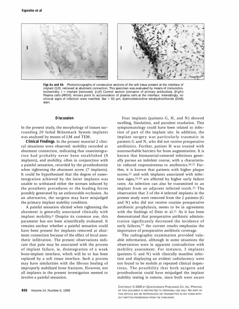
The major histologic findings in relation to thechronologic sequence of implant losses are summa-rized in Table 2. Regarding group 1, retrievedbefore abutment connection and in which clinicalsigns and symptoms of infection were present atretrieval, a sufficient amount of tissue was foundonly in the bottom hole of 1 implant (patient H).In this specimen the tissue consisted of well-vascu-larized connective tissue containing few inflamma-tory cells. Fragments of bone tissue, not displayingsigns of resorption, were embedded in stratifiedconnective tissue (Fig 4). The identity of bone wasconfirmed by TEM.
In group 2, retrieved at abutment connectionand where no clinical symptoms were apparent,bone was not found in direct contact with theimplant surface in any specimen. However, differ-ent histologic patterns were found in the retrievedtissues. The first pattern (patients A, B, and D) wastypical for a dense connective tissue capsule, withlayers of collagen and flattened fibroblasts (Fig 5).Apart from inactive (as judged from their ultra-structure) macrophages, inflammatory cells werenot present. In the bottom hole of implant B2, bonefragments were embedded in connective tissue, sim-ilar to that described above for specimen H.
A second histologic pattern was the presence ofepithelial cells in contact with the implant surface.This was verified by TEM in specimens from
804 Volume 14, Number 6, 1999
Esposito et al
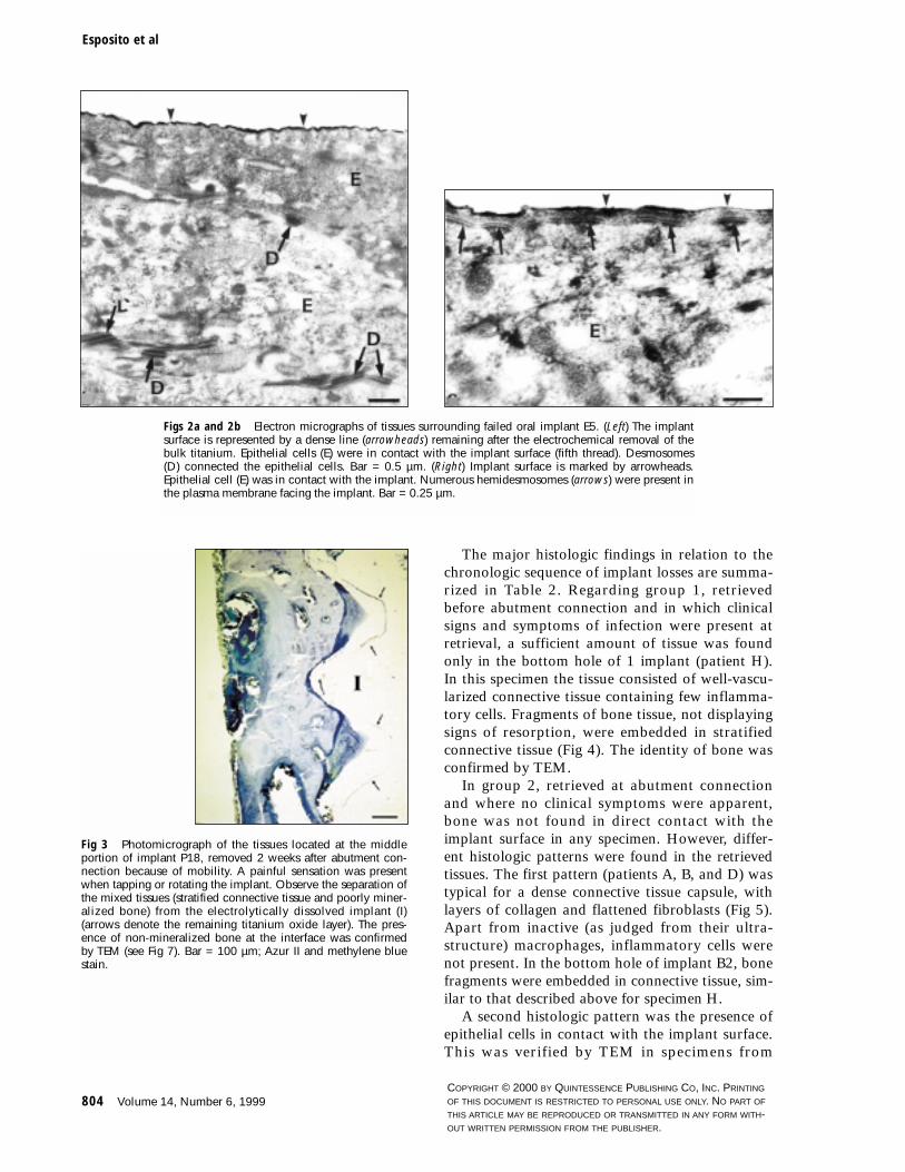
Figs 2a and 2b Electron micrographs of tissues surrounding failed oral implant E5. (Left) The implantsurface is represented by a dense line (arrowheads) remaining after the electrochemical removal of thebulk titanium. Epithelial cells (E) were in contact with the implant surface (fifth thread). Desmosomes(D) connected the epithelial cells. Bar = 0.5 µm. (Right) Implant surface is marked by arrowheads.Epithelial cell (E) was in contact with the implant. Numerous hemidesmosomes (arrows) were present inthe plasma membrane facing the implant. Bar = 0.25 µm.
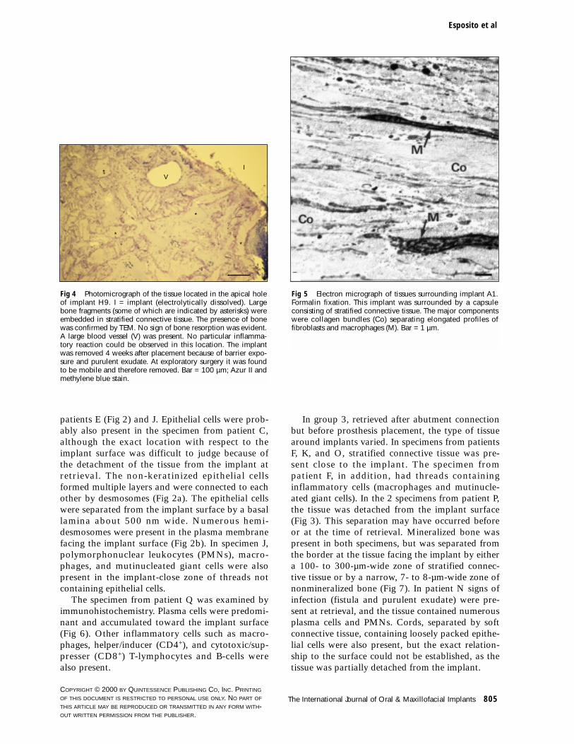
Fig 3 Photomicrograph of the tissues located at the middleportion of implant P18, removed 2 weeks after abutment con-nection because of mobility. A painful sensation was presentwhen tapping or rotating the implant. Observe the separation ofthe mixed tissues (stratified connective tissue and poorly miner-alized bone) from the electrolytically dissolved implant (I)(arrows denote the remaining titanium oxide layer). The pres-ence of non-mineralized bone at the interface was confirmedby TEM (see Fig 7). Bar = 100 µm; Azur II and methylene bluestain.
COPYRIGHT © 2000 BY QUINTESSENCE PUBLISHING CO, INC. PRINTING
OF THIS DOCUMENT IS RESTRICTED TO PERSONAL USE ONLY. NO PART OF
THIS ARTICLE MAY BE REPRODUCED OR TRANSMITTED IN ANY FORM WITH-OUT WRITTEN PERMISSION FROM THE PUBLISHER.
The International Journal of Oral & Maxillofacial Implants 805
Esposito et al
patients E (Fig 2) and J. Epithelial cells were prob-ably also present in the specimen from patient C,although the exact location with respect to theimplant surface was difficult to judge because ofthe detachment of the tissue from the implant atretrieval. The non-keratinized epithelial cellsformed multiple layers and were connected to eachother by desmosomes (Fig 2a). The epithelial cellswere separated from the implant surface by a basallamina about 500 nm wide. Numerous hemi-desmosomes were present in the plasma membranefacing the implant surface (Fig 2b). In specimen J,polymorphonuclear leukocytes (PMNs), macro-phages, and mutinucleated giant cells were alsopresent in the implant-close zone of threads notcontaining epithelial cells.
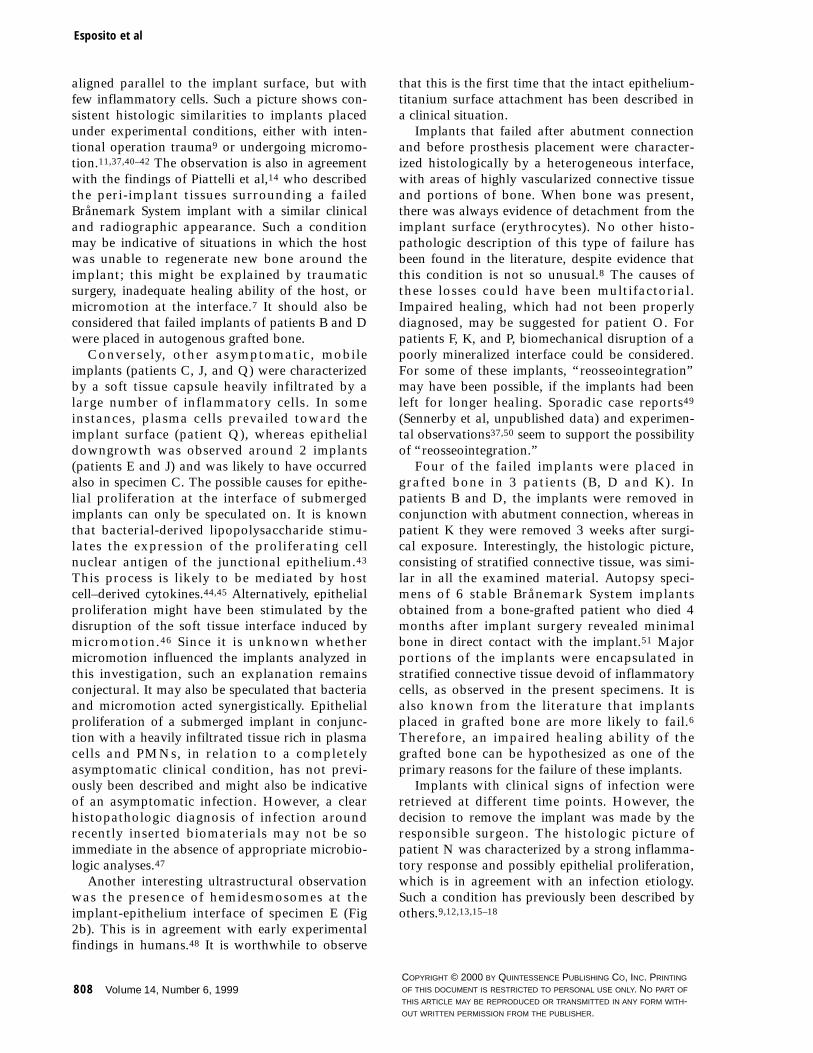
The specimen from patient Q was examined byimmunohistochemistry. Plasma cells were predomi-nant and accumulated toward the implant surface(Fig 6). Other inflammatory cells such as macro-phages, helper/inducer (CD4+), and cytotoxic/sup-presser (CD8+) T-lymphocytes and B-cells werealso present.
In group 3, retrieved after abutment connectionbut before prosthesis placement, the type of tissuearound implants varied. In specimens from patientsF, K, and O, stratified connective tissue was pre-sent close to the implant. The specimen frompatient F, in addition, had threads containinginflammatory cells (macrophages and mutinucle-ated giant cells). In the 2 specimens from patient P,the tissue was detached from the implant surface(Fig 3). This separation may have occurred beforeor at the time of retrieval. Mineralized bone waspresent in both specimens, but was separated fromthe border at the tissue facing the implant by eithera 100- to 300-µm-wide zone of stratified connec-tive tissue or by a narrow, 7- to 8-µm-wide zone ofnonmineralized bone (Fig 7). In patient N signs ofinfection (fistula and purulent exudate) were pre-sent at retrieval, and the tissue contained numerousplasma cells and PMNs. Cords, separated by softconnective tissue, containing loosely packed epithe-lial cells were also present, but the exact relation-ship to the surface could not be established, as thetissue was partially detached from the implant.
Fig 4 Photomicrograph of the tissue located in the apical holeof implant H9. I = implant (electrolytically dissolved). Largebone fragments (some of which are indicated by asterisks) wereembedded in stratified connective tissue. The presence of bonewas confirmed by TEM. No sign of bone resorption was evident.A large blood vessel (V) was present. No particular inflamma-tory reaction could be observed in this location. The implantwas removed 4 weeks after placement because of barrier expo-sure and purulent exudate. At exploratory surgery it was foundto be mobile and therefore removed. Bar = 100 µm; Azur II andmethylene blue stain.
Fig 5 Electron micrograph of tissues surrounding implant A1.Formalin fixation. This implant was surrounded by a capsuleconsisting of stratified connective tissue. The major componentswere collagen bundles (Co) separating elongated profiles offibroblasts and macrophages (M). Bar = 1 µm.
VI
*
*
*
*
COPYRIGHT © 2000 BY QUINTESSENCE PUBLISHING CO, INC. PRINTING
OF THIS DOCUMENT IS RESTRICTED TO PERSONAL USE ONLY. NO PART OF
THIS ARTICLE MAY BE REPRODUCED OR TRANSMITTED IN ANY FORM WITH-OUT WRITTEN PERMISSION FROM THE PUBLISHER.
806 Volume 14, Number 6, 1999
Esposito et al
Discussion
In the present study, the morphology of tissues sur-rounding 20 failed Brånemark System implantswas analyzed by means of LM and TEM.
Clinical Findings. In the present material 2 clini-cal situations were observed: mobility recorded atabutment connection, indicating that osseointegra-tion had probably never been established (9implants), and mobility, often in conjunction witha painful sensation, recorded by the prosthodontistwhen tightening the abutment screw (7 implants).It could be hypothesized that the degree of osseo-integration achieved by the latter implants wasunable to withstand either the stresses induced bythe prosthetic procedures or the loading forcespossibly generated by an unfavorable occlusion. Asan alternative, the surgeon may have misjudgedthe primary implant stability condition.
A painful sensation elicited when tightening theabutment is generally associated clinically withimplant mobility.6 Despite its common use, thisparameter has not been scientifically evaluated. Itremains unclear whether a painful sensation couldhave been present for implants removed at abut-ment connection because of the effect of local anes-thetic infiltration. The present observations indi-cate that pain may be associated with the processof implant failure, ie, disintegration of a weakbone-implant interface, which will be or has beenreplaced by a soft tissue interface. Such a processmay have similarities with the fibrous healing ofimproperly stabilized bone fractures. However, notall implants in the present investigation seemed toinvolve a painful sensation.
Four implants (patients G, H, and N) showedswelling, fistulation, and purulent exudation. Thissymptomatology could have been related to infec-tion of part of the implant site. In addition, theimplant surgery was particularly traumatic inpatients G and N, who did not receive preoperativeantibiotics. Further, patient H was treated withnonresorbable barriers for bone augmentation. It isknown that biomaterial-centered infections gener-ally pursue an indolent course, with a characteris-tic reduced responsiveness to antibiotics.30,31 Fur-ther, it is known that patients with higher plaquescores,32 and with implants associated with infec-tion signs,33,34 are affected by higher early failurerates. An infection can also be transmitted to animplant from an adjacent infected tooth.35 Theobservation that 3 of the 4 infected implants in thepresent study were removed from the 2 patients (Gand N) who did not receive routine preoperativeantibiotic prophylaxis, seems to be in agreementwith the findings of Dent et al.21 As it has beendemonstrated that preoperative antibiotic adminis-tration significantly decreased the incidence ofearly failures,21 the current results emphasize theimportance of preoperative antibiotic coverage.
The radiographic examination provided valu-able information, although in some situations theobservations were in apparent contradiction withmobility assessment. For instance, 3 implants(patients G and N) with clinically manifest infec-tion and displaying an evident radiolucency werenot found to be mobile at repeated clinical inspec-tions. The possibility that both surgeon andprosthodontist could have misjudged the implantstability testing is remote, since both were aware
Figs 6a and 6b Photomicrographs of consecutive sections of the soft tissue present at the interface ofimplant Q20, retrieved at abutment connection. This specimen was evaluated by means of immunohis-tochemistry. I = implant (removed). (Left) Control section (omission of primary antibodies). (Right)Plasma cells (RFD6). Arrows point to accumulation of plasma cells at the interface. Interestingly, noclinical signs of infection were manifest. Bar = 50 µm; diaminobenzidine tetrahydrochloride (DAB)stain.

COPYRIGHT © 2000 BY QUINTESSENCE PUBLISHING CO, INC. PRINTING
OF THIS DOCUMENT IS RESTRICTED TO PERSONAL USE ONLY. NO PART OF
THIS ARTICLE MAY BE REPRODUCED OR TRANSMITTED IN ANY FORM WITH-OUT WRITTEN PERMISSION FROM THE PUBLISHER.
of the clinical and radiographic pictures. Manualtesting has been shown to be a reliable parameterfor assessing implant stability, and the use of Peri-otest did not offer significant advantages.36 Thismay indicate that part of the implant was still inte-grated in bone.
Radiographs obtained after abutment connec-tion (patients F, K, and O) were unable to disclosewith any degree of certainty the implants that weregoing to become mobile a few weeks later. Ahypothesis is that weak integration or an insuffi-ciently large area of bone-implant interface mayhave been damaged, either by prosthetic manipula-tion or by occlusal overloading, resulting in theformation of a soft tissue interface.
Methodologic Aspects. Only 4 implants wereremoved en bloc with a trephine bur (patients F, O,and P), while all the others lost part of the interfacetissue during the retrieval procedures as a result ofthe attempts of the surgeon to be as conservative aspossible and not create large bone defects byremoving too much tissue. In all cases, implantsretrieved with the trephine bur had enough attachedtissue to allow adequate observations, whereas thetissue surrounding 6 of 16 implants that had beenremoved without a trephine bur was inadequate forhistologic analysis (Table 2). Three other implants(patients C, J, and N) had their surrounding soft tis-sue capsule completely or partially detached fromthe surface at removal. The observation of erythro-cytes in the threads of several implants, also when atrephine bur was used, indicates that the implant-tissue interface was broken and that bleedingoccurred at the interface during implant retrieval.However, for the implants removed after abutmentconnection, the mobility testing procedure may alsohave caused bleeding because of mechanical disrup-tion of the interface, as occasionally observed byAspenberg and Herbertsson.37
Previous investigations have shown the suitabil-ity of the electropolishing technique for studies onmetal-soft tissue interfaces. No alterations of thesoft tissue morphology, induced by electropolish-ing, have been observed27,38 (Källtorp et al, unpub-lished data). In some specimens, a fracture tech-nique was used to separate tissues from implants.29
However, in the present samples, dominated bysoft tissue at the interface, portions of tissues wereleft on the implant surface. Ground sections28 weremore difficult to evaluate than electropolished sec-tions, since they were up to 20 times thicker.Therefore, the electropolishing technique wasfound to be the best procedure available for a lightmicroscopic and ultrastructural analysis of anintact interfacial zone. Nevertheless, because of the
larger sizes and geometric complexities of clinicalimplants (internal threads, cover or abutmentscrews) as compared to experimental implants, theprocessing of clinical implants was technicallyhighly demanding. The electropolishing technique,however, has been found to induce artifacts in thebone-implant interface in the form of demineral-ization and impregnation of titanium ions.39
Therefore, it cannot be excluded that the bonedemineralization observed in specimens P18 (Fig7) and P19 could be artifactual.
Histopathologic Findings. The morphologicexamination of tissues around failed, asympto-matic implants removed at abutment connectionrevealed a lack of osseointegration and formationof a fibrous capsule. In particular, bone was absentfrom all the threads of clinically mobile implants.Two different histologic pictures were observed,which may represent different stages of the failureprocess. Some implants (patients A, B, and D)were surrounded by a dense connective tissue cap-sule rich in fibroblasts and collagen bundles
The International Journal of Oral & Maxillofacial Implants 807
Esposito et al
Fig 7 Electron micrograph of implant P18. In this specimenthe tissue was separated from the implant by erythrocytes indi-cating bleeding before or at retrieval. Mineralized bone (MB)and, closer to the implant, a 7- to 8-µm-wide zone of non-min-eralized bone (OB) was present. Bar = 1 µm.
aligned parallel to the implant surface, but withfew inflammatory cells. Such a picture shows con-sistent histologic similarities to implants placedunder experimental conditions, either with inten-tional operation trauma9 or undergoing micromo-tion.11,37,40–42 The observation is also in agreementwith the findings of Piattelli et al,14 who describedthe peri-implant tissues surrounding a failedBrånemark System implant with a similar clinicaland radiographic appearance. Such a conditionmay be indicative of situations in which the hostwas unable to regenerate new bone around theimplant; this might be explained by traumaticsurgery, inadequate healing ability of the host, ormicromotion at the interface.7 It should also beconsidered that failed implants of patients B and Dwere placed in autogenous grafted bone.
Conversely, other asymptomatic, mobileimplants (patients C, J, and Q) were characterizedby a soft tissue capsule heavily infiltrated by alarge number of inflammatory cells. In someinstances, plasma cells prevailed toward theimplant surface (patient Q), whereas epithelialdowngrowth was observed around 2 implants(patients E and J) and was likely to have occurredalso in specimen C. The possible causes for epithe-lial proliferation at the interface of submergedimplants can only be speculated on. It is knownthat bacterial-derived lipopolysaccharide stimu-lates the expression of the proliferating cellnuclear antigen of the junctional epithelium.43
This process is likely to be mediated by hostcell–derived cytokines.44,45 Alternatively, epithelialproliferation might have been stimulated by thedisruption of the soft tissue interface induced bymicromotion.46 Since it is unknown whethermicromotion influenced the implants analyzed inthis investigation, such an explanation remainsconjectural. It may also be speculated that bacteriaand micromotion acted synergistically. Epithelialproliferation of a submerged implant in conjunc-tion with a heavily infiltrated tissue rich in plasmacells and PMNs, in relation to a completelyasymptomatic clinical condition, has not previ-ously been described and might also be indicativeof an asymptomatic infection. However, a clearhistopathologic diagnosis of infection aroundrecently inserted biomaterials may not be soimmediate in the absence of appropriate microbio-logic analyses.47
Another interesting ultrastructural observationwas the presence of hemidesmosomes at theimplant-epithelium interface of specimen E (Fig2b). This is in agreement with early experimentalfindings in humans.48 It is worthwhile to observe
that this is the first time that the intact epithelium-titanium surface attachment has been described ina clinical situation.
Implants that failed after abutment connectionand before prosthesis placement were character-ized histologically by a heterogeneous interface,with areas of highly vascularized connective tissueand portions of bone. When bone was present,there was always evidence of detachment from theimplant surface (erythrocytes). No other histo-pathologic description of this type of failure hasbeen found in the literature, despite evidence thatthis condition is not so unusual.8 The causes ofthese losses could have been multifactorial.Impaired healing, which had not been properlydiagnosed, may be suggested for patient O. Forpatients F, K, and P, biomechanical disruption of apoorly mineralized interface could be considered.For some of these implants, “reosseointegration”may have been possible, if the implants had beenleft for longer healing. Sporadic case reports49
(Sennerby et al, unpublished data) and experimen-tal observations37,50 seem to support the possibilityof “reosseointegration.”
Four of the failed implants were placed ingrafted bone in 3 patients (B, D and K). Inpatients B and D, the implants were removed inconjunction with abutment connection, whereas inpatient K they were removed 3 weeks after surgi-cal exposure. Interestingly, the histologic picture,consisting of stratified connective tissue, was simi-lar in all the examined material. Autopsy speci-mens of 6 stable Brånemark System implantsobtained from a bone-grafted patient who died 4months after implant surgery revealed minimalbone in direct contact with the implant.51 Majorportions of the implants were encapsulated instratified connective tissue devoid of inflammatorycells, as observed in the present specimens. It isalso known from the literature that implantsplaced in grafted bone are more likely to fail.6Therefore, an impaired healing ability of thegrafted bone can be hypothesized as one of theprimary reasons for the failure of these implants.
Implants with clinical signs of infection wereretrieved at different time points. However, thedecision to remove the implant was made by theresponsible surgeon. The histologic picture ofpatient N was characterized by a strong inflamma-tory response and possibly epithelial proliferation,which is in agreement with an infection etiology.Such a condition has previously been described byothers.9,12,13,15–18
808 Volume 14, Number 6, 1999
Esposito et al
COPYRIGHT © 2000 BY QUINTESSENCE PUBLISHING CO, INC. PRINTING
OF THIS DOCUMENT IS RESTRICTED TO PERSONAL USE ONLY. NO PART OF
THIS ARTICLE MAY BE REPRODUCED OR TRANSMITTED IN ANY FORM WITH-OUT WRITTEN PERMISSION FROM THE PUBLISHER.
Conclusions
The clinical, radiographic, and histopathologic pic-tures of the tissues surrounding early implant fail-ures analyzed in the present investigation, takentogether, indicated that infection (in patients whounderwent complicated operations), impaired heal-ing, and disruption of a weak bone-implant inter-face seem to be the major etiologic factors involved.
Acknowledgments
The financial support from the National Research Council ofItaly (CNR), the Swedish Medical Research Council (9495), theFaculties of Odontology and Medicine at Göteborg University,the Swedish National Board for Technical Development(NUTEK), the C. M. Lerici Foundation, the Hjalmar SvenssonFoundation, the Greta and Einar Asker Foundation, and theAdlerbertska Research Fund is gratefully acknowledged. Wewish to thank Gunnel Bokhede, Lena Emanuelsson, and AnnKristin Blomgren for their skillful technical assistance. The sup-port of the staff at the Brånemark Clinic is greatly appreciated.
References
1. Albrektsson T, Brånemark P-I, Hansson H-A, Lindström J.Osseointegrated titanium implants. Requirements forensuring a long-lasting, direct bone-to-implant anchoragein man. Acta Orthop Scand 1981;52:155–170.
2. Lekholm U, van Steenberghe D, Herrmann I, Bolender C,Folmer T, Gunne J, et al. Osseointegrated implants in thetreatment of partially edentulous jaws: A prospective 5-year multicenter study. Int J Oral Maxillofac Implants1994;9:627–635.
3. Jemt T, Chai J, Harnet J, Heath MR, Hutton JE, Johns RB,et al. A 5-year prospective multicenter follow-up report onoverdentures supported by osseointegrated implants. Int JOral Maxillofac Implants 1996;11:291–298.
4. Henry PJ, Laney WR, Jemt T, Harris D, Krogh PHJ, PolizziG, et al. Osseointegrated implants for single-tooth replace-ment: A prospective 5-year multicenter study. Int J OralMaxillofac Implants 1996;11:450–455.
5. Friberg B, Nilson H, Olsson M, Palmquist C. Mark II: Theself-tapping Brånemark implant: 5-year results of aprospective 3-center study. Clin Oral Implants Res 1997;8:279–285.
6. Esposito M, Hirsch J-M, Lekholm U, Thomsen P. Biologi-cal factors contributing to failures of osseointegrated oralimplants. (I) Success criteria and epidemiology. Eur J OralSci 1998;106:527–551.
7. Esposito M, Hirsch J-M, Lekholm U, Thomsen P. Biologi-cal factors contributing to failures of osseointegrated oralimplants. (II) Etiopathogenesis. Eur J Oral Sci 1998;106:721–764.
8. Friberg B, Jemt T, Lekholm U. Early failures in 4,641 con-secutively placed Brånemark dental implants: A study fromstage 1 surgery to the connection of completed prostheses.Int J Oral Maxillofac Implants 1991;6:142–146.
9. Brånemark P-I, Breine U, Adell R, Hansson BO, LindströmJ, Ohlsson A. Intra-osseous anchorage of dental prostheses.I. Experimental studies. Scand J Plast Reconstr Surg 1969;3:81–100.
10. Meenaghan MA, Natiella JR, Armitage JE, Greene GWJ,Lipani CS. The crypt surface of blade-vent implants in clin-ical failure: An electron microscopic study. J Prosthet Dent1974;31:681–690.
11. Brunski JB, Moccia AFJ, Pollack SR, Korostoff E, Tracht-enberg DI. The influence of functional use of endosseousdental implants on the tissue-implant interface. I. Histolog-ical aspects. J Dent Res 1979;58:1953–1969.
12. Takeshita F, Kuroki H, Yamasaki A, Suetsugu T.Histopathologic observation of seven removed endosseousdental implants. Int J Oral Maxillofac Implants 1995;10:367–372.
13. Takeshita F, Suetsugu T, Higuchi Y, Oishi M. Histologicstudy of failed hollow implants. Int J Oral MaxillofacImplants 1996;11:245–250.
14. Piattelli A, Scarano A, Piattelli M. Microscopical aspects offailure in osseointegrated dental implants: A report of fivecases. Biomaterials 1996;17:1235–1241.
15. Takeshita F, Iyama S, Ayukawa Y, Suetsugu T, Oishi M.Abscess formation around a hydroxyapatite-coated implantplaced into the extraction socket with autogenous bonegraft. A histological study using light microscopy, imageprocessing, and confocal laser scanning microscopy. J Peri-odontol 1997;68:299–305.
16. Piattelli A, Scarano A, Piattelli M, Podda G. Implant peri-apical lesions: Clinical, histologic, and histochemicalaspects. A case report. Int J Periodontics Restorative Dent1998;18:181–187.
17. Piattelli A, Piattelli M, Mangano C, Scarano A. A histo-logic evaluation of eight cases of failed dental implants: Isbone overheating the most probable cause? Biomaterials1998;19:683–690.
18. Piattelli A, Scarano A, Dalla Nora A, De Bona G, FaveroGA. Microscopical features in retrieved human Brånemarkimplants: A report of 19 cases. Biomaterials 1998;19:643–649.
19. Esposito M, Hirsch J-M, Lekholm U, Thomsen P. Failurepatterns of four osseointegrated oral implant systems. JMater Sci Mater Med 1997;8:843–847.
20. Truhlar RS, Farish SE, Scheitler LE, Morris HF, Ochi S.Bone quality and implant design-related outcomes throughstage II surgical uncovering of Spectra System root formimplants. J Oral Maxillofac Surg 1997;55(suppl 5):46–54.
21. Dent CD, Olson JW, Farish SE, Bellome J, Casino AJ, Mor-ris HF, Ochi S. The influence of preoperative antibiotics onsuccess of endosseous implants up to and including stage IIsurgery: A study of 2,641 implants. J Oral Maxillofac Surg1997;55(suppl 5):19–24.
22. Morris HF, Manz MC, Tarolli JH. Success of multipleendosseous dental implant designs to second-stage surgeryacross study sites. J Oral Maxillofac Surg 1997;55(suppl 5):76–82.
23. Lambert PM, Morris HF, Ochi S. Positive effect of surgicalexperience with implants on second-stage implant survival.J Oral Maxillofac Surg 1997;55(suppl 5):12–18.
24. Lekholm U, Zarb GA. Patient selection and preparation.In: Brånemark P-I, Zarb GA, Albrektsson T (eds). Tissue-Integrated Prostheses. Chicago: Quintessence, 1985:199–209.
25. Adell R, Lekholm U, Brånemark P-I. Surgical procedures.In: Brånemark P-I, Zarb GA, Albrektsson T (eds). Tissue-Integrated Prostheses. Chicago: Quintessence, 1985:211–232.
The International Journal of Oral & Maxillofacial Implants 809
Esposito et al
COPYRIGHT © 2000 BY QUINTESSENCE PUBLISHING CO, INC. PRINTING
OF THIS DOCUMENT IS RESTRICTED TO PERSONAL USE ONLY. NO PART OF
THIS ARTICLE MAY BE REPRODUCED OR TRANSMITTED IN ANY FORM WITH-OUT WRITTEN PERMISSION FROM THE PUBLISHER.
COPYRIGHT © 2000 BY QUINTESSENCE PUBLISHING CO, INC. PRINTING
OF THIS DOCUMENT IS RESTRICTED TO PERSONAL USE ONLY. NO PART OF
THIS ARTICLE MAY BE REPRODUCED OR TRANSMITTED IN ANY FORM WITH-OUT WRITTEN PERMISSION FROM THE PUBLISHER.
810 Volume 14, Number 6, 1999
Esposito et al
26. Esposito M. Thomsen P, Mölne J, Gretzer C, Ericson LE,Lekholm U. Immunohistochemistry of soft tissues sur-rounding late failures of Brånemark implants. Clin OralImplants Res 1997;8:352–366.
27. Bjursten LM, Emanuelsson L, Ericson LE, Thomsen P,Lausmaa J, Mattson L, et al. A new method for ultrastruc-tural studies of the intact tissue-metal interface. Biomateri-als 1990;11:569–601.
28. Donath K, Breuner G. A method for the study of undecalci-fied bones and teeth with attached soft tissues. The Sage-Schliff (sawing and grinding) technique. J Oral Pathol1982;11:318–326.
29. Thomsen P, Ericson LE. Light and transmission electronmicroscopy used to study the tissue morphology close toimplants. Biomaterials 1985;6:421–424.
30. Dougherty SH, Simmons RL. Infections in bionic man: Thepathobiology of infections in prosthetic devices—Part II.Curr Probl Surg 1982;19:269–318.
31. Nishioka GJ, Jones J, Triplett RG, Aufdemorte TB. Therole of bacterial-laden biofilms in infections of maxillofa-cial biomaterials. J Oral Maxillofac Surg 1988;46:19–25.
32. Van Steenberghe D, Lekholm U, Bolender C, Folmer T,Henry P, Herrmann I, et al. The applicability of osseointe-grated oral implants in the rehabilitation of partial eden-tulism: A prospective multicenter study on 558 fixtures. IntJ Oral Maxillofac Implants 1990;5:272–281.
33. Quirynen M, Naert I, van Steenberghe D, Dekeyser C, Cal-lens A. Periodontal aspects of osseointegrated fixtures sup-porting a partial bridge. An up to 6-years retrospectivestudy. J Clin Periodontol 1992;19:118–126.
34. Lambert PM, Morris HF, Ochi S. The influence of 0.12%chlorhexidine digluconate rinses on the incidence of infec-tious complications and implant success. J Oral MaxillofacSurg 1997;55(suppl 5):25–30.
35. Sussman HI, Moss SS. Localized osteomyelitis secondary toendodontic-implant pathosis. A case report. J Periodontol1993;64:306–310.
36. Isidor F. Mobility assessment with the Periotest system inrelation to histologic findings of oral implants. Int J OralMaxillofac Implants 1998;13:377–383.
37. Aspenberg P, Herbertsson P. Periprosthetic bone resorption.J Bone Joint Surg [Br] 1996;78:641–646.
38. Eriksson AS, Bjursten LM, Ericson LE, Thomsen P. Hollowimplants in soft tissues allowing quantitative studies ofcells and fluid at the implant interface. Biomaterials 1988;9:86–90.
39. Ericson LE, Johansson BR, Rosengren A, Sennerby L,Thomsen P. Ultrastructural investigation and analysis ofthe interface of retrieved metal implants. In: Davies JE (ed).The Bone-Biomaterial Interface. Toronto: Univ of TorontoPress, 1991:425–437.
40. Cameron HU, Pilliar RM, Macnab I. The effect of move-ment on the bonding of porous metal to bone. J BiomedMater Res 1973;7:301–311.
41. Aspenberg P, Goodman S, Toksvig-Larsen S, Ryd L,Albrektsson T. Intermittent micromotion inhibits boneingrowth. Titanium implants in rabbits. Acta OrthopScand 1992;63:141–145.
42. Goodman S, Wang J-S, Doshi A, Aspenberg P. Difference inbone ingrowth after one versus two daily episodes ofmicromotion: Experiments with titanium chambers in rab-bits. J Biomed Mater Res 1993;27:1419–1424.
43. Takata T, Miyauchi M, Ogawa I, Ito H, Kobayashi J, NikaiH. Reactive change in proliferative activity of the junc-tional epithelium after topical application of lipopolysac-charide. J Periodontol 1997;68:531–535.
44. Ristow H-J. A major factor contributing to epidermal pro-liferation in inflammatory skin diseases appears to be inter-leukin-1 or a related protein. Proc Natl Acad Sci USA1987;84:1940–1944.
45. Lee SW, Morhenn VB, Ilnicka M, Eugui EM, Allison AC.Autocrine stimulation of interleukin-1 alpha and trans-forming growth factor alpha production in human ker-atinocytes and its antagonism by glucocorticoids. J InvestDermatol 1991;97:106–110.
46. Jansen JA, de Groot K. Guinea pig and rabbit model forthe histological evaluation of permanent percutaneousimplants. Biomaterials 1988;9:268–272.
47. Petty W, Spanier S, Shuster JJ, Silverthrone C. The influ-ence of skeletal implants on incidence of infection. J BoneJoint Surg [Am] 1985;67:1236–1244.
48. Gould TRL, Westbury L, Brunette DM. Ultrastructuralstudy of the attachment of human gingiva to titanium invivo. J Prosthet Dent 1984;52:418–420.
49. Engquist B, Nilson H, Åstrand P. Single-tooth replacementby osseointegrated Brånemark implants. A retrospectivestudy of 82 implants. Clin Oral Implants Res 1995;6:238–245.
50. Ivanoff C-J, Sennerby L, Lekholm U. Reintegration ofmobilized titanium implants. An experimental study in rab-bit tibia. Int J Oral Maxillofac Surg 1997;26:310–315.
51. Nyström E, Kahnberg K-E, Albrektsson T. Treatment ofthe severely resorbed maxillae with bone graft and tita-nium implants: Histologic review of autopsy specimens. IntJ Oral Maxillofac Implants 1993;8:167–172.