hormones digesting, absorbing and assimilating a meal requires precise coordination of a huge number...
TRANSCRIPT

Hormones Digesting, absorbing and assimilating a meal requires precise coordination of a huge
number of physiologic processes. Control over GI functions is
provided by nervous and endocrine systems.

Gastrointestinal (GI)
The hormones most important in controlling digestive function are
synthesized within the GI tract by cells scattered in the epithelium of the stomach
and SI. These endocrine cells and the hormones they secrete are referred to as the
enteric endocrine system. Interestingly, most if not all "GI hormones" are also
synthesized in the brain.

Overview of the Digestive SystemConsider for a moment a Big Mac. The purpose of eating a Big
Mac (besides hedonism), is to assimlate the nutrients it represents and make them available to build, repair and maintain your own
tissues, as well as provide energy for studying and occasional other pursuits.
You may have asked yourself - "Exactly what nutrients are present in a Big Mac that I can assimilate?" MacDonald's comes close to full disclosure in this regard- They don't tell you that in order to take advantage of these nutrients, you have to provide the means to carefully break them down into much smaller molecules that
can be imported into blood. Luckily, your digestive system takes care of this very complex
process so efficiently

Overview of the Digestive System
At its simplest, the digestive system it is a tube running from mouth to anus.
This tube is like an assembly line, or more properly-a disassembly line.
chief goal is to break down huge macromolecules (proteins, fats and starch), which cannot be
absorbed into smaller molecules (amino acids, fatty acids and glucose) that can be absorbed
across the wall of the tube, and into the circulatory system for dissemination around your
body.

The breakdown of foodstuffs is accomplished through a
combination of mechanical and enzymatic processes. -the digestive tube requires considerable assistance from
accessory digestive organs such as the salivary glands, liver and pancreas, which dump their secretions into the tube.
The name "accessory" should not be taken to mean dispensible-without pancreatic enzymes you would starve
to death in short order.

In many ways, the digestive system can be
thought of as a well-run factory in which a large number of complex tasks are performed. The
three fundamental processes that take place are:
Secretion: Delivery of enzymes, mucus, ions and the like into the lumen, and hormones into blood
Absorption: Transport of water, ions and nutrients from the lumen, across the epithelium
and into blood Motility: Contractions of smooth muscle in the wall of the tube that crush, mix and propel its
contents

Like any well-run factory, proper function of the digestive system requires robust control systems.
Control systems must facilitate communication among different sections of the digestive tract (i.e.
control on the factory floor), and between the digestive tract and the brain (i.e. between workers
and management). Control of digestive function is achieved through
a combination of electrical and hormonal messages which originate either within the
digestive system's own nervous and endocrine systems, as well as from the CNS and from
endocrine organs such as the adrenal gland.

Different parts of these systems are constantly talking to one another.
The basic messages are along the lines of "I just received an extraordinary load of food, so I suggest you get prepared" (stomach to large intestine) or
"For goodness sake, please slow down until I can catch up with what you've already given me"
(small intestine to stomach).

Finally, a note about differences in digestive anatomy and physiology among animals. The
digestive systems of humans, dogs, mice, horses, kangaroos and great white sharks are, to a first
approximation, virtually identical.
If you look more carefully however, it becomes apparent that each of these species has evolved
certain digestive specializations that have allowed it to adapt to a particular diet.

These differences become particularly apparent when you
compare a carnivore like a cat with a herbivore like a goat or a horse. Goats and horses evolved from
ancestors that subsisted on plants and adapted parts of their digestive
tracts into massive fermentation vats which enabled them to efficiently
utilize cellulose, the major carbohydrate of plants.
In contrast, cats evolved from animals that lived on the carcasses of other animals, and have digestive systems that reflect this history -
extremely small fermentation vats and essentially no ability to utilize cellulose. Bridging the gap between carnivores and herbivores are omnivores like humans and pigs, whose digestive tracts attest to a
historical diet that included both plants and animals. The image above shows a young omnivore in the company of herbivore and carnivore
friends.

Basic Functional Anatomy of the Digestive System

The digestive system depicted on previous slide-
a carnivore – is the simplest among mammals.
Other species, even humans, have a more or very much more extensive large intestine, and ruminants like cattle
and sheep have a large set of forestomachs through which food passes before it reaches the stomach.
Each of the organs contributes to the digestive process in several unique ways. If you were to describe their most
important or predominant function, and summarize shamelessly, the list would look something like this:

Mouth: Foodstuffs are broken down mechanically by chewing and saliva is added as a lubricant. In some
species, saliva contains amylase, an enzyme that digests starch.
Esophagus: A simple conduit between the mouth and stomach - clearly important but only marginally
interesting compared to other regions of the tube.
Stomach: Where the real action begins - enzymatic digestion of proteins initiated and foodstuffs reduced to
liquid form. Liver: The center of metabolic activity in the body - its
major role in the digestive process is to provide bile salts to the small intestine, which are critical for digestion and
absorption of fats.

Pancreas: Important roles as both an endocrine and
exocrine organ - provides a potent mixture of digestive enzymes to the small intestine which are critical for
digestion of fats, carbohydrates and protein.
Small Intestine: The most exciting place to be in the entire digestive system - this is where the final stages of chemical enzymatic digestion occur and where almost
almost all nutrients are absorbed.
Large Intestine: Major differences among species in extent and importance - in all animals water is absorbed, bacterial fermentation takes place and feces are formed.
In carnivores, that's about the extent of it, but in herbivores like the horse, the large intestine is huge and
of critical importance for utilization of cellulose.

Microbial Life in the Digestive Tract
The GI contains an immensely complex ecology of microorganisms.
A typical person harbors more than 500 distinct species of bacteria, representing dozens of
different lifestyles and capabilities. The composition and distribution of this
menagerie varies with age, state of health and diet.

The number and type of bacteria in the GI tract vary dramatically by region.
In healthy individuals the stomach and proximal SI contain few microorganisms, largely a result of the
bacteriocidal activity of GASTRIC ACID
In sharp contrast to the stomach and SI, the contents of the colon literally teem with bacteria, predominantly
strict anaerobes . Between these two extremes is a transitional zone,
usually in the ileum, where moderate numbers of both aerobic and anaerobic bacteria are found.
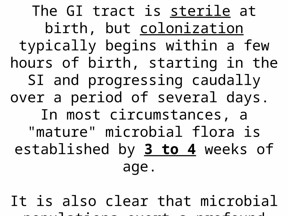
The GI tract is sterile at birth, but colonization
typically begins within a few hours of birth, starting in the SI and progressing caudally over
a period of several days. In most circumstances, a "mature" microbial flora is established by 3 to 4 weeks of age.
It is also clear that microbial populations exert a profound effect on structure and function of
the digestive tract.

For example:
The morphology of the intestine of germ-free animals differs considerably from
normal animals - villi of the SI are remarkably regular and the rate of epithelial cell renew is
reduced
The cecum of germ-free rats is roughly 10 times the size of that in a conventional rat.

Bacteria in the intestinal lumen metabolize a variety of sterols and
steroids. For example, bacteria convert the bile
salt cholic acid to deoxycholic acid.
Small intestinal bacteria also have a important role in sex steroid metabolism.

Finally, bacterial populations in the large intestine digest carbohydrates, proteins and lipids that escape digestion
and absorption in SI. This fermentation, particularly of cellulose, is of critical importance to herbivores like cattle and horses which
make a living by consuming plants. However, it seems that even species like humans and rodents derive significant benefit from the nutrients
liberated by intestinal microorganisms.

Overview of GI Hormones
If you are like most people, you eat several meals and occasional snacks each day, but rarely think about the immense number of tasks that must be performed by your digestive system to break
down, absorb and assimilate those nutrients. Robust control systems are required to coordinate digestive processes in man and animals, and are provided by both the
nervous and endocrine systems.
There are a bunch of hormones, neuropeptides and neurotransmitters that affect GI function.
Interestingly, a number of the classical GI hormones are also synthesized in the brain, and sometimes
referred to as "brain-gut peptides".

Major PLAYERS

Hormone -Major Activities-Stimuli for Release
Gastrin-Stimulates gastric acid secretion & proliferation of gastric epithelium Presence of peptides and amino acids in gastric lumen
Cholecystokinin-Stimulates secretion of pancreatic enzymes, and contraction and emptying of the gall bladder Presence of fatty acids and amino acids in the small intestine
Secretin-Stimulates secretion of water and bicarbonate from the pancreas and bile ducts Acidic pH in the lumen of the small SI
Ghrelin- a strong stimulant for appetite and feeding; also a potent stimulator of GH secretion. Not clear, but secretion peaks prior to feeding and diminishes with gastric filling

Hormone Major Activities Stimuli for Release
Motilin-Apparently involved in stimulating housekeeping patterns of motility in the stomach and small intestine-Not clear, but secretion is associated with fasting
Gastric inhibitory polypeptide-Inhibits gastric secretion and motility and potentiates release of insulin from beta cells in response to elevated blood glucose concentration-Presence of fat and glucose in the small intestine

Gastrin
Gastrin is a major physiological regulator of gastric acid secretion.
Also has an important trophic or growth-promoting influence on the gastric mucosa. Gastrin is synthesized in G cells, which are
located in gastric pits, primarily in the antrum region of the stomach and binds
receptors found predominantly on parietal and enterochromaffin-like cells.

4 major types of secretory epithelial cells cover the surface of the stomach and extend down into
gastric pits and glands:
Mucous cells: secrete an alkaline mucus that protects the epithelium
against shear stress and acid
Parietal cells: secrete hydrochloric acid!
Chief cells: secrete pepsin, a proteolytic enzyme
G cells: secrete the hormone gastrin

Structure of Gastrin and the Gastrin Receptor
Gastrin is a linear peptide that is synthesized as a preprohormone and is post-translationally cleaved to
form a family of peptides with identical carboxy termini.
The predominant circulating form is gastrin-34 ("big gastrin"), but full biologic activity is present in the
smallest peptide (gastrin-14 or minigastrin).
Further, full bioactivity is preserved in the 5 C-terminal aa’s of gastrin, which is known as pentagastrin. The five C-terminal amino acids of gastrin and
cholecystokinin are identical, which explains their overlapping biological effects.


The gastrin receptor is also one of the receptors that
bind cholecystokinin, and is known as the CCK-B receptor.
It is a member of the G protein-coupled receptor family. Binding of gastrin stimulates an increase in intracellular Ca++, activation of protein kinase C, and production of
inositol phosphate.

Control and Physiologic Effects of Gastrin
The primary stimulus for secretion of gastrin is the presence of certain foodstuffs, especially
peptides, certain amino acids and calcium, in the gastric lumen.
Also, as yet unidentified compounds in coffee, wine and beer are potent stimulants for gastrin
secretion. Secretion of this hormone is inhibited when the lumenal pH of the stomach becomes very low
(less than about 3).

Control and Physiologic Effects of Gastrin
Gastrin appears to have at least two major effects on gastrointestinal function:
Stimulation of gastric acid secretion: Gastrin receptors are found on parietal cells, and binding of gastrin, along
with histamine and acetylcholine, leads to fully-stimulated acid secretion by those cells.
Canine parietal cells have roughly 44,000 gastrin receptors each, and in that species, it has been
demonstrated that immunoneutralization of gastrin blocks secretion of acid in response to
intragastric administration of peptides.

Control and Physiologic Effects of Gastrin
Gastrin appears to have at least two major effects on gastrointestinal function:
Enterochromaffin-like (ECL) cells also bear gastrin receptors, and recent evidence
indicates that this cell may be the most important target of gastrin with regard to regulating acid secretion. Stimulation of ECL cells by gastrin leads to histamine release, and histamine binding to H2
receptors on parietal cells is necessary for full-blown acid secretion.

Control and Physiologic Effects of Gastrin
Gastrin appears to have at least two major effects on gastrointestinal function:
Promotion of gastric mucosal growth: Gastrin clearly has the ability to stimulate many aspects of mucosal
development and growth in the stomach. Treatment with gastrin stimulates DNA, RNA and
protein synthesis in gastric mucosa and increases the number of parietal cells.
Another observation supporting this function is that humans with hypergastrinemia (abnormally high blood
levels of gastrin) consistently show gastric mucosal hypertrophy.

In addition to parietal and ECL cell targets,
gastrin also stimulates pancreatic acinar cells via binding to CCK receptors, and
gastrin receptors have been demonstrated on certain populations of gastric smooth muscle cells, supporting pharmacologic
studies that demonstrate a role for gastrin in regulating gastric motility.

Disease States
Excessive secretion of gastrin, or hypergastrinemia, is a well-recognized cause of a severe disease known as
Zollinger-Ellison syndrome, which is seen at low frequency in man and dogs.
The hallmark of this disease is gastric and duodenal ulceration due to excessive and unregulated secretion of
gastric acid. Most commonly, hypergastrinemia is the result of
gastrin-secreting tumors (gastrinomas), which develop in the pancreas or duodenum.

Zollinger-Ellison Syndrome (ZES)
-rare disorder that causes tumors in the pancreas and duodenum and ulcers in the stomach and duodenum.
-Excesss gastrin cause the stomach to produce too much acid, which in turn causes stomach and duodenal ulcers
(peptic ulcers). The ulcers caused by ZES are less responsive to treatment than ordinary peptic ulcers.
-What causes people with ZES to develop tumors is unknown, but approximately 25 % of ZES cases are
associated with a genetic disorder called MEN 1.

Zollinger-Ellison Syndrome (ZES)
The symptoms of ZES include signs of peptic ulcers: gnawing, burning pain in the abdomen; diarrhea; nausea; vomiting; fatigue; weakness;
weight loss; and bleeding.
Physicians diagnose ZES through blood tests to measure levels of gastrin and gastric acid
secretion. They may check for ulcers by doing an
endoscopy.

Zollinger-Ellison Syndrome (ZES)
The primary treatment for ZES is medication to reduce
the production of stomach acid. Proton pump inhibitors that suppress acid production and promote healing are the first line of treatment. H-2 blockers (cimetidine, famotidine, ranitidine) may also be used, but are less effective in reducing stomach
acid. Surgery to treat peptic ulcers or to remove tumors in the
pancreas or duodenum are other treatment options. People who have been treated for ZES should be
monitored in case the ulcers or tumors recur.


Cholecystokinin (CCK)
Cholecystokinin plays a key role in facilitating digestion within the SI. It is secreted from
mucosal epithelial cells in the first segment of the small intestine (duodenum), and stimulates delivery into the small intestine of digestive
enzymes for the pancreas and bile from the gall bladder.
CCK is also produced by neurons in the enteric nervous system and is widely and abundantly
distributed in the brain.

Structure of Cholecystokinin and Its Receptors
CCK and gastrin are highly similar peptides.
Like gastrin, CCK is a linear peptide that is synthesized as a preprohormone, then
proteolytically cleaved to generate a family of peptides having the same carboxy ends.
Full biologic activity is retained in CCK-8 (8 amino acids), but peptides of 33, 38 and 59 amino acids are
also produced. In all of these CCK peptides, the tyrosine seven
residues from the end is sulfated, which is necessary for activity.

Structure of Cholecystokinin and Its Receptors
Two receptors that bind cholecystokinin have been identified.
CCKA receptor is found abundantly on pancreatic acinar cells.
CCKB receptor, which also functions as the gastrin receptor, is the predominant form in brain and
stomach. Both receptors are have 7TMD typical of G protein-
coupled receptors.

Control and Physiologic Effects of Cholecystokinin
Foodstuffs flowing into the SI consist mostly of large macromolecules (proteins, polysaccharides and
triglyceride) that must be digested into small molecules (amino acids, monosaccharides, fatty acids) in order to
be absorbed. Digestive enzymes from the pancreas and bile salts from the liver (which are stored in the gallbladder) are critical
for such digestion.

Control and Physiologic Effects of
Cholecystokinin
CCK is the principle stimulus for delivery of pancreatic enzymes and bile into the SI. Most potent stimuli for secretion of CCK are the presence of partially-digested fats
and proteins in the lumen of the duodenum (a particularly potent stimulus is pictured below).

An elevation in blood concentration of CCK has 2 major effects that facilitate digestion:
Release of digestive enzymes from the pancreas into the duodenum. Older
literature refers to CCK as pancreozymin, a term coined to describe this effect.

An elevation in blood
concentration of CCK has 2 major effects that facilitate
digestion:
Contraction of the gallbladder to deliver bile into the duodenum. The name cholecystokinin (to
"move the gallbladder") was given to describe this effect.
CCK is also known to stimulate secretion of bile
salts into the biliary system


Pancreatic enzymes and bile flow through ducts into the
duodenum, leading to digestion and absorption of the very molecules that stimulate CCK secretion.
Thus, when absorption is completed, CCK secretion ceases.
Injection of CCK into the ventricles of the brain induces
satiety (lack of hunger) in laboratory animals. In view of its pattern of secretion relative to feeding, it would make physiologic sense that this hormone might
participate in control of food intake. However, recent experiments suggest that CCK is at best
a minor player in regulation of food intake.

In addition to its synthesis in small intestinal epithelial cells, CCK has been clearly demonstrated in neurons
within the wall of the intestine and in many areas of the brain.
It seems, in fact, to be the most abundant neuropeptide in the CNS.
Secretion of CCK from neurons appears to modulate the activity of other hormones and neuropeptides, but it
seems safe to say the understanding its role in function of the brain is rudimentary at best.

Disease States
Diseases resulting from excessive or deficient secretion of CCK are rare.
CCK deficiency has been described in humans as part of autoimmune polyglandular syndrome, and was manifest as a malabsorption syndrome
clinically similar to pancreatic exocrine insufficiency.

Disease States
Diseases resulting from excessive or deficient secretion of CCK are rare.
Additionally, there is mounting evidence that aberrations in expression of CCK or its receptor within the human brain may play a part in the
pathogenesis of certain types of anxiety and schizophrenia.
Clearly, a much better understanding of the role of CCK in brain function is required.

Secretin
The SI is periodically assaulted by a flood of acid from the stomach, and it is important to put out
that fire in a hurry to avoid acid burns. Secretin functions as a type of fireman: it is released in response to acid in the SI, and
stimulates the pancreas to release a flood of bicarbonate base, which neutralizes the acid.
Secretin is also of some historical interest, as it was the first hormone to be discovered.

Structure of Secretin and Its Receptors
Secretin is synthesized as a preprohormone, then proteolytically processed to yield a single 27 aa peptide
by removal of the signal peptide plus amino and carboxy-terminal extensions.
The sequence of the mature peptide is related to that of glucagon, vasoactive intestinal peptide and gastric
inhibitory peptide.
The secretin receptor has seven membrane-spanning domains and characteristics typical of a G protein-
coupled receptor.

Control and Physiologic Effects of Secretin
Secretin is secreted in response to one known stimulus: acidification of the duodenum, which occurs most
commonly when liquified ingesta from the stomach are released into the SI.
The principal target for secretin is the pancreas-which responds by secreting a bicarbonate-rich fluid, which
flows into the first part of the intestine through the pancreatic duct.

Control and Physiologic Effects of Secretin
Bicarbonate ion is a base and serves to neutralize the acid, thus preventing acid burns and
establishing a pH conducive to the action of other digestive enzymes.

Control and Physiologic Effects of Secretin
A similar, but quantitatively less important
response to secretin is elicited by bile duct cells, resulting in additional bicarbonate
being dumped into the small gut. As acid is neutralized by bicarbonate, the
intestinal pH rises toward neutrality, and secretion of secretin is turned off.

Disease States
Diseases associated with excessive or deficient secretion of
secretin have not been identified.

Ghrelin
Ghrelin was discovered as the peptide hormone that potently stimulates release of GH from the
anterior pituitary. It was subsequently determined that ghrelin, along
with several other hormones, has significant effects on appetite and energy balance.
The predominant source of ghrelin is epithelial cells in the stomach.

Structure of Ghrelin and Its Receptor
Ghrelin is synthesized as a preprohormone, then proteolytically processed to yield a 28 aa
peptide. An interesting and unique modification is imposed
on the hormone during synthesis in the form of an n-octanoic acid bound to one of its amino
acids; this modification is necessary for biological activity.

Structure of Ghrelin and Its Receptor
Synthesis of ghrelin occurs predominantly in epithelial cells lining the fundus of the
stomach, with smaller amounts produced in the placenta, kidney, pituitary and
hypothalamus.

Structure of Ghrelin and Its Receptor
The ghrelin receptor was known well before ghrelin was discovered. Cells within the anterior pituitary bear a
receptor that, when activated, potently stimulates secretion of GH - that receptor was named the growth
hormone secretagoue receptor (GHS-R).
The natural ligand for the GHS-R was announced in 1999 as ghrelin, and ghrelin was named for its ability to provoke growth hormone secretion (the suffix ghre
means "grow").
Ghrelin receptors are present on the cells in the pituitary that secrete growth hormone, and also have been identified in the hypothalamus, heart and adipose
tissue.

Control and Physiologic Effects of Ghrelin
At least 2 major biologic activities have been ascribed to ghrelin:
Stimulation of GH secretion: Ghrelin, as the ligand for the growth hormone secretagogue receptor, potently stimulates secretion of GH. The ghrelin signal is integrated with that of GHRH and SS to
control the timing and magnitude of GH secretion.

Control and Physiologic Effects of Ghrelin
At least 2 major biologic activities have been ascribed to ghrelin:
Regulation of energy balance: In rodents and humans, ghrelin functions to increase hunger though its action
on hypothalamic feeding centers. This makes sense relative to increasing plasma ghrelin
concentrations observed during fasting. Additionally, humans injected with ghrelin reported sensations of intense hunger. Ghrelin also appears to suppress fat
utilization in adipose tissue, which is somewhat paradoxical considering that GH has the opposite effect. Overall, ghrelin seems to be one of several
hormonal signals that communicates the state of energy balance in the body to the brain.

Control and Physiologic Effects of Ghrelin
Other effects of ghrelin include stimulating gastric
emptying and having a variety of positive effects on cardiovascular function (e.g. increased
cardiac output).
It is not totally clear whether the cardiovascular effects are a direct effect of ghrelin or represent an indirect effect of ghrelin's ability to stimulate
GH secretion.

Blood concentrations of ghrelin are lowest
shortly after consumption of a
meal, then rise during the fast just
prior to the next meal. The figure to the right shows this
pattern based on assays of plasma
ghrelin in 10 humans during the
course of a day.

Disease States
Ghrelin concentrations in blood are reduced in obese humans compared to lean control subjects, but whether this is cause or effect is not defined.
Patients with anorexia nervosa have higher than normal plasma ghrelin levels, which decrease if
weight gain occurs.

Disease States
Prader-Willi syndrome is another disorder relevant to ghrelin science. Affected patients
develop extreme obesity associated with uncontrollable and voracious appetite. The
plasma ghrelin levels are exceptionally high in comparison to patients similarly obese due to
other causes. Prader-Willi syndrome is clearly a complex disease with many defects; it may be
that excessive ghrelin production contributes to the appetite and obesity components.

Disease States
PWS is an uncommon inherited disorder characterized by mental retardation, decreased
muscle tone, short stature, emotional lability and an insatiable appetite which can lead to
life-threatening obesity.
The syndrome was first described in 1956 by Drs. Prader, Labhart, and Willi.

PWS is caused by the absence of segment 11-13 on
the long arm of the paternally derived chromosome 15.
In 70-80% of PWS cases, the region is missing due to a deletion. Certain genes in this region are
normally suppressed on the maternal chromosome, so, for normal development to
occur, they must be expressed on the paternal chromosome. When these paternally derived
genes are absent or disrupted, the PWS phenotype results.

When this same segment is missing from the
maternally derived chromosome 15, a completely different disease, Angelman
syndrome, arises. This pattern of inheritance — when
expression of a gene depends on whether it is inherited from the mother or the father
— is called genomic imprinting. The mechanism of imprinting is uncertain,
but, it appears to involve DNA methylation.


Genes found in the PWS chromosomal region code for the small ribonucleoprotein N (SNRPN).
SNRPN is involved in mRNA processing.
A mouse model of PWS has been developed with a large deletion which includes the SNRPN region and the
PWS 'imprinting centre' (IC) and shows a phenotype similar to infants with PWS.
These and other molecular biology techniques may lead to a better understanding of PWS and the mechanisms of
genomic imprinting.