howard a. kuhn - additive manufacturing in the biomedical space
TRANSCRIPT

Additive Manufacturing Of Bioresorbable Scaffolds
R3D@TRI-C September 9, 2016
Howard A. Kuhn PhD FASM Adjunct Professor, University of Pittsburgh
Technical Advisor, America Makes
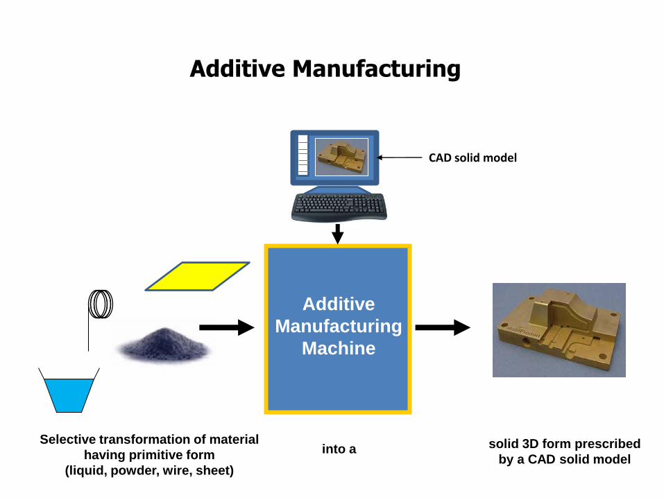
Selective transformation of material
having primitive form
(liquid, powder, wire, sheet)
Additive Manufacturing
Additive
Manufacturing
Machine
solid 3D form prescribed
by a CAD solid model into a
0
CAD solid model
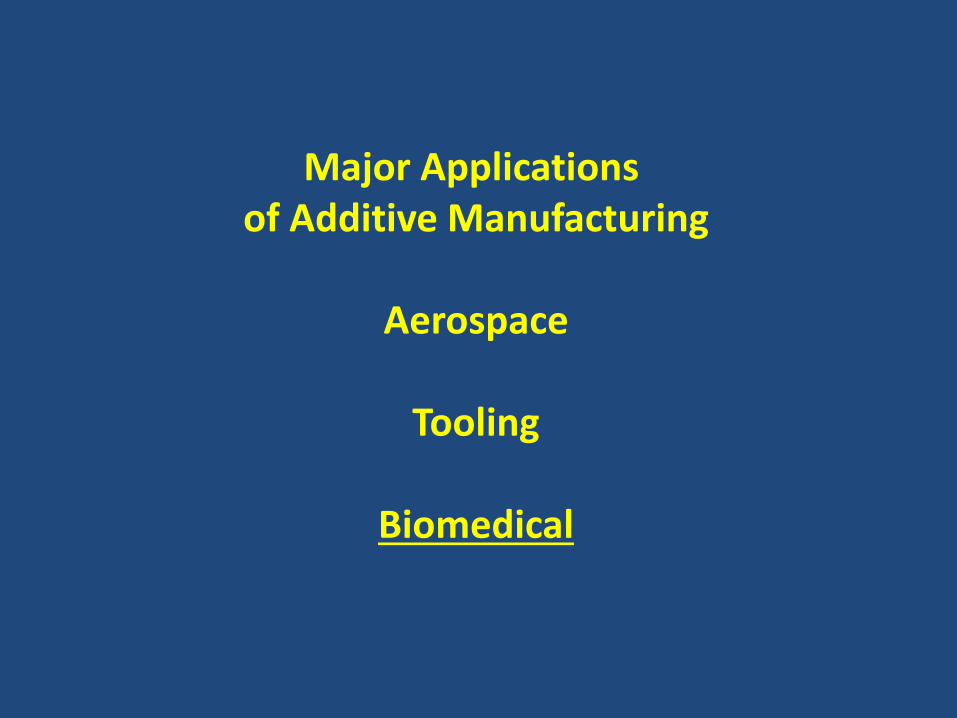
Major Applications of Additive Manufacturing
Aerospace
Tooling
Biomedical

Biomedical Applications
Surgery Planning Models
Splints
Exoskeleton Components
Prostheses
Limbs Hearing Aids Dental Aligners
Implants (Replacement Therapy)
Bioresorbable implants (Regenerative Therapy)
Functional Tissue Generation (Organ Replacement)
Taking advantage of additive manufacturing/3DPrinting capabilities for production of patient specific parts:
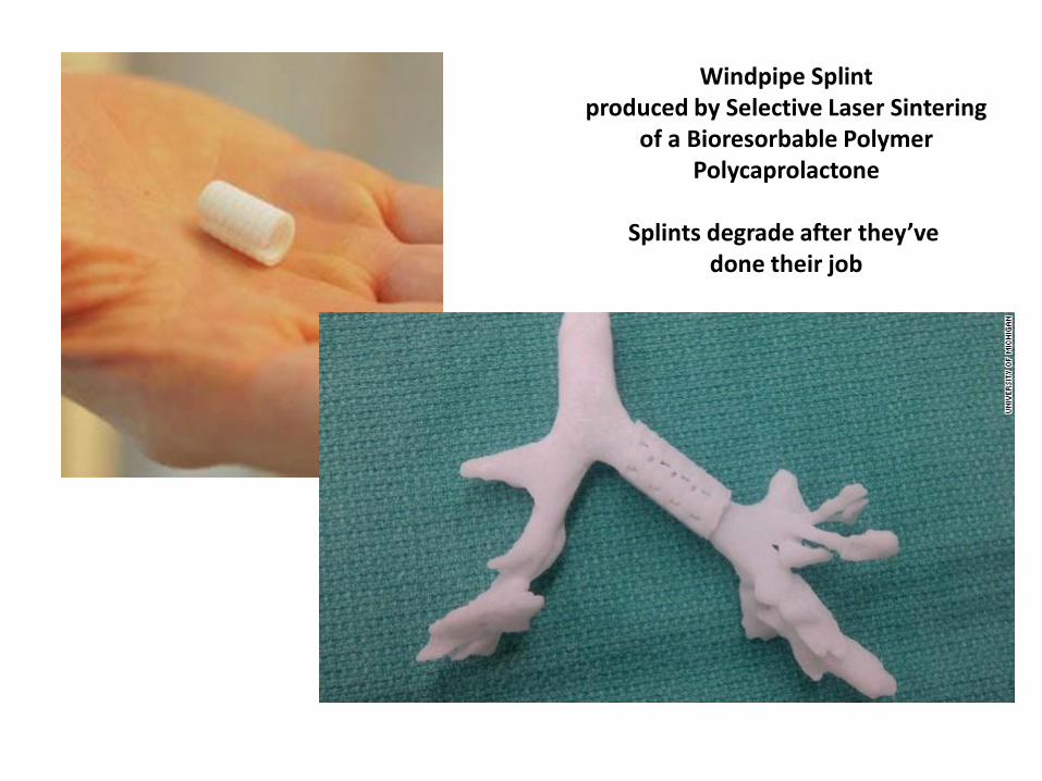
Windpipe Splint produced by Selective Laser Sintering
of a Bioresorbable Polymer Polycaprolactone
Splints degrade after they’ve
done their job
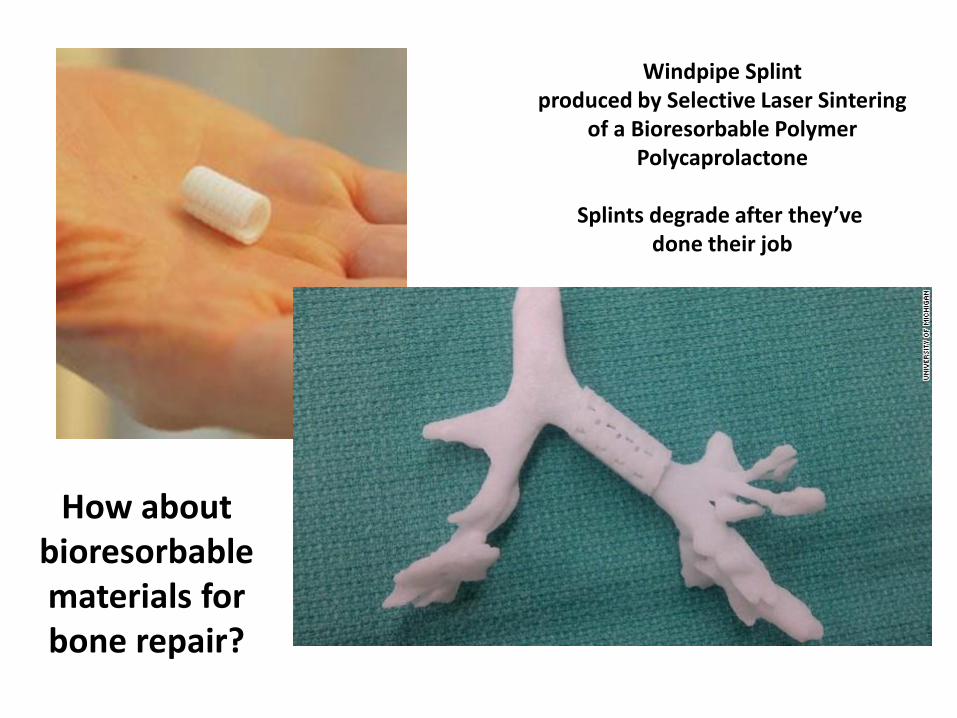
How about bioresorbable materials for bone repair?
Windpipe Splint produced by Selective Laser Sintering
of a Bioresorbable Polymer Polycaprolactone
Splints degrade after they’ve
done their job
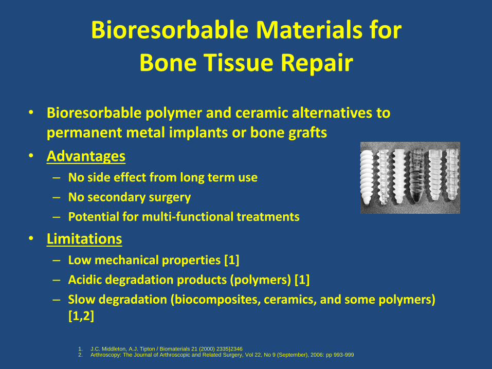
Bioresorbable Materials for Bone Tissue Repair
• Bioresorbable polymer and ceramic alternatives to permanent metal implants or bone grafts
• Advantages – No side effect from long term use
– No secondary surgery
– Potential for multi-functional treatments
• Limitations – Low mechanical properties [1]
– Acidic degradation products (polymers) [1]
– Slow degradation (biocomposites, ceramics, and some polymers) [1,2]
1. J.C. Middleton, A.J. Tipton / Biomaterials 21 (2000) 2335}2346 2. Arthroscopy: The Journal of Arthroscopic and Related Surgery, Vol 22, No 9 (September), 2006: pp 993-999

Bioresorbable Materials for Bone Tissue Repair
• Bioresorbable polymer and ceramic alternatives to permanent metal implants or bone grafts
• Advantages – No side effect from long term use
– No secondary surgery
– Potential for multi-functional treatments
• Limitations – Low mechanical properties [1]
– Acidic degradation products (polymers) [1]
– Slow degradation (biocomposites, ceramics, and some polymers) [1,2]
How about Bioresorbable Metals ?
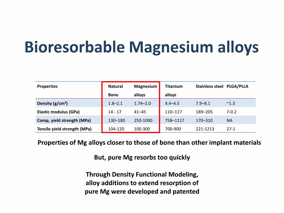
Bioresorbable Magnesium alloys
Properties Natural
Bone
Magnesium
alloys
Titanium
alloys
Stainless steel PLGA/PLLA
Density (g/cm3) 1.8–2.1 1.74–2.0 4.4–4.5 7.9–8.1 ~1.3
Elastic modulus (GPa) 14 - 17 41–45 110–117 189–205 7-0.2
Comp, yield strength (MPa) 130–180 250-1000 758–1117 170–310 NA
Tensile yield strength (MPa) 104-120 100-300 700-900 221-1213 27-1
But, pure Mg resorbs too quickly
Through Density Functional Modeling, alloy additions to extend resorption of pure Mg were developed and patented
Properties of Mg alloys closer to those of bone than other implant materials
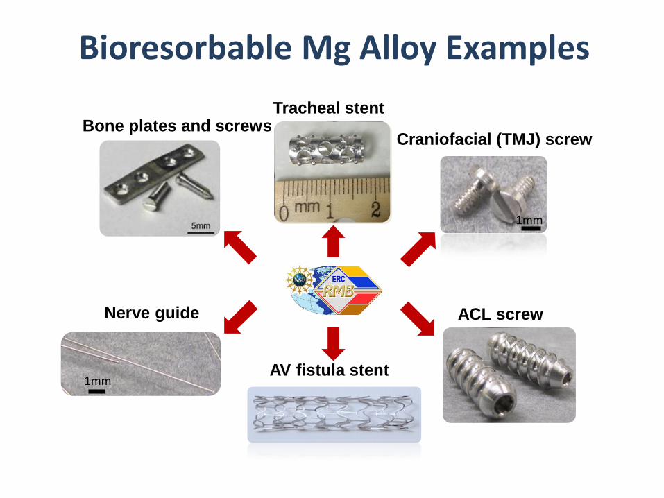
Bioresorbable Mg Alloy Examples
Bone plates and screws
ACL screw
AV fistula stent 1mm
1mm
Nerve guide
Craniofacial (TMJ) screw
Tracheal stent

Mg Ti
Mg degradation does not inhibit fracture healing
In vivo Tests
Mg enhances bone growth
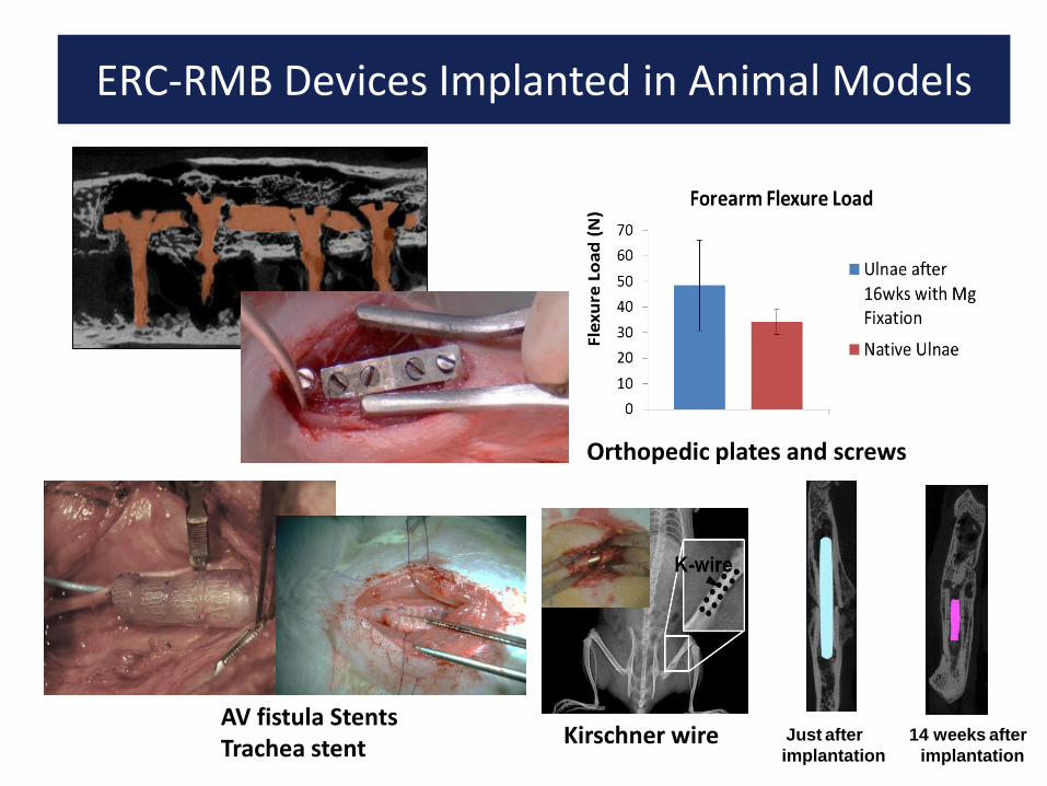
ERC-RMB Devices Implanted in Animal Models
Orthopedic plates and screws
AV fistula Stents Trachea stent
Kirschner wire Just after
implantation
14 weeks after
implantation
ERC-RMB Devices Implanted in Animal Models
What about Additive Manufacturing
of Bioresorbable Metals?
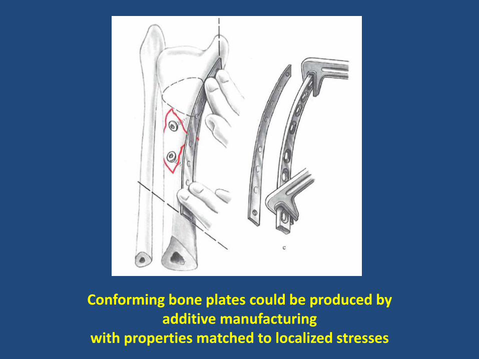
Conforming bone plates could be produced by additive manufacturing
with properties matched to localized stresses
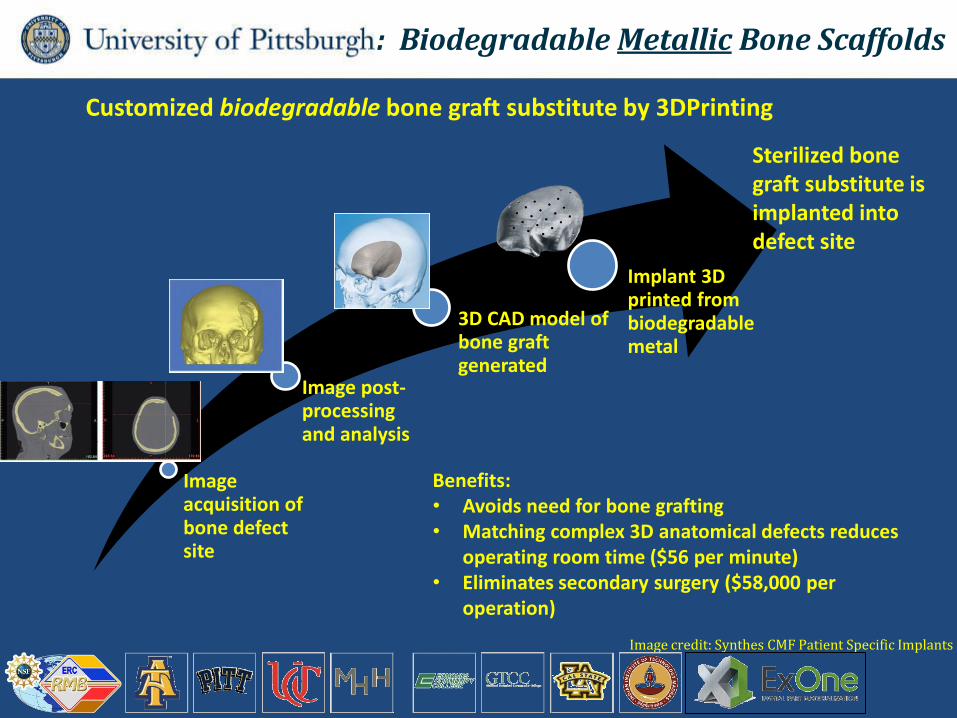
Image acquisition of bone defect site
Image post-processing and analysis
3D CAD model of bone graft generated
Implant 3D printed from biodegradable metal
Customized biodegradable bone graft substitute by 3DPrinting
Sterilized bone graft substitute is implanted into defect site
Image credit: Synthes CMF Patient Specific Implants
Benefits: • Avoids need for bone grafting • Matching complex 3D anatomical defects reduces
operating room time ($56 per minute) • Eliminates secondary surgery ($58,000 per
operation)
: Biodegradable Metallic Bone Scaffolds
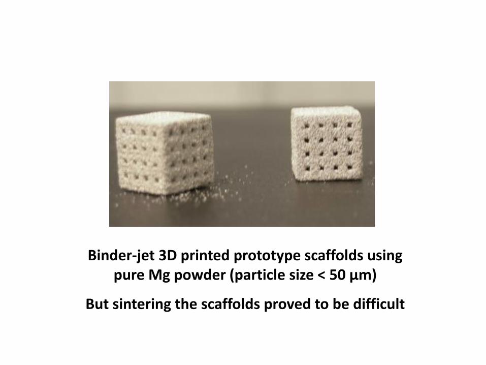
Binder-jet 3D printed prototype scaffolds using pure Mg powder (particle size < 50 μm)
But sintering the scaffolds proved to be difficult
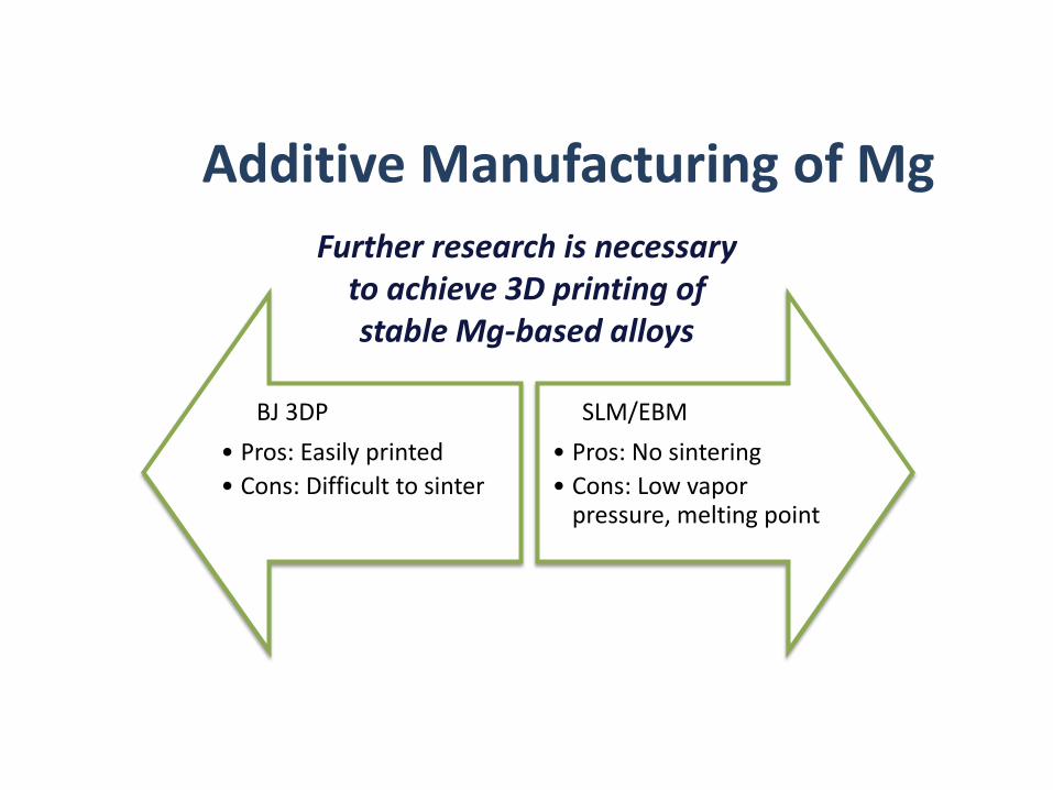
Additive Manufacturing of Mg
BJ 3DP
• Pros: Easily printed
• Cons: Difficult to sinter
SLM/EBM
• Pros: No sintering
• Cons: Low vapor pressure, melting point
Further research is necessary to achieve 3D printing of stable Mg-based alloys

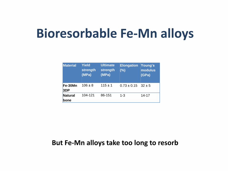
Bioresorbable Fe-Mn alloys
Material Yield
strength
(MPa)
Ultimate
strength
(MPa)
Elongation
(%)
Young’s
modulus
(GPa)
Fe-30Mn
3DP
106 ± 8 115 ± 1 0.73 ± 0.15 32 ± 5
Natural
bone
104-121 86-151 1-3 14-17
Bioresorbable Fe-Mn alloys
Material Yield
strength
(MPa)
Ultimate
strength
(MPa)
Elongation
(%)
Young’s
modulus
(GPa)
Fe-30Mn
3DP
106 ± 8 115 ± 1 0.73 ± 0.15 32 ± 5
Natural
bone
104-121 86-151 1-3 14-17
But Fe-Mn alloys take too long to resorb

ThermoCalc determination of suitable alloying elements to accelerate resorption of Fe-Mn
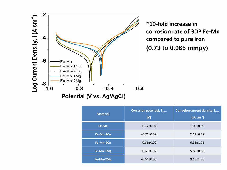
Material Corrosion potential, Ecorr
[V)
Corrosion current density, icorr
[µA cm-2]
Fe-Mn -0.72±0.04 1.00±0.06
Fe-Mn-1Ca -0.71±0.02 2.12±0.92
Fe-Mn-2Ca -0.66±0.02 6.36±1.75
Fe-Mn-1Mg -0.65±0.02 5.89±0.80
Fe-Mn-2Mg -0.64±0.03 9.16±1.25
~10-fold increase in corrosion rate of 3DP Fe-Mn compared to pure iron
(0.73 to 0.065 mmpy)
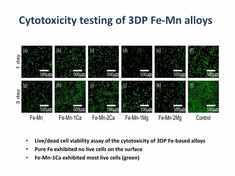
Cytotoxicity testing of 3DP Fe-Mn alloys
• Live/dead cell viability assay of the cytotoxicity of 3DP Fe-based alloys
• Pure Fe exhibited no live cells on the surface
• Fe-Mn-1Ca exhibited most live cells (green)

Fe-Mn
3DPrinting of Fe-Mn alloys
Fe-Mn-1Ca
3D printing
& Sintering
20µm 20µm
• ExOne’s RX1 BJ printer was used for this study
• Sintered at 1200 ºC, 3 hours
20µm 20µm
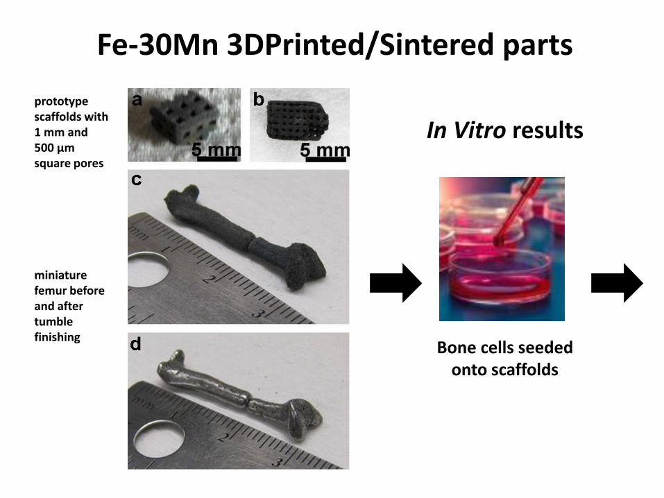
Fe-30Mn 3DPrinted/Sintered parts
Bone cells seeded onto scaffolds
In Vitro results
prototype scaffolds with 1 mm and 500 µm square pores
miniature femur before and after tumble finishing
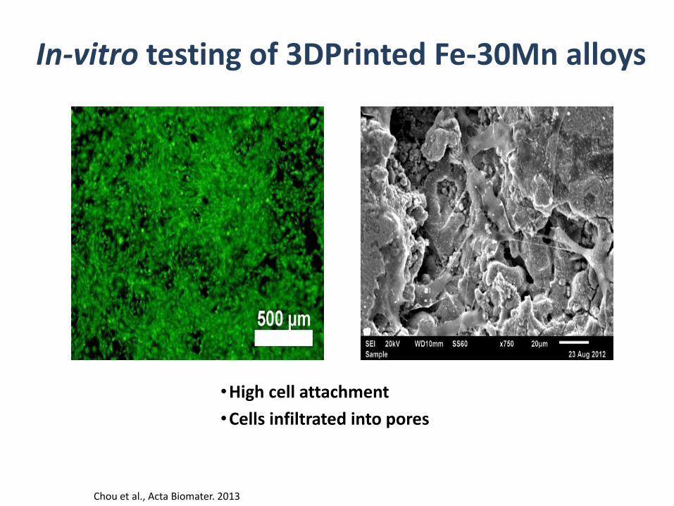
•High cell attachment
•Cells infiltrated into pores
Chou et al., Acta Biomater. 2013
In-vitro testing of 3DPrinted Fe-30Mn alloys

Technical feasibility
Goat mandible model
2. CT Scan
Goat mandible CT Scan STL file 3DP mandible
BJ/Sintering SLM - Renishaw
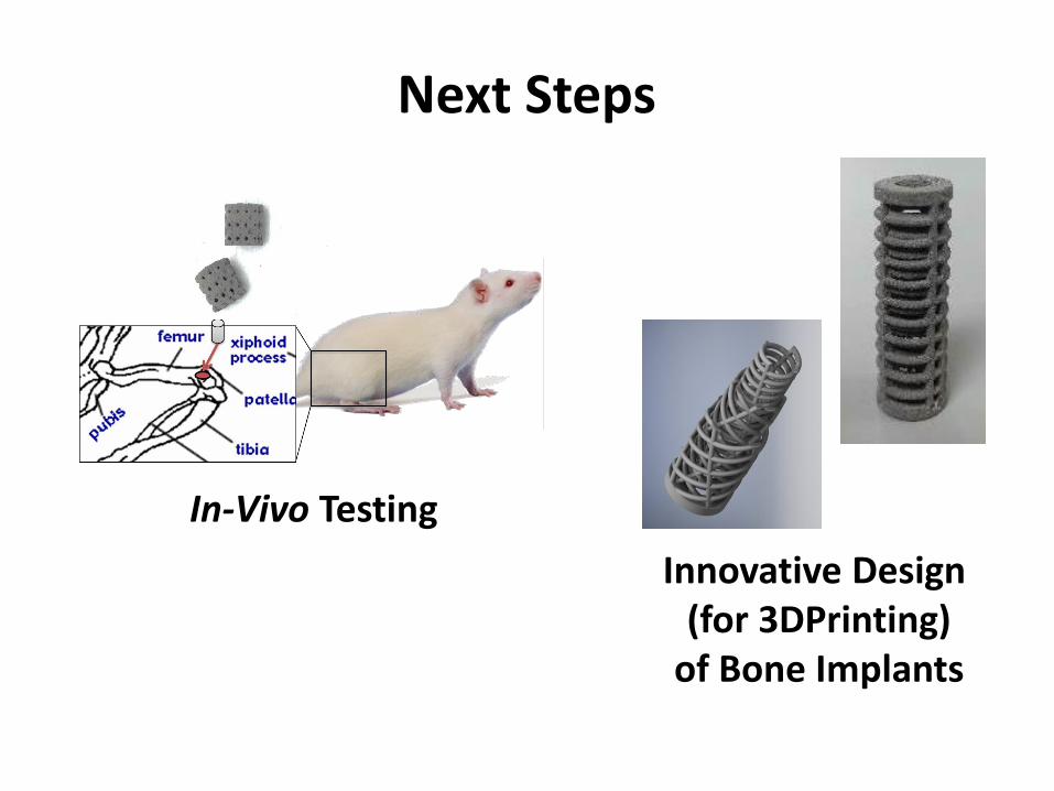
Next Steps
In-Vivo Testing
Innovative Design (for 3DPrinting)
of Bone Implants
To Your Good Health
Acknowledgements: Drs. Daeho Hong, Da-Tren Chou, Abhijit Roy, Prashant Kumta