imaging introduction - stanford universityotolic.stanford.edu/documents/imaging-introduction.pdf ·...
TRANSCRIPT
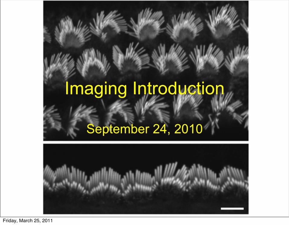
Imaging Introduction
September 24, 2010
Friday, March 25, 2011

What is a microscope?
• Merriam-Webster: an optical instrument consisting of a lens or combination of lenses for making enlarged images of minute objects; especially: compound microscope (a microscope consisting of an objective and an eyepiece mounted in a drawtube)
Friday, March 25, 2011

Why is it important to know how a microscope works?
Friday, March 25, 2011

Why is it important to know how a microscope works?
• Interpreting your images correctly
Friday, March 25, 2011
Why is it important to know how a microscope works?
• Interpreting your images correctly• Take the best images possible
Friday, March 25, 2011

Why is it important to know how a microscope works?
• Interpreting your images correctly• Take the best images possible• Work independently
Friday, March 25, 2011

Why is it important to know how a microscope works?
• Interpreting your images correctly• Take the best images possible• Work independently• Properly use it and not damage it
($35,000 - $70,000)
Friday, March 25, 2011

Why is it important to know how a microscope works?
• Interpreting your images correctly• Take the best images possible• Work independently• Properly use it and not damage it
($35,000 - $70,000)• Push the limits of the microscope and
use it for new applications
Friday, March 25, 2011

Components of microscope
Friday, March 25, 2011

Components of microscope
Friday, March 25, 2011

Why do we care about this?
Not Aligned Aligned w/ Smallest Aperture
Aligned w/ Largest apertureAligned w/ Medium Aperture
20x Objective on Upright Zeiss
Friday, March 25, 2011

63x ObjectiveNot Aligned Aligned w/ Smallest Aperture
Aligned w/ Largest apertureAligned w/ Medium Aperture
Friday, March 25, 2011

Friday, March 25, 2011
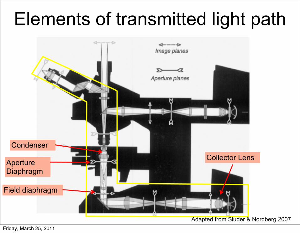
Elements of transmitted light path
Adapted from Sluder & Nordberg 2007
Collector Lens
Field diaphragm
Aperture Diaphragm
Condenser
Friday, March 25, 2011

From Jeff LichtmanFriday, March 25, 2011

Illumination Cone Diagram
Friday, March 25, 2011

Aperature Diaphram applethttp://www.microscopy.fsu.edu/primer/java/kohler/condensercones/index.html
What would happen if the field diaphram was vared?
Friday, March 25, 2011
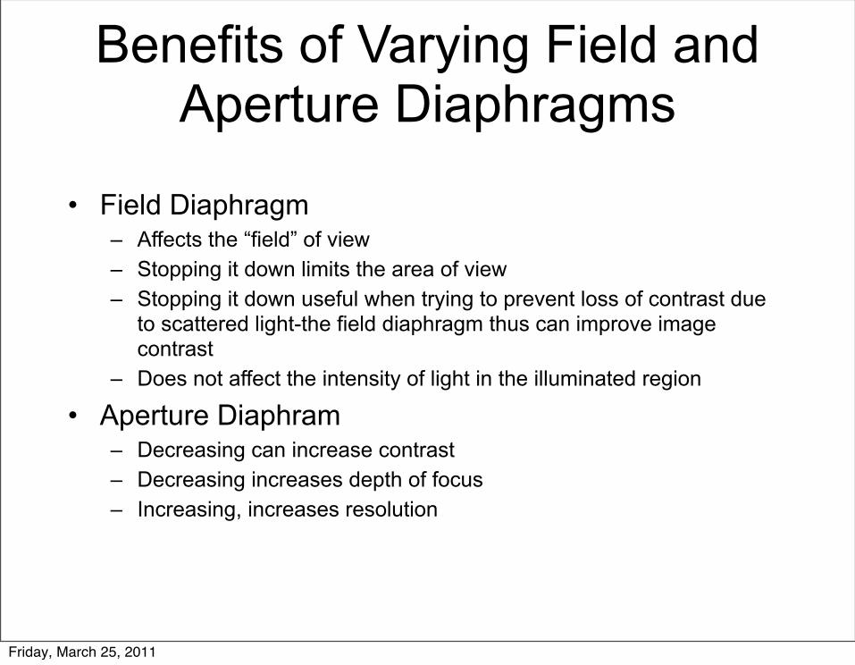
Benefits of Varying Field and Aperture Diaphragms
• Field Diaphragm– Affects the “field” of view– Stopping it down limits the area of view– Stopping it down useful when trying to prevent loss of contrast due
to scattered light-the field diaphragm thus can improve image contrast
– Does not affect the intensity of light in the illuminated region
• Aperture Diaphram– Decreasing can increase contrast– Decreasing increases depth of focus– Increasing, increases resolution
Friday, March 25, 2011
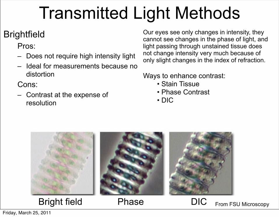
Transmitted Light MethodsBrightfield
Pros:– Does not require high intensity light– Ideal for measurements because no
distortionCons:– Contrast at the expense of
resolution
From FSU MicroscopyBright field Phase DIC
Our eyes see only changes in intensity, they cannot see changes in the phase of light, and light passing through unstained tissue does not change intensity very much because of only slight changes in the index of refraction.
Ways to enhance contrast:• Stain Tissue• Phase Contrast• DIC
Friday, March 25, 2011

Phase Contrast• Pros
– Can get contrast without losing resolution– On living tissue, dead cells are dark, live cells are
bright
• Cons– Phase images are usually surrounded by halos
around the outlines of details– Requires special objective– Phase annuli do limit the working numerical aperture
thus resolution– Not good with thick specimens– Requires much more light than brightfield
From FSU MicroscopyFriday, March 25, 2011

Differential Interference Contrast (DIC)
From FSU Microscopy
• Pros– Can get contrast without losing resolution– Prism does not need to be in the back focal plane of
the objective– Psuedo-3D shadow casting effect– Good for thicker specimens because of optical
sectioning– Smaller specimen features are not obscured by
adjoining regions having large optical gradients
• Cons– Cost– Sensitive only to gradients in a specific direction, so
the image of a given object can change as it is rotated.
– Analyzer must be removed in fluorescence imaging– Requires much more light than brightfield
Friday, March 25, 2011

Next Up: Objective
Adapted from Sluder & Nordberg 2007
Collector Lens
Field diaphragm
Aperture Diaphragm
Condenser
Friday, March 25, 2011

Next Up: Objective
Adapted from Sluder & Nordberg 2007
Collector Lens
Field diaphragm
Aperture Diaphragm
Condenser
Objective
Friday, March 25, 2011

How do you choose an objective?
From Zeiss BrochureFriday, March 25, 2011

Objective Innerds – Why so many lenses?
From Zeiss BrochureFriday, March 25, 2011

We don’t live in an ideal worldSpherical Aberrations
Chromatic Aberrations
Field Curvature
From Zeiss BrochureFriday, March 25, 2011

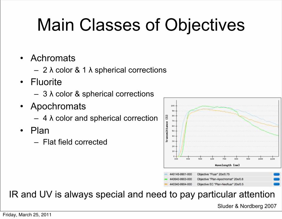
Main Classes of Objectives
• Achromats– 2 λ color & 1 λ spherical corrections
• Fluorite– 3 λ color & spherical corrections
• Apochromats– 4 λ color and spherical correction
• Plan– Flat field corrected
Sluder & Nordberg 2007 Friday, March 25, 2011
Main Classes of Objectives
• Achromats– 2 λ color & 1 λ spherical corrections
• Fluorite– 3 λ color & spherical corrections
• Apochromats– 4 λ color and spherical correction
• Plan– Flat field corrected
Sluder & Nordberg 2007
IR and UV is always special and need to pay particular attention
Friday, March 25, 2011
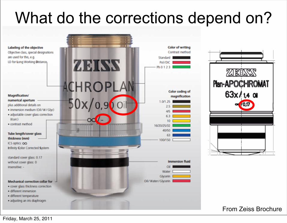
What do the corrections depend on?
From Zeiss BrochureFriday, March 25, 2011

Why do corrections matter? Co-localization
North 2006
Beads Fission Yeast
Friday, March 25, 2011

Image Sharpness
Improper Coverslip thickness
North 2006; FSU MicroscopyFriday, March 25, 2011

Image Sharpness
Improper Coverslip thickness
North 2006; FSU MicroscopyFriday, March 25, 2011

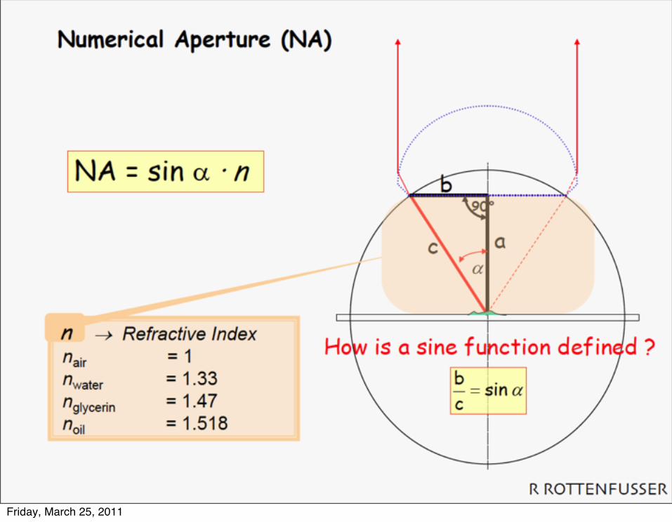
Numerical Aperature
From Zeiss BrochureFriday, March 25, 2011
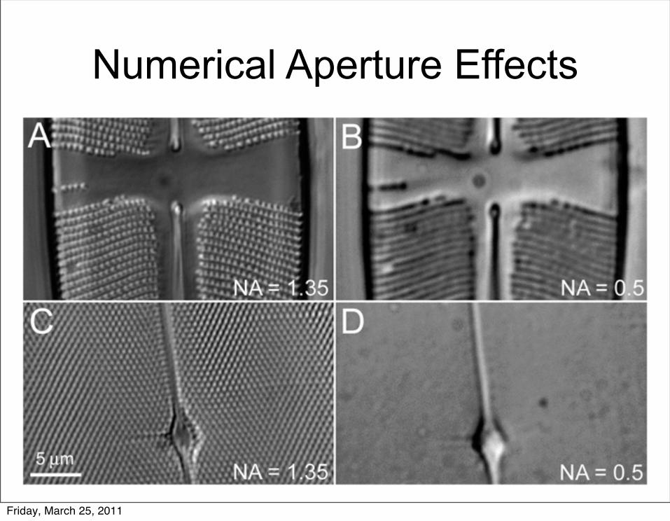
Numerical Aperture Effects
Friday, March 25, 2011

Resolution: 1.22 λ / (NAobj + Nacondenser)
More Next Week!
From Jeff LichtmanFriday, March 25, 2011
Friday, March 25, 2011

Tube Length
From Zeiss BrochureFriday, March 25, 2011

Ray Optics
Friday, March 25, 2011

From Jeff LichtmanFriday, March 25, 2011

Infinity Corrected Microscope
Adapted from Sluder & Nordberg 2007
Collector Lens
Field diaphragm
Aperture Diaphragm
Condenser
Objective
Friday, March 25, 2011

Infinity Corrected Microscope
Adapted from Sluder & Nordberg 2007
Collector Lens
Field diaphragm
Aperture Diaphragm
Condenser
Objective
Tube Lens
Friday, March 25, 2011

Benefits of Infinity Corrected Microscopes
• Flexibility: Inserting pieces of glass in Infinity space do not cause a lot of chromatic aberrations
• More correction lenses can go in the objective, so less aberrations
Friday, March 25, 2011

Elements of reflected light path
Adapted from Sluder & Nordberg 2007
Collector Lens
Field diaphragm
Aperture Diaphragm
Condenser
Objective
Tube Lens
Friday, March 25, 2011

Elements of reflected light path
Adapted from Sluder & Nordberg 2007
Collector Lens
Field diaphragm
Aperture Diaphragm
Condenser
Objective
Tube Lens
Friday, March 25, 2011

Elements of reflected light path
Adapted from Sluder & Nordberg 2007
Collector Lens
Field diaphragm
Aperture Diaphragm
Condenser
Objective
Tube Lens
Filter Cube
Light manipulation are the same just comes from above!Field diaphragm in the incident light path-really useful to remove background!!!
Friday, March 25, 2011
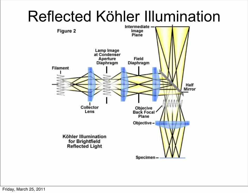
Reflected Köhler Illumination
Friday, March 25, 2011

Reflected Köhler Illumination
Why can we come from above in Fluorescence?
Friday, March 25, 2011
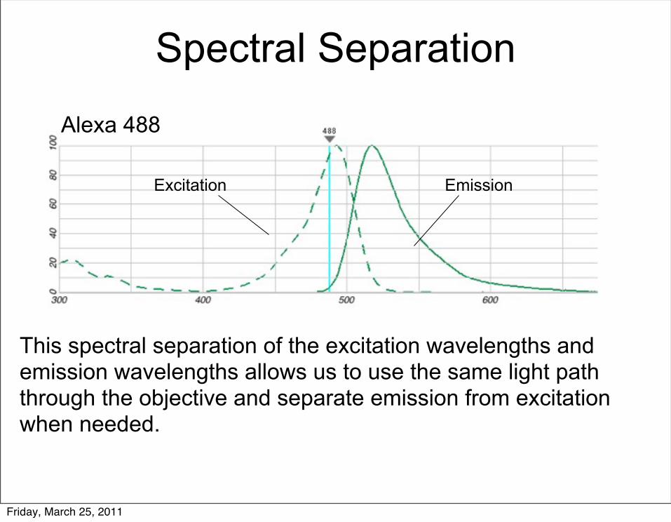
Spectral Separation
Alexa 488
Excitation Emission
This spectral separation of the excitation wavelengths and emission wavelengths allows us to use the same light path through the objective and separate emission from excitation when needed.
Friday, March 25, 2011
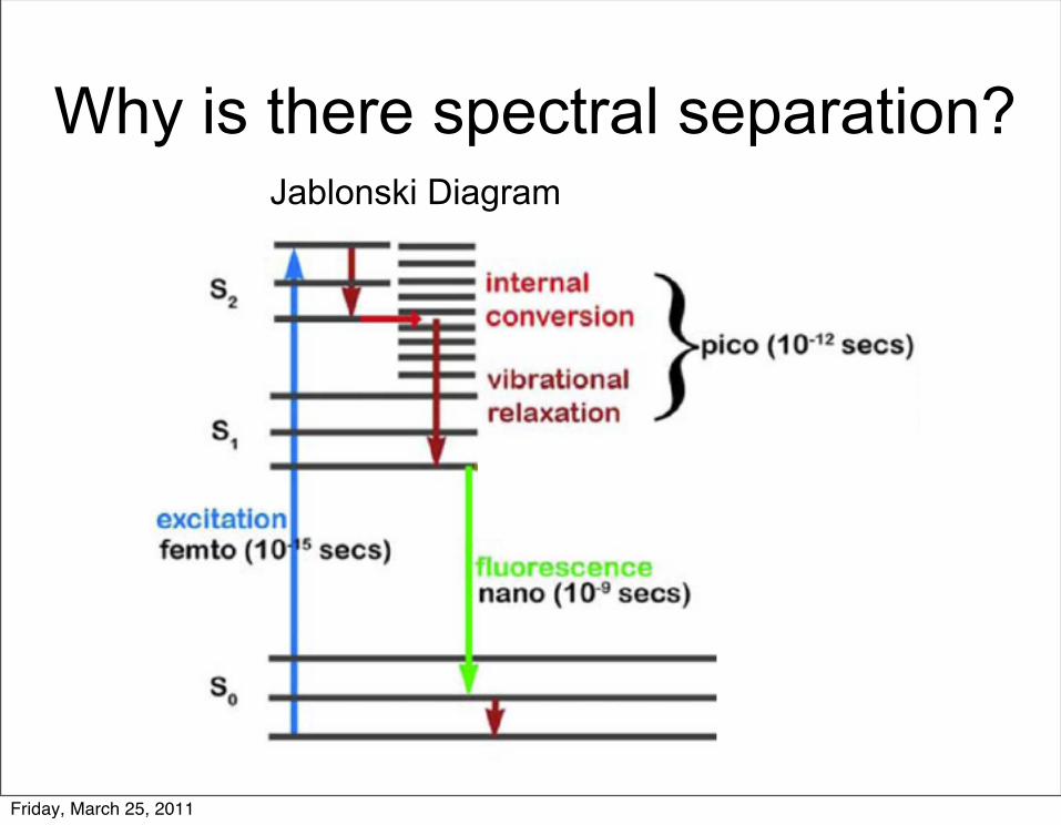
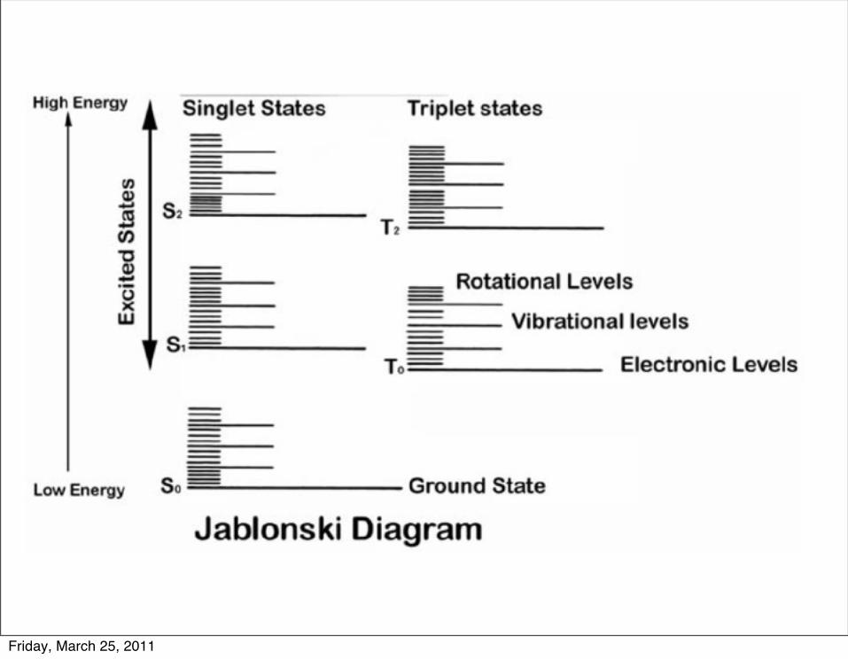
Why is there spectral separation?Jablonski Diagram
Friday, March 25, 2011

Where does the spectral spread come from?
Friday, March 25, 2011

The Filter Cube:How we separate light paths
From FSU Microscopy
To DetectorEmission
Filter
FromIlluminatorDichroic Mirror
ExcitationFilter
Friday, March 25, 2011

Dichroic Mirror
DM 565Designed to pass light above a certain wavelength and reflect light below it
Normal Mirror
Friday, March 25, 2011

Dichroic Mirror Reality%
Tra
nsm
ittan
ce
IdealReality
Friday, March 25, 2011

Filter Cube Specs%
Tra
nsm
ittan
ce
Excitation EmissionDichroic
Friday, March 25, 2011

2-photon considerations
Friday, March 25, 2011

Light Source
Fluorescent Specimen
Excitation lightFluorecence
To Detector
How the Cube works
Friday, March 25, 2011

Filter Design
Friday, March 25, 2011

Fluorescent Lamps
Friday, March 25, 2011

4 Major Fluorescent Lamp Sources• Mercury• Metal halide Xenon
LED
Friday, March 25, 2011

4 Major Fluorescent Lamp Sources• Mercury• Metal halide Xenon
LED
Friday, March 25, 2011

Friday, March 25, 2011
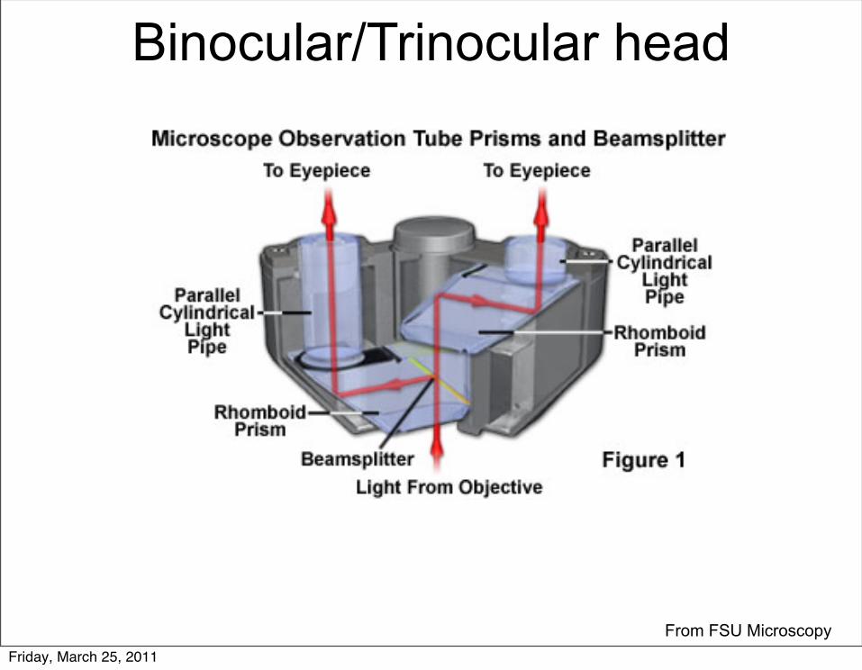
Binocular/Trinocular head
From FSU MicroscopyFriday, March 25, 2011

Function of EyepiecesImage formation
Friday, March 25, 2011

Sluder & Nordberg 2007
EyepiecesEver get your specimen into focus using your eyes and then you take the image with the computer and it is blurry or you see nothing (for confocal)?
Friday, March 25, 2011

Sluder & Nordberg 2007
EyepiecesEver get your specimen into focus using your eyes and then you take the image with the computer and it is blurry or you see nothing (for confocal)? Parfocality!
Friday, March 25, 2011
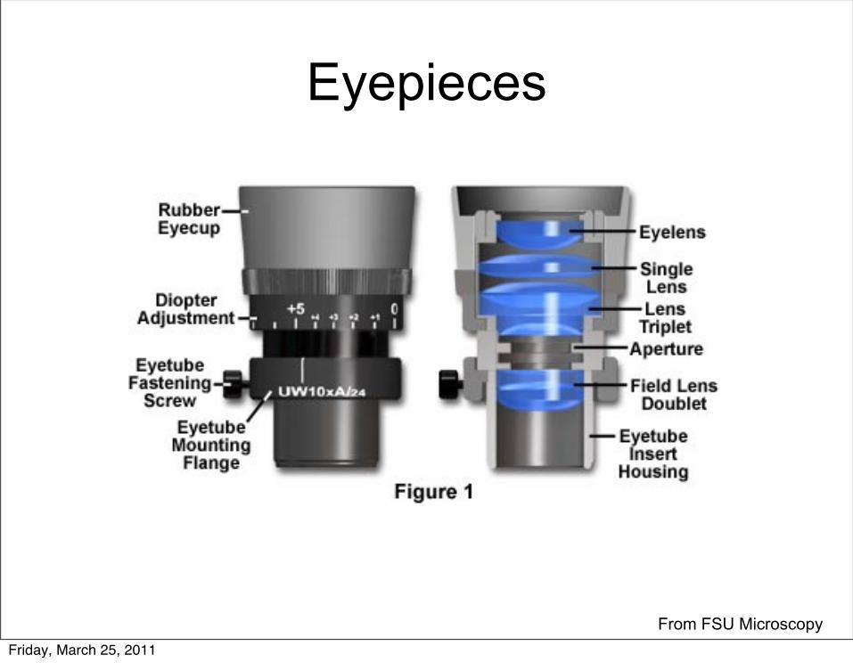
Eyepieces
From FSU MicroscopyFriday, March 25, 2011

Questions?
• Next Time– Confocal Microscopy– PSF– 2-photon Microscopy
Friday, March 25, 2011
References• Sluder, Greenfield, and Joshua J Nordberg. 2007. Microscope basics. Methods in
cell biology 81, no. 06: 1-10. doi:10.1016/S0091-679X(06)81001-0. http://www.ncbi.nlm.nih.gov/pubmed/17519159.
• North, Alison J. 2006. Seeing is believing? A beginners' guide to practical pitfalls in image acquisition. The Journal of cell biology 172, no. 1: 9-18. doi:10.1083/jcb.200507103. http://www.pubmedcentral.nih.gov/articlerender.fcgi?artid=2063524&tool=pmcentrez&rendertype=abstract.
Friday, March 25, 2011

Imaging Path Illumination Path
From Jeff LichtmanFriday, March 25, 2011

Friday, March 25, 2011

Numerical Aperature
1.4NA Oil Lens1.4 = (1.5) sin(u)u = 69 degrees
Friday, March 25, 2011

Friday, March 25, 2011

Friday, March 25, 2011

Friday, March 25, 2011
Friday, March 25, 2011
Friday, March 25, 2011
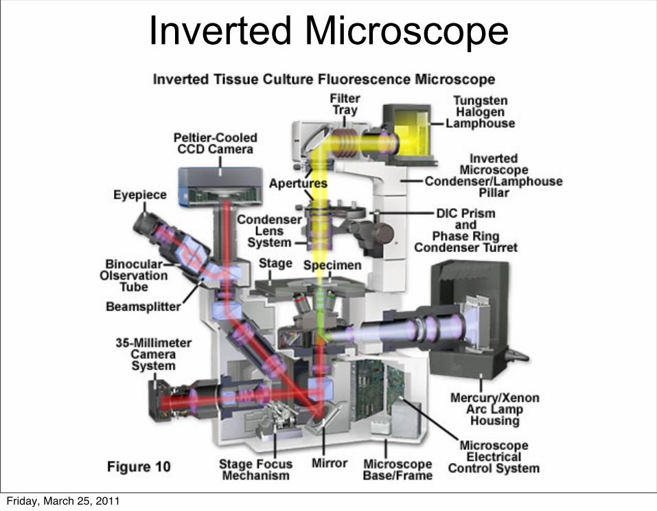
Inverted Microscope
Friday, March 25, 2011

From Jeff LichtmanFriday, March 25, 2011

What is the main difference between these two?
Olympus BX51$35,000
Barska Compound Microscope$300
Friday, March 25, 2011

What is the main difference between these two?
Olympus BX51$35,000
Barska Compound Microscope$300
Infinity Correction!Friday, March 25, 2011
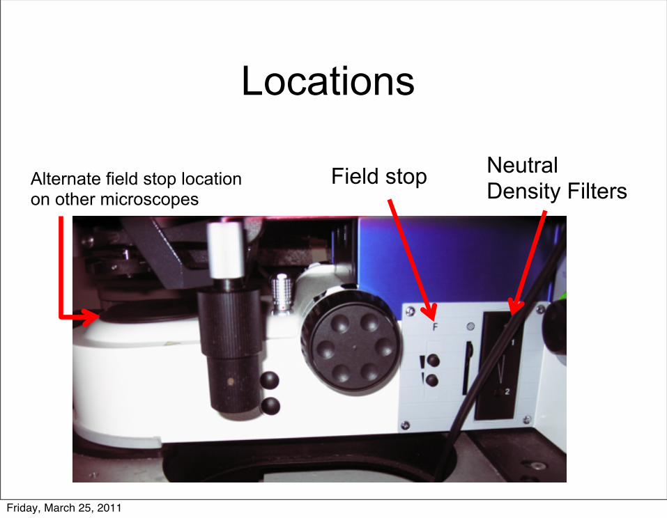
Locations
Field stop Neutral Density FiltersAlternate field stop location
on other microscopes
Friday, March 25, 2011

Aperature Diaphragm Condenser
Turret
Polarizer
Friday, March 25, 2011