immunoglobulin g4-related disease mimicking unresectable
TRANSCRIPT
Case report
Gut and Liver, Vol. 7, No. 5, September 2013, pp. 616-620
Immunoglobulin G4-Related Disease Mimicking Unresectable Gallbladder Cancer
Yoon Suk Lee*, Sang Hyub Lee†, Min Geun Lee*, Seung-June Lee*, Jin-Hyeok Hwang*, Eun Shin‡, and Yoon Jin Lee§
*Department of Internal Medicine, Seoul National University Bundang Hospital, Seoul National University College of Medicine, Seongnam, †Department of Internal Medicine and Liver Research Institute, Seoul National University Hospital, Seoul National University College of Medicine, Seoul, Departments of ‡Pathology and §Radiology, Seoul National University Bundang Hospital, Seoul National University College of Medicine, Seongnam, Korea
Immunoglobulin G4 (IgG4)-related disease is a novel dis-ease entity that can involve diverse organs, causing specific diseases, including autoimmune pancreatitis, sclerosing cholangitis, cholecystitis, inflammatory aortic aneurysm, and inflammatory pseudotumor. IgG4-related disease is charac-terized by elevated serum IgG4 concentrations, abundant IgG4 lymphoplasmacytic infiltration, and dramatic steroid responses. It is clinically important to differentiate this rare disease from primary sclerosing cholangitis and cholangio-carcinoma, because the treatment and prognosis of these two diseases are completely different. However, the preoper-ative diagnosis is challenging, and the disease is frequently misdiagnosed. If the serum level of IgG4 is within the normal range, the diagnosis of IgG4-related disease is more difficult. This article reports on a 59-year-old man with IgG4-related disease mimicking unresectable gallbladder cancer with nor-mal serum IgG4 concentrations. (Gut Liver 2013;7:616-620)
Key Words: Immunoglobuin G; Cholangitis, sclerosing; Gall-bladder neoplasms; Cholecystitis
INTRODUCTION
Immunoglobulin G-related disease (IgG4-RD) is a novel dis-ease entity which is a steroid responsive, autoimmune disorder. And it is well known that IgG4-RD can involve diverse organs
Correspondence to: Sang Hyub LeeDepartment of Internal Medicine and Liver Research Institute, Seoul National University Hospital, Seoul National University College of Medicine, 101 Daehak-ro, Jongno-gu, Seoul 110-744, KoreaTel: +82-2-2072-2228, Fax: +82-2-762-9662, E-mail: [email protected]
Current affiliation: Min Geun LeeDepartment of Internal Medicine, Hanmaeum Hospital, Jeju, KoreaSeung-June LeeDepartment of Internal Medicine, Handoh Hospital, Ansan, Korea
Received on February 24, 2013. Revised on April 5, 2013. Accepted on April 7, 2013.pISSN 1976-2283 eISSN 2005-1212 http://dx.doi.org/10.5009/gnl.2013.7.5.616
This is an Open Access article distributed under the terms of the Creative Commons Attribution Non-Commercial License (http://creativecommons.org/licenses/by-nc/3.0) which permits unrestricted non-commercial use, distribution, and reproduction in any medium, provided the original work is properly cited.
causing specific diseases including autoimmune pancreatitis, sclerosing cholangitis, cholecystitis, inflammatory aortic aneu-rysm, and inflammatory pseudotumor.1,2 There are many reports of misdiagnosis of IgG4-related sclerosing cholangitis (SC) as pancreatic and biliary cancer.3-5 However, clinical presentation mimicking gallbladder (GB) cancer is rarely reported. We report herein a case of IgG4-RD mimicking GB cancer that was suc-cessfully treated with an oral steroid.
CASE REPORT
A 59-year-old man was referred to our emergency room with jaundice and right upper abdominal discomfort for 1 month. He complained of nausea, fatigue, loss of appetite, and yellowish discoloration of the skin. However, he did not complain of chill-ing sense and his body temperature was within normal limit.
Laboratory data were as follows (values in parentheses indi-cate normal range): white blood cell count 7,780/mm3 (range, 4,000 to 10,000/mm3), hemoglobin 14.3 g/dL (range, 13 to 17 g/dL), hematocrit 40.1% (range, 39% to 52%), platelet count 233,000/mm3 (range, 130,000 to 400,000/mm3), asparatate ami-notransferase 80 IU/L (range, 0 to 40 IU/L), alanine aminotrans-ferase 138 IU/L (range, 0 to 40 IU/L), alkaline phosphatase 402 IU/L (range, 30 to 115 IU/L), total bilirubin 6.0 mg/dL (range, 0.2 to 1.2 mg/dL), direct bilirubin 5.0 mg/dL (range, 0 to 0.5 mg/dL), amylase 55 U/L (range, 30 to 100 U/L), lipase 60 U/L (range, 23

Lee YS, et al: IgG4-Related Disease 617
to 300 U/L), total protein 8.0 g/dL (range, 6 to 8 g/dL), albumin 4.3 g/dL (range, 3.3 to 5.2 g/dL), hepatitis B surface antigen negative, hepatitis B surface antibody negative, and urine biliru-bin 3 positive (negative). The serum levels of carcinoembryonic antigen and a-fetoprotein were within normal limits; however, carbohydrate antigen 19-9 was 90 U/mL (range, 0 to 37 U/mL).
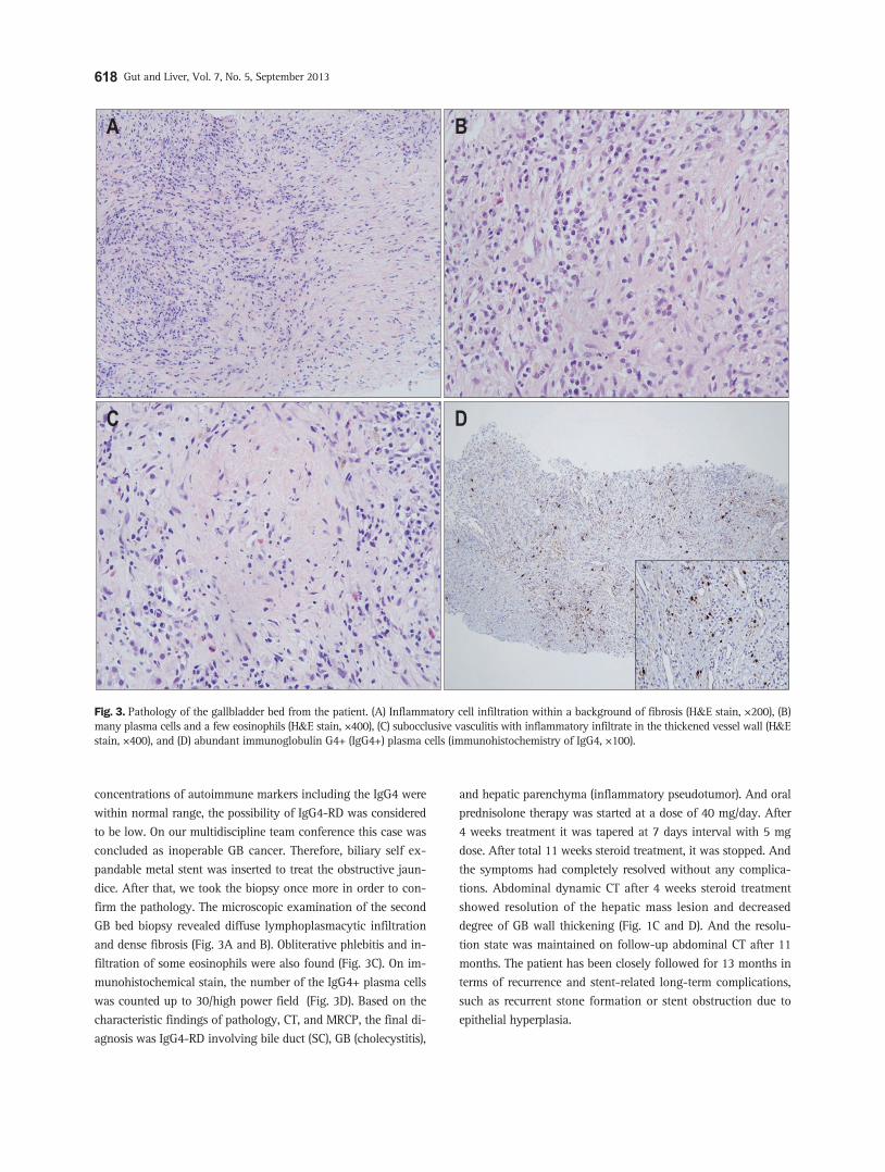
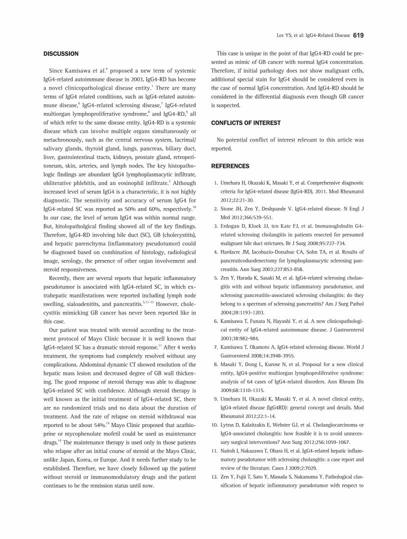
Abdominal computed tomography (CT) revealed an infiltra-tive low density mass involving the neck portion of GB and adjacent hepatic parenchyma along with multiple radio-opaque gallstones in the fundus and neck portion (Fig. 1A and B). How-ever, there were not any abnormalities in pancreas and other or-gans on abdominal CT scan. As there was contiguous soft tissue infiltration involving the right and common hepatic artery, right and main portal vein, it was initially interpreted as unresectable GB cancer. The mass showed mild enhancement in the arterial phase and further delayed enhancement. In addition, there was diffuse symmetrical wall thickening of the extrahepatic bile duct and cystic duct. Magnetic resonance cholangiopancreatography (MRCP) revealed short segmental stricture at the proximal com-mon hepatic duct with upstream bile duct dilatation (Fig. 2). Under the impression of GB cancer with direct hepatic invasion, ultrasonography-guided 18-gauge core needle biopsy was done at the hypoechoic lesion at S5 of the liver adjacent to the thick-ened wall of GB for pathologic confirmation. Histologic exami-nation showed no malignant cells but only moderate periportal fibrosis with portal inflammation. In order to differentiate IgG4-
related disease, we performed additional laboratory tests for autoimmune disorders as follows (values in parentheses indicate normal range): antinuclear antibody was negative: IgG 1,280 mg/dL (range, 700 to 1,700 mg/dL), IgA 381 mg/dL (range, 90 to 400 mg/dL), IgM 71.7 mg/dL (range, 45 to 230 mg/dL), IgG subtype IV 75 mg/dL (range, 6 to 121 mg/dL). Since the serum
Fig. 1. Computed tomography (CT) image (A, B) before and (C, D) after treatment. (A, B) CT before treat-ment with steroids reveals an infil-trative low density mass involving the neck portion of the gallbladder and adjacent hepatic parenchyma, along with multiple radio-opaque gallstones and diffuse wall thicken-ing of the common bile duct (an-notated with a black solid arrow). (C, D) Abdominal CT after 4 weeks of treatment with steroids indicates resolution of the hepatic mass le-sion and a decreased degree of GB wall thickening. The biliary metal stent can also be observed. (A, C) Axial image. (B, D) Coronal image.
Fig. 2. Magnetic resonance cholangiopancreatography reveals a short segmental stricture at the proximal common hepatic duct (annotated with a white solid arrow) with upstream bile duct dilatation.
618 Gut and Liver, Vol. 7, No. 5, September 2013
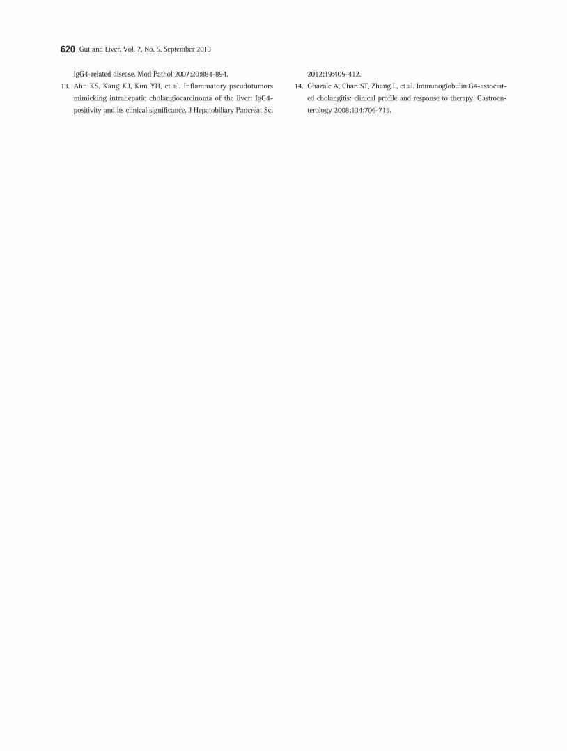
concentrations of autoimmune markers including the IgG4 were within normal range, the possibility of IgG4-RD was considered to be low. On our multidiscipline team conference this case was concluded as inoperable GB cancer. Therefore, biliary self ex-pandable metal stent was inserted to treat the obstructive jaun-dice. After that, we took the biopsy once more in order to con-firm the pathology. The microscopic examination of the second GB bed biopsy revealed diffuse lymphoplasmacytic infiltration and dense fibrosis (Fig. 3A and B). Obliterative phlebitis and in-filtration of some eosinophils were also found (Fig. 3C). On im-munohistochemical stain, the number of the IgG4+ plasma cells was counted up to 30/high power field (Fig. 3D). Based on the characteristic findings of pathology, CT, and MRCP, the final di-agnosis was IgG4-RD involving bile duct (SC), GB (cholecystitis),
and hepatic parenchyma (inflammatory pseudotumor). And oral prednisolone therapy was started at a dose of 40 mg/day. After 4 weeks treatment it was tapered at 7 days interval with 5 mg dose. After total 11 weeks steroid treatment, it was stopped. And the symptoms had completely resolved without any complica-tions. Abdominal dynamic CT after 4 weeks steroid treatment showed resolution of the hepatic mass lesion and decreased degree of GB wall thickening (Fig. 1C and D). And the resolu-tion state was maintained on follow-up abdominal CT after 11 months. The patient has been closely followed for 13 months in terms of recurrence and stent-related long-term complications, such as recurrent stone formation or stent obstruction due to epithelial hyperplasia.
Fig. 3. Pathology of the gallbladder bed from the patient. (A) Inflammatory cell infiltration within a background of fibrosis (H&E stain, ×200), (B) many plasma cells and a few eosinophils (H&E stain, ×400), (C) subocclusive vasculitis with inflammatory infiltrate in the thickened vessel wall (H&E stain, ×400), and (D) abundant immunoglobulin G4+ (IgG4+) plasma cells (immunohistochemistry of IgG4, ×100).
Lee YS, et al: IgG4-Related Disease 619
DISCUSSION
Since Kamisawa et al.6 proposed a new term of systemic IgG4-related autoimmune disease in 2003, IgG4-RD has become a novel clinicopathological disease entity.1 There are many terms of IgG4 related conditions, such as IgG4-related autoim-mune disease,6 IgG4-related sclerosing disease,7 IgG4-related multiorgan lymphoproliferative syndrome,8 and IgG4-RD,9 all of which refer to the same disease entity. IgG4-RD is a systemic disease which can involve multiple organs simultaneously or metachronously, such as the central nervous system, lacrimal/salivary glands, thyroid gland, lungs, pancreas, biliary duct, liver, gastrointestinal tracts, kidneys, prostate gland, retroperi-toneum, skin, arteries, and lymph nodes. The key histopatho-logic findings are abundant IgG4 lymphoplasmacytic infiltrate, obliterative phlebitis, and an eosinophil infiltrate.2 Although increased level of serum IgG4 is a characteristic, it is not highly diagnostic. The sensitivity and accuracy of serum IgG4 for IgG4-related SC was reported as 50% and 60%, respectively.10 In our case, the level of serum IgG4 was within normal range. But, hitolopatholgical finding showed all of the key findings. Therefore, IgG4-RD involving bile duct (SC), GB (cholecystitis), and hepatic parenchyma (inflammatory pseudotumor) could be diagnosed based on combination of histology, radiological image, serology, the presence of other organ involvement and steroid responsiveness.
Recently, there are several reports that hepatic inflammatory pseudotumor is associated with IgG4-related SC, in which ex-trahepatic manifestations were reported including lymph node swelling, sialoadenitits, and pancreatitis.5,11-13 However, chole-cystitis mimicking GB cancer has never been reported like in this case.
Our patient was treated with steroid according to the treat-ment protocol of Mayo Clinic because it is well known that IgG4-related SC has a dramatic steroid response.11 After 4 weeks treatment, the symptoms had completely resolved without any complications. Abdominal dynamic CT showed resolution of the hepatic mass lesion and decreased degree of GB wall thicken-ing. The good response of steroid therapy was able to diagnose IgG4-related SC with confidence. Although steroid therapy is well known as the initial treatment of IgG4-related SC, there are no randomized trials and no data about the duration of treatment. And the rate of relapse on steroid withdrawal was reported to be about 54%.14 Mayo Clinic proposed that azathio-prine or mycophenolate mofetil could be used as maintenance drugs.14 The maintenance therapy is used only in those patients who relapse after an initial course of steroid at the Mayo Clinic, unlike Japan, Korea, or Europe. And it needs further study to be established. Therefore, we have closely followed up the patient without steroid or immunomodulatory drugs and the patient continues to be the remission status until now.
This case is unique in the point of that IgG4-RD could be pre-sented as mimic of GB cancer with normal IgG4 concentration. Therefore, if initial pathology does not show malignant cells, additional special stain for IgG4 should be considered even in the case of normal IgG4 concentration. And IgG4-RD should be considered in the differential diagnosis even though GB cancer is suspected.
CONFLICTS OF INTEREST
No potential conflict of interest relevant to this article was reported.
REFERENCES
1. Umehara H, Okazaki K, Masaki Y, et al. Comprehensive diagnostic
criteria for IgG4-related disease (IgG4-RD), 2011. Mod Rheumatol
2012;22:21-30.
2. Stone JH, Zen Y, Deshpande V. IgG4-related disease. N Engl J
Med 2012;366:539-551.
3. Erdogan D, Kloek JJ, ten Kate FJ, et al. Immunoglobulin G4-
related sclerosing cholangitis in patients resected for presumed
malignant bile duct strictures. Br J Surg 2008;95:727-734.
4. Hardacre JM, Iacobuzio-Donahue CA, Sohn TA, et al. Results of
pancreaticoduodenectomy for lymphoplasmacytic sclerosing pan-
creatitis. Ann Surg 2003;237:853-858.
5. Zen Y, Harada K, Sasaki M, et al. IgG4-related sclerosing cholan-
gitis with and without hepatic inflammatory pseudotumor, and
sclerosing pancreatitis-associated sclerosing cholangitis: do they
belong to a spectrum of sclerosing pancreatitis? Am J Surg Pathol
2004;28:1193-1203.
6. Kamisawa T, Funata N, Hayashi Y, et al. A new clinicopathologi-
cal entity of IgG4-related autoimmune disease. J Gastroenterol
2003;38:982-984.
7. Kamisawa T, Okamoto A. IgG4-related sclerosing disease. World J
Gastroenterol 2008;14:3948-3955.
8. Masaki Y, Dong L, Kurose N, et al. Proposal for a new clinical
entity, IgG4-positive multiorgan lymphoproliferative syndrome:
analysis of 64 cases of IgG4-related disorders. Ann Rheum Dis
2009;68:1310-1315.
9. Umehara H, Okazaki K, Masaki Y, et al. A novel clinical entity,
IgG4-related disease (IgG4RD): general concept and details. Mod
Rheumatol 2012;22:1-14.
10. Lytras D, Kalaitzakis E, Webster GJ, et al. Cholangiocarcinoma or
IgG4-associated cholangitis: how feasible it is to avoid unneces-
sary surgical interventions? Ann Surg 2012;256:1059-1067.
11. Naitoh I, Nakazawa T, Ohara H, et al. IgG4-related hepatic inflam-
matory pseudotumor with sclerosing cholangitis: a case report and
review of the literature. Cases J 2009;2:7029.
12. Zen Y, Fujii T, Sato Y, Masuda S, Nakanuma Y. Pathological clas-
sification of hepatic inflammatory pseudotumor with respect to
620 Gut and Liver, Vol. 7, No. 5, September 2013
IgG4-related disease. Mod Pathol 2007;20:884-894.
13. Ahn KS, Kang KJ, Kim YH, et al. Inflammatory pseudotumors
mimicking intrahepatic cholangiocarcinoma of the liver: IgG4-
positivity and its clinical significance. J Hepatobiliary Pancreat Sci
2012;19:405-412.
14. Ghazale A, Chari ST, Zhang L, et al. Immunoglobulin G4-associat-
ed cholangitis: clinical profile and response to therapy. Gastroen-
terology 2008;134:706-715.