importin beta and crm1 control a ranbp2 spatiotemporal...
TRANSCRIPT

© 2017. Published by The Company of Biologists Ltd.
Importin beta and CRM1 control a RANBP2 spatiotemporal switch
essential for mitotic kinetochore function
Eugenia Gilistro1, Valeria de Turris2, Michela Damizia1, Annalisa Verrico1, Sara Moroni1,
Riccardo De Santis2,3, Alessandro Rosa2,3, and Patrizia Lavia1
1. Institute of Molecular Biology and Pathology (IBPM), CNR National Research Council of Italy, Via degli
Apuli 4, 00185, Rome, Italy; 2 Center for Life Nanoscience@Sapienza, Istituto Italiano di Tecnologia, Viale
Regina Elena 291, 00161 Rome, Italy, 3. Department of Biology and Biotechnology "Charles Darwin",
Sapienza University of Rome, Piazzale Aldo Moro 5, 00185 Rome
ABSTRACT
Protein conjugation with SUMO is a post-translational modification that modulates protein
interactions and localisation. RANBP2 is a large nucleoporin endowed with SUMO E3 ligase
and SUMO-stabilising activity and is implicated in some cancer types. RANBP2 is part of a
larger complex, comprising SUMO-modified RANGAP1, the GTP-hydrolysis activating
factor for the GTPase RAN. During mitosis, the RANBP2/SUMO-RANGAP1 complex
localises to the mitotic spindle and to kinetochores after microtubule attachment. Here we
have addressed the mechanisms that regulate this localisation and how they affect
kinetochore functions. Using proximity ligation assays, we find that nuclear transport
receptors importin beta and CRM1 play critical roles in localising the RANBP2/SUMO-
RANGAP1 complex away from, or at KTs, respectively. Using newly generated inducible
cell lines, we show that overexpression of nuclear transport receptors affects the timing of
RANBP2 localisation in opposite manners. Concomitantly, kinetochore functions are also
affected, including the accumulation of SUMO-conjugated topoisomerase II alpha and
kinetochore-fibre stability. These results delineate a novel mechanism through which nuclear
transport receptors govern the functional state of kinetochores by regulating the timely
deposition of RANBP2 at kinetochores.
Keywords:
RANBP2, importin beta, CRM1/exportin 1, kinetochore function, proximity ligation assay
Jour
nal o
f Cel
l Sci
ence
• A
dvan
ce a
rtic
le
JCS Advance Online Article. Posted on 9 June 2017

List of abbreviations: CRM1, chromosome region maintenance-1; dox, doxycycline; IF,
Immunofluorescence; FG, phenyl/glyicine; K-fibre, kinetochore fibre; KT, kinetochore;
LMB, Leptomycin B; MT, microtubule; NOC, nocodazole; NE, nuclear envelope; NES,
nuclear export signal; NPC, nuclear pore complex; NUP, nucleoporin; PLA, proximity
ligation assay; RBD, RAN GTPase-binding domain; RANBP, RAN-binding protein; RRSU,
RANBP2/RANGAP1-SUMO/UBC9; siRNA, small interfering RNA; SIM, SUMO-
interacting motif; SUMO, small ubiquitin-related modifier; TOP2A, topisomerase II alpha
Jour
nal o
f Cel
l Sci
ence
• A
dvan
ce a
rtic
le

INTRODUCTION
Protein conjugation with SUMO (small ubiquitin-related modifier) peptides is a post-
translational modification of growing importance in cell division (reviewed by Wan et al.,
2012; Flotho and Melchior, 2013; Eifler et al., 2015). SUMO addition modifies the
interaction surfaces of proteins and can modulate their interaction profile, localisation, or
function. Indeed, SUMO conjugation often targets and "rewires", and/or relocates, proteins
acting in rapid responses and dynamic signalling processes.
RAN-binding protein 2 (RANBP2), or Nucleoporin358 (NUP358, nucleoporin of 358
kDa), is the largest nucleoporin (NUP) in nuclear pore complexes (NPC). It is endowed with
SUMO E3-type ligase activity (Pichler et al., 2002). Overlapping with the SUMO ligase
domain is a binding domain for SUMO-conjugated proteins (SUMO-interacting motif, SIM),
which RANBP2 uses to associate with, and stabilise, SUMO-conjugated proteins (Werner et
al., 2012). RANBP2 has a modular structure (Wu et al., 1995; Yokoyama et al. 1995) that
includes four RAN GTPase-binding domains (RBDs), phenyl/glyicine (FG)-rich regions
shared with other NUPs, a zinc finger region and a cyclophilin-homologous domain.
A major RANBP2 target is RANGAP1, the GTP-hydrolysis activating factor for the
GTPase RAN. RANBP2 associates with and stabilises sumoylated RANGAP1 (SUMO-
RANGAP1) through the SIM domain and tethers it to NPCs (Matunis et al., 1996; Mahajan
et al., 1997; Matunis et al., 1998), while unconjugated RANGAP1 is soluble in the
cytoplasm. In turn, RANGAP1 association with RANBP2 reinforces its SUMO E3 activity:
RANBP2 and RANGAP1 are actually viewed as components of a multimeric SUMO ligase
unit that also includes the E2 SUMO-conjugating enzyme UBC9, called RRSU
(RANBP2/RANGAP1-SUMO/UBC9) complex (Werner et al., 2012).
RANBP2 and SUMO-RANGAP1 associate throughout the cell cycle (Swaminathan et al.,
2004). After nuclear envelope (NE) breakdown and NPC disassembly, they both localise to
mitotic microtubules (MTs) and a fraction accumulates at kinetochores (KTs) after MT
attachment (Joseph et al., 2002). RANGAP1 localisation to KTs requires sumoylation and
RANBP2 function (Joseph et al., 2004).
In addition to RANBP2/SUMO-RANGAP1, SUMO-specific isopeptidases also reside at
centromeres and KTs (Zhang et al., 2008; Cubeñas-Potts et al., 2015) and play roles in
centromere/KT functions (Mukhopadhyay et al., 2010; Cubeñas-Potts et al., 2013). These
findings suggest that cycles of sumoylation and desumoylation modulate proteins in KT-
directed mitotic processes that ultimately govern chromosome segregation (Wan et al. 2012).
Jour
nal o
f Cel
l Sci
ence
• A
dvan
ce a
rtic
le

Both RANGAP1 and RANBP2 have roles in MT nucleation and stability. RANGAP1
decreases the local RANGTP concentration at KTs. In physiological mitosis, RANGTP
activates KT-directed nucleation of MTs (Tulu et al., 2006) that contribute to the spindle
organisation (reviewed by Cavazza and Vernos 2015; Prosser and Pelletier, 2017). KT-
associated RANGAP1 is critical to RANGTP turn-over at KTs, and hence to modulation of
KT-directed MT nucleation and K-fibre stability (Torosantucci et al., 2008). RANBP2 has
also roles in MT nucleation and stabilisation. That role is critical in the presence of specific
mutations found in certain cancers (e.g., BRAF mutations). Remarkably, RANBP2 confers a
"vulnerability" to those cancers by rendering them sensitive to the MT-targeting drug
vinorelbine (Vecchione et al., 2016).
The RSSU complex also facilitates the disassembly of export complexes (Ritterhoff et al.,
2016) formed by the nuclear export vector exportin-1/CRM1 (chromosome region
maintenance 1), cargo proteins carrying nuclear export signals (NES), and RANGTP, which
stabilises the complex. In nuclear export, RANGAP1 activates RANGTP hydrolysis at the
NPC and initiates export complex disassembly and release of the NES cargo. RSSU
complexes at MT-attached KTs (Joseph 2004) may similarity facilitate the release of KT
proteins harbouring NES signals. Understanding how the RRSU complex is itself regulated
in space and time during mitosis is therefore a relevant question.
Both RANBP2 and RANGAP1 interact with nuclear transport receptors during nuclear
transport cycles. RANBP2 interacts via FG-rich domains with importin beta, the main vector
of protein import in interphase nuclei. RANBP2 is the most cytoplasmic NUP, and interacts
with importin beta in the initial steps of the import cycle, i.e. when import complexes
assembled in the cytoplasm dock to the NPC to traverse it (Bednenko et al., 2003; Christe et
al., 2015). After NE breakdown, importin beta associates with mitotic MTs (Ciciarello et al.,
2004) and interacts with spindle-associated and MT-regulatory factors. The binding to
importin beta keeps these factors inactive (rev. Ciciarello et al., 2007; Clarke and Zhang
2008; Kalab and Heald, 2008), hence keeping mitotic progression in schedule by preventing
the premature onset of mitotic events: importin beta acts therefore as a global negative
regulator of mitosis (reviewed by Forbes et al., 2015). When overexpressed, importin beta
causes an array of mitotic abnormalities (Nachury et al., 2001; Ciciarello et al., 2004; Kalab
et al., 2006), including the inhibition of RANGAP1 localisation to KTs (Roscioli et al.,
2012).
RANBP2 and RANGAP1 also interact with CRM1, the export vector for proteins out f
the nucleus. The former uses a zinc finger-containing domain (Singh et al., 1999). The latter
Jour
nal o
f Cel
l Sci
ence
• A
dvan
ce a
rtic
le
contains several NES motifs and requires CRM1 for its cytoplasmic localisation (Cha et al.,
2015). During mitosis, CRM1 fractions localise at centrosomes (Forgues et al., 2003; Budhu
et al., 2005), MTs, and KTs (Arnaoutov et al., 2005; Zuccolo et al., 2007; Wu et al. 2013).
The KT-associated CRM1 fraction is required to localise several proteins therein, including
SUMO-RANGAP1 and RANBP2 (Arnaoutov et al. 2005).
These findings implicate nuclear transport receptors in the mitotic localisation of the
RRSU complex. The underlying mechanisms, and the functional consequences for mitotic
progression, remain however unclear. If nuclear transport receptors contribute to determine
the RRSU complex localisation, they should also determine the sites of RANBP2-dependent
SUMOylation of mitotic proteins and influence processes that depend upon it.
To address these questions, we have visualised the interactions between transport factors
and the RANBP2/SUMO-RANGAP1 complex during mitosis. Using proximity ligation
assays (PLA) and inducible cell lines to modulate the expression of each transport factor, we
find that importin beta and CRM1 play opposite functions in a finely-tuned mechanism that
regulates the RANBP2/RANGAP1 complex at mitotic MTs and at KTs, respectively.
Through this mechanism, importin beta and CRM1 influence mitotic processes that require
RANBP2 activity, including the KT localisation of SUMO-conjugated topoisomerase II
alpha (TOP2A), and the stability of KT microtubules, or K-fibres.
RESULTS
RANBP2 interactions with nuclear transport receptors are spatially and temporally
regulated before and after MT attachment to chromosomes
Most RAN network components and effectors are shuttling proteins and establish
dynamic interactions that have been difficult to depict in whole cells without resorting to
expressing exogenous tagged proteins. The PLA technique visualises interactions between
native proteins located in close proximity (within 30 nm) in situ (see Materials and
Methods). In human mitotic cells, it was employed to visualise interacting members of the
chromosomal passenger complex (Vuoriluoto et al., 2011).
We took advantage of the PLA technology to analyse RRSU interactions in mitotic HeLa
cells. In pilot experiments, we selected known interacting pairs, i.e. RANBP2/RANGAP1
and RAN/CRM1. PLA signals for both combinations accumulated at the nuclear rim, and, to
a lesser extent in the cytoplasm for RANBP2/RANGAP1 and in the nucleus for RAN/CRM1
(Fig. S1), as expected from the localisation of single proteins. No signal was observed in
Jour
nal o
f Cel
l Sci
ence
• A
dvan
ce a
rtic
le
PLA reactions using antibodies to RANBP2 and a non-expressed protein, i.e. GFP (data not
shown).
We then investigated PLA products formed by RANBP2 and nuclear transport receptors.
In interphase, RANBP2 PLA products with both importin beta (Fig. 1A) and CRM1 (Fig.
1C) were restricted to the NE region, visualised by lamin B1, consistent with the resident
nature of RANBP2 at NPCs.
To follow the redistribution of RANBP2/importin beta PLA products from interphase to
mitosis, we stained cells for lamin B1, MTs (alpha-tubulin), and KTs (CREST antibody)
(Fig. S2A). That showed a progressive relocalisation of RANBP2/Importin beta PLA signals
from the interphase NE rim to MTs: as the NE deforms under centrosomal pushing, and
eventually breaks down, PLA products are released in the cytoplasm and gradually associate
with the growing mitotic MTs. In prometaphase, abundant PLA signals accumulate at MTs.
Most signals can be inscribed within the profiles of the forming half spindles and excluded
from chromosomes (Fig. 1B, top row). As MT/KT attachments are established and
chromosome become biorented, PLA signals decrease. In anaphase fewer PLA signals are
visualised, mostly associated with interpolar rather than KT-bound MTs (stage-specific
panels are shown in Fig. S2A).
We next examined the RANBP2/CRM1 combination. Low abundance PLA products were
detected at MTs in early mitosis, which increased in metaphase. At that stage, PLA signals
began to concentrate at the outer CREST-stained KTs and remained associated with KTs
during segregation in anaphase (detailed mitotic progression in Fig. S2B). In telophase, when
the spindle disassembled, PLA products for both the RANBP2/importin beta and
RANBP2/CRM1 combinations relocalised around chromatin, indicating that the NE was
reforming. The PLA patterns are consistent with the immunofluorescence (IF) localisation of
individual components (Joseph et al., 2002 and 2004; Ciciarello et al., 2004, Arnaoutov et
al., 2005, Wu et al., 2013). Thus, the PLA approach visualises interacting, or closely
associated, endogenous RANBP2 and nuclear transport receptors, and can depict variations
in these interactions at the intracellular site at which they occur.
Although PLA cannot measure the absolute amount of interacting proteins within cells, as
it entails an amplification step, the PLA patterns suggested that RANBP2 interactions varied
before and after MT attachment to chromosomes. We preliminarily compared quantitative
estimates of PLA signals either manually or automatically (counting every single PLA signal
as an object (details in Materials and Methods), with consistent results; henceforth the
automatic mode was used unless specified otherwise. We counted PLA signals either
Jour
nal o
f Cel
l Sci
ence
• A
dvan
ce a
rtic
le

throughout the cells, or at MTs, or in the chromosome/KT area, then grouped the cells in
discrete classes according to their PLA signal content, and compared the profile of
prometaphases and metaphases in unperturbed cell populations. In RANBP2/importin beta
PLA reactions, we counted significantly fewer signals in metaphases compared to
prometaphases, both at spindle MTs (Fig. 1B) and in whole cells (data not shown): thus,
metaphase marked a real quantitative decrease of RANBP2 interaction with importin beta,
rather than delocalisation from MTs. RANBP2 PLA products with CRM1 showed the
opposite trend, with a significant increase from prometaphase to metaphase in whole cells,
which coincided with the appearance of PLA products in the chromosome/KT region in
metaphase (Fig.1D). In addition, we observed a clear shift in the modal class of abundance of
RANBP2-containing PLA products with each one of nuclear transport receptors before and
after metaphase alignment.
These results suggest that RANBP2 takes part in two distinct associations that include
importin beta and CRM1, respectively. These variations suggest that a RANBP2 fraction
“switches partners” from prometaphase, when it forms abundant interactions with importin
beta along the spindle MTs, to metaphase, when its association with MTs decreases and PLA
signals with CRM1 increase at the level of MT attached-KTs.
To control the PLA specificity, we silenced RANBP2, which should prevent the
formation of PLA products. RANBP2-specific siRNAs effectively down-regulated RANBP2
abundance compared to controls treated with control siRNAs (GL2, targeting the luciferase
gene) (Fig. S3A). As a read-out, SUMOylated RANGAP1 decreased in RANBP2-silenced
compared to controls (Fig. S3A), which coincided with the absence of RANGAP1 from
metaphase KTs in IF images (Fig. S3B). RANBP2-interfered cultures showed lengthened
mitotic duration, paralleled by the appearance of multipolar spindles and misaligned
chromosomes (Fig. S3C), as expected (Salina et al. 2003; Joseph et al., 2004; Hashizume et
al., 2013). In RANBP2-silenced cultures we found no, or very few, PLA signals, for both
importin beta/RANBP2 (Fig. S3D) and CRM1/RANBP2 (Fig. S3E) at MTs, KTs, and in
whole cells. Although importin beta abundance was reported to decrease in RANBP2-
interfered cells in other experiments (Hashizume et al., 2013), neither importin beta nor
CRM1 abundance displayed significant variations under the conditions used in this work
(Fig. S3A): thus, down-regulation of PLA products truly reflects the loss of RANBP2 protein
in silenced cultures, indicating that the PLA technique depicts genuine RANBP2 interactions
in mitotic cells.
Jour
nal o
f Cel
l Sci
ence
• A
dvan
ce a
rtic
le

RANBP2 and RANGAP1 are engaged as a unit in regulated interactions with nuclear
transport receptors
Given that SUMO-RANGAP1 associates with RANBP2 throughout the cell cycle
(Swaminathan et al., 2004), the localisation of PLA products between RANBP2 and each
nuclear transport receptor should parallel that of RANGAP1, or at least a fraction thereof. To
test this, we repeated the PLA reactions with nuclear transport receptors using RANGAP1.
As RANBP2, RANGAP1 also formed abundant PLA products with importin beta at the MT
level in prometaphase, which decreased in metaphase (Fig. S4A). That suggests that a
RANGAP1 fraction associates, directly or via RANBP2, with importin beta in early mitosis
and detaches from it when MTs attach to KTs.
Conversely, RANGAP1/CRM1 PLA products increased from prometaphase to
metaphase, at which stage they became visible in the chromosome area (Fig. S4B).
Interestingly, RANGAP1/CRM1 PLA products were barely detectable when using a CRM1
antibody targeting the CRM1 N-terminal region, which contains the RANGTP-binding
domain; the N-terminal CRM1 antibody also failed to detect CRM1 at KTs (Fig. S4C).
These observations suggest that CRM1 requires a free RAN-binding domain in order to
localise at KTs and recruit RANGAP1 therein; once anchored, the domain is probably not
available to the antibody. Direct examination of RANBP2/RANGAP1 PLA products finally
revealed signals along MTs in prometaphase, with a fraction accumulating at KTs in
metaphase (Fig. S4D).
In summary, both RANBP2 and RANGAP1 establish parallel interactions with importin
beta at MTs, which decrease at metaphase, and with CRM1 at MTs and at metaphase KTs.
This suggests they engage as a unit in complementary interactions with nuclear transport
receptors with mitotic stage-specific patterns. Because the partner switch becomes evident
after chromosome alignment, rather than in late prometaphase, when MT-KT interactions are
first established, we conclude that the event of biorentation, rather than MT/KT contact per
se, marks a key step in the “switch partners” model for the RANBP2/RANGAP1 complex.
Jour
nal o
f Cel
l Sci
ence
• A
dvan
ce a
rtic
le

CRM1 post-transcriptional silencing or functional inhibition down-regulate RANBP2-
containing PLA products at kinetochores
To assess whether the KT-associated CRM1 fraction plays an active role in the RANBP2
metaphase switch, we inactivated CRM1 in different manners and asked how that would
affect PLA patterns during mitotic progression. We first silenced CRM1 post-
transcriptionally using siRNAs (Fig. 2A). That yielded multipolar mitoses and significant
chromosome misalignment and missegregation (Fig. 2B), as expected (Arnaoutov et al.,
2005). Under these conditions, RANBP2/CRM1 PLA products were severely down-
regulated, and a substantial proportion of cells displayed no, or rare, PLA signals both in the
chromosome/KT region (Fig. 2C) and throughout the cell. Thus, although RNA interference
protocols may not completely deplete the targeted proteins, and the PLA technique includes
an amplification step that can amplify products generated by the residual protein, the CRM1
silencing experiments (Fig. 2A-C) indicate that the stage-specific variations previously
depicted using the PLA approach were genuine.
Because the RNA interference protocol took 48-72 hours to achieve effective CRM1
silencing, we sought to rule out possible indirect effects caused by long-term alterations in
interphase nuclear export. We therefore repeated the PLA assays after short-term treatment
with leptomycin B (LMB) to inhibit CRM1 function. CRM1 inhibition was demonstrated by
the nuclear retention of RANBP1, a characterised NES-containing export cargo, in
interphase (Fig. 2D), and induction of mitotic abnormalities similar to those observed after
CRM1 silencing (Fig. 2E). We found that a 2 hour-pulse of LMB was sufficient to inhibit the
formation of CRM1/RANBP2 PLA products in mitotic cells (Fig. 2F). Interestingly, LMB
did not affect the formation of RANBP2/RANGAP1 PLA products, yet prevented their
accumulation at KTs (Fig. S4E). Thus, LMB blocks the "switch partners" step occurring in
normal mitotic cells and prevents the localisation of RANBP2/CRM1 PLA products to
metaphase KTs.
In summary, loss-of-function experiments validate the "switch partners" model depicted
in PLA assays: they substantiate the conclusion that both RANBP2 and RANGAP1 interact
with nuclear transport receptors during mitosis, and highlight a specific shift coinciding with
chromosome biorientation: at this time, their interaction with importin beta decreases along
the spindle MTs and they complexes with CRM1 increase and accumulate at KTs.
Jour
nal o
f Cel
l Sci
ence
• A
dvan
ce a
rtic
le

Induction of importin beta overexpression alters RANBP2 interactions and localisation
in mitotic cells
At this point we asked whether unbalancing transport factors would perturb the RRSU
complex mitotic localisation. Previous studies examined the consequences of importin beta
overexpression in mitosis in transient assays, consistently reporting multipolar spindles and
chromosome missegregation (Nachury et al., 2001; Ciciarello et al., 2004; Kalab et al.,
2006), as well as inhibition of SUMO-RANGAP1 accumulation at KTs (Roscioli et al.,
2012). If RRSU moves as a complex, then importin beta overexpression should also
influence the localisation of RANBP2 and impact on mitotic processes that depend upon it.
To verify this we needed to eliminate the variability associated with transient expression. We
established a HeLa cell line with stably integrated EGFP-tagged importin beta under the
control of a doxycycline (dox)-inducible promoter (Materials and Methods). In time-lapse
microscopy, all cells expressed importin beta-EGFP with similar intensity starting 3-4 h after
dox addition. Overall, importin beta abundance increased by about 1.8-fold 6 h after dox
induction, and 2.5-fold after 24 h (Fig. 3A), and overexpressed importin beta-EGFP
reproduced the localisation of the endogenous protein at the spindle MTs (Fig. 3B). In time-
lapse analysis, mitotic cells underwent lengthened prometaphase and metaphase duration
early after importin beta induction (6 h), associated with severe alterations in the spindle
bipolarity and/or function; over time (24 h), a significant proportion died during mitosis or in
the next interphase (Fig. 3C). Consistent with this, fixed IF-stained cultures showed
significant chromosome misalignment and missegregation (Fig. 3D). Thus, importin beta
overexpression, albeit mild, hinders the spindle organisation and function.
We next examined RANBP2 interactions with transport receptors in the importin beta-
inducible cell line. We found significantly increased RANBP2/importin beta PLA signals
compared to controls. These signals mostly localised at the spindle MTs and, importantly,
remained abundant in metaphase (Fig. 3E), when they normally decrease in non-induced
cells. Concomitantly, RANBP2/CRM1 PLA products were down-regulated in whole cells,
and hardly any signal was seen at metaphase chromosomes, in induced compared to non-
induced cultures (Fig. 3F). Thus, increased importin beta levels retain RANBP2 at MTs, and
prevent the shift in the balance between MT-bound and KT-bound RRSU complex that takes
place in metaphase.
Jour
nal o
f Cel
l Sci
ence
• A
dvan
ce a
rtic
le

CRM1 overexpression alters the timing of RANBP2-dependend interactions in mitosis
We asked whether inducing elevated CRM1 levels would symmetrically affect RANBP2
mitotic interactions. We generated a dox-inducible CRM1-EGFP HeLa cell line using the
same vector as for importin beta (Materials and Methods). CRM1-EGFP expression was
detected by time-lapse microscopy about 3-4 hours after dox administration in stable cell
lines. Dox induction yielded a mild but reproducible increase in CRM1 expression (about
1.4-fold after 6 h, and 2-fold after 24 h) (Fig. 4A), and overexpressed CRM1 reproduced the
localisation of the endogenous protein at the spindle, with a fraction at KTs in metaphase
(Fig. 4B).
Time-lapse recording of CRM1-induced cells showed significant prometaphase and
metaphase delay, with a statistically significant fraction of recorded mitoses developing
micronucleated and multinucleated phenotypes after 24 hours of recording (Fig. 4C).
Consistent with the micro/multinucleation phenotypes recorded in live cells, IF analysis of
fixed cells depicted misaligned chromosomes in metaphase, as well as abnormal anaphase
and telophase figures with chromosomes that failed to segregate (Fig. 4D).
We found that mitotic RANBP2/importin beta PLA products were significantly reduced
in CRM1-induced cultures (Fig. 4E), while, in parallel cultures, RANBP2/CRM1 products
increased already in prometaphase compared to non-induced controls (Fig. 4F). In
physiological mitoses, RANBP2/CRM1 PLA products reach highest abundance in
metaphase and CRM1 induction did not increase their abundance any further (Fig. 4G).
Thus, increased abundance of CRM1 impaired the formation of RANBP2/importin beta PLA
products and caused a premature RANBP2 recruitment to prometaphase KTs. Concomitant
with this, cells develop severe segregation abnormalities, many of which are not corrected
and originate micronucleated cells.
MTs are essential to RANBP2/RANGAP1 localisation in mitotic cells
Previous assays showed that RANBP2 co-immunoprecipitated with importin beta, with or
without MTs (Roscioli et al. 2012), suggesting that MTs are dispensable for their interaction
to occur, at least in mitotic cell extracts. To assess whether MTs played any role in
supporting RANBP2 interactions in intact cells, we pre-synchronised cells by thymidine
arrest and release, and when they reached the G2/M transition, we applied NOC to prevent
mitotic MT assembly. Controls were pre-synchronised and left untreated after the block
release.
Jour
nal o
f Cel
l Sci
ence
• A
dvan
ce a
rtic
le
We compared RANBP2/importin beta PLA patterns in NOC-arrested and in untreated
prometaphases. As a standard we analysed BubR1, a spindle checkpoint component that
remains associated to unattached KTs. The results show sparse importin beta/RANBP2 PLA
signals throughout mitotic-arrested cells in the NOC samples. The overall abundance of PLA
signals however did not change in NOC-treated vs. untreated cultures (Fig. 5A, left panel),
indicating that MTs were not essential for importin beta/RANBP2 PLA product formation.
RANBP2/CRM1 PLA products also showed disperse patterns. We found that manual
counting of PLA spots relative to the KTs in single planes was more accurate than automated
counting in cells devoid of MTs with widespread chromosomes. After manual counting, the
amount of RANBP2/CRM1 PLA signals was again comparable in NOC-treated vs. untreated
cells (Fig. 5A, right panel). However, differently from BubR1 signals that associated with
CREST-stained KTs in all planes (Fig. 5B, left), only some of the CRM1/RANBP2 PLA
signals genuinely localised at KTs, while a large fraction laid in different planes from KTs
(Fig. 5B, right). Thus, NOC decreased the association of CRM1/RANBP2 PLA signals with
KTs (Fig. 5C), though not modifying their abundance. In summary, MTs are not required as
an assembly platform for RANBP2 PLA product formation with nuclear transport receptors,
but they are necessary for their localisation.
Overexpression of CRM1 or Importin beta affect SUMOylation of Topoisomerase II
alpha in opposite manners
The results thus far delineate a dynamic scenario in which the mitotic localisation of
RANBP2 depends on the antagonistic activities of importin beta, which retains it at the
spindle MTs, vs. CRM1, which drives its transfer to KTs after MT attachment. To
understand whether this RANBP2 switch might affect KT function, we decided to assess
potential RANBP2 SUMOylation substrates after induction of either importin beta or CRM1.
TOP2A has important roles in decatenation of chromatids to enable segregation. TOP2A
localises to chromosomes in early mitosis: in Fig. 6A, TOP2A (leftmost panel) is distributed
along the entire chromosome length in prometaphase (upper row) and gradually concentrates
at KTs in metaphase (Fig. 6A, lower row) to act in decatenation of sister centromeres. Cells
in which this is prevented form chromatin bridges that cannot be resolved in anaphase,
originating aneuploid daughter cells (reviewed by Chen et al., 2015). In physiological
mitosis, a fraction of TOP2A is SUMOylated (Azuma et al., 2003), and impaired
SUMOylation affects the centromere decatenation function of TOP2A (Dawlaty et al., 2008).
Both PIAS-gamma (Ryu et al. 2010) and RANBP2 (Dawlaty et al., 2008) are implicated in
Jour
nal o
f Cel
l Sci
ence
• A
dvan
ce a
rtic
le

TOP2A SUMO conjugation: indeed, TOP2A fails to accumulate at inner centromeres in
animal models that express lowered RANBP2 levels (Dawlaty et al., 2008).
We wondered whether SUMO conjugation of TOP2A, besides being affected by
RANBP2 abundance, is also affected by RANBP2 localisation. To that aim we adapted the
PLA protocol to detect intra-molecular reactions between TOP2A and SUMO-2/3 peptides:
this protocol enabled us to visualise SUMO/TOP2A PLA products (representative
prometaphase and metaphase PLA patterns are shown in Fig. 6B). By measuring the fraction
of SUMO/TOP2A PLA localized at KTs (visualized by CREST) over the entire pool of
SUMO/TOP2A PLA throughout the cell (plotted in the graph), it was evident that
SUMOylated TOP2A concentrates at KTs in metaphase.
It was interesting at this point to establish whether disrupting the importin beta- and
CRM1-dependent system of RANBP2 localisation affected the spatial pattern of SUMO-
TOP2A. We first examined CRM1-overexpressing cultures. Because CRM1 overexpression
anticipates the formation of RANBP2/CRM1 complexes at prometaphase KTs (see Fig. 4),
we focussed our analysis on scoring SUMO-TOP2A intra-molecular PLA signals in
prometaphase figures that showed clearly identifiable isolated KTs, hence MT-unattached.
We found that CRM1-induced cultures displayed significant SUMO-TOP2A accumulation at
MT-unattached KTs compared to non-induced controls (Fig. 6C). We then examined the
importin beta-induced cell line, in which the accumulation of RANBP2/CRM1 products at
metaphase KTs is impaired (see Fig. 3F). We found that the presence of SUMO-TOP2A
signals was also significantly reduced at metaphase KTs in this cell line (Fig. 6D). In
summary, therefore, conditions that prevent the RANBP2 "switch partners" model yield a
correspondingly altered timing of SUMO-TOP2A localisation at KTs during mitotic
progression.
CRM1 and importin beta overexpression affect K-fibre stability
Several studies have implicated both importin beta and CRM1 in MT dynamic activity
and stability (reviewed by Dasso, 2006; Ciciarello et al., 2007; Kalab and Heald, 2008;
Forbes et al., 2015). Based on the data obtained thus far, we wondered whether these effects
attributed to nuclear transport factors might involve RANBP2. To address that question, we
decided to analyse cold-induced MT depolymerisation, which are used as informative assays
to measure MT stability. After 20 min on ice, most control metaphases displayed partially
polymerised MTs and K-fibres forming disorganised spindles (Fig. 7A, left panel). In the
importin beta-induced cell line, most mitotic cells showed only short residual MT fragments
Jour
nal o
f Cel
l Sci
ence
• A
dvan
ce a
rtic
le

(Fig. 7Bb); MT destabilisation was associated with a lack of RANBP2 localisation at KTs
(Fig. 7Ba, d). We next analysed MTs in the CRM1 cell line. After 20 min of cold incubation,
most mitotic cells retained partially polymerised MTs, with or without CRM1 induction.
When the incubation was prolonged to 35 min to induce extensive MT depolymerisation,
most non-induced cells lacked resistant K-fibres and displayed short MT fragments or
tubulin spots over a diffuse background (Fig. 7D, left column). CRM1 induction resulted in
more extensive protection from cold depolymerisation and persistence of K-fibres (Fig.
7Db), associated with RANBP2 accumulation to KTs (Fig. 7Dd).
In summary, importin beta and CRM1 affect in opposite manners the sensitivity of K-
fibres to cold-induced depolymerisation, and their ultimate effect over K-fibre stability
correlates with their ability to induce the presence or absence of RANBP2 from KTs.
DISCUSSION
RAN GTPase network members, nuclear transport receptors, and components of the NPC
- through which transport is operated – are now well known to play regulatory roles in
mitosis when nucleocytoplasmic transport ceases. They regulate MT nucleation, dynamics,
and interactions with KTs, largely via physical association with mitotic structures
(centrosomes, spindle poles, microtubules and KTs), and ultimately contributing to
orchestrate chromosome segregation.
Here we focus on RANBP2, a NUP with well-established mitotic roles (Salina et al.,
2003; Joseph et al., 2004, Hashizume et al., 2013, Vecchione et al. 2016). In interphase
transport, the interplay between RANBP2 and nuclear transport receptors is crucial for the
correct distribution of protein cargoes within cells. Here we show that the spatiotemporal
pattern of RANBP2 in mitosis depends on interactions with importin beta and CRM1, and
impacts on relevant processes in mitotic progression.
The PLA approach developed here provides a valuable tool to investigate these
interactions in space and time. We find that RANBP2 PLA products with either importin
beta, or with CRM1, exhibit spatially and temporally regulated, mutually dependent patterns
that have an inverse trend before and after chromosome biorientation. RANBP2/importin
beta PLA products are abundant in prometaphase and localise at MTs, then are down-
regulated from metaphase onwards. Conversely, RANBP2/CRM1 PLA signals increase in
metaphase, when a fraction becomes visible at KTs, which remain KT-associated during
segregation (Fig. 8). Our PLA studies also show that RANBP2 interactions trace the RRSU
complex, because similar, parallel changes were observed when testing RANGAP1 instead
Jour
nal o
f Cel
l Sci
ence
• A
dvan
ce a
rtic
le

of RANBP2 in PLA reactions. The variations detected in this study suggest an active role for
importin beta in retaining the RRSU complex along MTs before chromosome biorientation,
and for CRM1 in recruiting a fraction of the complex once KTs become bioriented.
How may these interactions form and vary during mitotic progression? In structural
studies, importin beta interacts with RANBP2 via a NUP-binding domain in an extended N-
terminal region, and FG-rich regions in RANBP2 (reviewed by Bednenko et al., 2003;
Christie et al., 2015). On the other hand, CRM1 - together with RANGTP - interacts with
RANGAP1 (Arnauotov et al., 2005; Wu et al., 2013). CRM1 can also bind RANBP2 in a
zinc-finger domain outside the importin beta-interacting FG-rich regions (Singh et al.,
1999). Based on these data, the RANBP2/RANGAP1 complex likely "switches partners" via
specific contacts with nuclear transport receptors: importin beta with RANBP2, CRM1 with
RANGAP1 and possibly with a specific RANBP2 region. The switch does not disrupt the
integrity within the RRSU complex itself. The "preferred" RRSU partner in mitotic stages
may simply reflect its physical proximity to transport vectors. Importin beta associates with
mitotic MTs and poles via dynein (Ciciarello et al., 2004). CRM1 comprises fractions at
centrosomes, MTs, and a fraction anchored to KTs via the NPC subcomplex NUP107-160
(Zuccolo et al., 2007). The RRSU may interact with one or the other transport receptor
depending on its distance from them. Phosphorylation may further modulate local
interactions. Indeed, importin beta interactions are influenced by the phosphorylation state of
partners (Nardozzi et al., 2010). In addition, cdk1/cyclin B1 activity is involved in mitotic
phosphorylation of CRM1, and phosphorylated CRM1 shows higher affinity for RANGAP1
compared to non-phosphorylatable forms (Wu et al., 2013).
We also find that importin beta/RANBP2 PLA products form in cells lacking MTs, but
their distribution is spread. Similarly, the lack of MT integrity did not affect
RANBP2/CRM1 PLA formation, yet prevented their localisation to KTs: thus, MTs are an
integral part of the RANBP2 "switch partners" model during mitosis.
We have generated HeLa cell lines to induce overexpression of either importin beta, or
CRM1, in a controlled manner. High levels of overexpression of either receptor prevents
mitotic entry altogether (Ciciarello et al., 2004; Roscioli et al., 2012), whereas mild
overexpression of importin beta or CRM1, such as actually assessed in cancer samples (van
der Watt et al., 2009), does not prevent mitosis and induces genetic instability during
abnormal cell divisions. In our cell lines, induction of moderate importin beta overexpression
retained RANBP2/importin beta PLA products along the spindle MTs and prevented their
decrease in metaphase, unlike in physiological mitosis. Concomitant with this, the
Jour
nal o
f Cel
l Sci
ence
• A
dvan
ce a
rtic
le

accumulation of CRM1/RANBP2 PLA products at metaphase KTs was impaired.
Importantly, these cells failed to accumulate SUMO-TOP2A at centromeres, a feature that
has been associated with the origin of aneuploidy. SUMO has been found to regulate both
the centromere decatenation function of TOP2A (Dawlaty et al., 2008) and its interaction
with haspin (Yoshida et al, 2016), which in turn acts in a regulatory cross-talk with the
Aurora B kinase. The defective accumulation of SUMO-TOP2A at centromeres, associated
with RANBP2 retention at MTs, might underlie at least some of the segregation defects
recorded in importin beta-overexpression cells. Furthermore, associated with defective
RANBP2 interaction with CRM1 at KTs, these cells also display decreased stability of
mitotic MTs revealed by impaired K-fibre resistance to cold-induced depolymerisation.
Conversely, CRM1 overexpressing cells prematurely recruited RANBP2 to prometaphase
KTs. That was associated with missegregation and generation of multinucleated cells. CRM1
overexpression therefore overrides the control mechanism that normally retains
RANBP2/SUMO-RANGAP1 at prometaphase MTs and enables their delivery to KTs only
after biorientation. This suggests that, under these conditions, RANBP2 is transferred to KTs
at a stage at which MT attachments are not yet fully "mature". These attachments may be
stabilised prematurely: indeed, cold assays revealed hyperstable K-fibres associated with
precocious RANBP2 localisation at KTs. Interestingly, we observed in parallel the premature
accumulation of SUMO-TOP2A at centromeres, which may cause deregulated sister
chromatid decatenation before chromosomes are fully bioriented.
In conclusion, a model is beginning to emerge from the present data (Fig. 8), whereby the
mitotic localisation of RANBP2/SUMO-RANGAP1 depends on the antagonistic actions of
importin beta and CRM1. In turn, this affects functional features of centromeres/KTs. In this
study, we have characterised SUMO-conjugated TOP2A and K-fibre stability as two
paradigmatic targets of properly localised RANBP2 activity. It is reasonable to expect that
other processes that depend on protein SUMOylation may be similarly altered under elevated
levels of importin beta or CRM1.
Of note, several cancer types overexpress these karyopherins (Rensen et al., 2008; van der
Watt et al, 2009) and inhibitors of nuclear transport processes are being developed with
therapeutic purposes (Stelma et al., 2016; Mahipal and Malafa, 2016). The present data
suggest that the relative abundance of importin beta and CRM1 is crucial to KT functions
that depend on the RRSU complex.
Jour
nal o
f Cel
l Sci
ence
• A
dvan
ce a
rtic
le

MATERIALS AND METHODS
Cell culture, synchronisation and treatments
Human HeLa cells (American Tissue Culture Collection, CCL-2) were grown in DMEM
supplemented with 10% fetal bovine serum, 2% L-glutamine, 2.5% HEPES and 2%
penicillin/streptomycin at 37°C in 5% CO2. Where indicated, cells were synchronised in 2
mM thymidine for 20-24 hours to arrest the cell cycle at the G1/S transition, then releasing in
medium containing 30 μM deoxycytidine. Cells were treated with 400 ng/ml NOC (Sigma-
Aldrich) 10 h after thymidine release and harvested 4 h later. LMB (Enzo Life Sciences) was
used 20 nM in asynchronous cultures for 2 h.
Generation of stable cell lines for importin beta and CRM1
Inducible expression vectors for importin beta-EGFP and CRM1-EGFP were derived from
the enhanced piggyBac (ePiggyBac) vector, carrying a tetracycline-responsive promoter
element followed by a multicloning site. To generate epB-Bsd-TT-importin beta-EGFP, the
sequence encoding importin beta-EGFP (Ciciarello et al., 2004) was PCR-amplified using
oligonucleotides pEGFP-N1_Fw_ClaI (GGCATCGATAGCGCTACCGGACTC) and
pEGFP-N1_Rv (ACCTCTACAAATGTGGTATGGC). The PCR fragment was then digested
and cloned between the ClaI and NotI sites in the epB-Bsd-TT plasmid, in which the
Puromycin resistance gene in the original epB-Puro-TT (Rosa et al., 2014) was replaced with
a Blasticidin resistance gene. The epB-Bsd-TT-CRM1-EGFP vector was generated by
subcloning the CRM1-EGFP sequence (Roscioli et al., 2012) between the BamHI and NotI
sites of epB-Bsd-TT. HeLa cells were co-trasfected with vector and hypb7 (encoding the
transposase gene) using Lipofectamine (Invitrogen). 24 h after transfection, the medium was
replaced with Tet-free DMEM supplemented with 3μg/ml blasticidine-S hydrochloride
(Sigma). Blasticidine-S-resistant foci were expanded and tested for expression after
administration of 1 μg/ml doxycyline hyclate (dox, Santa Cruz Biotechnology).
RNA interference
RNA oligonucleotides were: RANBP2, 5’- GGACAGUGGGAUUGUAGUGTT-3’
(Ambion); GL2 (luciferase gene), 5’-CGUACGCGGAAUACUUCGA TT-3’ (Ambion). For
CRM1 a pool of three siRNAs was used (sc-35116, Santa Cruz Biotechnology). siRNA
duplexes were diluted in serum-free OptiMem and transfected using Oligofectamine
(Invitrogen), at 150 nM (RANBP2) and 20 nM (CRM1 and GL2) final concentrations.
Jour
nal o
f Cel
l Sci
ence
• A
dvan
ce a
rtic
le

Immunofluorescence
Cells grown on polylysine-coated coverslips were fixed in 3.7% paraformaldehyde/30
mM sucrose, permeabilised in 0.1% Triton X-100 and incubated with antibodies. Primary
antibodies are listed in Supplementary Table 1. Secondary antibodies were conjugated to
fluorescein isothiocyanate (FITC, green), Alexa 647 (far red), Cy3 (red) or 7-amino-4-
methylcoumarin-3-acetic acid (AMCA, blue) (Jackson Immunoresearch Laboratories), or
-diamidino-2-
phenylindole (DAPI, Sigma Aldrich). Coverslips were mounted in Vectashield (Vector
Laboratories).
Proximity ligation assay (PLA)
The PLA method is based on recognition of the proteins of interest by primary antibodies,
followed by secondary antibodies conjugated with oligonucleotide tails (called PLUS and
MINUS). Connector oligonucleotides complementary to the secondary antibody-conjugated
oligonucleotides are then added: when proteins are in close proximity (<30 nm), the
connector oligonucleotides can pair with the PLUS and MINUS tails in a ligation step. This
is followed by a rolling circle DNA amplification, visualised by a complementary fluorescent
probe. Intra-molecular PLA uses the same principle but uses primary antibodues directed
against different regions of the same protein or, against a protein and a candidate post-
translational modification (here, Top2A protein and SUMO2/3 peptides). Duolink PLA kits
were used following the Olink Bioscience protocol. Routinely, cells were blocked and
incubated with primary antibody as for IF; anti-mouse MINUS and anti-rabbit PLUS PLA
probes were then added and incubated in a pre-heated humidity chamber (60 min, 37°C).
Subsequent hybridisation, ligation and detection steps were performed using the Duolink
Detection kit according to the manufacturer's protocol. Primary antibodies are listed in
Supplementary Table 1.
MT depolymerisation assays
Uninduced (controls) or dox-induced (importin beta, CRM1) cultures were placed on ice
for 20 or 35 min. At the end of incubation they were washed in PTEMF buffer (20 mM
Pipes, 10 mM EGTA, 1mM MgCl2) to preserve resistant MTs, fixed (3.7% PFA, 0.2%
Triton X-100 in PTEMF), then processed for IF as described above.
Jour
nal o
f Cel
l Sci
ence
• A
dvan
ce a
rtic
le

Microscopy
Fixed samples were analysed under a Nikon Eclipse 90i microscope equipped with a
Qicam Fast 1394 CCD camera (Qimaging). Single-cell images were taken using an
immersion oil 100x objective (NA 1.3) and entire fields under a 40x objective (NA 0.75).
Images were acquired using NIS-Elements AR 3.2 and 4.0 software (Nikon); three-
dimensional deconvolution of 0.3-0.4 μm z-serial optical sections was performed using the
"AutoQuant" deconvolution module of NIS-Element AR 3.2/4.0. Creation of image
projections was performed using the Maximum Intensity Projection (MIP, for quantitative
analyses), and Extended Depth of Focus (EDF) functions of NIS-Element AR 3.2/4.0. IF
signals were quantitatively analysed using NIS-Element AR 3.2/4.0 (nd2 file format);
external background correction was applied and the sum intensity of signals on indicated
selected areas was measured. PLA spots were counted on images acquired on three
dimensions. In the manual count mode, PLA spots were counted in each individual plane. In
the automatic mode, images were processed using the MIP method (therefore loosing
quantitative information for separate planes), activating the "spot detection" and “count
objects” tools of NIS-Element AR 3.2/4.0. All figures shown in this work represent MIP
images unless specified otherwise. Images were processed with Adobe Photoshop CS 8.0.
Time-lapse imaging
Cells were seeded in µ-Slide (chambered coverslip) with 4/8 wells (80426/ 80821,
IbiTreat; Ibidi) in red phenol-free DMEM supplemented as above. During recording, cell
cultures were kept at 37°C in a T° and CO2-controlled microscope stage incubator (Okolab).
Cultures were recorded under an automated inverted microscope (Ti Eclipse; Nikon)
equipped with a DS-Qi1MC camera, an Intensilight C-HGFIE lamp, and the NIS-Elements
3.1 software (all from Nikon). Phase-contrast (60x, 0.7 NA) objective was used. Cells were
recorded for 6-24 h; phase-contrast images were taken every 15 min and GFP-fluorescence
images every 60 min.
Western immunoblotting
HeLa cells were lysed in RIPA buffer (50 mM Tris-HCl pH 8, 150 mM NaCl, 1% NP40, 1
mM EGTA, 1 mM EDTA, 0.1% SDS, 0.25% sodium deoxycholate) supplemented with
protease (05892791001, Roche) and phosphatase (PhoSTOP, 04906837001, Roche)
inhibitors. Proteins (40 μg per lane) were separated through SDS-PAGE and transferred to
Jour
nal o
f Cel
l Sci
ence
• A
dvan
ce a
rtic
le

nitrocellulose filters (Protran BA83, Whatman) in a semi-dry system (BIO-RAD). Blocking
and antibody incubations were in TBS (10 mM Tris-HCl pH 7.4, 150 mM NaCl) containing
0.1%Tween 20 and 5% low fat milk. Primary and secondary antibodies were incubated 1
hour at room T°. Primary antibodies are listed in Supplementary Table 1. HRP-conjugated
secondary antibodies (Santa Cruz Biotechnology) were revealed using the ECL detection
system (GE Healthcare) on Hyperfilm-ECL films (GE Healthcare).
ACKNOWLEDGEMENTS
We are grateful to Paola Rovella for experimental contributions to this work. This work
was supported by Associazione Italiana per la Ricerca sul Cancro (AIRC-IG14534) and
Consiglio Nazionale delle Ricerche (InterOmics Flagship Project, grant IBISA).
The authors declare no competing interests.
Jour
nal o
f Cel
l Sci
ence
• A
dvan
ce a
rtic
le
REFERENCES
1. Arnaoutov, A., Azuma, Y., Ribbeck, K., Joseph, J., Boyarchuk, Y., Karpova, T.,
McNally, J., and Dasso, M. (2005) Crm1 is a mitotic effector of Ran-GTP in somatic cells. Nat Cell
Biol. 7:626-32.
2. Azuma, Y., Arnaoutov, A., and Dasso, M. (2003) SUMO-2/3 regulates topoisomerase II in
mitosis. J Cell Biol. 163:477–487
3. Bednenko, J., Cingolani, G., and Gerace, L. (2003) Nucleocytoplasmic transport:
navigating the channel. Traffic 4:127-35.
4. Budhu, A.S., and Wang, X.W. (2005) Loading and unloading: orchestrating centrosome
duplication and spindle assembly by Ran/Crm1. Cell Cycle 4:1510–1514.
5. Cavazza, T., and Vernos, I. (2015) The Ran GTPase pathway: from nucleo-cytoplasmic
transport to spindle assembly and beyond. Front Cell Dev Biol. 11:3:82.
6. Cha, K., Sen, P., Raghunayakula, S., and Zhang, X.D. (2015) The cellular distribution of
RanGAP1 is regulated by CRM1-mediated nuclear export in mammalian cells. PLoS One.
27;10:e0141309.
7. Chen, T., Sun, Y., Ji, P., Kopetz, S., and Zhang, W. (2015) Topoisomerase IIα in
chromosome instability and personalized cancer therapy. Oncogene. 34:4019-31.
8. Christie, M., Chang, C.W., Róna, G., Smith, K.M., Stewart, A.G., Takeda, A.A., Fontes,
M.R., Stewart, M., Vértessy, B.G., Forwood, J.K., and Kobe, B. (2016) Structural biology and
regulation of protein import into the nucleus. J Mol Biol. 428: 2060-90.
9. Ciciarello, M., Mangiacasale, R., and Lavia, P. (2007) Spatial control of mitosis by the
GTPase Ran. Cell Mol Life Sci. 64:1891-914.
10. Ciciarello, M., Mangiacasale, R., Thibier, C., Guarguaglini, G., Marchetti, E., Di Fiore,
B., and Lavia, P. (2004) Importin β is transported to spindle poles during mitosis and regulates Ran-
dependent spindle assembly factors in mammalian cells. J Cell Sci. 117, 6511-6522.
11. Clarke, P.R., and Zhang, C. (2008) Spatial and temporal coordination of mitosis by Ran
GTPase. Nat Rev Mol Cell Biol. 9:464-77.
12. Cubeñas-Potts, C., Goeres, J.D., and Matunis, M.J. (2013) SENP1 and SENP2 affect
spatial and temporal control of sumoylation in mitosis. Mol Biol Cell 24:3483-95.
13. Cubeñas-Potts, C., Srikumar, T., Lee, C., Osula, O., Subramonian, D., Zhang, X.D.,
Cotter, R.J., Raught, B., and Matunis, M.J. (2015) Identification of SUMO-2/3 modified proteins
associated with mitotic chromosomes. Proteomics 15: 763–772.
14. Dasso, M. (2006) Ran at kinetochores. Biochem. Soc. Trans. 34:711–715.
Jour
nal o
f Cel
l Sci
ence
• A
dvan
ce a
rtic
le

15. Dawlaty, M.M., Malureanu, L., Jeganathan, K.B., Kao, E., Sustmann, C., Tahk, S.,
Shuai, K., Grosschedl, R., and van Deursen, J.M. (2008) Resolution of sister centromeres requires
RanBP2-mediated SUMOylation of topoisomerase IIalpha. Cell 133:103-15.
16. Eifler, K., and Vertegaal, A.C. (2015) SUMOylation-mediated regulation of cell cycle
progression and cancer. Trends Biochem Sci. 40:779-93.
17. Flotho, A., and Werner, A. (2012) The RanBP2/RanGAP1*SUMO1/Ubc9 complex: a
multisubunit E3 ligase at the intersection of sumoylation and the Ran GTPase cycle. Nucleus 3:429-
32.
18. Forbes, D.J., Travesa, A., Nord, M.S., and Bernis, C. (2015) Nuclear transport factors:
global regulation of mitosis. Curr Opin Cell Biol. 35:78-90.
19. Forgues, M., Difilippantonio, M.J., Linke, S.P., Ried, T., Nagashima, K., Feden, J.,
Valerie, K., Fukasawa, K., and Wang X.W. (2003) Involvement of Crm1 in hepatitis B virus X
protein-induced aberrant centriole replication and abnormal mitotic spindles. Mol Cell Biol. 23:
5282–92.
20. Hashizume, C., Kobayashi, A., and Wong, R.W. (2013) Down-modulation of nucleoporin
RanBP2/Nup358 impaired chromosomal alignment and induced mitotic catastrophe. Cell Death Dis.
4:e854.
21. Joseph, J., Liu, S.T., Jablonski, S.A., Yen, T.J., and Dasso, M. (2004) The RanGAP1-
RanBP2 complex is essential for microtubule-kinetochore interactions in vivo. Curr Biol. 14:611-7.
22. Joseph, J., Tan, S.H., Karpova, T.S., McNally, J.G., and Dasso, M. (2002) SUMO-1
targets RanGAP1 to kinetochores and mitotic spindles. J Cell Biol. 156:595-602.
23. Kalab, P., and Heald, R. (2008) The RanGTP gradient - a GPS for the mitotic spindle. J
Cell Sci. 121:1577-86.
24. Kaláb, P., Pralle, A., Isacoff, E.Y., Heald, R., and Weis, K. (2006) Analysis of a RanGTP-
regulated gradient in mitotic somatic cells. Nature 440:697-701.
25. Mahajan, R., Delphin, C., Guan, T., Gerace, L. and Melchior, F. (1997). A small
ubiquitin-related polypeptide involved in targeting RanGAP1 to nuclear pore complex protein
RanBP2. Cell 88:97–107.
26. Mahipal, A., and Malafa, M. (2016) Importins and exportins as therapeutic targets in
cancer. Pharmacol Ther. 164:135-43.
27. Matunis, M.J., Coutavas, E., and Blobel, G. (1996) A novel ubiquitin-like modification
modulates the partitioning of the Ran-GTPase-activating protein RanGAP1 between the cytosol and
the nuclear pore complex. J Cell Biol. 135:1457-70.
28. Matunis, M.J., Wu, J., and Blobel, G. (1998) SUMO-1 modification and its role in
targeting the Ran GTPase-activating protein, RanGAP1, to the nuclear pore complex. J Cell Biol.
140:499-509.
Jour
nal o
f Cel
l Sci
ence
• A
dvan
ce a
rtic
le

29. Mukhopadhyay, D., and Dasso, M. (2010) The fate of metaphase kinetochores is weighed
in the balance of SUMOylation during S phase. Cell Cycle 15:3194-201.
30. Nachury, M.V., Maresca, T.J., Salmon, W.C., Waterman-Storer, C.M., Heald, R., and
Weis, K. (2001) Importin beta is a mitotic target of the small GTPase Ran in spindle assembly. Cell
104:95-106.
31. Nardozzi, J.D., Lott, K., and Cingolani, G. (2010) Phosphorylation meets nuclear import: a
review. Cell Commun Signal. 8:32.
32. Pichler, A., Gast, A., Seeler, J.S., Dejean, A., and Melchior, F. (2002) The nucleoporin
RANBP2 has SUMO E3 ligase activity. Cell 108: 109-120.
33. Prosser, S.L., and Pelletier, L. (2017) Mitotic spindle assembly in animal cells: a fine
balancing act. Nat Rev Mol Cell Biol. 18:187-201.
34. Rensen, W.M., Mangiacasale, R., Ciciarello, M., and Lavia, P. (2008) The GTPase Ran:
regulation of cell life and potential roles in cell transformation. Front Biosci. 1;13:4097-121.
35. Ritterhoff , T., Das, H., Hofhaus, G., Schröder, R.R., Flotho, A., and Melchior, F.
(2016). The RanBP2/RanGAP1*SUMO1/Ubc9 SUMO E3 ligase is a disassembly machine for Crm1-
dependent nuclear export complexes. Nat Commun. 10;7:11482.
36. Rosa, A., Papaioannou, M.D., Krzyspiak, J.E., and Brivanlou, A.H. (2014) miR-373 is
regulated by TGFβ signaling and promotes mesendoderm differentiation in human Embryonic Stem
Cells. Dev Biol 391: 81-88
37. Roscioli, E., Di Francesco, L., Bolognesi, A., Giubettini, M., Orlando, S., Harel, A.,
Schininà, M.E., and Lavia, P. (2012) Importin beta negatively regulates multiple aspects of mitosis
including RANGAP1 recruitment to kinetochores. J Cell Biol. 196:435-50.
38. Ryu, H., Furuta, M., Kirkpatrick, D., Gygi, S.P., and Azuma, Y. (2010) PIASy-dependent
SUMOylation regulates DNA topoisomerase IIalpha activity. J Cell Biol. 191:783-94.
39. Salina, D., Enarson, P., Rattner, J.B., and Burke, B. (2003) Nup358 integrates nuclear
envelope breakdown with kinetochore assembly. J Cell Biol. 162:991-1001.
40. Singh, B.B., Patel, H.H., Roepman, R., Schick, D., and Ferreira, P.A. (1999) The zinc
finger cluster domain of RanBP2 is a specific docking site for the nuclear export factor, exportin-1. J
Biol Chem. 274:37370-8.
41. Stelma, T., Chi, A., van der Watt, P.J., Verrico, A., Lavia, P., and Leaner V.D. (2016)
Targeting nuclear transporters in cancer: Diagnostic, prognostic and therapeutic potential. IUBMB
Life. 68:268-80.
42. Swaminathan, S., Kiendl, F., Körner, R., Lupetti, R., Hengst, L., and Melchior, F.
(2004). RanGAP1*SUMO1 is phosphorylated at the onset of mitosis and remains associated with
RanBP2 upon NPC disassembly. J Cell Biol. 164:965-71
Jour
nal o
f Cel
l Sci
ence
• A
dvan
ce a
rtic
le

43. Torosantucci, L., M. De Luca, G. Guarguaglini, P. Lavia, and F. Degrassi. (2008)
Localized RanGTP accumulation promotes microtubule nucleation at kinetochores in somatic
mammalian cells. Mol. Biol. Cell 19:1873–1882.
44. Tulu, U.S., C. Fagerstrom, N.P. Ferenz, and P. Wadsworth. (2006) Molecular
requirements for kinetochore-associated microtubule formation in mammalian cells. Curr. Biol.
16:536–541.
45. van der Watt, P.J., Maske, C.P., Hendricks, D.T., Parker, M.I., Denny, L., Govender,
D., Birrer, M.J., and Leaner, V.D. (2009) The Karyopherin proteins, Crm1 and Karyopherin beta1,
are overexpressed in cervical cancer and are critical for cancer cell survival and proliferation. Int J
Cancer. 124:1829-40.
46. Vecchione, L., Gambino, V., Raaijmakers, J., Schlicker, A., Fumagalli, A., Russo, M.,
Villanueva, A., Beerling, E., Bartolini, A., Mollevi, D.G., et al. (2016) A vulnerability of a subset
of colon cancers with potential clinical utility. Cell 165:317-30.
47. Vuoriluoto, M., Laine., L.J., Saviranta, P., Pouwels, J., and Kallio, M.J. (2011) Spatio-
temporal composition of the mitotic Chromosomal Passenger Complex detected using in situ
proximity ligation assay. Mol Oncol. 5:105-11.
48. Wan, J., Subramonian, D., and Zhang, X.D. (2012) SUMOylation in control of accurate
chromosome segregation during mitosis. Curr Protein Pept Sci. 13:467-81.
49. Werner, A., Flotho, A., and Melchior, F. (2012) The RANBP2/RanGAP1*SUMO1/Ubc9
complex is a multisubunit SUMO E3 ligase. Mol Cell 46:287-98.
50. Wu, J., Matunis, M.J., Kraemer, D., Blobel, G., and Coutavas, E. (1995) Nup358, a
cytoplasmically exposed nucleoporin with peptide repeats, Ran-GTP binding sites, zinc fingers, a
cyclophilin A homologous domain, and a leucine-rich region. J Biol Chem. 270:14209-13.
51. Wu, Z., Jiang, Q., Clarke, P.R., and Zhang, C. (2013) Phosphorylation of Crm1 by
CDK1-cyclin-B promotes Ran-dependent mitotic spindle assembly. J Cell Sci 126:3417-28.
52. Yokoyama, N., Hayashi, N., Seki, T., Panté, N., Ohba, T., Nishii, K., Kuma, K.,
Hayashida, T., Miyata, T., Aebi, U., et al. (1995). A giant nucleopore protein that binds Ran/TC4.
Nature. 376:184-8.
53. Yoshida, M.M., Ting, L., Gygi, S.P., and Azuma, Y. (2016) SUMOylation of
DNA topoisomerase IIα regulates histone H3 kinase Haspin and H3 phosphorylation in mitosis. J
Cell Biol. 213:665-78.
54. Zhang, X.D., Goeres, J., Zhang, H., Yen, T.J., Porter, A.C., and Matunis, M.J. (2008).
SUMO-2/3 modification and binding regulate the association of CENPE with kinetochores and
progression through mitosis. Mol Cell. 29, 729–741.
55. Zuccolo, M., Alves, A., Galy, V., Bolhy, S., Formstecher, E., Racine, V., Sibarita, J.B.,
Fukagawa, T., Shiekhattar, R., Yen, T., and Doye, V. (2007). The human Nup107-160 nuclear
pore subcomplex contributes to proper kinetochore functions. EMBO J. 26:1853-64.
Jour
nal o
f Cel
l Sci
ence
• A
dvan
ce a
rtic
le
Figures
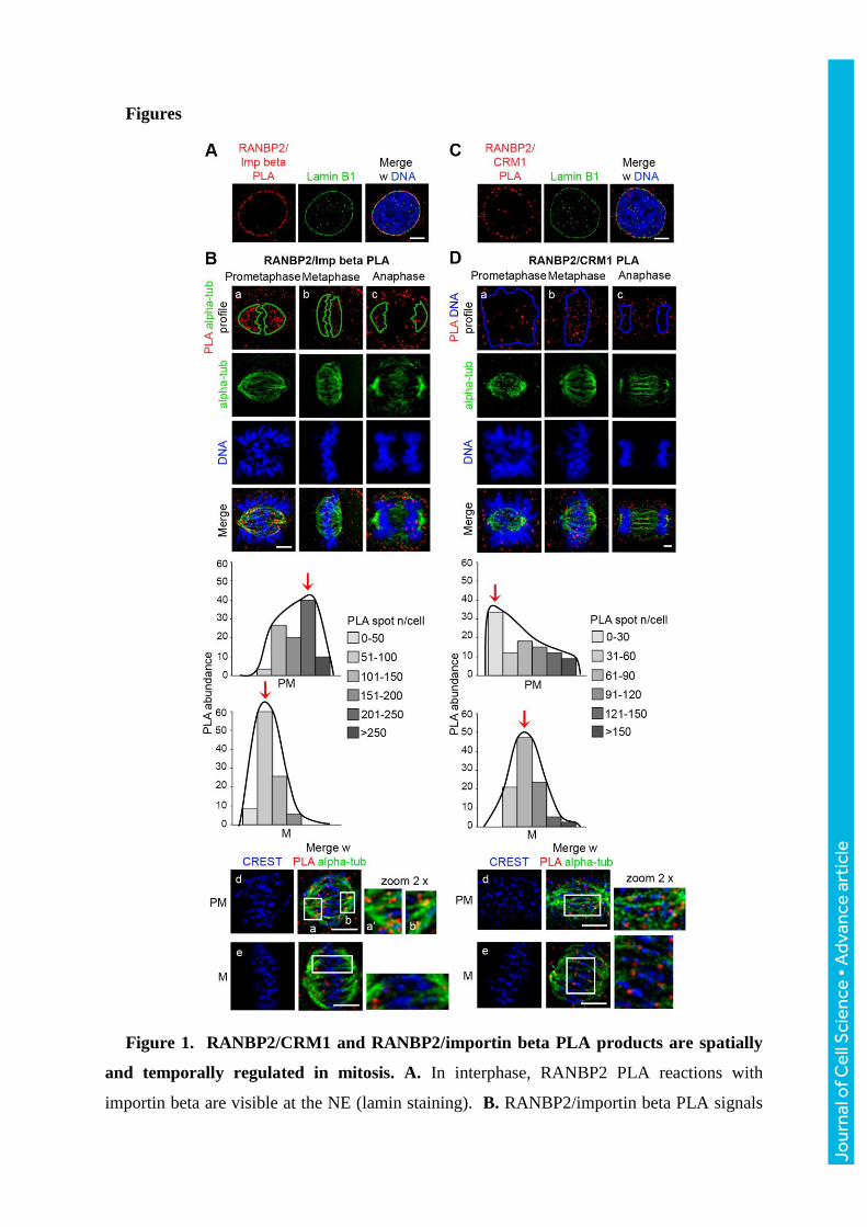
Figure 1. RANBP2/CRM1 and RANBP2/importin beta PLA products are spatially
and temporally regulated in mitosis. A. In interphase, RANBP2 PLA reactions with
importin beta are visible at the NE (lamin staining). B. RANBP2/importin beta PLA signals
Jour
nal o
f Cel
l Sci
ence
• A
dvan
ce a
rtic
le

during mitotic progression. PLA products localise mainly to MTs (delimited by the green
profile; MT and chromosome staining are shown below). The graph quantifies
RANBP2/importin beta PLA products associated with MTs in mitotic stages: prometaphase
(upper panel) and metaphase (lower panel) cells are grouped in discrete classes according to
their content of MT-associated PLA products (legend); the histograms represent the
frequency of cells in each class. Red arrows indicate modal classes. The decrease in
metaphase vs. prometaphase cells was highly significant (p<0.0001, Χ2 test); n, 180
prometaphases and 320 metaphases (9 experiments). The IF panels show the detail of
RANBP2/Importin beta PLA products along MTs in prometaphase (insets, 2x zoom-in). C.
Interphase RANBP2/CRM1 PLA products at the NE (lamin staining). D. RANBP2/CRM1
PLA signals localise at MTs and, after metaphase, in part also at chromosomes (delimited by
the blue profile). The histograms represent the frequency of prometaphase vs. metaphase cells
grouped in classes of abundance of RANBP2/CRM1 PLA products counted in the
chromosome/KT area. The IF panels show that RANBP2/CRM1 PLA products become KT-
associated in metaphase (insets, 2x zoom-in), with a highly significant increase compared to
prometaphase (p<0.0005, Χ2 test); n, 140 prometaphases and 540 metaphases, 9
experiments). Counting the overall number of PLA products in whole cells revealed the same
trend and level of statistical significance shown in graphs B and D. Bars, 5 μm. Extended
images for both pairs during mitotic progression are shown in Fig. S2.
Jour
nal o
f Cel
l Sci
ence
• A
dvan
ce a
rtic
le

Figure 2. CRM1 silencing or functional inhibition abolish RANBP2-containing PLA
products. A. CRM1 silencing substantially reduces CRM1 protein levels. B. Induction of
mitotic abnormalities in CRM1-silenced cultures compared to GL2-interfered controls
(***p<0.0001, Χ2 test); n, at least 3000 mitotic cells, 3 experiments. The IF panels exemplify
frequent defects: multipolar spindles (top), misaligned (middle) and missegregating
chromosomes (bottom). C. Histograms represent the frequency of mitotic cells grouped in
classes of abundance of KT-associated RANBP2/CRM1 PLA products in CRM1-silenced
and control (GL2) cells (p<0.025, Χ2 test in 2 experiments, n, 50 cells per condition). D.
Jour
nal o
f Cel
l Sci
ence
• A
dvan
ce a
rtic
le

LMB abolishes nuclear export of RANBP1 in interphase. E. Induction of mitotic
abnormalities in LMB-treated cultures compared to controls (***p <0.0001, Χ2 test); n, at
least 3400 cells, 2 experiments. F. Distribution of mitotic cells in classes of abundance of
RANBP2/CRM1 PLA products at KTs, in LMB-treated and control cultures (p<0.0001, Χ2
test); n, 60 cells per condition, 2 experiments. Red arrows indicate modal classes. Bars, 5
μm; except in D, 20 μm.
Jour
nal o
f Cel
l Sci
ence
• A
dvan
ce a
rtic
le

Figure 3. Importin beta retains RANBP2 at MTs and decreases its association with
CRM1. A. Cell extracts were probed with importin beta antibody after dox induction. The
upper importin beta band corresponds to dox-induced EGFP chimaera, the lower band is the
endogenous protein. B. Dox-induced importin beta-EGFP reproduces the endogenous protein
localisation at spindle MTs. C. Time-lapse imaging data, showing the evolution of mitotic
abnormalities recorded 6 or 24 h after importin beta induction (*p <0.01 compared to
controls, Χ2 test); n, at least 115 mitoses per time point, 3 experiments. D. The IF panels
exemplify mitotic abnormalities in fixed dox-induced cell samples (multipolar mitosis,
misaligned metaphase chromosome, lagging chromosome in telophase), quantified in the
Jour
nal o
f Cel
l Sci
ence
• A
dvan
ce a
rtic
le

histograms (*p <0.01, **p<0.0005 compared to controls, Χ2 test); n, at least 500 mitotic cells,
2 experiments. E. Histograms represent the distribution of metaphases according to their
abundance of importin beta/RANBP2 PLA signals in the MT area (blue spindles), showing a
highly significant increase in importin beta-induced compared to control cells (p<0.0001, Χ2
test); n, at least 128 cells per condition, 3 experiments. F. Histograms represent the
distribution of metaphases according to their content of CRM1/RANBP2 PLA signals
localising at KTs (blue), showing a highly significant decrease in importin beta-induced
compared to control metaphases (p<0.005, Χ2 test); n, 215 metaphases per condition, 4
experiments. Examples are shown in the IF panels. Bars, 5 μm.
Jour
nal o
f Cel
l Sci
ence
• A
dvan
ce a
rtic
le
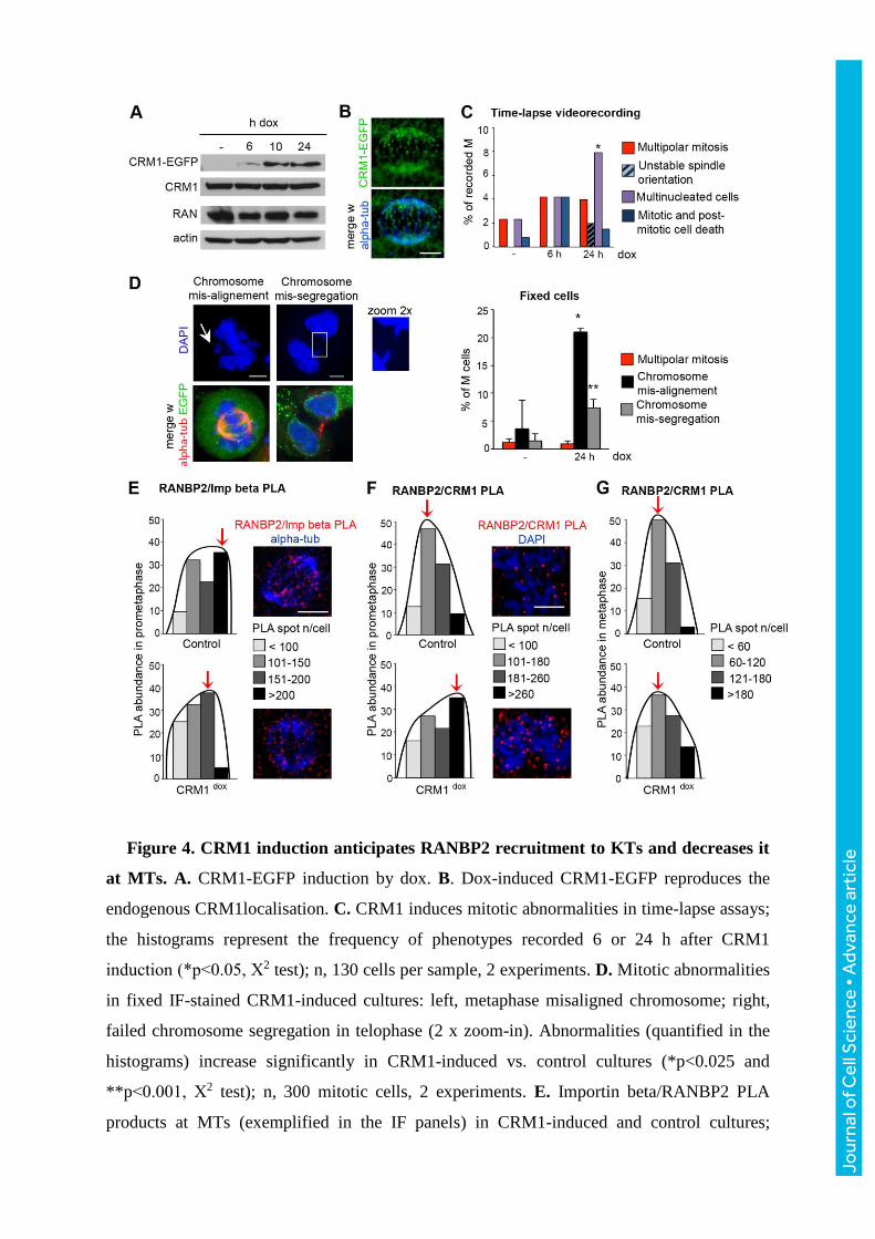
Figure 4. CRM1 induction anticipates RANBP2 recruitment to KTs and decreases it
at MTs. A. CRM1-EGFP induction by dox. B. Dox-induced CRM1-EGFP reproduces the
endogenous CRM1localisation. C. CRM1 induces mitotic abnormalities in time-lapse assays;
the histograms represent the frequency of phenotypes recorded 6 or 24 h after CRM1
induction (*p<0.05, Χ2 test); n, 130 cells per sample, 2 experiments. D. Mitotic abnormalities
in fixed IF-stained CRM1-induced cultures: left, metaphase misaligned chromosome; right,
failed chromosome segregation in telophase (2 x zoom-in). Abnormalities (quantified in the
histograms) increase significantly in CRM1-induced vs. control cultures (*p<0.025 and
**p<0.001, Χ2 test); n, 300 mitotic cells, 2 experiments. E. Importin beta/RANBP2 PLA
products at MTs (exemplified in the IF panels) in CRM1-induced and control cultures;
Jour
nal o
f Cel
l Sci
ence
• A
dvan
ce a
rtic
le

histograms represent the distribution of metaphases in classes of PLA abundance, and
indicate a significant decrease after CRM1 induction (p<0.01, Χ2 test); n, 40 metaphases per
condition, 3 experiments. F. RANBP2/CRM1 PLA signals at KTs significantly increase in
CRM1-induced vs. non-induced prometaphases. Histograms represent the distribution of
prometaphases according to their content of RANBP2/CRM1 PLA products at KTs (p<0.05,
Χ2 test); n, 40 prometaphases per condition, 2 experiments. G. Distribution of metaphase
cells according to their content of RANBP2/CRM1 PLA products at KTs: no significant
variation (Χ2 test) was observed (45 analysed metaphases per condition, 2 experiments).
Bars, 5 μm.
Jour
nal o
f Cel
l Sci
ence
• A
dvan
ce a
rtic
le

Figure 5. NOC disrupts the localisation but not the formation of RANBP2-containing
PLA products. A. Intracellular abundance of RANBP2/importin beta (left) and
RANBP2/CRM1 (right) PLA products in NOC-arrested and control prometaphases: no
statistical difference was observed for either interaction in whole cells, with or without MTs.
B. Co-localisation analysis of RANBP2/CRM1 PLA products and KTs in single planes in
NOC-treated cultures. Alpha-tubulin (green) is diffuse, indicating that NOC treatment was
Jour
nal o
f Cel
l Sci
ence
• A
dvan
ce a
rtic
le
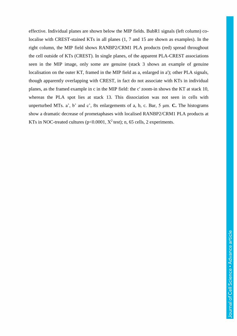
effective. Individual planes are shown below the MIP fields. BubR1 signals (left column) co-
localise with CREST-stained KTs in all planes (1, 7 and 15 are shown as examples). In the
right column, the MIP field shows RANBP2/CRM1 PLA products (red) spread throughout
the cell outside of KTs (CREST). In single planes, of the apparent PLA-CREST associations
seen in the MIP image, only some are genuine (stack 3 shows an example of genuine
localisation on the outer KT, framed in the MIP field as a, enlarged in a'); other PLA signals,
though apparently overlapping with CREST, in fact do not associate with KTs in individual
planes, as the framed example in c in the MIP field: the c' zoom-in shows the KT at stack 10,
whereas the PLA spot lies at stack 13. This dissociation was not seen in cells with
unperturbed MTs. a’, b’ and c’, 8x enlargements of a, b, c. Bar, 5 μm. C. The histograms
show a dramatic decrease of prometaphases with localised RANBP2/CRM1 PLA products at
KTs in NOC-treated cultures (p<0.0001, Χ2 test); n, 65 cells, 2 experiments.
Jour
nal o
f Cel
l Sci
ence
• A
dvan
ce a
rtic
le

Figure 6. CRM1 and importin beta affect SUMO-TOP2A accumulation at KTs. A. In
physiological mitosis, TOP2A co-localises with chromosomes in prometaphase (upper row)
and concentrates at CREST-stained KTs in metaphase (lower row). B. Examples of SUMO-
TOP2A intramolecular PLA patterns in prometaphase (upper) and metaphase (lower) cells,
showing that SUMOylated TOP2A concentrates at KTs in metaphase. In the scatter plot,
Jour
nal o
f Cel
l Sci
ence
• A
dvan
ce a
rtic
le
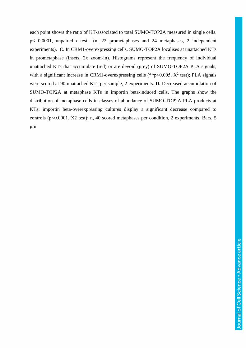
each point shows the ratio of KT-associated to total SUMO-TOP2A measured in single cells.
p< 0.0001, unpaired t test (n, 22 prometaphases and 24 metaphases, 2 independent
experiments). C. In CRM1-overexpressing cells, SUMO-TOP2A localises at unattached KTs
in prometaphase (insets, 2x zoom-in). Histograms represent the frequency of individual
unattached KTs that accumulate (red) or are devoid (grey) of SUMO-TOP2A PLA signals,
with a significant increase in CRM1-overexpressing cells (**p<0.005, Χ2 test); PLA signals
were scored at 90 unattached KTs per sample, 2 experiments. D. Decreased accumulation of
SUMO-TOP2A at metaphase KTs in importin beta-induced cells. The graphs show the
distribution of metaphase cells in classes of abundance of SUMO-TOP2A PLA products at
KTs: importin beta-overexpressing cultures display a significant decrease compared to
controls (p<0.0001, Χ2 test); n, 40 scored metaphases per condition, 2 experiments. Bars, 5
μm.
Jour
nal o
f Cel
l Sci
ence
• A
dvan
ce a
rtic
le
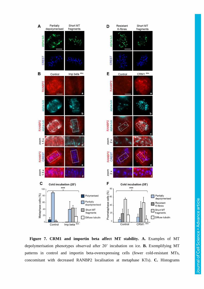
Figure 7. CRM1 and importin beta affect MT stability. A. Examples of MT
depolymerisation phenotypes observed after 20’ incubation on ice. B. Exemplifying MT
patterns in control and importin beta-overexpressing cells (fewer cold-resistant MTs,
concomitant with decreased RANBP2 localisation at metaphase KTs). C. Histograms
Jour
nal o
f Cel
l Sci
ence
• A
dvan
ce a
rtic
le

represent the frequency of MT phenotypes after cold incubation and show a significant shift
towards severe MT depolymerisation phenotypes in importin beta vs. control cultures
(***p<0.0001, Fisher’s exact test); n, 240 cells per sample, 5 experiments. D. Typical MT
depolymerisation phenotypes after 35’ incubation on ice. E. CRM1-overexpressing cells
show more resistant K-fibres juxtaposed to RANBP2 at prometaphase KTs compared to
controls. F. Quantification of MT phenotypes in CRM1-induced, with predominant resistant
K-fibres, vs. control cultures (***p <0.0001, Fisher’s exact test); n, 220 cells per group, 3
experiments. Bars, 5 μm.
Jour
nal o
f Cel
l Sci
ence
• A
dvan
ce a
rtic
le

Figure 8. Model for RANBP2/RANGAP1 recruitment to KTs. A. Schematic
representation of the spatial and temporal variations of RANBP2/SUMO-RANGAP1
(RRSU) interactions with transport factors during mitosis. In a physiological mitosis,
RRSU recruitment to MT-attached KTs with CRM1 modulates local cycles of protein
SUMOylation and of nucleotide turnover on RAN. B. In CRM1-induced cells RANBP2
recruitment at KTs is anticipated in prometaphase. C. In importin beta-induced cells,
RANBP2/importin beta PLA products are retained at MTs, while RANBP2/CRM1
decrease at KTs in metaphase. The altered timing (B), or impairment (C), of RANBP2
localisation at KTs affects in turn the accumulation of SUMO-TOP2A at centromeres and
the stability of K-fibres.
Jour
nal o
f Cel
l Sci
ence
• A
dvan
ce a
rtic
le

Primary antibodies used in immunofluorescence assays
Primary antibodies used in Proximity Ligation Assays Protein Host Source Catalog # Dilution CRM1 mouse Santa Cruz sc-74455 1:100 CRM1 rabbit Santa Cruz sc-5595 1:50 GFP rabbit Abcam ab6556 1:1000 Importin beta mouse Abcam ab2811 1:3000 RANGAP1 mouse Abcam ab28322 1:100 RANGAP1 rabbit Santa Cruz sc-25630 1:200 RANBP2 mouse Santa Cruz sc-74518 1:50 RANBP2 rabbit Abcam ab64276 1:2000 SUMO 2/3 mouse MBL M114-3 1:200 Topoisomerase II alpha rabbit TopoGEN TG2011-1 1:300
Primary antibodies used in Western immunoblotting (conditions were as recommended by the manifacturer) Protein Host Source Catalog # actin goat Santa Cruz sc1862 alpha-tubulin mouse Sigma-Aldrich T5168 CRM1 rabbit Santa Cruz sc-5595 importin beta mouse Abcam ab2811 importin beta mouse Becton Dickinson 610560 RAN mouse Becton Dickinson 61034 RANBP2 goat Santa Cruz sc-15442 RANGAP1 goat Santa Cruz sc-1862
Supplementary Table 1
Protein Host Source Catalog # Dilution alpha-tubulin chicken Abcam ab89984 1:50 alpha-tubulin mouse Sigma T5168 1:3000 BUBR1 rabbit Bethyl A300-995A 1:1000 CREST human Antibodies Inc. 15-234-0001 1:20 CRM1 mouse Becton Dickinson 611833 1:100 CRM1 rabbit Santa Cruz sc-5595 1:50 Importin beta mouse Abcam ab2811 1:3000 RANBP1 goat Santa Cruz sc-1160 1:25 RANBP2 mouse Santa Cruz sc-74518 1:50 RANBP2 rabbit Abcam ab64276 1:2000 RANGAP1 rabbit Santa Cruz sc-25630 1:200 Topoisomerase II alpha rabbit TopoGEN TG2011-1 1:300
J. Cell Sci. 130: doi:10.1242/jcs.197905: Supplementary information
Jour
nal o
f Cel
l Sci
ence
• S
uppl
emen
tary
info
rmat
ion

Figure S1. PLA testing on two characterised interactions, RANBP2/RANGAP1 and RAN/CRM1,
in time-course assays to identify amplification conditions for optimal signal-to-noise ratio in HeLa
cells. Upper bar, 20 µm; lower bar, 10 µm.
J. Cell Sci. 130: doi:10.1242/jcs.197905: Supplementary information
Jour
nal o
f Cel
l Sci
ence
• S
uppl
emen
tary
info
rmat
ion

J. Cell Sci. 130: doi:10.1242/jcs.197905: Supplementary information
Jour
nal o
f Cel
l Sci
ence
• S
uppl
emen
tary
info
rmat
ion

Figure S2. RANBP2 interactions with transport receptors during mitotic progression. A.
RANBP2/Importin beta PLA products from G2 to mitotic exit. The upper panel shows PLA signals
(red) in samples stained for lamin B1 (cyano), alpha-tubulin (green), and CREST (blue); in the
lower panel the anti-lamin antibody was omitted. RANBP2/Importin beta PLA signals (red)
concentrate at the NE in G2 cells (a); panel b shows PLA product delocalisation to the cytoplasm,
coinciding with the NE deformation and breakdown under centrosomal pushing; in c-d, mitotic
MTs gradually assemble and capture PLA complexes (PLA spots over forming MTs are arrowed).
In prometaphase (d), as the spindle takes shape, RANBP2/Importin beta PLA signals can be
appreciated along MTs. Their abundance decreases in late prometaphase (e)/metaphase (f); in
anaphase (g), residual signals mostly localise along polar MTs, but not at KT-bound MTs. B.
RANBP2/CRM1 PLA products during mitotic progression. PLA signals associate with MTs in
prometaphase (a), including at growing ends that make contacts with KTs; during prometaphase (b-
c) they associate with the outer KT face, then remain associated with KTs in metaphase (d) and
anaphase (d). In telophase (panels Ah and Be), both PLA pairs relocalise around decondensing
nuclei. Bars, 5 µm.
J. Cell Sci. 130: doi:10.1242/jcs.197905: Supplementary information
Jour
nal o
f Cel
l Sci
ence
• S
uppl
emen
tary
info
rmat
ion

J. Cell Sci. 130: doi:10.1242/jcs.197905: Supplementary information
Jour
nal o
f Cel
l Sci
ence
• S
uppl
emen
tary
info
rmat
ion

Figure S3. RANBP2 silencing assays validate the RANBP2 "switch partners" model. A.
Reduced RANBP2 abundance after 72 h of incubation with RANBP2-specific siRNAs compared to
controls (incubated with GL2 siRNAs). Bar, 20 µm. In parallel, SUMO-conjugated RANGAP1
decreases. B. RANGAP1 recruitment to KTs fails in RANBP2-interfered cells. C. RANBP2
silencing yields an increased mitotic index and mitotic abnormalities (a multipolar spindle and a
chromosome remaining near one pole, probably representing a monotelic or syntelic attachment, are
shown), with a highly significant difference compared to controls (***p<0.0001, Χ2 test, at least
660 mitotic cells per condition, 2 experiments). D. Histograms show that importin beta/RANBP2
PLA signals significantly decrease in RANBP2-interfered compared to control prometaphase cells
(p< 0.005, Χ2 test, 35 cells per condition, 2 experiments). E. In parallel, CRM1/RANBP2 PLA
signals also decrease in a highly significant manner in RANBP2-interfered compared to control
metaphases (p< 0.0005, Χ2 test, at least 35 cells per condition, 2 experiments). Bars in B-E, 5 µm.
J. Cell Sci. 130: doi:10.1242/jcs.197905: Supplementary information
Jour
nal o
f Cel
l Sci
ence
• S
uppl
emen
tary
info
rmat
ion

J. Cell Sci. 130: doi:10.1242/jcs.197905: Supplementary information
Jour
nal o
f Cel
l Sci
ence
• S
uppl
emen
tary
info
rmat
ion

Figure S4. RANGAP1 PLA products with nuclear transport factors. A. RANGAP1/importin
beta PLA products are superimposed to the MT area (green profile). B. RANGAP1/CRM1 PLA
products localise to MTs; after metaphase, a fraction localises to the chromosome area (blue
profile), reproducing the PLA pattern seen with RANBP2. C. Antibodies to different CRM1
epitopes detect distinct protein fractions: an antibody to the C-terminal region (left) decorates the
nuclear rim, and, in mitosis, depicts CRM1 fractions at spindle poles, MTs and metaphase KTs; an
antibody to the N-terminal region (right) depicts a more intranuclear interphase pattern and MT-
associated, but not KT-associated, CRM1 in metaphase. The region in which KTs align is framed
and enlarged (1.4x) below. D. RANGAP1/RANBP2 PLA products (rightmost column) localise at
MTs, with a fraction at the chromosome area in metaphase. E. LMB does not inhibit
RANGAP1/RANBP2 PLA product formation, yet prevents their recruitment to KTs.
J. Cell Sci. 130: doi:10.1242/jcs.197905: Supplementary information
Jour
nal o
f Cel
l Sci
ence
• S
uppl
emen
tary
info
rmat
ion