indications and surgical techniques dual plating of the
TRANSCRIPT
Received 12/16/2019 Review began 12/21/2019 Review ended 12/22/2019 Published 12/27/2019
© Copyright 2019Sain et al. This is an open accessarticle distributed under the terms ofthe Creative Commons AttributionLicense CC-BY 3.0., which permitsunrestricted use, distribution, andreproduction in any medium, providedthe original author and source arecredited.
Dual Plating of the Distal Femur:Indications and Surgical TechniquesArnab Sain , Vijay Sharma , Kamran Farooque , Muthukumaran V , KirubakaranPattabiraman
1. Orthopaedics, All India Institute of Medical Sciences, New Delhi, IND
Corresponding author: Arnab Sain, [email protected]
AbstractDual-plating of the distal femur is required in some cases to achieve stable fixation. Theindications of a medial plate in addition to the lateral plate are medial supracondylar bone loss,low trans-condylar bicondylar fractures, medial Hoffa fracture, peri-prosthetic distal femurfractures, non-union after failed fixation with single lateral plate, poor bone quality andcomminuted distal femur fractures (AO type C3). We recommend orthogonal plateconfiguration with locked plates by a single incision or dual incision approach as per surgeonchoice.
Categories: OrthopedicsKeywords: dual-plating, distal femur, medial plate fixation
Introduction And BackgroundSupracondylar femur fractures are commonly associated with severe comminution andsignificant soft tissue injury. Distal femoral fractures are mostly caused by high-energy injuries,such as falling injury and traffic accidents, and fractures are often severely comminuted.Despite the recent advances in techniques and implants, the treatment of intra-articular multi-fragmentary distal femoral fractures remains a challenge. Long-term disability can occur inpatients with extensive articular cartilage damage and marked comminution. Distal femurfractures in the elderly are complicated by poor bone quality (severe osteoporosis), a distalsegment that is too short for adequate fixation, blood loss, malunion and non-union, andincreased mortality [1-4].
Locked plating is one of the best and modern options for treating supracondylar femurfractures with relatively low failure rates. Single lateral plating of distal femur fractures wasoften found to have a relatively higher failure rate [2].
A medial plate in addition to lateral plating reduces the chances of failure of fixation [2]. Thisarticle will focus on the indications and techniques of using an additional medial plate fixationin fractures of the distal femur.
ReviewPre-operative planningDecision making before surgical stabilization is important. The treating surgeon shouldexamine the injured limb to check for the soft tissue status and any distal neurovascular deficit.Also, other injuries should be ruled out. Good quality radiographs in two planes are necessary.
1 1 1 1
1
Open Access ReviewArticle DOI: 10.7759/cureus.6483
How to cite this articleSain A, Sharma V, Farooque K, et al. (December 27, 2019) Dual Plating of the Distal Femur: Indicationsand Surgical Techniques. Cureus 11(12): e6483. DOI 10.7759/cureus.6483
Computed tomography of the distal femur with coronal and sagittal reformations helps indelineating fracture patterns. Proper pre-operative planning will guide towards the need fordual plating of distal femur fractures [5].
Indications for dual platingSupracondylar femur fractures were previously treated with condylar buttress plates [6].Subsequently, fixed-angle implants, angle-blade plates, intramedullary retrograde nails, anddynamic supracondylar screws were found to have a better biomechanical design for preventingthe varus collapse compared to condylar buttress plates [7,8]. Locking plates are known forhaving increased biomechanical resistance with the advantage of greater numbers of fixationscrews in the distal femur metaphysis [9,10]. Locking plates provide increased stability andresistance to failure compared to retrograde nails in elderly patients with poor bone stock [11-13]. The additional application of a medial plate in the following circumstances may help inachieving a stable anatomically aligned reduction.
Medial Supracondylar Bone Loss
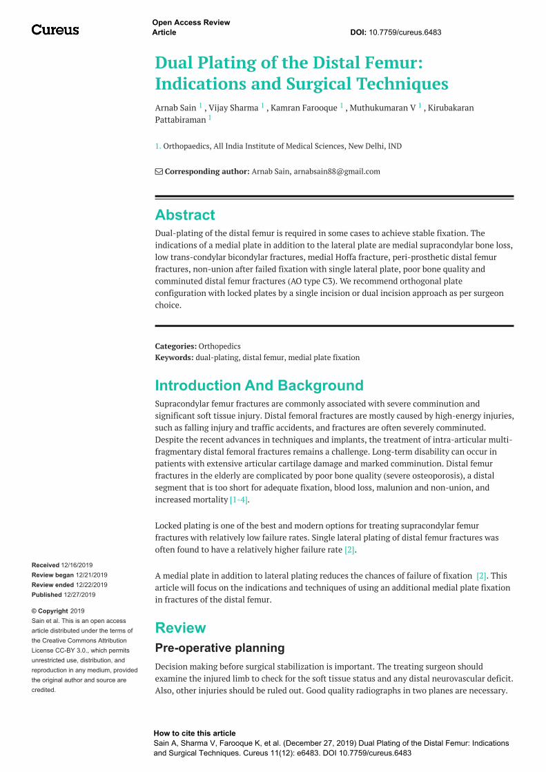
In distal femur fractures with extensive metaphyseal comminution, osteopenic bone and inhigh-energy or open fractures, there is functional loss of medial cortical buttress. In thesesituations, there is less chance of healing of the medial column. The addition of a medial platehelps in giving additional stability and reduces the chances of implant failure [1,14].Rajasekaran et al. found that additional medial plating and bone grafting was needed for non-union distal femur with medial bone loss more than 2 cm (Figures 1, 2) [15].
2019 Sain et al. Cureus 11(12): e6483. DOI 10.7759/cureus.6483 2 of 17
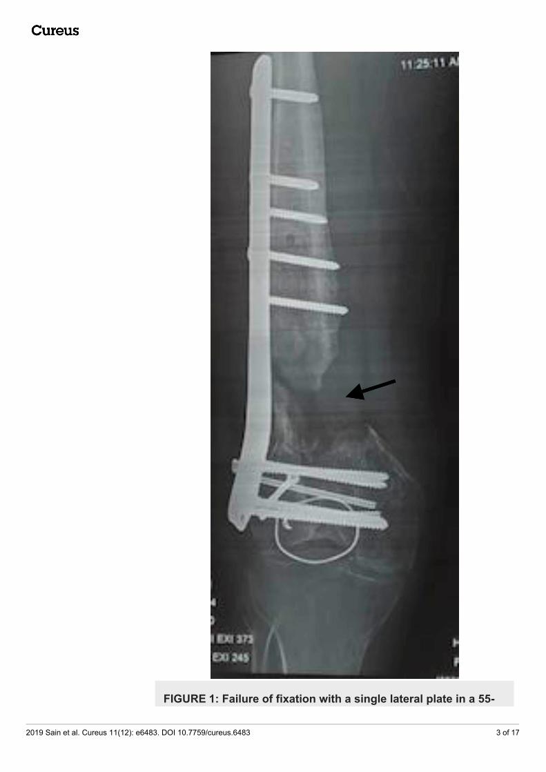
FIGURE 1: Failure of fixation with a single lateral plate in a 55-
2019 Sain et al. Cureus 11(12): e6483. DOI 10.7759/cureus.6483 3 of 17

year-old patient with an open fracture and medialsupracondylar bone loss. Note the medial bone loss of morethan 2 cm.
2019 Sain et al. Cureus 11(12): e6483. DOI 10.7759/cureus.6483 4 of 17

FIGURE 2: Revision with a medial plate and bone grafting.
2019 Sain et al. Cureus 11(12): e6483. DOI 10.7759/cureus.6483 5 of 17

Low Trans-condylar Bicondylar Fractures
High energy injuries sometimes lead to a low horizontal trans-condylar fracture pattern whichis associated with an intercondylar split. In this fracture pattern, fixation with a single lateralplate may not achieve stable fixation as sufficient screw purchase will not be possible becauseof the intercondylar notch. A medial buttress plate in addition to the lateral plate is helpful inthis situation [5,16].
Medial Hoffa Fracture
Anatomical restoration of the articular surface is necessary for comminuted distal femurfractures. In fractures with a large medial Hoffa fragment, the application of a medialneutralization plate is necessary to achieve stable fixation. This fixation provides stability inaddition to interfragmentary lag screws [5].
Peri-prosthetic Distal Femur Fractures
In peri-prosthetic fractures of the distal femur, dual plating offers stable fixation, especially inosteoporotic bone. This allows for early mobilization and rehabilitation. Stable fixation of peri-prosthetic fractures is necessary to avoid failures of fixation. Cicek et al. advocated dual lockedplate fixation of peri-prosthetic distal femur fractures with osteoporotic bones [17].
Non-union after Failed Fixation with Single Lateral Plate
There is an increased risk of non-union in patients with comminuted distal femur fracturestreated with single lateral locking plate [18]. In these cases of non-union of distal femurdefinitive treatment involves stabilization of non-union site with an additional medial plateand bone-grafting at the non-union site [19]. Non-union with medial bone loss more than 2 cmis an indication for medial plate augmentation and bone grafting (Figures 3, 4) [15].
2019 Sain et al. Cureus 11(12): e6483. DOI 10.7759/cureus.6483 6 of 17
FIGURE 3: Non-union after single lateral plate fixation.
2019 Sain et al. Cureus 11(12): e6483. DOI 10.7759/cureus.6483 7 of 17

2019 Sain et al. Cureus 11(12): e6483. DOI 10.7759/cureus.6483 8 of 17
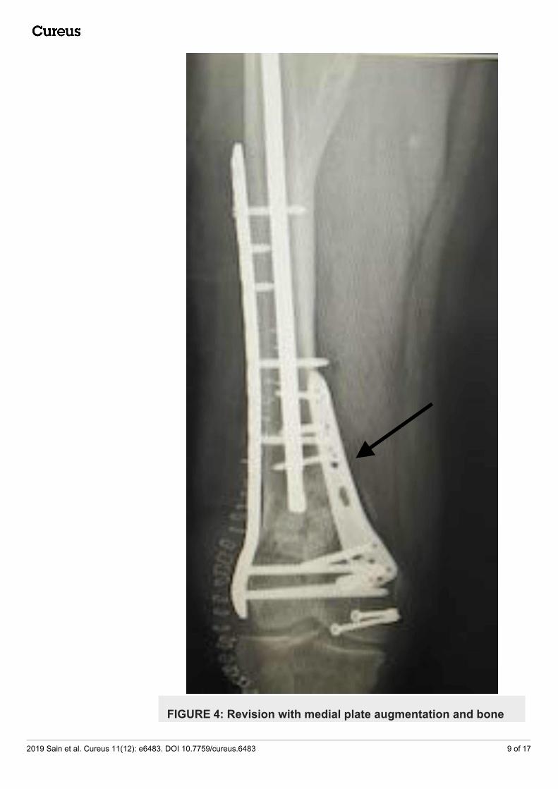
FIGURE 4: Revision with medial plate augmentation and bone
2019 Sain et al. Cureus 11(12): e6483. DOI 10.7759/cureus.6483 9 of 17

grafting.
Poor Bone Quality
In patients with distal femur fracture with osteoporotic bone, there is a high incidence offailure of fixation with a single lateral plate due to poor purchase of screw in osteoporotic bone.Metwaly and Zakaria demonstrated that dual plating in osteoporotic distal femur fractures by asingle incision offers the stability of fixation with resultant early mobility and acceleratedrehabilitation [20]. In the study by Todorov et al., medial augmented LISS (less invasivestabilization system) plating was found to have higher union rates compared to conventionalLISS plating in osteoporotic distal femur fractures [21]. Steinberg et al. also found higher unionrates with dual plating in fractures with osteoporotic bone [2].
Comminuted Distal Femur Fractures (AO type C3)
In patients with AO type C3 distal femoral fractures, dual plating offers a more stable construct.Imam et al. demonstrated that dual plating fixation using anterior approach for type C3 distalfemoral fractures is an efficient and safe method of management. It has several advantages suchas precise exposure, easy manipulation, anatomical reduction, and stable fixation [1]. Steinberget al. demonstrated higher rates of union with dual plating in AO type C3 distal femur fractures(Figures 5-8) [2].
2019 Sain et al. Cureus 11(12): e6483. DOI 10.7759/cureus.6483 10 of 17

FIGURE 5: 3D CT image showing a comminuted distal femurfracture in a 35-year-old male patient.
2019 Sain et al. Cureus 11(12): e6483. DOI 10.7759/cureus.6483 11 of 17

FIGURE 6: Failure following single lateral plate fixation.
2019 Sain et al. Cureus 11(12): e6483. DOI 10.7759/cureus.6483 12 of 17

FIGURE 7: Revision with dual plating and bone grafting was
2019 Sain et al. Cureus 11(12): e6483. DOI 10.7759/cureus.6483 13 of 17

done.
FIGURE 8: Knee flexion beyond 90 degrees in the follow-up.
Surgical techniquePosition
The patient is positioned supine on a radiolucent operating table with the knee flexed at 30degrees. Tourniquet use depends on the surgeon and may be used in very distal fractures. Theuse of a lateral femoral distractor with pins placed in the femoral diaphysis and proximal tibiametaphysis facilitates fracture alignment and disimpacts intra-articular fragments. Intra-operative use of C-arm is necessary for proper plate placement and fracture reduction andalignment [5].
Approach
The choice of approach depends on the surgeon. There are two approaches, the dual incisionapproach and the single incision approach. The dual incision approach involves placing amedial and lateral incision for exposure of fracture and articular surface [2]. The single incisionapproach involves the use of a single para-patellar incision (lateral or medial) for exposure offracture and articular surface (Figure 9). Imam et al. used a single midline incision and anextended medial para-patellar approach for dual plating of type C3 distal femoral fractures [1].
2019 Sain et al. Cureus 11(12): e6483. DOI 10.7759/cureus.6483 14 of 17
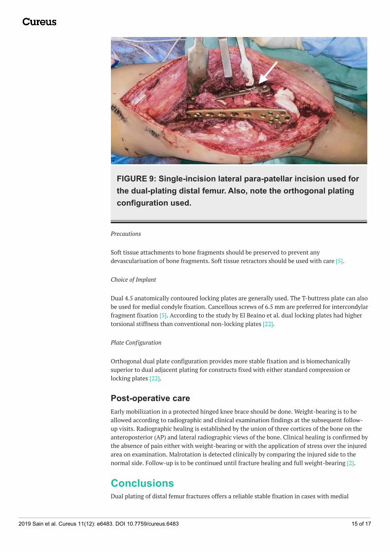
FIGURE 9: Single-incision lateral para-patellar incision used forthe dual-plating distal femur. Also, note the orthogonal platingconfiguration used.
Precautions
Soft tissue attachments to bone fragments should be preserved to prevent anydevascularisation of bone fragments. Soft tissue retractors should be used with care [5].
Choice of Implant
Dual 4.5 anatomically contoured locking plates are generally used. The T-buttress plate can alsobe used for medial condyle fixation. Cancellous screws of 6.5 mm are preferred for intercondylarfragment fixation [5]. According to the study by El Beaino et al. dual locking plates had highertorsional stiffness than conventional non-locking plates [22].
Plate Configuration
Orthogonal dual plate configuration provides more stable fixation and is biomechanicallysuperior to dual adjacent plating for constructs fixed with either standard compression orlocking plates [22].
Post-operative careEarly mobilization in a protected hinged knee brace should be done. Weight-bearing is to beallowed according to radiographic and clinical examination findings at the subsequent follow-up visits. Radiographic healing is established by the union of three cortices of the bone on theanteroposterior (AP) and lateral radiographic views of the bone. Clinical healing is confirmed bythe absence of pain either with weight-bearing or with the application of stress over the injuredarea on examination. Malrotation is detected clinically by comparing the injured side to thenormal side. Follow-up is to be continued until fracture healing and full weight-bearing [2].
ConclusionsDual plating of distal femur fractures offers a reliable stable fixation in cases with medial
2019 Sain et al. Cureus 11(12): e6483. DOI 10.7759/cureus.6483 15 of 17
supracondylar bone loss, low trans-condylar bicondylar fractures, medial Hoffa fracture, peri-prosthetic distal femur fractures, non-union after failed fixation with single lateral plate, poorbone quality and comminuted distal femur fractures (AO type C3). Single-incision or dualincision approach may be used. Orthogonal plate configuration with locked plates providesstable fixation and allows for early rehabilitation. Early mobilization is necessary to preventjoint stiffness.
Additional InformationDisclosuresConflicts of interest: In compliance with the ICMJE uniform disclosure form, all authorsdeclare the following: Payment/services info: All authors have declared that no financialsupport was received from any organization for the submitted work. Financial relationships:All authors have declared that they have no financial relationships at present or within theprevious three years with any organizations that might have an interest in the submitted work.Other relationships: All authors have declared that there are no other relationships oractivities that could appear to have influenced the submitted work.
References1. Imam MA, Torieh A, Matthana A: Double plating of intra-articular multifragmentary C3-type
distal femoral fractures through the anterior approach. Eur J Orthop Surg Traumatol. 2018,28:121-130. 10.1007/s00590-017-2014-9
2. Steinberg E, Elis J, Steinberg Y, Salai M, Ben-Tov T: A double-plating approach to distal femurfracture: a clinical study. Injury. 2017, 48:2260-2265. 10.1016/j.injury.2017.07.025
3. Meneghini RM, Keyes BJ, Reddy KK, Maar DC: Modern retrograde intramedullary nails versusperiarticular locked plates for supracondylar femur fractures after total knee arthroplasty. JArthroplasty. 2014, 29:1478-1481. 10.1016/j.arth.2014.01.025
4. Streubel P, Ricci W, Wong A, Gardner M: Mortality after distal femur fractures in elderlypatients. Clin Orthop Relat Res. 2011, 469:1188-1196. 10.1007/s11999-010-1530-2
5. Chapman J, Henley M: Double plating of distal femur fractures: indications and technique .Tech Orthop. 1994, 9:210-216. 10.1097/00013611-199400930-00008
6. Davison BL: Varus collapse of comminuted distal femur fractures after open reduction andinternal fixation with a lateral condylar buttress plate. Am J Orthop. 2003, 32:27-30.
7. Christodoulou A, Terzidis I, Ploumis A, Metsovitis S, Koukoulidis A, Toptsis C: Supracondylarfemoral fractures in elderly patients treated with the dynamic condylar screw and theretrograde intramedullary nail: a comparative study of the two methods. Arch Orthop TraumaSurg. 2005, 125:73-79. 10.1007/s00402-004-0771-5
8. Vallier H, Immler W: Comparison of the 95-degree angled blade plate and the lockingcondylar plate for the treatment of distal femoral fractures. J Orthop Trauma. 2012, 26:327-332. 10.1097/bot.0b013e318234d460
9. Frigg R, Appenzeller A, Christensen R, Frenk A, Gilbert S, Schavan R: The development of thedistal femur less invasive stabilization system (LISS). Injury. 2001, 32:24-31. 10.1016/s0020-1383(01)00181-4
10. Kregor P, Stannard J, Zlowodzki M, Cole P, Alonso J: Distal femoral fracture fixation utilizingthe Less Invasive Stabilization System (L.I.S.S.): the technique and early results. Injury. 2001,32:32-47. 10.1016/s0020-1383(01)00182-6
11. Assari S, Kaufmann A, Darvish K, Park J, Haw J, Safadi F, Rehman S: Biomechanicalcomparison of locked plating and spiral blade retrograde nailing of supracondylar femurfractures. Injury. 2013, 44:1340-1345. 10.1016/j.injury.2013.04.016
12. Salas C, Mercer D, DeCoster TA, Reda Taha M: Experimental and probabilistic analysis ofdistal femoral periprosthetic fracture: a comparison of locking plate and intramedullary nailfixation. Part A: experimental investigation. Comput Methods Biomech Biomed Engin. 2011,14:157-164. 10.1080/10255842.2010.535816
13. Salas C, Mercer D, DeCoster T, Reda Taha M: Experimental and probabilistic analysis of distalfemoral periprosthetic fracture: a comparison of locking plate and intramedullary nail
2019 Sain et al. Cureus 11(12): e6483. DOI 10.7759/cureus.6483 16 of 17

fixation. Part B: probabilistic investigation. Comput Methods Biomech Biomed Engin. 2011,14:175-182. 10.1080/10255842.2010.539207
14. Gwathmey W, Jones-Quaidoo S, Kahler D, Hurwitz S, Cui Q: Distal femoral fractures: currentconcepts. Am Acad Orthop Surg. 2010, 18:597-607. 10.5435/00124635-201010000-00003
15. Rajasekaran RB, Jayaramaraju D, Palanisami D, Agraharam D, Perumal R, Kamal A,Rajasekaran S: A surgical algorithm for the management of recalcitrant distal femurnonunions based on distal femoral bone stock, fracture alignment, medial void, and stabilityof fixation. Arch Orthop Trauma Surg. 2019, 139:1057-1068. 10.1007/s00402-019-03172-0
16. Sanders R, Swiontkowski M, Rosen H, Helfet D: Double-plating of comminuted, unstablefractures of the distal part of the femur. J Bone Joint Surg. 1991, 73:341-346.10.2106/00004623-199173030-00004
17. Çiçek H, Tuhanioğlu Ü, Oğur H, Seyfettinoğlu F, Bozkurt M: An alternative treatment forosteoporotic Su Type III periprosthetic supracondylar femur fractures: double locking platefixation. Acta Orthop Traumatol Turc. 2018, 52:92-96. 10.1016/j.aott.2017.09.010
18. Karam J, Campbell P, David M, Hunter M: Comparison of outcomes and analysis of risk factorsfor non-union in locked plating of closed periprosthetic and non-periprosthetic distal femoralfractures in a retrospective cohort study. J Orthop Surg Res. 2019, 14:150. 10.1186/s13018-019-1204-z
19. Ebraheim N, Buchanan G, Liu X, Cooper M, Peters N, Hessey J, Liu J: Treatment of distal femurnonunion following initial fixation with a lateral locking plate. Orthop Surg. 2016, 8:323-330.10.1111/os.12257
20. Metwaly R, Zakaria Z: Single-incision double-plating approach in the management ofisolated, closed osteoporotic distal femoral fractures. Geriatr Orthop Surg Rehabil. 2018,9:215145931879985. 10.1177/2151459318799856
21. Todorov D, Zderic I, Richards R, et al.: Is augmented LISS plating biomechanicallyadvantageous over conventional LISS plating in unstable osteoporotic distal femoralfractures?. J Orthop Res. 2018, 36:2604-2611. 10.1002/jor.24047
22. El Beaino M, Morris R, Lindsey R, Gugala Z: Biomechanical evaluation of dual plateconfigurations for femoral shaft fracture fixation. BioMed Res Int. 2019, 2019:7.10.1155/2019/5958631
2019 Sain et al. Cureus 11(12): e6483. DOI 10.7759/cureus.6483 17 of 17