information to users the quality of this...
TRANSCRIPT

Characterization of white spot syndrome virus ofpenaeid shrimp: Genomic cloning and sequencing,
structural protein analyzing and sequencing,genetic diversity, pathology and virulence
Item Type text; Dissertation-Reproduction (electronic)
Authors Wang, Qiong
Publisher The University of Arizona.
Rights Copyright © is held by the author. Digital access to this materialis made possible by the University Libraries, University of Arizona.Further transmission, reproduction or presentation (such aspublic display or performance) of protected items is prohibitedexcept with permission of the author.
Download date 22/06/2018 15:36:06
Link to Item http://hdl.handle.net/10150/284292

INFORMATION TO USERS
This manuscript has been reproduced from the microfihn master. UMI
films the text directly from the original or copy submitted. Thus, some
thesis and dissertation copies are in typewriter &ce, while others may be
from any type of computer printer.
The quality of this reproduction is dependent upon the quality of the
copy submitted. Broken or indistinct print, colored or poor quality
illustrations and photographs, print bleedthrough, substandard margins,
and improper alignment can adversely afifect reproduction.
In the unlikely event that the author did not send UMI a complete
manuscript and there are missing pages, these will be noted. Also, if
unauthorized copyright material had to be removed, a note will indicate
the deletion.
Oversize materials (e.g., maps, drawings, charts) are reproduced by
sectioning the original, b^inning at the upper left-hand comer and
continuing from left to right in equal sections with small overiaps. Each
original is also photographed in one exposure and is included in reduced
form at the back of the book.
Photographs included in the original manuscript have been reproduced
xerographically in this copy. Higher quality 6" x 9" black and white
photographic prints are available for any photographs or illustrations
appearing in this copy for an additional charge. Contact UMI directly to
order.
UMI A Bell & Howell Inclination Company
300 North Zed> Road, Ann Aibor MI 48106-1346 USA 313/761-4700 800/521-0600


NOTE TO USERS
This reproduction is the best copy available
UMI


\
CHARACTERIZATION OF WHITE SPOT SYNDROME VIRUS OF PENAEID
SHRIMP: GENOMIC CLONING AND SEQUENCING, STRUCTURAL
PROTEIN ANALYZING AND SEQUENCING, GENETIC DIVERSITY,
PATHOLOGY AND VIRULENCE
By
Qiong Wang
A Dissertation Submitted to the Faculty of the
DEPARTMENT OF VETERINARY SCIENCE AND MICROBIOLOGY
In Partial Fulfillment of the Requirements For the Degree of
DOCTOR OF PHILOSOPHY WITH A MAJOR IN PATHOBIOLOGY
In the Graduate College
THE UNIVERSITY OF ARIZONA
1 9 9 9
UMl Number: 9927506
UMI Microform 9927506 Copyrigiit 1999, by UMI Company. All rights reserved.
This microform edition is protected against miauthorized copying under Title 17, United States Code.
UMI 300 North Zeeb Road Ann Arbor, MI 48103

2
THE UNIVERSITY OF ARIZONA « GRADUATE COLLEGE
As members of the Final Examination Committee, we certify that we have
Oiong Fang read the dissertation prepared by_
entitled Characterization of tTIiite Spot Syndrone Virus of Penaeid Shrimp:
Genomic Cloning and Sequencing, Structural Protein Analyzing
and Secuencing, Genetic Diversity, Pathology and Virulence
and recommend that it be accepted as fulfilling the dissertation
requirement for the Degree of Doctor of Philosophy
Ph.i/.
nn J.
lus J
Date
lartinez Ji Hev/lett, Ph.D.
Final approval and acceptance of this dissertation is contingent upon the candidate's submission of the final copy of the dissertation to the Graduate College.
I hereby certify that I have read this dissertation prepared under my direction and recommend that it be accepted as fulfilling the dissertation reqy q^Dsement. ^
Dissertation Director Donald V. Lightner Date
3
STATEMENT BY AUTHOR
This dissertation has been submitted in partial fulfillment of requirements for an advanced degree at The University of Arizona and is deposited in the University Library to be made available to borrowers under rules of the library.
Brief quotations from this dissertation are allowable without special permission, provided that accurate acknowledgement of source is made. Requests for permission for extended quotation firom or reproduction of this manuscript in whole or in part may be granted by the head of the major department or the Dean of the Graduate College when in his or her judgment the proposed use of the material is in the interests of scholarship. In all other instances, however, permission must be obtained from the author.
SIGNED:

4
ACKNOWLEDGEMENTS
My greatest gratitude is due to my mentor. Dr. Donald V. Lightner, for giving me the privilege of being your student, for your teachings, guidance and trust. You not only equip me with the priceless scientific knowledge and skills but also serve as a role model exemplifying ethic integrity and professionalism.
I thank Bonnie T. Poulos, Linda M. Nunan, Rita M. Redman, and Brenda L. White for their technical assistance with the experiments and valuable discussions about my research.
To my graduate advisory conunittee members. Dr. Donald V. Lightoer, Dr. Glenn J. Songer, Dr. Cornelius J. Mare, Dr. Raymond B. Nagle and Dr. Martinez J. Hewlett. Thank you for being in my committee, for willingness to give advice and for donating your precious time to give me guidance.
To my colleague Kenneth W. Hasson, Carlos Pantoja, Jeffrey R. Garza, Leone L. Mohney, Kathy Tang, Wanda C. McCormack, Heidi S. Erickson, and Stephanie Durand for your friendship and cooperation.
Thanks are due to my family and friends for their spiritual support. They are my motivation to be a good researcher, and a good person.
I thank Wallace Clark for amino acid analysis and protein sequencing. Skip Vaught for DNA sequencing, and the Imaging Facility Laboratory at the University of Arizona for access to the electron microscope.
The original isolates of WSSV were kindly provided by Dr. Paul Frelier (Department of Veterinary Pathology, College of Veterinary Medicine, Texas A & M University, College Station, Texas), Dr. Craig Browdy (South Carolina Department of Natural Resources, Waddell Mariculture Center, Bluflfton, South Carolina), Dr. Laura K. Richman (Department of Pathology, National Zoological Park, Washington D. C.), Dr. Eileen Reddington (Diagxotics, Ltd.), Dr. Ramana Murty (Andhra University, Visakhapatnam, India), and Mr. Jie Huang (Yellow Sea Fishery Research Institute, Qingdao, Shangdong, China).
This research was partially funded by the Gulf Coast Research laboratory Consortium Marine Shrimp Farming Program, CREES, USDA under Grant No. 95-38808-1420, the National Sea Grant Program, USDC under the Grant No. NA56RG0617, USDC under Grant No. NA56FD0621, and a special grant from the U. S. National Fisheries Institute.

5
TABLE OF CONTENTS
LIST OF FIGUEIES 7
LIST OF TABLES 8
ABSTRACT 9
1. INTRODUCTION II
1.1 Problem Definition II
1.2 Literature Review 15
1.2.1 Major Viral Diseases of Cultured Penaeid Shrimp 15 1.2.1.1 Baculoviruses 20 1.2.1.2 Parvoviruses 25 1.2.1.3 Iridoviruses 29 1.2.1.4 Picomaviruses 29 1.2.1.5 Rhabdoviruses 31 1.2.1.6 Coronaviruses 32 1.2.1.7 Togaviruses 33 1.2.1.8 Reoviruses 34
1.2.2 The White Spot Syndrome Virus 35 1.2.2.1 History of white spot syndrome disease 35 1.2.2.2 Viral characterization 38 1.2.2.3 Diagnostic techniques 42 1.2.2.4 Host range 44 1.2.2.5 Geographic distribution and dissemination 45 1.2.2.6 Economic impact and disease management
methods 49
1.2.3 Baculovirus diversity and molecular biology 51 1.2.3.1 General biology of baculoviruses 51 1.2.3.2 Life cycle 52 1.2.3.3 Viral replication and regulation of gene
expression 56 1.2.3.4 Baculovirus structural proteins and genes 59 1.2.3.5 Genetic relatedness of baculoviruses 62
1.3 Dissertation Format 64
2. PRESENT STUDY 67
6
TABLE OF CONTENTS - Continued
APPENDIX I. PARTIAL CLONING OF THE GENOME OF THE WHITE SPOT SYNDROME VIRUS AND SEQUENCING OF CLONED Clal RESTRICTION FRAGMENTS 71
APPENDIX 2. SODIUM DODECYL SULFATE POLYACRYLAMINE GEL ELECTROPHORESIS OF THE STRUCTURAL PROTEINS OF SLX GEOGRAPHIC ISOLATES OF THE WHITE SPOT SYNDROME VIRUS AND PARTIAL AMINO ACID SEQUENCING OF THREE OF THE MAJOR STRUCTURAL POLYPEPTIDES 112
APPENDIX 3. IDENTIFICATION OF GENOMIC VARIATIONS AMONG GEOGRAPHIC ISOLATES OF WHITE SPOT SYNDROME VIRUS USING RESTRICTION ANALYSIS AND SOUTHERN BLOT HYBRIDIZATION 139
APPENDIX 4. Per os CHALLENGE OF Litopenaeus vannamei POSTLARVAE AND Farfantepenaeus duorarum JUVENILES WITH SIX GEOGRAPHIC ISOLATES OF WHITE SPOT SYNDROME VIRUS 172
REFERENCES 191

7
LIST OF FIGURES
FIGURE 1. Structural components of the two baculovirus virion phenotypes: budded virus (BV), and polyhedra derived virus (PDV) 53
FIGURE 2. Cellular infection cycle of a nuclear polyhedrosis virus 54

8
LIST OF TABLES
TABLE I. Major viral pathogens of cultured penaeid shrimp 18
TABLE 2. Diagnostic methods for the penaeid viral diseases 19
TABLE 3. Possible transmission routes of white spot syndrome disease M
9
ABSTRACT
The purpose of this dissertation was to characterize virulence, genomic and
protein composition of a newly emerged virus of penaeid shrimp: white spot syndrome
virus (WSSV).
A partial genomic library, covering approximately 30-50% of the genome, of
WSSV isolated from crayfish Orconectes punctimanits, was constructed by digesting
viral DNA with endonuclease Clal and cloning into the system pBluescript-JMl09. Three
viral inserts of approximately 2.2 kb, 2.8 kb and 6.3 kb, named as QW245, CR44, and
QW237 respectively, were sequenced and analyzed.
Six geographic isolates of WSSV, from China, India, Thailand, South Carolina,
Texas, as well as from crayfish obtained from the US National Zoo in Washington D. C.
were compared by electron microscopy (TEM) and SDS-PAGE. All viral isolates
contained three major polypeptides of 25, 23 and 19 kDa. A fourth major polypeptide at
the 14.5 kDa position was observed in four of the viral isolates. The 19 kDa polypeptide
of the crayfish WSSV appeared larger in size than that of the other isolates. Amino acid
composition of four of the major structural polypeptides of the South Carolina WSSV
was analyzed. The NH2 terminal amino acids of the 25, 23 and 14.5 kDa polypeptides of
the SC WSSV were sequenced as MDLSFTLSVVTA, MEFGNLTNLDVA, and
VARGGKTKGRRG, respectively.

10
The genomic composition of the six geographic isolates of WSSV were compared
by combining the methods of restriction analysis using nine endonucleases AccI, Bglll,
Clal, BamHI, EcoRJ, Hindll, Hael, Sad, Xhol and Southern blot hybridization applying
three digoxigenin-I I-dUTP labeled WSSV genomic probes LN4, C42 and A6. No
distinctive difference among five WSSV isolated from penaeid shrimp was detected;
differences were observed in the crayfish isolate of WSSV.
The virulence of the six geographic isolates of WSSV were compared by per os
challenge of Litopenaeus vannamei postlarvae and juveniles, and Farfantepenaeiis
diiorarum juveniles. The Texas WSSV caused higher and more rapid mortalities; the
crayfish WSSV caused lower and less rapid mortalities. L. vannamei postlarvae and
juveniles were very susceptible to WSSV infection, while Fa. diioranim juveniles
showed moderate resistance.

11
1. INTRODUCTION
1.1 Problem Definition
White spot syndrome disease of penaeid shrimp was first recognized in 1992
(Chen, 1995) and it has caused the most serious ongoing pandemic in shrimp growing
countries of Asia, including China, India, Thailand, Japan, Taiwan, Korea, Indonesia,
Malaysia, and Vietnam (Cai et al., 1995; Flegel et al., 1995; Huang et al., 1995a; Inouye
et al., 1994, 1996; Kimura et al., 1996; Mohan et al., 1998; Momoyama et al., 1994;
Nakano et al., 1994; Takahashi et al., 1994; Wang et al., 1995; Wongteerasupaya et al.,
1995). Several outbreaks of white spot syndrome disease have also taken place in the
Gulf of Mexico and Southeast of the United States (Lightner et al., 1997a; Lo et al., 1999;
Wangetal., 1999).
Several names have been given to the etiological agent by researchers in different
laboratories around the world. White spot syndrome virus (WSSV) or white spot
baculovirus (WSBV) (Chen, 1995; Kasomchandra and Boonyaratpalin, 1996; Lightner,
1996), systemic ectodermal and mesodermal baculovirus (SEMBV) (Wongteerasupaya et
al., 1995), rod-shaped virus of Marsupenaeus japonicus (RV-PJ) (Inouye et al., 1994),
penaeid rod-shaped DNA virus (PRDV) (Inouye et al., 1996, Kimura et al., 1996),
penaeid acute viremia (PAV) (Inouye et al., 1996) and hypodermal and hematopoietic
necrosis baculo-like virus of P. chinensis (HHNBV) (Huang et al., 1995) are all thought
12
to be the same or very closely related viruses. For the purpose of this dissertation, the
name WSSV will be used.
Almost all the species of penaeid shrimp are susceptible to WSSV infection.
According to the new taxonomy of penaeid shrimp by Perez-Farfante and Kensley
(1997), the major species naturally infected by the virus in Asian countries include
Penaeus monodon, Fenneropenaeits chinensis , Fe. indicus, Fe. penicillatus, and
Marsupenaeus japonicus (Chang et al., 1996; Chou et al., 1995, 1998; Flegel et al., 1995;
Huang et al., 1995a; Inouye et al., 1994, 1996; Kasomchandra et al., 1998; Kimura et al.,
1996; Lo et al., 1996a; Mohan et al., 1998; Nakano et al., 1994; Nunan et al., 1998; Park
et al., 1998; Takahashi et al., 1994; Wang et al., 1995; Wongteerasupaya et al., 1995).
Severe WSSV-induced mortalities have been observed in Litopenaeus setifenis stocks
from the states of Texas and South Carolina in USA (Lightner et al., 1997a; Lo et al.,
1999; Wang et al., 1999). Additional penaeid species infected by WSSV include
Metapenaeus ensis, Farfantepenaeiis aztecus. Fa. duorarum, Fe. merginensis, P.
semisulcatus, L. stylirostris, L. vannamei, Trachypenaeus curvirostris (Cai et al., 1995;
Chang et al., 1998; Lightner et al., 1997a; Lightner et al., 1998; Nunan and Lightner,
1997; Nunan et al., 1998; Tapay et al., 1997; Wang et al., 1998; Wang et al., 1999).
Among these penaeid species, the cumulative mortality caused by this disease can reach
as high as 100%. Severe mortalities among several non-penaeid species, including
Exopalaemon orientalis, Macrobrachium rosenbergii (caridean shrimp), Orconectes
punctimanus and Procambarus sp. (crayfish), were also reported (Chang et al., 1998;
13
Peng et al., 1998; Richman et al., 1997; Wang et al., 1998). WSSV has also been detected
by PCR, in situ hybridization or monoclonal antibody assays, in wild crabs {Calappa
lophos, Portunus sanguinolentus, Charybdis sp., Helice tridens), wild lobsters {Paniilinis
sp.), palaemonidian "pest" shrimp, a planktonic copepod, Artemia spp. and pupae of an
ephydridian insect (Chang et al., 1998; Kanchanaphum et al., 1998; Lo et al., 1996b;
Huang et al., 1995b; Wang et al., 1998).
Efforts have been directed to compare virulence, protein and DNA composition of
WSSV isolated from different geographic isolations (Lo et al., "in press"; Nadala et al.,
1998; Nadala and Loh, 1998; Wang et al., 1999). Results showed that all the geographic
isolates of WSSV are very closely related, almost identical at the aspects of protein and
DNA composition, and virulence. Slight differences of the crayfish isolate of WSSV have
been detected in previous studies (Wang et al., 1999; unpublished data). Virulence
studies have shown that, in less resistant hosts such as Litopenaeiis vannamei, all isolates
of WSSV were extremely virulent, with the cumulative mortalities reaching 100%
(Lightner et al., 1998; Wang et al., 1999). However, in the more resistant host,
Litopenaeiis diioranim, the crayfish isolate of WSSV caused less and slower developing
mortality (Wang et al., 1999).
The taxonomy of WSSV is unsettled. According to morphological characteristics
(Durand et al., 1996; Huang et al., 1995a; Inouye et al., 1994; Momoyama et al., 1994;
Takahashi et al., 1994; Wang et al., 1995; Wongteerasupaya et al., 1995), WSSV could

14
have been assigned to the subfamily Nudibacuiovirinae, family Baculoviridae (Blissard
and Rohmiann 1990; Francki et al., 1991). Limited subsequent DNA sequence analysis
of WSSV, however, showed a low degree of homology with other baculoviruses
(Kobayashi, 1997). Furthermore, in marked contrast to other baculoviruses which are
highly host specific (Blissard and Rohrmaim, 1990), WSSV has a distinctively wide host
range. In the sixth report of the International Committee of Taxonomy of Virus (1. C. T.
V.) (Murphy et al., 1995), the non-occluded baculoviruses (which includes WSSV) were
excluded from the family Baculoviridae and were assigned to the non-classified virus
groups.
Because of the large size of the genome and the complex member of proteins,
WSSV has not yet been fully characterized, although diagnostic tools, such as gene
probes, PCR, and immunological methods have been developed (Chang et al., 1996;
Chou et al., 1998; Huang et al., 1995c; Kasomchandra et al., 1998; FCimura et al., 1996;
Lightner and Redman, 1998; Nunan and Lighmer, 1997; Sahul-Hameed et al., 1998).
Currently, genomic sequencing is being conducted in several laboratories around the
world (Flegel's in Thailand; Lightner's in USA; Lo's in Taiwan; Bonami's in France;
Lorenzen's in Netherlands), but to date, very little sequence data has been published.
The purposes of the research reported in this dissertation were to obtain DNA and
protein sequence information in order to better understand and characterize this newly
discovered virus. Based on the DNA sequences, putative genes can be illustrated and
15
compared with other viruses in the database. In the future, after completion of sequencing
the whole genome, the protein sequences can be utilized to locate the ORP of structural
proteins in the genome. In vitro transcription and translation of the viral structural
proteins can be conducted to produce peptides for monoclonal antibody development or
other applications.
Other objectives of this dissertation are to compare virulence, genomic and
protein composition of six geographic isolates of WSSV. The goal was to understand
how the geographic isolates are related, and what is the homology among them. A final
goal was to develop genomic or protein markers to differentiate WSSV from different
regions or with different virulence attributes. This information will be applied in
epizootiological studies.
1.2 Literature Review
1.2.1 Major Viral Diseases of Cultured Penaeid Shrimp
From an experimental base in the 1960's and the early I970's, the aquaculture of
penaeid shrimp quickly developed into a major industry, which had generated
employment for hundreds of thousands of people, billions of U. S. dollars in revenue, and
high quality food products. The industry was so profitable that it was referred as a "gold
rush" during the I970's to the early 1990's. As an example, in the year of 1997, the world

16
production of shrimp from fisheries and farms was ~ 3,000,000 metric ton (MT), among
which ~ 931,000 MT was from farmed shrimp. Farmed shrimp account for ~ 30% of all
shrimp produced since 1994. About 60% of the shrimp consumed in the U. S. in 1997
were from farms. The Eastern hemisphere contributed 70% of the world's farmed shrimp;
while the Western hemisphere contributed 30%. The top five shrimp farming countries in
1997 included: Thailand (150,000 MT), Ecuador (130,000 MT), Indonesia (80,000 MT),
China (80,000 MT), and India (60,000 MT) (Rosenberry, 1997).
Shrimp farms and hatcheries were built up so quickly to seek instant profit that
they were often poorly planned. Serious environmental and social consequences have
often resulted. Loss of mangrove swamp and coastal saltwater marsh lands;
eutrophication and nitrification of coastal waters that receive shrimp farm effluents; salt
water intrusion into freshwater aquifers and croplands (such as the nearby rice fields)
from shrimp farm ponds and canals; loss of artisanal fishing grounds and human rights
violations have been among the negative consequences. Pathogens, pollution, poor
plarming and poor management have brought crop failure. Many farms were abandoned,
bank loans were unpaid (e. g. ~ $500 million World Bank loan to China in the 1993-
1994 WSSV pandemic). The "gold rush" of shrimp aquaculture began to be viewed as a
"rape and run" business.
Almost from the beginning of shrimp aquaculture, disease was recognized as a to
threat to the industry. It did not lead to serious catastrophes until the late 1980's (first in

17
Taiwan) when certain epizootic and panzootic disease caused the collapse or near
collapse of the industries of certain regions (Taiwan, Gulf of Thailand near Bangkok,
areas of India, Indonesia and China, Taura region of Ecuador, etc.).
Among many infectious or noninfectious etiologies that cause shrimp diseases, viruses
have been the most important pathogens. At least 17 different viruses have been
recognized, which putatively belong to the family Baculoviridae, Parvoviridae,
Iridoviridae, Picomaviridae, Rhabdoviridae, Togaviridae and Reoviridae (Lighmer, 1996,
1998) (Table 1.). Eight of the viruses are known to be enzootic in western hemisphere
penaeid shrimp, among which the infectious hypodermal and hematopoietic necrosis
virus (IHHNV) and Taura syndrome virus (TSV) are the two most serious pathogens that
caused significant economic losses to the shrimp aquaculture industry in the western
hemisphere. In the Eastern hemisphere, about 12 viruses, or groups of closely related
viruses, are recognized in cultured penaeid shrimp. Two of the viruses, white spot
syndrome virus (WSSV) and yellow head syndrome virus (YHV) have caused massive
pandemics in the Indo-Pacific region and cumulative economic losses of billions of US
dollars. Several Asian viruses, such as WSSV and YHV, have shown infectivity on some
important American species of penaeid shrimp in the laboratory. The international trade
of shrimp product may have provided a means to introduce viruses accidentally into a
new geographic region and spread into the shrimp culture industry or into wild stocks.
Diseases can be transferred within and between adjacent regions through live shrimp.

18
TABLE I. Major viral pathogens of cultured penaeid shrimp (modified from Lightner,
1996).
Genome type
Virus family /Genome
Abbreviation of viruses
Major geographic regions impacted
Key references
DNA viruses
Baculoviruses (ds DNA)
WSSV Eastern hemisphere Huang et al., 1995a; Takahashi et al., 1994; Wongteerasupaya et al., 1995
DNA viruses
Baculoviruses (ds DNA)
BP Western hemisphere
Brock etal., 1986; Couch, 1974a, b; Liehmer et al.. 1985
DNA viruses
Baculoviruses (ds DNA)
MBV Eastern hemisphere Bovo, 1984 in Lighmer et al., 1985; Lester etal., 1987; Lighmer et al., 1983b
DNA viruses
Baculoviruses (ds DNA)
BMN Eastern hemisphere Brock and Lighmer, 1990; San et al., 1981
DNA viruses
Baculoviruses (ds DNA)
PHRV Eastern hemisphere Owens, 1993
DNA viruses
Parvoviruses (ss DNA, majority in negative sense)
IHHNV World wide Bonamietal., 1990; Lighmer et al., I983a,c
DNA viruses
Parvoviruses (ss DNA, majority in negative sense)
HPV World wide Lighmer and Redman, 1985
DNA viruses
Parvoviruses (ss DNA, majority in negative sense) LPV Eastern hemisphere Owens etaL, 1991
DNA viruses
Parvoviruses (ss DNA, majority in negative sense)
SMV Eastern hemisphere Fraserand Owens, 1996
DNA viruses
Iridovirus (ds DNA)
IRIDO Western hemisphere
Lighmer and Redman, 1993
EINA viruses
Picomavirus (ss RNA, positive-sense)
TSV Western hemisphere
Brock etal., 1995; Hassonetal., 1995; Lighmer etaL, 1995
EINA viruses
Rhabdoviruses (ss RNA, negative-sense)
RPS Western hemisphere
Nadala et a!., 1992
EINA viruses
Coronavirus (ss RNA, positive-sense)
YHV Eastern hemisphere Boonyaratpalin et al., 1993; Felgeletal., 1995 Cowley et al., "in press"
EINA viruses
Togavirus (ss RNA, positive-sense)
LOW Western hemisphere
Bonamietal., 1992
EINA viruses
Reo viruses (ds RNA)
REO-III World wide Tsing and Bonami, 1987
EINA viruses
Reo viruses (ds RNA) REO-rV Eastern hemisphere Adams and Bonami, 1991
WSSV; white spot syndrome virus BP; Baculovirus penaei-type virus MBV: Penaeus monodon-type baculovirus BMN: baculoviral mid-gut gland necrosis-type virus PHRV: haemocyte-infecting non-occluded baculovirus IHHNV: infectious hypodermal and haematopoietic
necrosis virus HPV: hepatopancreatic parvovirus
LPV: lymphoidal parvo-like virus SMV: spawner-isolated mortality virus IRJDO: shrimp iridovirus TSV: Taura syndrome virus YHV: yellow head virus RPS: rhabdovirus of penaeid shrimp LOW: lymphoid organ vacuolization vims REO: reo-like virus

TABLE 2. Diagnostic methods for the penaeid viral diseases (modified from Lightner, 1996),
METHODS wssv BP MBV BMN PHRV IHHNV HFV LPV IRIDO TSV YHV LOW REO Direst BP LM ++ ++ ++ ++ - - ++ - + ++ ++ - -
Phase LM + ++ ++ ++ - - - - + - - - -
Darkfield LM ++ ++ ++ ++ + - - - + - - - -
Histopathology ++ ++ ++ ++ + ++ ++ ++ ++ +-H- +++ +
Enhancement/ Histology
- - ++ - - ++ ++ - - - - • -
Bioassay/ Histology
•H- ++ - + - ++ - - - +++ +++ - -
Transmission EM
+ + + + + + + ++ ++ + + +*f
Scanning EM - + - - - - - - + - - - -
Fluorescent Antibody
- + - ++ - r&d - - - ++/r&d - - -
ELISA with PABs
+ + - - - r&d - - - ++/r&d r&d - -
ELISA with MABs
- + - + - r&d r&d - - r&d r&d - -
DNA probes +++/C + ++/C + - +++/C +-H-/C - - +++/c r&d - -
PCR +++ ++/c + - - +++ +++ - - +/r&d r&d - -
PCR in situ +++/r&d - - - - - - - - - - - -
-= no known or published application of technique; += application of technique known or published; ++= technique with sufficient diagnostic accuracy; +++= technique provides a higher degree of sensitivity in pathogen detection; c= commercial kit available; r&d; technique in research and development phase. Methods: BF= bright fleld LM of tissue impression smears, wet-mount, stained whole mounts; LM=light microscopy:
EM= electron microscopy of sections or of purified or semi-purified virus; ELISA= enzyme-linked immunosorbent assay; PABs= polyclonal antibodies; MABs= monoclonal antibodies; PCR= polymerase chain reaction.

20
shrimp-eating gulls, other seabirds, and aquatic insects. The trade of live or frozen
shrimp may spread exotic viruses to the importing countries (Lightner, 1998). Many
techniques, including histopathological methods (Bell and Lightner, 1988), bioassay,
electron microscopy, gene probes, antibodies, and PCR, have been developed to diagnose
shrimp viral diseases (Table 2.). The major shrimp viral pa±ogens and the currently
available diagnostic methods are reviewed as follows.
A new taxonomic system for penaeid shrimp (Perez-Farfante and Kensley, 1997)
will be used throughout this dissertation.
1.2.1.1 Baculoviruses
Baculoviruses are large, rod-shaped, enveloped double stranded DNA viruses
which infect only invertebrates, particularly insects. According to a previous taxonomy
(Francki et al., 1991), there were three groups of baculoviruses: nuclear polyhedrosis
viruses (NPV), granulosis viruses (GV), and non-occluded baculoviruses. However, the
recent sixth report of the International Committee of Taxonomy of Virus (I. C. T. V.)
excluded the non-occluded baculoviruses from the family Baculoviridae, and assigned
them as non-classified viruses. Since the non-occluded baculoviruses resemble other
baculoviruses in respect to morphology and structural components, they will be reviewed
in this dissertation as members of the baculovirus family following the old taxonomic
system (Francki et al., 1991). The biology of baculoviruses is reviewed in section: 1.2.3.
There are at least five distinctive viral entities in the family of Baculoviridae that infect

21
penaeid shrimp: white spot syndrome virus (WSSV), Baculovirus penaei type viruses
(BP), Penaeiis monodon-type baculovirus (MBV), baculoviral midgut gland necrosis type
virus (BMN), and penaeid hemocyte rod-shaped virus (PHRV) (Lightner, 1996).
White spot syndrome virus (WSSV), also named as: baculoviral hypodermal and
hematopoietic necrosis virus (HHNBV); rod-shaped nuclear virus of Marsupenaeiis
japoniciis (RV-PJ); penaeid acute viremia (PAV) and systemic ectodermal and
mesodermal baculovirus (SEMBV), is one of the most serious diseases of penaeid
shrimp. Since it is the virus studied in this dissertation, it is thoroughly reviewed in
section: 1.2.2.
Bacidovinis penaei type virus (BP) was the first shrimp virus described (Couch,
1974a, b). It is a NPV with a virion size of approximately 56-79 x 286-337 nm, and it
forms distinctive tetrahedral occlusions. According to the guidelines for virus
nomenclature published by the Intemational Committee on Nomenclature of Viruses (I.
C. N. V.) (Murphy et al., 1995), the name PvSNPV (means P. vannamei, in a single
enveloped nuclear polyhedrosis virus group) was suggested. BP has caused a sporadic,
but sometimes serious hatchery disease in the Americas (Couch, 1974a, b; Lightner et al.,
1985). It has also been observed from wild shrimp in Hawaii (Brock et al., 1986). Many
American and one introduced Asian species of cultured or wild peaneid shrimp, including
Litopenaeus vannamei, L. setifenis, L stylirostris, L. schmitti, Farfantepenaeus
duoranim. Fa. aztecus. Fa. paulensis. Fa. subtilis, Melicertus marginatiis.

22
Fenneropenaeus penicillatus (an introduced species from Asia), Trachypenaeus similis,
and Protrachypene precipua have been found to be infected by BP. Geographically, BP
is widespread in the Americas, ranging from the Northern Gulf of Mexico, south through
the Caribbean to the State of Bahia in Central Brazil. On the Pacific coast, BP ranges
from Peru to Mexico. One unique strain of BP was found in wild shrimp (Melicertus
marginatus) in Hawaii (Brock et al., 1986). However, BP has never been reported outside
of the Americas and Hawaii. Epizootics of BP in hatcheries among larval at early
postlarval stages of shrimp are often acute with high mortality rates. The disease may
first appear in protozoea stage 2, but the stage with the most serious mortalities, which
could be as high as 90%, is the mysis stage. When shrimp reach PL5, the mortalities
caused by BP decrease rapidly. Severely affected mysis stage larvae and postlarvae
exhibit a white midgut line through the abdomen. In postlarvae and juveniles, the disease
may be subacute or chronic. The shrimp may show reduced feeding and growth rates and
increased surface and gill fouling due to epibiotic and epicommensal organisms. The
target tissues for BP include the hepatopancreatic and anterior midgut epithelial cells
(Lightner et al., 1985, 1996). In addition to the presence of distinctive eosinophilic
tetrahedral occlusions of a size of 0.1- 20 |^m, the virus causes cytopathological changes
that include hypertrophied nuclei, chromatin diminution and margination, karyolysis,
highly vacuolated cytoplasm and formation of a membranous labyrinth in the cytoplasm
(Couch, 1989). A BP infection can be diagnosed by wet mount, bioassay,
histopathological method, and transmission and scanning electron microscopy (Lightner,
1996; Lightner and Redman, 1992). Specific gene probes have been developed to detect

23
BP in dot blot or in situ hybridization (Bruce et al., 1993). Polyclonal antibodies were
used to provide an immunological diagnosis in an ELISA test (Lewis, 1986). A PCR
method has been developed and it provides perhaps the most sensitive diagnosis
(Lightner, 1996).
Penaeiis monodon-Vfpc baculovirus (MBV) constitutes a group of closely related
nuclear polyhedrosis viruses (NPVs) with a size of approximately 75 X 324 nm, it
produces spherical occlusions of the size of O.l - 20 |im in diameter. The name of
plebejus baculovirus (PBV) refers to a geographic stain of MBV from M plebejiis in
Australia (Lester et al., 1987). PmSNPV was suggested as MBV's official name
according to the guidelines set forth in I. C. N. V (Murphy et al., 1995). MBV are widely
distributed in the eastern hemisphere including P. R.China, Taiwan, Philippines,
Malaysia, Singapore, Thailand, Sri Lanka, India, Indonesia and Australia (Lightner et al.,
1983b). MBV caused disease has also been observed in several countries in the Middle
East, Tahiti, Hawaii, Mexico, Ecuador, Brazil, Puerto Rico, and in several southeastern
states of the U. S. (Bovo in Lighmer et al., 1985). Many penaeid shrimp species including
Penaeiis monodon, P. semisulcatus, P. esculentus, Fenneropenaeus merguiensis, Fe.
penicillatus, Melicertus plebejus. Me. kerathurus, and possibly Litopenaeus vannamei,
can be infected by MBV. The virus can cause moderate to very heavy infection of the
hepatopancreas and anterior midgut of all life stages of shrimp. Many shrimp populations
can tolerate MBV infection very well, so the shrimp may survive well even with a heavy
infection. However, MBV infected shrimp grow poorly, and they may be predisposed to

24
infections by other pathogens. Diagnosis of MBV can be made by histopathological
examination of the characteristic eosinophilic spherical occlusion bodies within the
hypertrophied nuclei of hepatopancreas tubule or midgut epithelial cells. Wet-mount,
bioassay, TEM, gene probes and PCR methods can also be applied to make a diagnosis
(Lightaer, 1996).
Baculoviral midgut gland necrosis type virus (BMN) disease of the Kuruma
shrimp, P. japonicus, has had a variety of names, including midgut cloudy disease, white
turbid liver disease, and white turbidity disease, P. Japonicus. The same (or a similar)
virus in P. monodon was named type C baculovirus disease of P. monodon (TCBV)
(Brock and Lightner, 1990). BMN was later given the official name of PjNOB (non-
occluded baculovirus of Penaeus japonicus) according to Francki et al. (1991). However,
the sixth I. C. N. V (Murphy et al., 1995) removed all non-occluded baculoviruses from
the Baculoviridae. The size of an enveloped BMNV is approximately 77 x 310 nm. The
BMN disease has been reported in the Eastem hemisphere, including: Japan, Korea,
Indonesia, Philippines, and Australia (Sano et al., 1981). BMNV can infect
Marsitpenaeus japonicus, Fe. chinensis, P. monodon and P. semisulcatiis. The virus can
cause serious acute disease in hatcheries among the protozoeal, mysis, and postlarval
stages of susceptible host species. The cumulative mortalities among postlarvae (PL9-
PLIO) is the highest, reaching up to 98%, and then decreasing rapidly by PL20. BMN
attacks hepatopancreatic (HP) tubule epithelial cells, causing necrosis. Hypertrophied
nuclei of infected HP cells are filled with viral particles. The BMN disease can be
25
diagnosed by bioassay, wet-mount, and TEM. Specific antibodies and genomic probes
have been developed to provide a sensitive diagnosis (Arimoto et al., 1995; Lightner,
1996).
Penaeid hemocyte rod-shaped virus (PHRV) is a hemocyte-infecting non-
occluded baculo-like virus. The virions are atypically large (588 x 119 nm, with
extraordinarily long virions being 888 nm) relative to typical baculoviruses (Owens,
1993). Some PHRV virions were encapsulated by a small envelope, which forced the
long virions to become either V- or U- shaped. PHRV was only reported in a hybrid P.
monodon and P. esculentus that were being grown at an Australian shrimp farm (Owens,
1993). The significance of this virus, if any, is unknown.
1.2.1.2 Parvoviruses
Parvoviruses are the smallest and simplest, non-enveloped, single-stranded
(majority in negative-sense), DNA viruses. The viral particles are icosahedral, with a size
of about 20-25 nm in diameter, and they have the weight of about 6 x 10^ Da. The
parvovirus virions contain capsomers on their nucleocapsids, and contain no viral or
cellular enzymes within the nucleocapsids (Levy et al., 1994). At least four different
parvoviruses or parvo-like viruses have been found to infect penaeid shrimp. They are
infectious hypodermal and hematopoietic necrosis virus (IHHNV), hepatopancreatic

26
parvovirus (HPV), lymphoid parvo-like virus (LPV), and spawner-isolated mortality
virus (SMV) (Lightner, 1996).
Infectious hypodermal and hematopoietic necrosis virus (IHHNV) is the smallest
of the known shrimp viruses. The icosahedral viral particles average 22 nm in diameter
have a density of 1.40 g/ml, contain linear single-stranded DNA of about 4.1 kb, and
have a capsid composed of polypeptides with molecular weights of 74, 47, 39 and 37.5
kDa (Bonami et al., 1990; Lightoer et al., 1983 a, c). IHHNV is widely distributed in
culture facilities and wild populations of shrimp in the Americas and Asia. The virus can
infect Litopenaeus stylirostris, L. vannamei, L. setifems, Farfantepenaeus californiensis.
Fa. diioranim. Fa. aztecus. P. monodon, P. semisiilcatus, and Ma. japonicus. (Lightner,
1996). However, the disease occurrence from infection is different among different hosts.
In L. stylirostris, IHHNV can cause an acute disease with very high mortalities in
juveniles. It does not cause serious disease among larvae and early postlarvae, but at PL
35 or older, patent disease occurs with high mortalities. Adults seldom show signs of the
disease or mortalities. In L. vannamei, IHHNV infection and disease is typically chronic.
Diseased shrimp often have runt deformity syndrome (RDS). Juvenile shrimp with RDS
display bent or deformed rostrums and antenna, and other cuticular deformities.
Population of juvenile shrimp with RDS display a wide distribution of sizes (Bell and
Lightner, 1984). Histologically, infected shrimp demonstrate prominent Cowdry A,
eosinophilic, intranuclear inclusion bodies (CAI) within chromatin-marginated,
hypertrophied nuclei of cells in tissues of ectodermal and mesodermal origin, including

27
epidermis, hypodermal epithelium of fore and hindgut, nerve cord, hematopoietic organs,
antermal gland, gonads, lymphoid organ, cormective tissue and muscle. Histopathological
methods, bioassay, gene probes and PCR have been applied to diagnose this disease
(Lightoer, 1996).
Hepatopancreatic parvovirus (HPV) is a small virus with the size of 22-24 nm in
diameter. It has a cosmopolitan distribution in East and SE Asia, East Africa, Australia
and on the Pacific side of the Americas. Many shrimp species, including Fe. mergiiiensis,
Fe. chinensis, Fe. penicillatus. P. semisulcatus, P. esculentus, P. monodon, L. vannamei,
L. stylirostris and Macrobrachium rosenbergii have been reported to be infected by HPV
(Lightner and Redman, 1985, 1992). Severe HPV infection may cause a whitish and
atrophied hepatopancreas, poor grow rate, anorexia, and gill and surface fouling. The
virus infection itself normally does not cause severe mortalities. However, HPV makes
shrimp more prone to secondary infection by bacterial or other viral agents, and the
infection by multiple agents can cause mortalities up to 50-100% among juveniles.
Histologically, single prominent basophilic intranuclear inclusion bodies are observed in
the hypertrophied nuclei of hepatopancreatic tubule epithelial cells after HPV infection.
Early in the infection, HPV inclusions are small eosinophilic bodies centrally located
within the nucleus and closely associated with the nucleolus. Histopathological methods
and gene probes have recently been developed to diagnose this disease (Lightner, 1996).

28
Ljnnphoid parvo-Iike virus (LPV) has been found only in shrimp cultured in
Australia. Affected shrimp, including P. monodon, P. escidentiis, and Fe. mergiiiensis
exhibited multinucleated giant cell formation in the hypertrophied lymphoid organ (Owen
et al., 1991). The icosahedral virion of LPV is about 20 nm in diameter (Owens et al.,
1991, 1992). The nuclei in these giant cells displayed mild nuclear hypertrophy and
marginated chromatin, and formed discrete fibrocyte-encapsulated spherical structure
called a "lymphoid organ spheroid". Infected cells in the lymphoid organ, hematopoietic
organ, various connective tissues, and gills showed unique eosinophilic to basophilic,
distinctive, spherical intranuclear inclusion bodies. The histopathological lesions caused
by LPV resemble that caused by IHHNV, however, an IHHNV-specific genomic probe
failed to react with LPV (Lightner, 1996).
Spawner-isolated mortality virus (SMV) has caused a serious epizootic in captive
P. monodon at a research facility in northem Queensland, Australia (Fraser and Owens,
1996). Affacted shrimp became lethargic and anorexic. The carapace and pleopods
showed red discoloration. Red faeces was a hallmark of this disease. Increased mortality
rate was observed among affected shrimp. Histologically, eosinophillic refractile material
was observed in the subcuticular epithelium, on the basement membrane, and in the
capsule surrounding the hepatopancreas. Small (20nm), nonenveloped, icosahedral,
parvo-like virions were observed in the cytoplasm of midgut cells with transmission
electron microscopy (Fraser and Owens, 1996).

29
1.2.1.3 Iridoviruses
Iridoviruses are large, icosahedral, double-stranded DNA viruses, with a size of
125-300 nm in diameter (Levy et al., 1994). The capsids of iridoviruses contain 5-9%
lipid as an integral part of their structure. Some iridoviruses contain a host plasma-
derived envelope surrounding the capsid. Virions consist of 13-35 structural
polypeptides, one or two molecule(s) of linear, circularly permuted double-stranded DNA
(100-250 X 10^ Da), and several virion-associated enzymes. Iridoviruses enter the cell by
pinocytosis and are uncoated in phagocytic vacuoles. Transcription of the viral DNA
appears limited to the nucleus, but DNA replication occurs in both the nucleus and
cytoplasm. Paracystalline arrays of viruses form inclusions in the cytoplasm (Levy et al.,
1994). Only one iridovirus, IRIDO, was reported to infect penaeid shrimp. IRJDO has
caused high mortality in Protrachypene precipua (Lightner and Redman, 1993),
however, infection of IRIDO has never observed in commercially important penaeid
shrimp. Histologically, infected shrimp displayed basophilic cytoplasmic inclusions in
the gill cells and in a varity of connective cells (Lightner and Redman, 1993).
1.2.1.4 Picornaviruses
Picomaviruses are non-enveloped, icosahedral viruses with single-stranded
positive-sense RNA. They are about 27 nm in diameter, and consist of a single molecule
of polyadenylated RNA containing 7-8.5 kb nucleotides surrounded by an icosahedral

30
protein capsid. Taura syndrome virus (TSV) is the only picoma-Iike virus so far found in
shrimp (Brock et al., 1995; Hasson et al., 1995; Lightner et al., 1995).
Taura syndrome virus (TSV) is the most serious biological threat to penaeid
shrimp aquaculture in the United States, as well as to the aquaculture industries and
commercial shrimp farms of the Americas. The virus is a small, icosahedral, RNA virus
with a size of approximately 30 nm in diameter. Its genome consists of a linear, positive-
sense single-stranded RNA of approximately 9 to 10 Kb. The virus has three major
polypeptides of 55,40 and 24 KDa, and a minor polypeptide of 58 KDa making its capsid
(Bonami et al., 1997; Hasson et al., 1995; Lighmer, 1996, 1997; Mari et al., 1998). TSV
is distributed (or has been detected) in many countries in the western hemisphere,
including: Ecuador, Peru, Colombia, Honduras, El Salvador, Guatemala, Brazil,
Venezeula, Mexico, Nicaragua, Belize, Costa Rica, Panama, and the United States. Many
species of penaeid shrimp can be infected by TSV. The late postlarval, juvenile and adult
life stages of L. vannamei are very susceptible to TSV infection. L. stylirostris, L.
schmitti, L. setiferus. Fa. duorarum, and Fa. aztecus can also be infected by TSV, but of
these only the PL and juvenile stage of L. setiferus., and juvenile Fa. schmitti were found
to be severely affected in laboratory studies (Lightner, 1996; Overstreet et al., 1997). P.
monodon. Ma. japoniciis, and Fe. chinensis were found to be moderately susceptible to
TSV (Brock et al., 1997). Shrimp with an acute TSV infection display a distinctive
histopathological lesion that consists of multifocal areas of necrosis of the cuticular
epithelium and variety of connective tissues, which are characterized by the presence of

31
several to extremely numerous, variable sized eosinophilic to basophilic cj^oplasmic
inclusion bodies that give TSV lesions a characteristic "peppered" or "buckshot"
appearance, which is considered as pathognomic for this disease (Lightner et al., 1995).
Histopathology, infectivity bioassay, TSV-specific cDNA probes, RT-PCR, and
monoclonal antibodies have been developed to diagnose TSV disease (Lightner, 1996).
1.2.1.5 Rhabdoviruses
The Rhabdoviruses are enveloped viruses with negative-sense, single-stranded
RNA genome. The virus usually has a bullet-shaped appearance of 70 x 180 nm in size.
The helical nucleocapsid is made up of N (nucleocapsid protein), L (large ElNA-directed
RNA polymerase) and P proteins. The lipid-rich envelope contains an external
glycoprotein G and internal matrix protein M, and look like pike-like projections (Levy et
al., 1994). Rhabdovirus of penaeid shrimp (RPS) are the only rhabdovirus or rhabdo-like
virus to infect shrimp (Lightner, 1996).
Rhabdovirus of penaeid shrimp (RPS) is a cytoplasmic, bullet-shaped RNA vims
with a size of45 x 160 nm (Nadala et al., 1992). The RPS virus has been isolated only by
one laboratory from the American penaeids L. vannamei, and L. stylirostris from Hawaii
and Ecuador (Nadala et al., 1992). The virus does not cause distinctive histopathology
other than changes to the lymphoid organ. No consistent gross or clinical signs has been
related to this virus. Infected shrimp showed hypertrophied lymphoid organ with
32
numerous large hypertrophied nodules which consist of foci of necrosis and
inflammation, and cells with hypertrophied nuclei, cytoplasmic vacuolization, basophilic
cytoplasmic inclusions. The RPV disease can be diagnosed by histopathological methods,
antibody tests and TEM examination (Lightner, 1996, Nadala et al., 1992). A carp cell
line can be used to grow this virus (Nadala et al., 1992).
1.2.1.6 Coronaviruses
Coronaviruses are enveloped viruses containing a single stranded, positive-sense
RNA genome of about 20 kb in size (Murphy et al., 1995). Virions are commonly 120-
160 nm in diameter, pleomorphic (spherical, disc-, kidney- or rod-shaped). Two to four
proteins are associated with the envelope. The largest surface projections (S) are
glycoproteins. The viral nucleocapsid is helical (coronavirus), or tubular (torovirus). The
yellow head virus is a corona-like virus that infects shrimp (Lightner, 1996).
Yellow head virus (YHV) is a RNA virus with a virion size of approximately 44 x
173 nm, and nucleocapsid size of 15 x 800 nm. The YHV disease is one of the most
serious shrimp diseases in Asian countries including: Thailand, Taiwan, China,
Indonesia, Malaysia, and the Philippines (Lightner, 1996). P. monodon is the most
affected species (Boonyaratpalin et al., 1993; Flegel et al., 1995). Juvenile to subadult P.
monodon may show yellowish color of their cephalothorax, whitish, yellowish or
brownish gills, and die quickly after displaying the clinical signs. Histopathologically.
33
YHV infections show a generalized multifocal to difilise severe necrosis, with prominent
nuclear pyknosis and karyorrhexis. Basophilic, spherical, perinuclear cytoplasmic
inclusions may occur in affected tissues, especially in hemocytes, lymphoid organ,
hematopoietic tissue, gill, and variety of connective tissues. The YHV disease can be
diagnosed by histopathological method, TEM, specific cDNA probes, and RT-PCR
(Lightner, 1996, 1998). Previously, YHV was believed to be a rhabdovirus based one its
morphology and the RNA nature of its genome. However, recently, the YHV was
reclassified as a toro- or corona-like virus because the obtained RNA sequences
suggested a positive-sense RNA genome of this virus, and the sequences showed
homology with genomes of coronaviruses (Cowley et al., in press)
1.2.1.7 Togaviruses
Togaviruses are the simplest among the enveloped viruses. The enveloped virions
are about 55 nm in size, and contain single-stranded, positive-sense RNA which is about
12 kb in length, polyadenylated at 3' end, capped with 7-methylguanosine at the 5' end.
The RNA genome is encapsidated by a single species of capsid protein, and the
nucleocapsid is surrounded by a lipid envelope derived from the host plasma membrane
and embedded with two viral-encoded glycoproteins (Levy et al., 1994).
Only one toga-like virus, lymphoid organ vacuolization virus (LOW), has been
found in the penaeid shrimp (Lightner, 1996). LOW is a poorly understood virus of

34
penaeid shrimp. It is an enveloped, cytoplasmic RNA virus resembling members of the
togavirus family (Bonami et al., 1992). Whole virions have a diameter of 52-54 nm,
while the nucleocapsids are about 30 nm and contain four polypeptides of 70, 60, 38 and
37 Kda (Bonami et al., 1992). While LOW infection has only been described in the
American penaeid L. vannamei, it may also infect L stylirostris in the Americas and P.
monodon in Asia because similar pathological lesions have been observed among these
species. Histopatho logically, infected shrimp had multifocal necrosis of sheath cells of
the lymphoid organ, and the formation of "spheroids" which contains lymphoid organ
cells with hypertrophied nuclei, pyknotic or karyorrhectic nuclei. Affected cells also
displayed eosinophilic to densly basophilic cytoplasmic inclusion bodies, and a highly
vacuolated cytoplasm. LOW infections can be diagnosed by histopathological methods
and TEM examination (Lightner, 1996, 1998).
1.2.1.8 Reoviruses
Reoviruses are non-enveloped, double-stranded RNA viruses of about 70 nm in
diameter. The virions of reoviruses appear spherical, but are probably of icosahedral
symmetry with double shells. They do not contain lipid. The outer capsid contains three
proteins, and the core contains 4-6 proteins and 10-12 double-stranded RNAs inside. One
of the core protein is the RNA-dependent RNA polymerase. All the genomic RNA of
reoviruses carry methylated caps on the 5' end of the positive-sense strands, and ppG on
the negative-sense strands (Levy et al., 1994). Two shrimp viruses, type III reo-like virus

35
(REO-III) and type IV reo-like virus (REO-IV), were found to be reoviruses (Lightner,
1996).
Type III reo-like virus (REO-III) and type-IV reo-like virus (REO-IV) are two
morphologically distinctive reo-like viruses from penaeid shrimp. Type III has been
found in Ma. japoniciis, P. monodon., and L vannamei (Tsing and Bonami, 1987); while
type IV was found only in Fe. chinensis (Adams and Bonami, 1991). The principal target
for reo-like viruses is hepatopancreas. Histopathologically, Feulgen-negative cytoplasmic
inclusion bodies are observed in F- or R- type cells of an atrophied hepatopancreas. TEM
showed that these cytoplasmic inclusion bodies consist of aggregates or crystalline arrays
of viral particles of 50-60 nm in diameter (Tsing and Bonami, 1987; Adams and Bonami,
1991). I^O infections have been found in shrimp with mixed infections by other viruses,
rickettsia, fungi, or other serious disease syndromes. Therefore, the significance of REO
as penaeid pathogen is poorly understood (Lightner, 1996).
1.2.2 The White Spot Syndrome Virus
1.2.2.1 History of white spot syndrome disease
The first reported episode of white spot syndrome disease took place in the north
eastern protion of Asia in 1992 (Chen, 1995), but it did not draw too much attention

36
because only a few sporadic incidences of the disease were noticed. However, in 1993, an
"explosive epidemic disease of shrimp" (EEDS) collapsed the shrimp aquaculture
industry in P. R. China. In the summer of 1993, from May to August, this disease
suddenly appeared throughout the entire coastal area of China. With the passing of its
"footsteps" from south to north, almost no shrimp survived in the culture ponds. The
shrimp production of 1993 in P. R. China only achieved about one-fourth of it stocking
expectation. Because shrimp production was a very important source of foreign currency,
this serious shrimp disease quickly drew the attention of the government, and it was even
listed as a high national security issue that was potentially harmful to China's
international trade. Because the onset of this disease was so sudden, and because its
course was so quick, many people panicked and blamed a variety of things: pollution,
poor management, bacterial diseases, antibiotic abuse, insecticide poisoning, and some
"old" shrimp viruses (e.g. MBV, HPV, Reo-like virus etc.). A research group in the
Yellow Sea Fisheries Institute, Qingdao, P. R. China, started to work on this disease after
the initial outbreak. After doing histopathological examination, bioassay, virus
purification, they concluded that this disease was caused by a baculo-like virus that was
distinctively different from other shrimp viruses (Cai et al., 1995; Huang et al., 1995a). In
the ensuing 2-3 years, this disease caused serious problems in Japan, Thailand, Taiwan,
Korea, Indonesia, Malaysia, India, and Vietnam (Flegel et al., 1995; Huang et al., 1995a;
Inouye et al., 1994, 1996; Kimura et al., 1996; Mohan et al., 1998; Momoyama et al.,
1994; Nakano et al., 1994; Takahashi et al., 1994; Wang et al., 1995; Wongteerasupaya et
al., 1995). The same or very similar etiological agents were discovered in several

37
research laboratories in these countries. Different names were given to this virus by the
different laboratories: White spot syndrome virus (WSSV) or white spot bacuiovirus
(WSBV) (Chen, 1995; Kasomchandra and Boonyaratpalin, 1996; Lightner, 1996),
systemic ectodermal and mesodermal bacuiovirus (SEMBV) (Wongteerasupaya et al.,
1995), rod-shaped virus of Penaeus japonicus (RV-PJ) (Inouye et al., 1994), penaeid rod-
shaped DNA virus (PEIDV) (Inouye et al., 1996, Kimura et al., 1996), penaeid acute
viremia (PAV) (Inouye et al., 1996) and hypodermal and hematopoietic necrosis baculo-
like virus of P. chinensis (HHNBV) (Huang et al., 1995). For the purpose of this
dissertation, the term "white spot syndrome virus " (WSSV) will be used to describe this
virus.
In the western hemisphere, WSSV disease is not as prevalent as in Asia.
However, since 1995, several serious WSSV incidences have been observed every year in
shrimp-growing facilities in Texas and South Carolina, and it has resulted in serious
economic impacts to affected farms (Lightner et al., 1997a; Lo et al., 1999; Wang et al.,
1999). Sporadic WSSV disease was reported in crayfish kept in the National Zoo, and in
wild "bait" shrimp. Very recently, severe WSSV infection was observed in shrimp
sampled from a shrimp farm in Honduras. All of the above occurrences of this virus have
led to a growing concern that WSSV may cause a very serious adverse impact on the
shrimp aquaculture industry in the westem hemisphere. Vigilance must be increased to
prevent WSSV from spreading further within the westem hemisphere.
38
1.2.2.2 Viral characterization
Several laboratories around the world have worked on the biology, pathology, and
classification of WSSV including aspects of its histopathology, physiochemical
properties, morphology, DNA composition, structural proteins, and virulence.
Histopathologically, WSSV produces a systemic infection which causes cytopathological
changes characterized by hypertrophied nuclei and vacuolated cytoplasm in various
tissues which originate from the ectoderm and mesoderm; e. g. the cuticular epithelial
cells, various cormective tissues, the lymphoid organ, hemocytes, antennal gland,
hematopoietic tissue, and nerve tissue. Prominent eosinophilic to pale basophilic (H & E
stain), Feulgen-positive intranuclear inclusion bodies were observed in hypertrophied
nuclei of target tissues (Chou et al., 1995; Huang et al., 1995a; Lightner, 1996;
Momoyama et al., 1994). The virus is assembled completely in the nucleus in the absence
of occluding protein such as polyhedrin. Time course studies showed that WSSV may
enter the host through the oral route, then spread to other target tissues via infected
hemocytes. At 12 hr post injection, WSSV infected cells can be detected in the cuticular
epithelium and lymphoid organ. The infection may become lethal after 40 h (Chang et al.,
1996; Wongteerasupaya et al., 1995). In farms, typical symptoms showed up in 2 days,
and cumulative mortalities reached 100% within 3 to 10 days.
WSSV can be purified using Renografin, sucrose, or CsCl gradients. The buoyant
density of intact virions is about 1.21 g/ml, while the nucleocapsid of WSSV has a

39
buoyant density of a proximately 1.28 g/ml. Morphologically, WSSV is a bacilliform,
enveloped, nonoccluded virus (Durand et al., 1996; Huang et al., 1995a; Inouye et al.,
1994; Kimura et al., 1995; Momoyama et al., 1994; Takahashi et al., 1994; Wang et al.,
1995; Wongteerasupaya et al., 1995). Multifibrillar appendages have been frequently
observed from one end of the virion. The naked nucleocapsid shows very unique banding
pattern perpendicular to its long axis. In ultra-thin sections, the virion has a size of
approximately 83-120 nm x 275-360 nm; by negative staining, the virion can be as large
as 150 X 450 nm (Durand et al., 1996; Huang et al., 1995a; Inouye et al., 1996; Kimura et
al., 1995; Wongteerasupaya et al., 1995). However, the sizes of nucleocapsid and nucleic
acid vary depending on the methods used to prepare the samples.
Research results showed that WSSV contains a DNA genome which is double-
stranded, covalently closed, and supercoiled. The molecular weight (mw) of the genome
was estimated to be from 153 to 295 Kb by adding up the MW of the DNA bands
resulting from endonuclease digestion (Durand et al., 1996; Inouye et al., 1996; Wang et
al., 1995; Wongteerasupaya et al., 1995). Because of the large size of the genome, the
DNA composition of WSSV has not yet to be fully analyzed. Currently, genomic
sequencing is being conducted in several laboratories around the world (e. g. Flegel's in
Thailand; Lightner's in USA; Lo's in Taiwan; Bonami's in France; Lorenzen's in
Netherlands, and others), but to date, very little sequence data has been published. Gene
probes and PCR methods have been developed, based on cloned fragments of the WSSV
genome and their sequence information. These methods have provided the means to
40
develop DNA-based sensitive diagnostic tests (Kimura et al., 1996; Lightner and
Redman, 1998; Lo et al., 1996a, b; Nunan and Lightner, 1997; Peng et al., 1998).
Restriction analysis has been utilized to compare the genomes of different geographic
isolates of WSSV, and it seems that there is very high homology among them (Lo et al.,
1999). However a few differences have been detected (see Appendix 3).
Some studies have been done on the protein composition of WSSV. Polyclonal
antibodies have been developed in research laboratories to provide immunological
methods to diagnose this disease (Huang et al., 1995c; Sahul-Hameed et al., 1998). By
SDS-PAGE, WSSV showed very complex structural protein profiles of 20 to 30
polypeptides in the virion, with three to four of them being major structural polypeptides
(Huang et al., 1995c; Nadala and Loh, 1998; Nadala et al., 1997, 1998; Sahual-Hameed et
al., 1997). In Appendix 2 of this dissertation, we compared the structural polypeptide
profiles of six geographic isolates of WSSV and sequenced three of the major
polypeptides fi-om their N-termini (Wang et al., "in press").
The virulence of several geographic isolates of WSSV has been compared (Wang
et al., 1999). Although all the isolates appeared to be highly virulent to penaeid shrimp,
slight differences in resulted cumulative mortalities were detected among the WSSV
isolates.

41
Because the genomic and protein composition of WSSV is not fully understood,
the taxonomy of WSSV is unsettled. Based on morphological characteristics (Durand et
al., 1996; Huang et al., 1995a; Inouye et al., 1994; Momoyama et al., 1994; Takahashi et
al., 1994; Wang et al., 1995; Wongteerasupaya et al., 1995), WSSV was previously
assigned to the subfamily Nudibaculovirinae, family Baculoviridae (Blissard and
Rohrmann 1990; Francki et al., 1991). However, limited DNA and protein sequence
information did not provide support of this classification. Furthermore, in the sixth report
of the International Committee of Taxonomy of Viruses (1. C. T. V.) (Murphy et al.,
1995), the non-occluded baculoviruses and baculo-like viruses (which includes WSSV)
were excluded from the family Baculoviridae and were assigned to non-classified virus
groups.
The purpose of the dissertation presented here was to perform research on the
DNA and protein composition of WSSV, and to compare different geographic isolates of
WSSV, in order to better understand and characterize this virus. Some significant results,
which have been published or submitted for publication, are appended to the dissertation.
The results reported here have contributed to the knowledge about the molecular biology
and protein structure of WSSV.

42
1.2.2 J Diagnostic techniques
The current diagnostic and detection methods for WSSV include observation of
clinical signs, the use of wet-mount preparations, bioassay with susceptible SPF L
vannamei (or other species), histopathology, transmission electron microscopy (TEM),
dot blot and in situ hybridization using WSSV-specific DNA probes, polymerase chain
reaction, and immunological methods (Chang et al., 1996; Chou et al., 1998; Huang et
al., 1995c; Kasomchandra et al., 1998; Kimura et al., 1996; Lightaer, 1996; Lighmer and
Redman, 1998; Nunan and Lightner 1997; Sahul-Hameed, 1998). A presumptive
diagnosis can be made by observing clinical signs. Acutely affected shrimp show a rapid
reduction in food consumption, and become lethargic. Diseased shrimp may show a red
discoloration of the cuticle. White spots of 0.5 to 2.0 mm in diameter may be observed on
the inner surface of the carapace. The cumulative mortalities of an infected shrimp
population can reach as high as 100% within 1 to 10 days of the onset of clinical signs. A
history of the culture facility, species cultured, and culture region can provide important
information in making a presumptive diagnosis. A rapid field test using Trypan blue-
Eosin stain was developed to demonstrate the hypertrophied or vacuolated nuclei in
squashes of epithelial and connective tissues of gills or stomach of WSSV infected
shrimp (Huang et al., 1995b).
Hematoxylin and eosin (H&E) histopathology is the most commonly used method
to provide a reliable diagnosis of this disease. Prominent eosinophilic to pale basophilic.

43
Feulgen-positive intranuclear inclusion bodies are displayed in hypertrophied nuclei of
the cuticular epithelial cells and connective tissue cells, and also in the antennal gland
epithelium, lymphoid organ sheath cells, hematopoietic tissue, and fixed phagocytes of
the heart (Huang et al., 1995a; Lightner 1996; Momoyama et al., 1994; Wongteerasupaya
et al., 1995). Further confirmation of WSSV infection can be made by TEM
demonstration of WSSV cytopathology in the target tissues, or by the presence of the
large rod-shaped non-occluded virions in the intranuclear inclusion bodies of affected
cells or in the hemolymph (Inouye et al., 1994, 1996; Kasomchandra et al., 1998;
Lightner 1996; Takahashi et al., 1994).
Non-radioactive, digoxigenin (DIG) labeled WSSV specific DNA probes have
been developed at the University of Arizona in US, France, Taiwan, China, Japan, and
Thailand (Chang et al.,1996, 1998; Durand et al., 1996; Lo et al., I996a,b ; Nunan and
Lightner, 1997). These gene probes provide excellent sensitivity when used in in situ
hybridization with Davidson's fixed tissue, or in dot blot hybridization on shrimp
hemolymph or extracted DNA samples. These non-radioactive gene probes were
developed employing the non-radioactive Genius TMI Kit (Boehringer Mannheim,
Indianapolis, IN), which contains digoxigenin-11-dUTP (DIG) as the DNA label, and it
uses an ELISA-based system for final detection. Commercially available dot blot and in
situ hybridization kit for WSSV detection is currently being marketed (DiagXotics,
Wilton, CT, USA). These WSSV specific probes can detect the virus in low
44
concentration, and thus, it can be used for screening the broodstock and in finding carrier
shrimp.
PCR methods have been developed to provide a highly sensitive diagnostic
method for WSSV (Kimura et al., 1996; Lighmer and Redman, 1998; Lo et al., 1996a;
Nunan and Lightner, 1997; Peng et a!., 1998). The PCR method has been applied to
pathology and epizootiological studies, and it has also been used to confirm WSSV
infection after histopathological diagnosis. In Asian countries, PCR methods have been
widely used to screen broodstock and postlarvae (PL) to confirm that they are WSSV-
free, or to detect WSSV-infected lots before they are stocked into culture facilities.
Research efforts have been invested on development of polyclonal and
monoclonal antibodies to WSSV (Huang et al., 1995c; Sahul-Hameed et al., 1998).
However, commercialized products have not yet been developed.
1.2.2.4 Host range
The WSSV infects many species of penaeid shrimp and a variety of other decapod
crustaceans (Lightner et al., 1997a). The Asian penaeids reported to be infected by
WSSV are Penaetis monodon, P. semisulcatus, Marsupenaexis japonicus,
Fenneropenaeus chinensis, Fe. penicillatus, Fe. indices, Fe. merguiensis, Trachypenaeus
curvirostris, and Metapenaeus ensis (Chang et al., 1998; Lightner, 1996; Wang et al..

45
1998). In the American penaeids, natural infections by WSSV have been reported in
among Litopenaeus setifenis cultured in Texas (Lightner, 1996; Rosenberry, 1996). Other
American penaeids, including the PL and juvenile stages of L. vannamei, L stylirostris,
L. setifenis, Farfantepenaeus azteciis, and Fa. duoranim have been experimentally
infected and some of these species suffer disease as a result of experimental infection
(Lightner 1996, Tapay et al., 1997). WSSV has the ability to infect other marine and
freshwater decapods (Chang et al., 1998; Wang et al., 1998). Work conducted in Taipei
indicates that WSSV infects and causes serious disease in Macrobrachiiim spp. and in the
North American crayfish, Procambams clarki, and it can cause infection, but not serious
disease, in a variety of marine crabs {Calappa iophos, Portunus sangiiinolentiis,
Charybdis sp., and Helice tridens) and spiny lobsters (Panulinis sp.) (Chang et al., 1998;
Lo et al., 1996; Lightner 1996, Wang et al., 1998). The diagnosis of natural infections by
WSSV in the North American crayfish Orconectes punctimanus and Procambams spp.
being held at the US National Zoo confirms that these species are potential hosts for
WSSV (Richman et al., 1997).
1.2.2.5 Geographic distribution and dissemination
Following its appearance in 1992 to 1993 in northeast Asia, WSSV has spread
very rapidly throughout most of the shrimp-growing regions of Asia and the Indo-Pacific,
presumably with transfers of infected PL or broodstock. Documented reports of the
WSSV epizootics in Asia include: P. R. China, India, Thailand, Japan, Taiwan, Korea,
46
Indonesia, Malaysia, Vietnam, and Bangladesh (Flegel et al., 1995; Huang et al., 1995a;
Inouye et al., 1994, 1996; Kimura et al., 1996; Mohan et al., 1998; Momoyama et al.,
1994; Nakano et al., 1994; Park et al., 1998; Takahashi et al., 1994; Wang et al., 1995;
Wongteerasupaya et al., 1995).
In the Western hemisphere, the first documented case caused by WSSV appeared
in November 1995 in a L. setiferus reared in Texas. After this episode, WSSV infections
among L setiferus have been observed every year since 1995 in shrimp farms in Texas
and South Carolina. An infection and disease due to WSSV was documented in crayfish
Orconectes punctimanus and Procambarus spp. held at the National Zoo in Washington
D. C. (Richman et al., 1997). In 1998, bait shrimp Fa. duoranim caught from the Gulf of
Mexico were detected with WSSV (unpublished data). Very recently (January of 1999),
L. styiirostris sampled from a shrimp farm in Honduras were found to display extremely
severe WSSV infections. As of the writing, the 1999 WSSV epizootic in Central America
has been confirmed in Panama, Honduras, El Salvador, Guatemala and Nicaragua
(Lightner, unpublished). This is the first WSSV case from Central America, and thus, it
represents the first introduction of WSSV (presumptively of Asian origin) into the major
shrimp-farming region of the Western Hemisphere. This case was reported to the 0.1. E.
The means by which WSSV is disseminated has not been thoroughly investigated.
Several possible routes of transmission have been known or suggested (Table 3). In
shrimp farm ponds, the disease spreads very rapidly through a shrimp population by

47
TABLE 3. Possible transmission routes of white spot syndrome disease.
Short range transmission (within ponds) - Cannibalism
Waterbome
Medium Range transmission
(Between ponds or farms)
- Chronically infected shrimp
- Carriers, such as crabs, lobsters, insects
People / equipment
- Sea birds (?)
Waterbome
Long range transmission
(Between countries)
Frozen infected shrimp
Infected postlarvae or broodstock
Bait shrimp from infected sources
Water discharge and solid wastes from
processing plants
Ship ballast (water)

48
cannibalism. Aquatic animals such as crabs and insects may carry this virus and transmit
WSSV from pond to pond. This possibility is supported by the detection of WSSV in
these aquatic animals despite their showing no obvious signs of disease (Chang et al.,
1998; Kanchanaphum et al., 1998; Wang et al., 1998). In other words, these aquatic
animals are passively carry or are tolerant to WSSV. However, they may carry this virus
for a long period of time and infect shrimps that are in the same or nearby ponds. Other
transport vectors include pond or tank water, contaminated equipment and personnel, and
sea birds. Chronic infection by WSSV in penaeids has not been well studied. However,
histological observation among shrimp that survive WSSV epizootics, suggests a possible
carrier state among the surviving shrimp. That WSSV infections have been observed in
the gonads of surviving shrimp further suggests the possibility of vertical transmission
(Wang et al., 1999). The international transfer of live penaeid shrimp for aquaculture
purposes, as well as the international trade in frozen shrimp between geographic regions,
provides a mechanism for the interregional transfer and introduction of WSSV. The
United States is a major importer of wild and farm-raised penaeid shrimp. WSSV
enzootic countries such as Thailand, India, and China have supplied the largest share of
imported shrimp to U. S. Some of these shrimp were emergency harvested during viral
epizootics. Some shrimp on the US market have displayed classic gross clinical signs of
WSSV, and some of these shrimp have tested positive for WSSV by PCR or bioassay
methods (Nunan et al., 1998). The following practices could have introduced Asian
WSSV into the US: reprocessing of imported shrimp at processing plants located in
fishing ports and the release of untreated liquid and solid wastes from these plants into

49
coastal waters (which are the nursery grounds for juvenile penaeid shrimp); the disposal
of solid waste in land fills where seagulls and other shrimp-eating birds consume virus-
infected tissues and then transport the virus to shrimp farms or coastal estuaries by
reguritation or through their faeces; the use of imported infected shrimp as bait by sport
fishermen in coastal waters; or the use of imported shrimp as a fresh food for the
maintenance of other aquatic species (Lightner et al., 1997a).
1.2.2.6 Economic impact and disease management methods
The WSSV was first recorded in northeast Asia in 1992 (Chen, 1995). By 1993, it
had caused a huge economic impact in Japan and China. China was hit especially hard.
The farmed shrimp production in P. R. China in 1992 was 200,000 metric ton, and P. R.
China was stocked to produce 300,000 metric ton in 1993. Because of this virus, the
production in 1993 crashed to 50,000 metric tons. In that year alone, a $500 million
World Bank loan to China went unpaid (Rosenberry, 1996). Since 1993, WSSV has
continued to cause problems in many Asian countries including: P. R. China, India,
Thailand, Japan, Taiwan, Korea, Indonesia, Malaysia, Vietnam, and Bangladesh (Flegel
et al., 1995; Huang et al., 1995a; Inouye et al., 1994, 1996; Kimura et al., 1996; Mohan et
al., 1998; Momoyama et al., 1994; Nakano et al., 1994; Takahashi et al., 1994; Wang et
al., 1995; Wongteerasupaya et al., 1995). Starting from 1995, WSSV-caused disease has
been observed in cultured shrimp in Texas, and South Carolina, and it caused the collapse

50
of production in some infected farms. The worldwide economic impact worldwide since
its discovery in 1992, may exceed 6 billion U. S. dollars in cumulative losses.
The management strategies for WSSV include avoidance of the disease through
screening the broodstock and PL by DNA probes or the PCR method, to modifying farm
management methods to reduce the impact of the disease, and to the development of
WSSV resistant species or stocks (Rosenberry, 1996). DNA probes and PCR methods are
routinely used in the Asian countries to check broodstock before they are used in
hatcheries and to screen PLs before they are stocked into farm ponds. If WSSV is
suspected, 20-70 ppm of formalin is used to wash nauplii and postlarvae. Although no
commercial success has been reported, some efforts have been invested to develop
WSSV-free domesticated broodstock in the Asian countries. In North America, specific-
pathogen-free (SPF) stocks of L. vannamei have been developed (Wyban et al., 1992) and
used by shrimp farmers to prevent the introduction and spread of several serious
pathogens. Management strategies to stop the introduction of WSSV include preparation
of ponds with disinfectant to eliminate potential carriers, the use of fine screens, such as
crab net, to exclude WSSV carriers, avoiding the use of fresh feed, especially
crustaceans, and operating farms as a closed or semiclosed system in an attempt to reduce
water exchange. The use of feeds supplemented with immunostimulants (e.g.
peptidoglycan) has been tested against WSSV infection in Japan (Itami et al., 1998), and
it seems that these feeds can significantly increase the survival rate of the Japanese
Kuruma shrimp, Ma. japonicus. However, additional testing is needed to determine the
51
merits of this method in a large-scale aquaculture application. Another strategy is to
develop WSSV resistant shrimp stocks. Some differences have been detected among the
shrimp species and family lines, with some extremely susceptible, and others being
moderately resistant to WSSV infection and disease (Lightner et al., 1998; Wang et al.,
1999). In Asian countries, developing resistance among shrimp has been observed in
shrimp farms, and even some WSSV infected ponds have produced good to excellent
harvests. These findings are encouraging for the development of WSSV resistant shrimp
stocks. The viral-host interaction is another approach to control the disease. Infection by
a less virulent shrimp viruses (e. g. IHHNV) induced moderate to excellent protection
against the extremely virulent WSSV infection in a well designed and monitored
laboratory study (unpublished data). However, this approach needs further testing before
it might be applied to an aquaculture practice.
1.2 J Baculovirus diversity and molecular biology
Since WSSV resembles baculoviruses at the aspects of morphology and genomic
composition, a review of baculovirus diversity and molecular biology will provide
information to select different approaches to study this virus.
1.2.3.1 General biology of baculoviruses
Baculoviruses have been widely used as insecticides, and as expression vectors
for the production of proteins for medical research and biotechnology (Jasny, 1988).

52
Baculoviruses are a group of large enveloped viruses with covalently closed, supercoiled,
double-stranded DNA genome of 88-153 kbp (Burgess, 1977; Schafer et al., 1979),
which are pathogenic only for invertebrates. Baculoviruses infect over 600 species of
insects (Martignoni and Iwai, 1986), and several crustacean species(Couch, 1974a, b;
Lightner, 1996). Taxonomically, baculoviruses were previously divided into three
subgroups (Francki et al., 1991): A, the nuclear polyhedrosis viruses (NPVs) that have
many virions occluded within single intranuclear crystals called polyhedra; B, the
granulosis viruses (GVs) that have only a single virion within each crystal or "granule";
C, the nonoccluded baculoviruses (NOBs), also called nudibaculoviruses, that have no
occlusion body surrounding the virions; WSSV resembles viruses in the NOBs group.
However, in the sixth report of the International Committee of Taxonomy of Viruses (I.
C. T. V.) (Murphy et al., 1995), the non-occluded baculoviruses were excluded from the
family Baculoviridae and were assigned to the non-classified virus groups. For the
purpose this dissertation, the NOBs will be reviewed with other baculoviruses in the
following section. The Autographa califomica nuclear polyhedrosis virus (AcNPV)
(Ayres et al., 1994) is the stereotype of baculovirus and also the one best studied and
understood, thus, most of the information in this review comes from this virus.
1.23.2 Life cycle
In the environment, occluded baculoviruses are surrounded and protected by the
occlusion body that is composed predominately of a 29-kd polyhedrin protein. Polyhedra

53
Baculovirus Phenotypes
BV specific Coir.ponpnK
Peplomers-
Envelope glycoprotein (gp^)'
Virion envelope (derived from
plasma membrane)
Common Virion Components
Virus DNA
Capsid Protein (p39)
Basic DNA Binding Protein (p6.9)
PDV sperificComponpnK
Polyhedral Envelope (PE)
Protein (p32-34)
Virion envelope (assembled in the nucleus)
Polyhedrin(p29)
Budded Virus (BV) Polvhedra Derived Vims (PDV)
FIGURE 1. Structural components of the two baculovirus virion phenotypes: budded virus (BV), and polydedra derived virus (PDV). The BV phenotype is represented by a virion with a single nucleocapsid although multiple nucleocapsids are also sometimes observed in BV virions. The PDV phenotype is represented by a diagram of a multiple nuclear polyhedrosis virus (MNPV) (modified fi-om Blissard and Rolirmarm, 1990).

54
Virogenic Stroma
in.ASM/V N^E:MliKANh NtUC't l-AR Mb'MHRANt'E: NUCl.HAR I'ORF
Bu&tilcil Viru.% fli V)
I'oly^Mtra jr«? (vU^^'wU
*«»H» tty l(»Wk I >
>•> ii«.- rwTi.tf^.it
f.T> t\r • l'cily|%citr*i Oirrivotf •. Virus (Pf)V)
FIGURE 2. Cellular infection cycle of a nuclear polyhedrosis virus. Polyhedra are ingested by an insect and solubilized by the high pH in the host's midgut. Virions of the polyhedra derived virus (PDV) phenotype are released and enter midgut epithelial cells by fusion with microvilli. Nucieocapsids are transported to the nucleus where uncoating of the viral DNA occurs, followed by gene expression and viral DNA replication. Progeny nucieocapsids are observed assembling within and around a dense virogenic stroma. Some progeny nucieocapsids bud through the nuclear membrane and are transported to the plasma membrane but apparently lose the nuclear derived envelope in the cytoplasm. These nucieocapsids then bud through the cytoplasmic membrane into hemocoel acquiring the budded virus (BV) specific envelope that contains the virus-encoded envelope glycoprotein (gp64). These virions (of the BV phenotype) appear to be specialized for secondary infection of other host cells. A second group of progeny nucieocapsids become enveloped within the nucleus by a de novo assembled envelope. These virions are subsequently occluded within polyhedrin protein that crystallizes around them. Maturation of the polyhedra includes the addition of a polyhedral envelope around the periphery of the forming occlusion bodies. Upon insect death and cell lysis, the polyhedra are released into the environment (modified from Blissard and Rohrmann, 1990).

55
are very stable and can persist indefinitely in the environment. When subjected to high
pH (>10) in the midgut of insects, polyhedra dissolve and release the infectious virions.
A unique feature of the insect NPV life cycle is the production of two virion
phenotypes, polyhedra-derived virus (PDV) that are within polyhedra, and budded virus
(BV) that are in the hemolymph of the infected host. PDV and BV phenotypes are
genetically identical, however, they differ in virion morphology, protein composition,
tissue specificity, source of virion envelopes and mode of viral entry into host cells
(Fig.l) (Blissard and Rohrmann, 1989; Granados, 1980; Keddie and Volkman, 1985;
Stoltz et al., 1973; Summer and Volkman, 1976). After rapid dissolution of polyhedra in
the host midgut, the released virions in the PDV phenotype enter the host cell by fusion
of the virion envelope with microvilli of midgut epithelial cells. In the midgut cells,
nucleocapsids are transported into the nucleus where they uncoat as early as 1 hr post
infection (p. i.), and the virus undergoes a primary round of replication with progeny
nucleocapsids observed as early as 8 hr p.i. Cytopathic effects such as hypertrophied
nuclei and intranuclear viral inclusion are observed during this phase. At 12 p.i. some
progeny nucleocapsids begin to bud through the nuclear membrane. In the cytoplasm, the
envelope acquired from the nuclear membrane is lost, and the nucleocapsid is
subsequently transported to and buds through the plasma membrane. These BV
phenotyped virions may infect many cell types, and produce a second round of
replication. In the infected cell, progeny nucleocapsids produced in the nucleus may have
two fates (Fig. 2): they may move out of the nucleus into the cytoplasm and bud through

56
the plasma membrane becoming BV; or they may be enveloped de novo in the nucleus
and later become occluded within polyhedrin and become PDV. The nucleocapsid of BV
is individually enveloped; many nucleocapsids of PDV may be enveloped into single
envelope. The envelopes of PDV are derived de novo, are specialized for interaction with
polyhedrin and for infection of the columnar epithelial cells of the midgut, but are much
less infectious in the hemolymph. Conversely, the envelopes of B V are derived from host
cell plasma membrane, are specialized for interaction with cells within the host
hemolymph, and BV is highly infectious in the hemolymph and in the cell culture. The
protein composition on the envelope of the two phenotypes differs: the envelope of BV
contains a viral encoded envelope glycoprotein gp64, while the PDV envelope does not.
Because of the differences on the envelopes, the two phenotypes enter cells differently.
The PDV enters midgut epithelial cells by fusion, while the BV enters cells by absorptive
endocytosis.
1.2.3.3 Viral replication and regulation of gene expression
In the infected cells, the e.xpression of viral genes and DNA replication occur in
an ordered cascade of events in which each successive phase is dependent on the previous
phase. The cascade of viral gene expression is regulated at the transcription level with the
gene products of one temporal class of baculovirus genes transactivating transcription of
the genes of the next temporal class (Friesen and Miller, 1986). Baculovirus gene
expression is divided into two general phases, an early phase that precedes viral DNA
57
replication and a late phase that occurs during or after viral DNA replication. The early
phase is subdivided into immediate early (IE) and delayed early (DE): the IE genes can
be transcribed by uninfected insect cells and require no viral gene products for their
expression; the DE genes require other viral gene products for their transcription. The late
phase genes are also subdivided into late genes and hyperexpressed late genes. The
hyperexpressed late genes are distinguished from other late genes in that mRNAs of most
late genes decline at very late times post infection, whereas mRNA levels of
h3^erexpressed late genes, such as for polyhedrin and plO, remain high throughout the
infection cycles (Blissard and Rohrmarm, 1990).
Baculovirus IE and DE genes have promoter elements similar to those from
eukaryotic organisms, as would be expected for promoters that must be recognized by the
host cells. Since insect genes are regulated in a complex manner, many different
promoter motifs may be used by each of the baculovirus immediate early genes. Several
consensus sequences upstream of baculovirus early genes have been observed. A number
of early genes initiate transcription within a tetra-nucleotide "CAGT' motif which is
perfectly conserved (Blissard and Rohrmaim, 1989). A putative TATA box is also
conserved in sequence and position 21-24 nt upstream of this CAGT motif Similarity
also exists between a possible consensus repeat from the hr5 enhancer (T/A T/A
CGNGTR) and sequences upstream of several early genes (Nissen and Friesen, 1989).

58
Baculovirus enhancer sequences also play a role in the regulation of early gene
expression. Enhancers are c/5-acting DNA sequence elements that increase the rate of
utilization of the promoter by RNA polymerase and function relatively independent of
their orientation or distance from the promoter. For example, the AcMNPV genome
contains five regions termed homologous repeat (hrl-hr5) (Cochran and Faulkner, 1983).
The repeated sequence in each homologous repeat contains multiple copies of a
conserved 26 bp palindrome with EcoRJ sit at its core. The enhancer can enhance the
transcription of IE and DE genes by 10-20 fold in the absence of early gene products, by
1000-3000 fold in the presence of the early gene products.
All baculovirus late genes appear to be transcribed from the consensus late
promoter element, A/G TAAG, which functions as both promoter and mRNA start site.
In several baculovirus genes, multiple copies of this promoter motif are located upstream
of the open reading frame (ORF), and transcription may initiate from several or all
upstream ATAAGs (Blissard et al., 1989; Blissard and Rohrmann, 1989). The short
promoter motif of baculoviruses late genes is very different from bacterial and nuclear
eukaryotic promoters, but more similar to promoters recognized by RNA polymerase
specific to yeast mitochondrial DNA and certain bacteriophages. The transcription of late
baculovirus genes is conducted by either a viral-coded or a modified host RNA
polymerase that is similar to the RNA polymerase of yeast mitochodrial DNA or the
bacteriophage.

59
The levels of mRNA of hyperexpressed late genes remain high at the very late
time post infection. The reasons could be that the rate of transcription initiation from the
promoter is high, or the half-life of hyperexpressed late gene mRNA is long. A 12-nt
consensus sequence (AATAAGTATTTT) surrounding the late gene promoter ATAAG
may be important in the elevation of levels of the hyperexpressed mRNAs and proteins
(Rohrmann, 1986).
A viral encoded DNA polymerase, resembling eukaryotic DNA polymerase
delta, accomplishes baculovirus replication (Kelly, 1981, 1982; Miller et al., 1981; Oellig
et al., 1987). Another protein ET-L encoded by baculoviruses has similarities with
proliferative cell nuclear antigen (PCNA) of mammals that can stimulate the activity of
DNA polymerase delta (Crawford and Miller, 1988; Oellig et al., 1987). Cellular PCNA
expression and location are associate with the S phase of the cell cycle in mammalian
cells; therefore, it is suggested that the PCNA-like protein may establish an S-phase
environment and thus allow viral DNA replication independent of the host cell in which
DNA replication either is not occurring or is shut down (Oellig et al., 1987).
1.2.3.4 Baculovirus structural proteins and genes
Baculovirus displays a complex protein profile which is expected from its large
genome with a potential of approximately 340 ORFs (Ayres et al, 1994). A few of the
structural proteins have been well characterized.

60
The 29 kd polyhedrin proteins making up the crystalline matrix of the occlusion
body is the most highly conserved baculovirus protein so far characterized. The major
function of the polyhedrin protein is to stabilize the virions in the environment, and to
provide protection from UV light. Thus, this gene is subject to intense selection pressure.
Because of the high level of gene expression of this gene, the polyhedrin gene promoter
is used for foreign gene expression (Smith et aL, 1983).
PIO is a hyperexpressed protein associated with the assembly of polyhedrin. It
helps to form a network of rope-like structures that encircle and penetrate the nucleus of
infected cells (Quant-Russell et al., 1987). The pIO plays a role in the transport and
attachment of the polyhedrin envelope to polyhedra (Williams et al., 1989). The
polyhedron envelope protein with a size of 32 kd to 36 kd is a major component of the
polyhedron envelope (Whitt and Marming, 1988). This protein needs a reducing
environment for release. It is connected to the carbohydrate envelope by thiol linkages.
Within the nucleocapsid, viral DNA is associated with a very basic, arginine-rich
(40%), histone-like DNA-binding protein p6.9 that is encoded by the virus (Tweeten et
al., 1980; Wilson, 1988). This protein may be involved in the condensation of viral DNA
prior to or during packaging. Upon infection, the p6.9 protein may be phosphorylated,
causing the de-condensation of the packaged viral DNA (Tweeten et al., 1980; Wilson
and Consigli, 1985). The p6.9 gene is transcribed at high levels late in infection.
61
consistent with the production of nucleocapsids and virions. It is a highly conserved
protein among insect baculoviruses. The p39 capsid protein is one of the most intensely
stained bands in Coomassie blue stained SDS-PAGE profiles. It is well conserved among
baculoviruses (Thiem and Miller, 1989).
SDS-PAGE protein profiles of BV virions are more complex than PDV virions,
perhaps due to the presence of cellular proteins obtained during the budding process, or a
greater number of viral-encoded proteins. A major component of BV virions is a 64 kd
envelope glycoprotein (gp64), which is not detected in the PDV virions (Blissard and
Rohrmann, 1989; Volkman, 1986). The gp64 protein is found in the cytoplasm at early
times post infection and it moves to the plasma membrane where BV acquires it during
the budding process. The gp64 protein is phosphorylated and glycosylated, contains a
disulfide bridge, and appears to form tetramers in the membrane (Volkman and
Goldsmith, 1984). MAbs against the gp64 protein can neutralize infectivity of the BV,
which suggests that the gp64 protein plays a major role in the infection process (Keddie
and Volkman, 1985; Volkman and Goldsmith, 1985). BV entry into cells by the
endocytic pathway, and the gp64 plays a role in fusion of the viral envelope with the
endosomal membrane (Volkman and Goldsmith, 1985). The gp64 genes among
baculoviruses are well conserved (Blissard and Rohrmarm, 1989; Whitford et al., 1989).
The first 17-20 amino acids at the N-terminus of the envelope glycoprotein are
hydrophobic and probably serve as the signal sequence for membrane insertion which is
removed by cleavage after inserting into the membrane. The hydrophobic domain at the
62
C-terminus serves as the transmembrane anchor sequence, holding the glycoprotein in the
cellular membrane and later the virion envelope. The N- and C- terminal regions are not
highly conserved in amino acid sequence, but the hydrophobic nature of these regions is
conserved. The N-Iinked glycosylation sites and the internal regions of this protein that
are exposed on the surface of the virion are highly conserved.
1.2.3.5 Genetic relatedness of baculoviruses
Baculoviruses show conservation of some genes and extensive diversity of others
(Smith and Summers, 1982). The large differences in genome sizes may reflect the
incorporation of genes from insects or other viruses and may possibly give certain viruses
a selective advantage, while not being essential for their replication (Schafer et al., 1979).
Homologous repeated sequences have been found in the genome of several
baculoviruses, and may function as an enhancer of transcription of early genes (Arif and
Doerfler, 1984; Bicknell et al., 1987; Cochran and Faulkner, 1983; Kuzio and Faulkner,
1984).
Transposable elements have been found in several regions of the baculovirus
genome (Beames and Summers, 1989; Carstens, 1987; Gombart et al., 1989), and may
play a major role in the variation of baculoviruses because of their capacity to cause
insertions, deletions, and other DNA rearrangements (Shapiro, 1983). Transposable
elements in baculovirus genome have originated from highly or moderately repeated host

63
insect ceil DNA (Beames and Summers, 1988; Fraser et a!., 1983). Most transposable
elements in bacuioviruses lack open reading frames and are nonautonomous. However,
sometimes the transposable elements contain baculovirus late gene promoter sequence
ATAAG, therefore, the insertion or excision of transposable elements could cause
aberrant gene expression around the transposable elements (Gombart et al., 1989; Oellig
et al., 1987). Many transposons in bacuioviruses contain 4 bp direct repeats of the viral
target sequence TTAA and flanking inverted repeat sequence of 10 to 32 nt on either end
of the inserted DNA (Beames and Summers, 1988; Beams and Summers, 1989; Fraser et
al., 1985). This motif of inverted repeats flanked by direct repeats is characteristic of
transposable elements found in both eukaryotic and prokaryotic systems. The inverted
repeat is usually derived from the transposable element whereas the direct repeat is
commonly a duplication of the original sequence at the insertion site. Other transposable
elements such as copia and copia-like elements are flanked at both ends by long terminal
repeats (LTEls) that encode the information for transcription initiation and termination
(Lewin, 1987). The internal portions of such elements contain open reading frames
resembling retroviruses, including homology to reverse transcriptase. In summary,
transposon-mediated mutations of bacuioviruses provide functional inactivation of viral
genes and transcriptional activation. Another role transposable elements may play is the
introduction of insect promoters and enhancers to the baculovirus genome which could
affect virulence and host range. The diversity in bacuioviruses are caused by selection
pressures as well as the diversity of mobile DNA within the host.

64
1.3 Dissertation Format
The first Chapter is an introduction section: to define the problems to be studied;
to review the literature about the major viral diseases of penaeid shrimp, the white spot
syndrome disease and virus, and the baculoviruses; and to explain the dissertation format.
The second Chapter summarizes the methods, results, and conclusions of the research
described in the four appendices.
The principal research findings of this dissertation are presented as four
appendices following Chapter 1 and 2. These four appendices are manuscripts published
or submitted for publication. There were four research objectives of this work: I) to
construct a genomic library of WSSV and analyze its DNA sequence; 2) to characterize
structural proteins of WSSV; 3) to compare the genomic composition of geographic
isolates of WSSV; and 4) to compare the virulence of WSSV and susceptibility of several
species of penaeid shrimp.
Appendix 1 describes the construction of a partial genomic library of a WSSV
strain isolated from crayfish Orconectes punctimanus, sequencing and analysis of three of
the clones. This manuscript will be edited and submitted for publication, and the DNA
sequence information will be submitted to GenBank. This paper is co-authored only with
the graduate degree adviser D. V. Lightner. The first author, Q. Wang designed the
experiments, performed all the laboratory work, and wrote the manuscript. The graduate

65
degree adviser provided funds to support this research, advised on the research direction,
and edited the manuscript.
Appendix 2 compares the structural protein profiles of six geographic isolates of
WSSV. Three of the major structural polypeptides from the South Carolina isolate of
WSSV were partially sequenced from their N-termini. This manuscript has been
submitted to the journal "Archives of Virology", and followed the format required by that
journal. The first author (Q. Wang) is the degree candidate, and the paper is co-authored
with Bonnie Poulos and Donald Lightner. Q. Wang designed and performed most of the
laboratory work and wrote the manuscript; Bonnie Poulos provided technical advice and
did "trouble shooting"; Donald Lightner is the degree adviser who provided funds to
support this project and gave general guidance. The second and third authors edited the
manuscript for publication.
Appendix 3 details a molecular biological method to differentiate the crayfish
WSSV isolate from other geographic isolates of WSSV. This manuscript is to be
submitted to the journal "Disease of Aquatic Animals", and it follows the format required
by the journal. Q. Wang, as the first author, proposed the research concept, designed the
experiments, conducted the laboratory studies and wrote the manuscript. Linda Nunan, as
the second author, gave advice on construction of the genomic probes and edited the
manuscript. The third author, Donald Lighmer, as the degree program advisor, provided
research fund to support this research, set the goals for this project, and edited the
manuscript.

66
Appendix 4 is a reprint of a paper published in the journal "Aquacuiture". It
compares the virulence of six geographic isolates of WSSV in two representative
American species of penaeid shrimp: postlarvae and juvenile Litopenaeus vannamei and
juvenile Farfantepenaeiis duorarum. The degree candidate, as the first author, planned
the research, designed the experimental protocol, conducted most of the experimental
work, and composed the manuscript. Brenda White maintained the animal facility,
monitored the animals and recorded part of the research results. Rita Redman processed
part of the histological samples. Donald Lightner provided funding to support this
research, gave the guidance of research direction, discussed the results and edited the
manuscript.
The last part of the dissertation is the reference section that includes all the
references for the main body of this dissertation, the Chapter 1 and Chapter 2. The
references for appendices were included separately following the text of each appendix.
67
2. PRESENT STUDY
The methods, results, and conclusions of this study are presented in the papers
appended to this dissertation. The following is a summary of the most important findings
of these papers.
The four appendices included in the dissertation provide details for most of the
accomplishments of this degree project: construction of a partial genomic library of the
white spot syndrome virus and DNA sequence analysis; analysis of major structural
proteins of WSSV and partial sequencing; identification of genomic variations among
geographic isolates of WSSV; comparison of the virulence of WSSV isolates and the
susceptibility of different species of penaeid shrimp.
Appendix 1 describes the construction of a partial genomic library of a WSSV
strain isolated firom a crayfish species, Orconectes punctimaniis. The quantity of virus
was amplified in specific-pathogen-free Litopenaeus vannamei and it was purified using
a renografin gradient. Viral DNA was extracted from purified viral particles, digested
with an endonuclease Clal. Viral DNA restriction firagments were cloned in the system
pBIuescript-JM109. Some 130 of the 150 analyzed white clones appeared to carry
exogenous DNA inserts when the extracted plasmid DNA was electrophoresed. These
viral inserts may cover 30-50% of the total viral genome (-200-300 kb). Gene probes
were made firom three selected clones that have plasmids carrying viral inserts of

68
approximately 2.2 kb, 2.8 kb and 6.3 kb, and they were named QW245, CR44, and
QW237, respectively. That those inserts belong to the viral genome was confirmed in the
dot blot and in situ hybridization analysis. The whole sequences of each of the three
inserts were obtained by serial sub-sequencing. Presumptive exons were illustrated in the
sequences, and the sequences were further compared to sequences in GenBank. The
information reported here is valuable for diagnostic and epizoological studies. More
importantly, it provides basic knowledge to help classify this newly discovered virus, and
to understand the strain differences in this viral group.
Appendix 2 compares six geographic isolates of WSSV, from China, India,
Thailand, South Carolina, Texas, as well as from crayfish kept at the US National Zoo in
Washington D. C. by electron microscopy and sodium sulfate polyacrylamine gel
electrophoresis (SDS-PAGE). Amino acid composition of four of the major structural
polypeptides of the South Carolina WSSV was analyzed, and three of the four
polypeptides were partially sequenced from their NHi terminals. The morphologies of
purified virions of the six geographic isolates of WSSV were indistinguishable by
transmission electron microscopy. By SDS-PAGE, the protein profiles of the six isolates
were very similar, but not identical. They all contained three major polypeptides with
sizes of approximately 25, 23 and 19 kDa. A fourth major polypeptide at the 14.5 kDa
position was observed in four of the geographic isolates. The WSSV isolated from
crayfish presented a slightly different structural protein profile, particularly with regard to
the protein in the 19 kDa range that appeared larger in size than that of the other isolates.

69
The NHt terminal amino acids of the 25, 23 and 14.5 kDa polypeptides of the South
Carolina WSSV were sequenced as MDLSFTLSWTA, MEFGNLTNLDVA, and
VARGGKTKGRRG, respectively. The information presented in this paper may be
important for aiding in the eventual classification of this newly emerged virus.
Appendix 3 compares the genomic composition of six geographic isolates of
WSSV, from China, India, Thailand, Texas, South Carolina and crayfish kept at the
National Zoological Park in Washington D. C., by combining the methods of restriction
analysis and Southern blot hybridization. Viral and host total genomic DNA, extracted by
treating infected shrimp tissue with proteinase K and cetyltrimethylammonium bromide
(CTAB) followed by phenol-chloroform extraction and ethanol precipitation, were
digested with each of the nine endonucleases Accl, Bglll, Clal, BamHI, EcoRI, Hindll,
Hael, Sad and Xhol. DNA restriction fragments were electrophoresed, transferred to
nylon membrane and detected with three unique digoxigenin-11-dUTP labeled WSSV
genomic probes, LN4, C42 and A6. No distinctive differences among five WSSV
isolated from penaeid shrimp were detected, however, differences in the crayfish isolate
of WSSV were observed. The results proved that the genomic components of WSSV
from different geographic regions share a high homology. The method developed from
this work can be used to differentiate the crayfish isolate of WSSV from other WSSV
isolates.

70
Appendix 4 compares the virulence of six geographic isolates of WSSV using
Litopenaeus vannamei postlarvae and Farfantepenaeus duorarum juveniles. The six
geographic isolates of WSSV originated from China, India, Thailand, Texas, South
Carolina, as well as from crayfish maintained at the USA National Zoo. For challenge
studies, virus-infected tissues were given per os to L. vannamei postlarvae and Fa.
duorarum juveniles. Resultant WSSV infections were confirmed by histological
examination. The cumulative mortality of L. vannamei postlarvae reached 100% after
challenge with each of the six geographic isolates of WSSV. However, the Texas isolate
caused mortalities more rapidly than did the other shrimp isolates; the crayfish WSSV
isolate was the slowest. In marked contrast, cumulative mortalities of juvenile Fa.
duorarum reached only 35-60%, and varied among the geographic isolates of WSSV.
Interestingly, in Fa. duorarum, the Texas WSSV isolate was also the most virulent, while
the crayfish WSSV was the least virulent. The findings suggest that slight differences in
virulence exist among geographic isolates of WSSV, and that susceptibility may vary
with species and life stages of the host shrimp.

71
APPENDIX 1.
PARTIAL CLONING OF THE GENOME OF THE WHITE SPOT SYNDROME
VIRUS AND SEQUENCING OF CLONED Clal RESTRICTION FRAGMENTS
Qiong Wang* and Donald V. Lightner
Department of Veterinary Science and Microbiology, University of Arizona, Tucson,
Arizona 85721, USA
Correspondence should be addressed to:
Qiong Wang, Department of Veterinary Science and Microbiology, University of
Arizona, Tucson, Arizona 85721, USA
E-mail: qvvangfSu.arizona.edu
Fax: (520)6214899
Tel: (520) 6214438

72
ABSTRACT: White spot syndrome virus (WSSV) causes the most serious disease of
cultured penaeid shrimp. A strain of WSSV isolated from a crayfish species, Orconectes
punctimaniis, was amplified in specific pathogen free Litopenaeus vannamei and was
purified in a Renografin gradient. Viral DNA was extracted from purified viral particles,
digested with an endonuclease Clal. Viral DNA restriction fragments were cloned in the
system pBluescript-JM109. Electrophoresis revealed that 130 of the 150 analyzed white
clones appeared to carry exogenous DNA inserts in their plasmids. These viral inserts
were estimated to represent 30-50 % of the total 200-300 kb viral genome. Gene probes
were made from three selected clones that have plasmids carrying viral inserts of
approximately 2.2 kb, 2.8 kb and 6.3 kb, and they were designated as QW245, CR44, and
QW237, respectively. That these inserts belong to the viral genome was confirmed using
dot blot and in situ hybridization analysis. The sequences of each of the three inserts were
obtained by serial sub-sequencing. Presumptive exons were illustrated in the sequences,
and the sequences were further compared with sequences in GenBank. The information
reported here has application for diagnostic and epizoological studies. More importantly,
it provides basic knowledge that may aid in the classification of this newly discovered
virus, and to better understand strain differences in this viral group.
KEY WORDS: White spot syndrome virus, WSSV, genomic cloning, DNA sequence.
RUNNING TITLE: Construction of genomic library of WSSV and DNA sequencing

73
INTRODUCTION
White spot syndrome disease of penaeid shrimp was first recognized in Taiwan in
1992 (Chen, 1995) and it has caused the most serious ongoing pandemic in shrimp
growing countries of Asia, including China, India, Thailand, Japan, Taiwan, Korea,
Indonesia, Malaysia, and Vietnam (Flegei et al., 1995; Huang et al., 1995a; Inouye et al.,
1994, 1996; Kimura et al., 1996; Mohan et al., 1998; Momoyama et al., 1994; Nakano et
al., 1994; Takahashi et al., 1994; Wang et al., 1995; Wongteerasupaya et al., 1995).
Several significant outbreaks of white spot syndrome disease also have taken in the Gulf
of Mexico and Southeast of the United States (Lightner et al., 1997; Lo et al.,1999; Wang
etal., 1999).
The etiological agent of this disease has now been widely accepted to be a virus.
The viral agent of white spot syndrome has been given a number of different names.
Among them are: white spot syndrome virus (WSSV) or white spot baculovirus (WSBV)
(Chen, 1995; Kasomchandra and Boonyaratpalin, 1996; Lightner, 1996), systemic
ectodermal and mesodermal baculovirus (SEMBV) (Wongteerasupaya et al., 1995), rod-
shaped virus of Penaeiis japonicus (RV-PJ) (Inouye et al., 1994), penaeid rod-shaped
DNA virus (PRDV) (Inouye et al., 1996, Kimura et al., 1996), penaeid acute viremia
(PAV) (Inouye et al., 1996) and hypodermal and hematopoietic necrosis baculo-like virus
of P. chinensis (HHNBV) (Huang et al., 1995a). All of these are thought to be the same
or very closely related viruses.
74
The WSSV has been detected from many species of aquatic animals. The penaeid
shrimp, Penaeus monodon, Fenneropenaeiis chinensis , Fe. indicus, Fe. penicillatus, and
Marsupenaeus Japonicus, are the major species naturally infected by the virus in Asian
countries. Some infected shrimp show white spots on the inner surface of the cuticle
(Chen, 1995; Kasomchandra and Boonyaratpalin, 1996), and the cumulative mortality in
different stocks can reach as high as 100% after the appearance of the clinical signs. In
North America, severe WSSV-induced mortalities have been documented in Litopenaeus
setifenis stocks in shrimp-growing facilities or in wild shrimp in Texas and South
Carolina every year since 1995 (Lighmer et al., 1997; Nunan and Lighmer, 1997; Nunan
et al., 1998; Wang et al., 1999). In December 1995, a serious disease outbreak was
investigated in crayfish, Orconectes punctimanus and Procambanis sp., at the USA
National Zoo in Washington D.C. (Richman et al., 1997). Histological analysis and
transmission electron microscopy (TEM) showed that the pathogen closely resembled
WSSV from shrimp. That the isolate from the crayfish was a strain of WSSV was
confirmed by dot blot with specific gene probes, PCR methods, and bioassays with
shrimp (Richman et al., 1997). Additional hosts reported for WSSV include:
Metapenaeiis ensis, Farfantepenaeus azteciis, Fa. duoranim, Fenneropenaeiis
merguiensis, Penaeus semisulcatus, Litopenaeiis stylirostris, L. vannamei,
Trachypenaeus ciirvirostris (all penaeid shrimp), Exopalaemon orientalis,
Macrobrachiiim rosenbergii (a caridean shrimp) and Procambanis clarki (a crayfish). All
of the species showed high susceptibilities to WSSV under natural or experimental
conditions (Cai et al., 1995; Chang et al., 1998; Lightner et al., 1998; Wang et al., 1998).
75
WSSV has also been detected in wild crabs {Calappa lophos, Portuniis sanguinolentiis,
Charybdis sp., Helice tridens), wild lobsters {Panulinis sp.), palaemonid pest shrimp,
planktonic copepod, Artemia spp. and pupae of an ephydridian insect, by PCR, in situ
hybridization or monoclonal antibody assays ( Chang et al., 1998; Lo et al., 1996; Huang
et al., 1995b; Wang et al., 1998).
Efforts have been directed to compare virulence, protein and DNA composition of
WSSV isolated from different geographic locations (Lo et al.. 1999; Nadala et al., 1998;
Nadala-Jr. and Loh, 1998; Wang et al., 1999). Results have shown that all the geographic
isolates of WSSV are very closely related, almost identical at the aspects of protein and
DNA composition, and virulence. Slight differences of the crayfish isolate of WSSV have
been detected in previous studies (Wang et al., 1999; unpublished data). Virulence
studies have shown that, in less resistant hosts such as Litopenaeiis vannamei, all isolates
of WSSV were extremely virulent, with the cumulative mortalities reaching 100%
(Lightner et al., 1998; Wang et al., 1999). However, in the more resistant host,
Litopenaeiis duoranim, the crayfish isolate of WSSV caused less and slower developing
mortality (Wang et al., 1999).
The taxonomy of WSSV is unsettled. According to morphological characteristics
(Durand et al., 1996; Huang et al., 1995a; Inouye et al., 1994; Momoyama et al., 1994;
Takahashi et al., 1994; Wang et al., 1995; Wongteerasupaya et al., 1995), WSSV was
previously assigned to the subfamily Nudibaculovirinae, family Baculoviridae (Blissard

76
and Rohrmann 1990; Francki et al., 1991). Limited subsequent DNA sequence analysis
of WSSV, however, showed a low degree of homology with other baculoviruses (Nunan
an Lighmer, 1997; unpublished data). Furthermore, in marked contrast to other
baculoviruses which are highly host specific (Blissard and Rohrmaim, 1990), WSSV has
a distinctively wide host range. In the sixth report of the International Committee of
Taxonomy of Virus (I. C. T. V.) (Murphy et al., 1995), the non-occluded baculoviruses
(which includes WSSV) were excluded from the family Baculoviridae and were assigned
to the non-classified virus groups.
Because of the large size of the genome and the complex protein profiles, WSSV
has not yet been fully characterized, although diagnostic tools, such as gene probes, PCR,
and immunological methods have been developed (Chang et al., 1996; Chou et al., 1998;
Huang et al., 1995b; Kasomchandra et al., 1998; Kimura et al., 1996; Lightner and
Redman, 1998; Nunan and Lighmer, 1997; Sahul-Hameed et al., 1998). Currently,
genomic sequencing is being conducted in several laboratories around the world, but to
date, very little sequence data has been published. Since the crayfish WSSV showed
evidence of being different from other WSSV isolates in the previous studies (Wang et
al., 1999; unpublished data), our sequence information can be compared with the
sequence information fi-om other laboratories in order to recognize the mutant regions
that contribute to the strain differences.
77
The purposes of the work reported here were to construct a gene library of the
white spot syndrome virus and obtain DNA sequence information of this virus, in order
to better classify this virus by understanding the genes that it contains.
MATERIALS AND METHODS
Viral source: Diseased subadult crayfish, Orconectes punctimanus, were collected on
March 8 th., 1998, from US National Zoological park at Washington D. C., and were
designated as University of Arizona (UAZ) case No. 96-52. According to information
provided by the Zoo technician, these crayfish were bought from an outlet in Arkansas,
which were probably caught from the wild. Shortly after their arrival in the National Zoo,
high mortalities began. In three days, the cumulative mortalities among O. Punctimanus
and another species of crayfish Procambarus sp. co-cultured in the same holding tanks,
reached almost 100%. Moribund crayfish of both species were collected and sent to the
Shrimp Pathology Laboratory, at the University of Arizona.
Ampliflcation of viral quantity: Specific pathogen free (SPF) Litopenaeus vannamei
were used to amplify the quantity of virus for use in these studies. Fifteen 5-6 gram
shrimp were stocked into a 90 L tanks which was outfitted with previously activated
oyster shell biofilter and with an air diffuser. Five grams infected O. Punctimamis tissue
were placed in a tube with 5 ml sterilized I X TN (20 mM tris-Cl, 400 mM NaCl, pH
7.4), homogenized and clarified at low-speed (500 X g) centrifugation for 10 min. A 500
jxl aliquot of the resulting supernatant fluid was frozen at -70°C until used to prepare the

78
treatment inoculum. Just prior to injecting the treatment shrimp, the 500 (il aliquot of
homogenate was thawed and diluted 1:20 with 9.5 mL of 2% saline (NaCI). The diluted
solution was filtered sterilized using 0.22^1 filter. A volume of 0.2 ml of the inoculum
was used to inject into the third abdominal segment of each experimental shrimp. For
maintenance, 35% Rangen (Buhl, Idaho) pellet diet were fed ad lib to shrimp twice daily.
Hemolymph was drawn from moribund shrimp. A small volume of 10% sodium citrate
(in dH20) was used to wet the syringes to prevent coagulation of hemolymph. Moribund
shrimp and their hemolymph were stored at -70°C until used for virus purification.
Histological Procedures: For infection confirmation, two moribund shrimp were
preserved in Davidson's AFA fixative, followed by routine histological processing , and
hemato.xylin and eosin-phloxine staining (Bell and Lightner, 1988; Lightner 1996). All
the anatomical, histological and pathological nomenclature for normal and WSSV-
infected shrimp used were according to Bell and Lightner (1996). Severity of resultant
WSSV infections was graded according to Lighmer (1996).
Virus puriflcation: A Renografin gradient purification procedure was modified from the
method of Bonami et al. (1990). For each isolate, approximately 2 g WSSV infected
tissue was collected from the epidermis, stomach, appendages and gills of severely
infected shrimp, and homogenized. A tris-NaCl buffer (TN; 20 mM tris-Cl, 400 mM
NaCI, pH 7.4) was used throughout the purification procedure. Each of the homogenates
was clarified at 500 X g for 10 min to pellet cellular debris. The resulting supernatant

79
fluids were further clarified by centrifugation at 3000 X g for 15 min (4°C) twice. The
viruses in the resultant supernatant fluids were pelleted for 1 h, at 70,000 X g (4°C). The
virus pellets were resuspended in 2 ml I X TN and layered carefully onto the top of 15-
45% (w/w, in 1 X TN) linear Renografin gradients. The gradients were centrifuged at
153,200 X g for 1.5 hr at 4°C. The visible bands were harvested, mixed with 4-5 volumes
of 1 X TN, and then centrifuged at 124,100 X g for 1.5 hr at 4°C to pellet the viruses.
Each viral pellet was then resuspended in 500|al distilled H2O and refrigerated at 4°C.
Electron microscopy: The samples for transmission electron microscopic (TEM)
observation were prepared on carbon-coated copper grids by negative staining with 2%
phosphotungstic acid (PTA), pH 7.0, according to the method of Bonami et al. (1990).
The grids were examined with a Philips CM-12 TEM at a constant voltage of 75 volts
and photographed.
Extraction of viral DNA: The procedure used here was modified from that provided by
Lo et al. (1996) as follows. Purified virus (in 100 ^1 1 X TN) was mixed with 800 ^1
digestion buffer (100 mM NaCl; 10 mM Tris-HCl, pH 8; 25 mM EDTA, pH 8; 0.5% N-
lauryl sarcosine; 0.5 mg / ml proteinase K). After 1 h incubation at 65°C, 100 (il 5 M
NaCl and 70 [xl CTAB/NaCl (10% N-cetyl-N, N,N-trimethylammonium bromide in 0.7
M NaCl) were added into the tube and incubated for another 10 min. Then the DNA was
extracted with equal volume of chloroform/isoamyl alcohol (49:1) once, followed by an
equal volume of phenol 3 times, and a double volume of chloroform/isoamyl alcohol
80
once. The DNA was precipitated by adding I/IO volume of NaAc (sodium acetic acid)
and double volume of 100% ethanol (-20°C), and incubated at -70°C for 2 hours. The
DNA was pelleted by centrifligation at maximum speed (-33,000 x g) at 4°C for 15 min.
The supernatant was decanted, and the pellet was washed with 70% cold ethanol (-20®C).
The precipitate was pelleted, the ethanol was decanted, and the DNA pellet was dried.
The DNA was resuspended in 30 |il HPLC H2O at 65°C for 30 min. The resulting DNA
was used in the endonuclease digestion step.
Partial cloning of the genome and library construction: The endonuclease Clal was
used to digest the extracted viral DNA and the vector pBluescript II SK+, according to
the manufacture's protocol (Boehringer Mannheim, Indianapolis, IN). The digested
pBluescript plasmids were dephosphorylated by phosphatase (Boehringer Mannheim,
Indianapolis, IN), and then the plasmids were ligated with viral restriction fragments
using T4 DNA ligase (Boehringer Mannheim, Indianapolis, IN). Transformation was
performed using JM109 High Efficiency Competent Cells (1 X 10^ cfu/|ag DNA,
Promega) following the manufacture's protocol. Resulting white bacterial clones were
streaked for isolation, and then grown to a large quantity in Terrific Broths (12 g Bacto-
trypton, 24 g Yeast Extract, 4 ml Glycerol, 100 ml 0.17 M KH2PO4 / 0.72 M s K2HPO4 in
1 L H2O). Plasmids were quickly extracted by using INSTA-MINI-PREP™ (5 prime -> 3
Prime Inc) for initial screening.

81
Agarose gel electrophoresis: Electrophoresis was performed in TBE (Tris-Borate-
EDTA) buffer (Sambrook et ai. 1989). Gels ranging from 0.8 tol % agarose were used
and stained with incorporated ethidium bromide (0.5 ^ig/ml).
Extraction of plasmids for sequencing: A modified mini alkaline-Iysis/PEG
precipitation procedure was adapted from the PRISM™ ready reaction DyeDeoxy™
terminator cycle sequencing kit protocol of Applied Biosystems, Inc.
Genomic fragment labeling: The genomic fragments of CR44, QW237 and QW245
were DIG-labeled by the random primed labeling method using the Genius I kit TM
(Boehringer Marmheim, Indianapolis, IN) according to the protocol supplied by the
manufacturer. The viral inserts cut out of from the bacterial plasmids with Clal were used
as templates for labeling. Southern transfer (Sambroock et al., 1989) and dot-blots were
performed on positively charged nylon membranes (Borhringer Mannheim, Indianapolis,
IN) according to the methods suggested in the application manual.
In situ hybridization: WSSV infected shrimp and specific-pathogen-free shrimp were
processed as positive and negative controls to test the specificity of gene probes. Shrimp
were fixed in Davidson's fixative according to Bell & Lightner (1988) and paraffin-
embedded. Sections were mounted onto positively charged microscopic slides
(Superfrost/Plus, Fisher Scientific) and were used for in situ hybridization using
digoxigenin labeled probes according to the method of Bruce et al. (1993).

82
DNA sequencing: The double-stranded plasmids DNA extracted by alkaline-lysis/PEG
precipitation method were used for DNA sequencing which was performed on an Applied
Biosystem (ABI) 373 using fluorescently labeled dideoxynucleotides, and Taq
polymerase by the Division of Biotechnology of the University of Arizona. The terminal
sequence was determined for both strands by using the primers for pBluescript. The
internal sequence was obtained by using designing primers from the terminal sequences.
Gene feature search: The search launcher of Baylor College of Medicine (BCM)
(http://dot.imgen.bcm.tmc.edu:933l) was used to design primers for sequencing, to align
sequences and to search the gene features.
RESULTS
Viral amplification: The quantity of WSSV was successfully amplified in SPF L
vannamei which displayed very severe (G4) WSSV infection by histological examination
(Fig I).
Virus purification and viral ultrastructure: One distinct viral band was observed at
approximately 4/5 of the way down the centrifiigation tubes after the Renografin gradient
centrifugation (Fig 2). Another faint band was found slightly above the major band,
which contained intact virions of WSSV. After negative staining, the mean sizes of

83
virions and nucleocapsids were approximately 320 x 120 nm, and 350 x 80 nm,
respectively (Fig 3). Intact enveloped virions displayed appendage-like envelope
extensions at one end (Fig 3a). The "naked" nucleocapsids of WSSV demonstrate a
stacked ring-like structural appearance (Fig 3b).
Cloning of the Clal restriction fragments: Extracted viral DNA appeared to be a single
band in the 1% agarose gel (Fig 4), migrating at the same distance as the higher MW
marker (12 kb), which suggests a high MW for the viral DNA. The viral DNA was
digested well with the endonucelase Clal, and showed numerous bands in the 1% agarose
gel, ranging from approximately 0.7 kb to 15 kb (data not shown).
After the cloning, 150 clones were randomly picked up for further screening. 130 of the
150 white clones appeared to have inserts after electrophoresing extracted plasmids (Fig
5). By estimation, these clones may represent a total of 100 kb, about 30-50% of the
whole viral genome, which is estimated about 200-300 kb in length.
DNA sequencing: Three clones, designated as CR44, QW237 and QW245, with viral
inserts of 2.8 kb, 6.3 kb, and 2.2 kb, respectively, were sequenced. Since all three clones
have long sequences beyond PCR sequencing capability in one run, internal primers were
designed (Table 1) to do a serial PCR sequencing. The DNA fragments were aligned and
assembled (Fig 6, 7, 8). Putative open reading frames were illustrated in the three DNA

84
fragments, and their biological functions were estimated by comparison with sequences
in the GenBank (Fig 9).
To confirm that these fragments belong to the viral genome, inserts were cut out of the
plasmids (Fig 10), labeled with DIG, tested in the dot blot and in situ hybridization. All
three probes showed high specificity to WSSV (Fig. 11). In the Southern blot
hybridization, LN4 probe reacted with the CR44 fragment, which means that these two
fragments are overlapped in the genome that was proved by comparing these two
sequences.
DISCUSSION
The morphology of the WSSV isolated from crayfish closely resembles that of other
geographic isolates of WSSV (Wang et al., 1999). They all showed the similar virion size
and shapes. The intact enveloped virions have extensions at the end; the unenveloped
nucleocapsids exhibited a superficially segmented appearance (Chou et al., 1995; Durand
et al., 1996; Huang et al., 1994a; Inouye et al., 1994; Nakano et al., 1994; Takahashi et
al., 1994). However, osmotic pressure during the viral purification and negative staining
may contribute to the variation of virion morphology observed within each of the
samples. By morphology, WSSV is closely related to some baculo-Iike viruses observed
in crabs: e. g. baculovirus of the Carcinus maenas (Bazin et al., 1974; Bonami 1980),
baculovirus B2 in C. mediterraneus (Mari 1987), and baculovirus B in Callinectes
sapidus (Johnson, 1977).

85
The endonuclease Clal digested WSSV DNA completely, which solved the previous
problem that many endonucleases could not perform a complete digestion on the WSSV
DNA. Because of the efficiency of Clal on WSSV DNA, it was used to compare
geographic isolates of WSSV by restriction analysis and Southern blot hybridization
(unpublished data).
The three DIG-labeled genomic probes: CR44, QW237 and QW245 did not react with
negative control shrimp, but reacted with WSSV-infected cells in all the target tissues
equally well by in situ hybridization. And these three probes reacted with six geographic
isolates of WSSV indistinguishably.
Some putative open reading frames were illustrated in the three sequenced clones: CR44,
QW237, and QW245. However, these sequences did not show significant homology with
sequences in the GenBank. Putative biological functions were suggested for these
sequences.
The sequence of CR44 overlapps with a previously characterized sequence LN4 (Nunan
and Lightner, 1997). The crayfish isolate of WSSV showed a slight genomic difference
from other geographic isolates of WSSV by combining restriction analysis and Southern
blot hybridization, the mutant region was located relative the LN4 fragment (unpublished

86
data). After aligning the LN4 and CR44 fragment, the mutant region is further located
relative to the LN4+CR44 fragment (Fig 12).
The fragment QW237 included a Ikb region that has multiple 34 nt repeats (Fig. 8). In
the baculovirus genome, multiple repeat regions often have the function as an enhancer,
which greatly increase the transcription of the early gene expression (Guarino et al.,
1986; Guarino and Summers, 1986; Nissen and Friesen, 1989). Repeat fragments are also
often related with transposable elements in the baculovirus genome (Beames and
Summers, 1988, 1989; Carstens, 1987; Fraser et al., 1985; Gombart et al., 1989). Since
transcription regulation and transposable elements are important biological functions, it is
fruitful to do further research on the Qw237 fragments. Because of the repeats in the
QW237 fragment, it is more likely to demonstrate the strain differences among
geographic isolates of WSSV by using the DIG-labeled QW237 fragment as the probe in
the Southern blot hybridization after the restriction digestion by endonuclease such as
Clal.
ACKNOWLEDGEMENT
This research was funded by the Gulf Coast Research laboratory Consortium Marine
Shrimp Farming Program, CREES, USDA under Grant No. 95-38808-1420, the National
Sea Grant Program, USDC under the Grant No. NA56RG0617, USDC under Grant No.
NA56FD0621, and by a grant fi-om the U. S. National Fisheries institute. The original

87
isolates of WSSV were kindly provided by Dr. Laura K. Richman (Department of
Pathology, National Zoological Park, Washington D. C.). We thank Skip Vaught for
DNA sequencing, and the Imaging Facility Laboratory at the University of Arizona for
access to the electron microscope.
REFERENCES
Bazin, F., P. Monsarrat, J. R. Bonami, G. Croizier, G. Meynadier, J. M. Quoit, and C.
Vago. 1974. Particular virales de type baculovirus observees chez le crabe Carciniis
maenas. Rev. Trav. Inst. Peches. Marit. 38:205-208.
Beames, B., and M. D. Summers. 1988. Comparisons of host cell DNA insertions and
altered transcription at the site of insertion in few polyhedra baculo virus mutants.
Virology 162:206-220.
Beams, B., and M. D. Summers. 1989. Location and nucleotide sequence of 25k protein
missing from baculovirus few polyhedra (FP) mutants. Virology 168:344-353.
Bell, T. A., and D. V. Lightner. 1988. A handbook of normal shrimp histology. Special
publication No. I. World Aquaculture Society, Baton Rouge, LA, USA, 114 pp.
Blissard, G. W., and G. F. Rohrmarm. 1990. Baculovirus diversity and molecular biology.
Annu. Rev. Entomol. 35:127-155.

88
Bonami, J. R. 1980. Recherches sur les infections viraies des crustacee marins: etude des
maladies a etiologie simple et complexe chez les decapodes des cotes francaises. These
Doct. Etat. Universite des Sciences et Techniques du Languedoc, Montepellier.
Bonami, J. R., M. Brehelin, J. Mari, B. Trumper, and D. V. Lightner. 1990. Purification
and characterization of IHHN virus of penaeid shrimps. J. Gen. Virol. 71: 2657-2664.
Bruce LD, Redman RM, Lightner DV, and Bonami JR. 1993. Application of gene probes
to detect a penaeid shrimp baculovirus in fixed tissue using in situ hybridization. Dis.
Aquat. Org. 17: 215-221.
Cai, S., J. Huang, C. M. Wang, X. L. Song, X. Sun, J. Yu, Y. Zhang, and C. H. Yang.
1995. Epidemiological studies on the explosive epidemic disease of prawn in 1993-1994.
J. Fish. China 19: 112-117 (in Chinese).
Carstens, E. B. 1987. Identification and nucleotide sequence of the regions of Aiitographa
califomica nuclear polyhedrosis virus genome carrying insertion elements derived from
Spodopterafrugiperda. Virology 161:8-17.

89
Chang, P. S., C. F. Lo, Y. C. Wang, and G. H. Kou. 1996. Identification of white spot
syndrome associated baculovirus (WSBV) target organs in the shrimp Penaeiis monodon
by in situ hybridization. Dis. Aquat. Org. 27: 131-139.
Chang, P. S., H. C. Chen, and Y. C. Wang. 1998. Detection of white spot syndrome
associated baculovirus in experimentally infected wild shrimp, crab and lobsters by in
situ hybridization. Aquaculture 164: 233-242.
Chen, S. N. 1995. Current status of shrimp aquaculture in Taiwan. In: Browdy, C. L., and
J. S. Hopkins (ed.). Swimming through troubled water. Proceedings of special session on
shrimp farming, Aquaculture '95. World Aquaculture Society, Baton Rouge, LA, USA,
pp. 29-34.
Chou, H. Y., C. Y. Huang, C. H. Wang, and C. F. Lo. 1995. Pathogenicity of a
baculovirus infection causing white spot syndrome in cultured penaeid shrimp in Taiwan.
Dis. Aquat. Org. 23:165-173.
Chou, H. Y., C. Y. Huang, C. F. Lo, and G. H. K.ou. 1998. Studies on transmission of
white spot syndrome associated baculovirus (WSBV) in Penaeus monodon and P.
japonicus via waterbome contact and oral ingestion. Aquaculture 164: 263-276.

90
Durand, S., D. V. Lightner, L. M. Nunan, R. M. Redman, J. Mari, and J. R. Bonami.
1996. Application of gene probes as diagnostic tools for White Spot Baculovirus
(WSBV) of penaeid shrimp. Dis. Aquat. Org. 27:59-66.
Flegel, T. W., D. F. Fegan, and S. Sriurairatana. 1995. Environmental control of
infectious disease in Thailand. In: Browdy, C. L., and J. S. Hopkins (ed.). Swimming
through troubled water. Proceedings of the special session on shrimp farming,
Aquaculture '95. World Aquaculture Society, Baton Rouge, LA, USA, pp. 65-79.
Francki, R. I. B., C. M. Fauquet, D. L. Knudson, and F. Brown. 1991. Classification and
nomenclature of viruses. Arch. Virol, (suppl) 2: 1-450.
Fraser, M. J., J. S. Brusca, G. E. Smith, and M. D. Summers. 1985. Transposon mediated
mutagenesis of a baculovirus. Virology 145:356-361.
Gombart, A. F., G. W. Blissard, and G. F. Rohrmann. 1989. Characterization of the
genetic organization of the Hindlll M region of the multicapsid nuclear polyhedrosis
virus of Orgyia pseudotsugata reveals major differences among baculoviruses. J. Gen.
Virol. 70:1815-1828.

91
Guarino, L. A., M. A. Gonzalez, and M. D. Summers. 1986. Complete sequence and
enhancer function of the homologous DNA regions of Autographa califomica nuclear
polyhedrosis virus. J. Virol. 60: 224-229.
Guarino, L. A., and M. D. Summers. 1986. Interspersed homologous DNA of AcMNPV
enhancers delayed-early gene expression. J. Virol. 60:215-223.
Huang, J., X. L. Song, J. Yu, and C. H. Yang. 1995a. Baculoviral hypodermal and
hematopoietic necrosis, study on the pathogen and pathology of the shrimp explosive
epidemic disease of shrimp. Mar. Fish. Res. 16: I-IO (in Chinese).
Huang, J., J. Yu, X. H. Wang, X. L. Song, C. S. Ma, F. X. Zhao, and C. H. Yang. 1995b.
Survey on the pathogen and route of transmission of baculoviral hypodermal and
hematopoietic necrosis in shrimp by ELISA of monoclonal antibody. Mar. Fish. Res. 16:
40-50 (in Chinese).
Inouye, K., S. Miwa, N. Oseko, H. Nakano, T. Kimura, K. Momoyama, and M. Hiraoka.
1994. Mass mortalities of cultured Kuruma shrimp Penaeus japonicus in Japan in 1993:
electron microscopic evidence of the causative virus. Fish Pathol. 29: 149-158.

92
Inouye, K., K. Yamano, N. Ikeda, T. Kimura, H. Nakano, K. Momoyama, J. Kobayashi,
and S. Miyajima. 1996. The penaeid rod-shaped DNA virus (PEIDV), which causes
penaeid acute viremia (PAV). Fish Pathol. 31: 39-45 (in Japanese).
Johnson, P. T. 1977. A viral disease of the blue crab, Callinectes sapidus: histology and
differential diagnosis. J. Invertebr. Pathol. 29:201-209.
Kasomchandra, J., and S. Boonyaratpalin. 1996. Red disease with white patches or white
spot disease in cultured penaeid shrimp in Asia. Asian Shrimp News 273: 4.
Kasomchandra J, S. Boonyaratpalin, and T. Itami. 1998. Detection of white-spot
syndrome in cultured penaeid shrimp in Asia: microscopic observation and poljonerase
chain reaction. Aquaculture 164: 243-251.
tCimura, T., K. Yamano, H. Nakano, K. Momoyama, M. Hiraoka, and K. Inouye. 1996.
Detection of penaeid rod-shaped DNA virus (PRDV) by PCR. Fish Pathol. 31: 93-98 (in
Japanese).
Lightaer, D. V. (Ed.) 1996. A handbook of shrimp pathology and diagnostic procedures
for diseases of cultured penaeid shrimp. World Aquaculture Society, Baton Rouge,
Louisiana, USA, 305 pp.
93
Lightner, D. V., R. M. Redman, B. T. Poulos, L. M. Nunan, J. L. Mari, and K. W.
Hasson. 1997. Risk of spread of penaeid siirimp viruses in the Americas by the
international movement of live shrimp for aquaculture and frozen shrimp for commodity
markets. Revue Scientifique et technique Office International des Epizootics 16: 146-
160.
Lightner, D. V., K.. W. Hasson, B. L. White, and R. M. Redman. 1998. Experimental
infection of Western Hemisphere penaeid shrimp with Asian white spot syndrome virus
and Asian yellow head virus. J. Aquat. Anim. Health 10: 271-281.
Lightner, D. V., and R. M. Redman. 1998. Shrimp diseases and current diagnostic
methods. Aquaculture 164: 201-220.
Lo, C. F., J. H. Leu, C. H. Ho, C. H. Chen, S. E. Peng, Y. T. Chen, C. M. Chou, P. Y.
Yeh, C. J. Huang, H. Y. Chou, C. H. Wang, and G. H. Kou. 1996a. Detection of
baculovirus associated with white spot syndrome (WSBV) in penaeid shrimps using
polymerase chain reaction. Dis. Aquat. Org. 25:133-141.
Lo, C. F., C. H. Ho, S. E. Peng, C. H. Chen, H. C. Hsu, Y. L. Chiu, C. F. Chang, K. F.
Liu, M. S. Su, C. H. Wang, and G. H. Kou. 1996b. White spot syndrome baculovirus

94
(WSBV) detected in cultured and captured shrimp, crabs and other arthropods. Dis.
Aquat- Org. 27:215-225.
Lo, C. F., D. V. Lightner, C. H. Ho, and H. C. Hsu. (In Press). Specific genomic fragment
analysis of different geographic isolates of white spot syndrome associated virus. Dis.
Aquat. Org.
Mari, J. 1987. Recherches sur les maladies virales du crustace decapode marin Carcinus
mediterraneiis, Czeniaski 1884. These Doctorat. Universite des Sciences et Techniques
du Languedoc, Montpellier.
Mohan, C. V., K. M. Shankar, S. Kulkami, and P. M. Sudha. 1998. Histopathology of
cultured shrimp showing gross signs of yellow head syndrome and white spot syndrome
during 1994 India epizootics. Dis. Aquat. Org. 34: 9-12.
Momoyama, K., M. Hiraoka, H. Nakano, H. Koube, K. Inouye, and N. Oseka. 1994.
Mass mortalities of cultured Kuruma shrimp, Penaeus japonicus, in Japan in 1993:
histopathological study. Fish Pathol. 29: 141-148.
Murphy, F. A., C. M. Fauquet, D. H. L. Bishop, S. A. Ghabrial, A. W. Jarvis, G. P.
Martelli, M. A. Mayo, and M. D. Summers. 1995. Virus taxonomy. Classification and

95
nomenclature of viruses. Sixth report of the International Committee on Taxonomy of
Viruses. Arch. Virol, (suppl) 10: 1-586.
Nadala, E. C. B., Jr., L. M. Tapay, and P. C. Loh. 1998. Characterization of a non-
occluded baculovirus-like agent pathogenic to penaeid shrimp. Dis. Aquat. Org. 33:221-
229.
Nadala, E. C. B., Jr., and P. C. Loh. 1998. A comparative study of three different isolates
of white spot virus. Dis. Aquat. Org. 33: 231-234.
Nakano, H., H. Koube, S. Umezawa, K. Momoyama, M. Hiraoka, K. Inouye, and N.
Oseko. 1994. Mass mortalities of cultured Kuruma shrimp, Penaeiis japonicus, in Japan
in 1993: epizootiological survey and infection trials. Fish Pathol. 29: 135-139.
Nissen, M. S., and P. D. Friesen. 1989. Molecular analysis of the transcriptional
regulatory region of an early baculovirus gene. J. Virol. 63:493-503.
Nunan, L. N., and D. V. Lightner. 1997. Development of a non-radioactive gene probe by
PCR for detection of white spot syndrome virus (WSSV). J. Virol. Meth. 63: 193-201.

96
Nunan, L. M., B. T. Poulos, and D. V. Lightner. 1998. The detection of white spot
syndrome virus (WSSV) and yellow head virus (YHV) in imported commodity shrimp.
Aquacuiture 160:19-30.
Richman, L. K., R. J. Montali, D. K. Nichols, and D. V. Lighmer. 1997. A newly
recognized fatal baculovirus infection in freshwater crayfish. Proc. Am. Assoc. Zoo
Veter.
Sahul-Hameed, A. S., M. Anikumar, M. L. Stephen-Raj, and K. Jayaraman. 1998. Studies
on the pathogenicity of systemic ectodermal and mesodermal baculovirus and its
detection in shrimp by immunological methods. Aquacuiture 160:31-45.
Sambrook, J, E. F. Fritsch, and T. Maniatis. 1989. Molecular cloning. A laboratory
manual, 2 nd ed. Vol 1-3. Cold Spring Harbor Laboratory, Cold Spring Harbor, NY.
Takahashi, Y., T. Itami, M. Kondo, M. Maeda, R. Fujii, S. Tomonaga, K. Supamattaya,
and S. Booyaratpalin. 1994. Electron microscopic evidence of bacilliform virus infection
in Kuruma shrimp {Penaeus japonicus). Fish Pathol. 29: 121-125.
Wang, C. H., C. F. Lo, J. H. Leu, C. M. Chou, P. Y. Yeh, H. Y. Chou, M. C. Tung, C. F.
Chang, M. S. Su, and G. H. Kou. 1995. Purification and genomic analysis of baculovirus

91
associated with white spot syndrome (WSBV) of Penaeus monodon. Dis. Aquat. Org. 23:
239-242.
Wang, Q., B. L. White, R. M. Redman, and D. V. Lightner. 1999. Per os challenge of
Litopenaeus vannamei postlarvae and Farfantepenaeus duoranim juveniles with six
geographic isolates of White Spot Syndrome Virus (WSSV). Aquaculture 170: 179-194.
Wang, Y. C., C. F. Lo, P. S. Chang, and G. H. Kou. 1998. Experimental infection of
white spot baculovirus in some cultured and wild decapods in Taiwan. Aquaculture 164:
221-231.
Wongteerasupaya, C., J. E. Vickers, S. Sriurairatana, G. L. Nash, A. Akarajamom, V.
Boonsaeng, S. Panyim, A. Tassanakajon, B. Withyachumnamkul, and T. W. Flegel.
1995. A nonoccluded, systemic baculovirus that occurs in cells of ectodermal and
mesodermal origin and causes high mortality in the black tiger prawn, Penaeus monodon.
Dis. Aquat. Org. 21: 69-77.

98
Table 1. Primers designed for sequencing the clones CR44, Qw237 and QW245.
Clones Designated primer ID Sequences of primers (5'-3')
CR44 44ksl CTTATAAGTGTGTGTGTTAACGCACG
44ks2 TGCTAACTTGAACGACCGAA
44skl CCAATATGGACCTAATTGCATACG
44slc2 GTCATGGAl 11 1 I IGAGACCGTAT
QW237 237ksl GGAAGATAAACATCTCGCATTCG
237ks2 TCGAGGACAGACCTATCCGA
237ks3 TGTTTTGTATGGTAAAGTCGTTGA
237ks4 CAGTAAGTGCCCTTCTCGACAA
237ks5 GATGATGTTAACAAGAAGAGGTACG
237ks6 TGTTATTGCACCACGATTCGT
237ks7 AAATGGACTGATTGGGCGTAA
237skl GGAGTAGAACAACAGCAACCGT
237sk2 GGCAGAAGTAACAACATCAATAACG
237sk3 CAAAGCAACAGGTGCGTAA
QW245 245ksl ATCTGCATTCCTACGCATAG
245ks2 GTGACCGAATTAATATCGCCG
245skl GACAGAGACGAGTCTGCAGATATCG

Fig 1. Typical WSSV histological lesions observed in a specific pathogen free (SPF) Litopenaeus vannamei that is used to amplify the crayfish isolate of WSSV. Arrows indicate hypertrophied nuclei of viral infected cells in the stomach region.

100
Fig 2. Purification of WSSV using 15-45% (w/w) Renografin gradient centrifugation. Top arrow indicates the viral band containing most of enveloped virions, the bottom arrows indicates the viral band containing most of naked viral nucieocapsids.

101
Fig 3. Transmission electron micrographs of WSSV after negative staining with 2% phosphotungstic acid (PTA). A: intact virions with envelope and tail-like structure at the end; B: nucleocapsids presenting unique superficial banding pattern on the surface.

1 2 3 102
Fig 4. Electrophoresis of DNA samples. Lane 1:1 kd molecular weight marker X (75 bp-12216 bp). Lane 2: Low mass DNA ladder, the bands in the gel from upper to lower position contain DNA mass of 200ng, 120ng, 80ng, 40ng, 20 ng and lOng respectively. Lane 3: Extracted DNA of purified WSSV.

103
Fig 5. Electrophoresis of plasmid DNA extracted from white clones. A: Clone 1-20; B: Clone 21-55; C: Clone 56-110; D: Clone 231-171. The first left lane of every gel is the 1 Kd molecular weight ladder; the second left lane of every gel is the uncut plasmid pBluescript.

104
ATCGATGCTATATCGGTAAAGGTACCATTGATTCGCCCCCTAAAATACTACCCATACTTAACAGTTTCTTT TCGACTTTGTTGTTGGTTATCCCACAACCCATCAATAAATCAGCAGTTACAATTATGTACTTGCAGTTGTG AATTGCTGCATTGAATAGTCTATCAGTGAGAATTCTATATAGAGGGGAGAAAGTGAAAGACTTGTCATGGG GTAAAGCTTCTCTTTTCACTATATTGTTGATATTCCTCAAATCTTCTGGTAGTCTTTTGAGCCAATCAGAT TCAGATTCAGCACCTTTCCTTGTTAGAAATTCCTCATTTGTGAATGATGTTGAAGAAATAATGACGTTTTT TCCAAGCAGGGGTTGCCTGCTGAGAGGCGTATTTTATTCACAATTTCATCCACCTCATGTTTTGGAGTATT TTTATGCAAAGAGACGATATATACACCTTCAGCGAGTTCGCTTCTCCGTATACTTTTGATAGACTCGATAA TGGACGGTATTCTTCTATAGTTTTTAACGATTCCCTTGATAGCAAACCCGGACGCCATCTTTCATGACTCA TTTTGGAGGGTGAGATCATGCAATGAAAAAAACTTATAAGTGTGTGTGTTAACGCACGTAAATCCAGCCAT TGACGTCATATCCGCCTAGTTTTTATCCTCAACTTCTGAGAATTGCTCAACGTACTCATCTCTTTTCCTGG TACATGACGTCAATGGAAATGCCGATGACGTCATGTTAGGGGTATAAAACGACTGGCTCGAGCCCTCGGAT TCACACTTTGTCTGTCCTGAGCAGTAGAGAAGGACCCAGTGAAGAAAATACTCCTCAACTTCCAACAAGAA TGGGCTCTGCTAACTTGAACGACCGAACTCTACTCGTGGAGGAAGGGCAAGTAAACGACTACGGGTCTACC AACAAGAAACAACCAGTGTGCTGTCACATTTGCTATCTAGGAAGAGGAGGAAGAATTGCATGTGTCATCGC ATCTGTTCTGGGCTGCGTCCTCCTGCTGGTGACTGTCATGACTTTGTTGATTGTGGTACTGGGAACTGCAC CAGTTAATTGTGATGTGAGCCCACAGAGCTACTCGCCGCCGCCGCAGCCGCCGGTGCAGTTTCATCCTTAC CATTCTTCTTCCACAACCACCACTACTTCCACTACTACTACTACTACACCAACTCCACCAGATACTAAAAA AGTTGACGACGACTATGATGACGACGTCAATATTGGAGGGCAATCAGTTACTGTGAATAATGGAGGTGTTT TCATCAATGGAAGAAAACTCTCAAAAGAAGAAGAAAAAGCAATGGGTATCAATACAGATAATGGAGGATTT GTTTGGAAGAATGGTGTTTTTTCTCAATATGGCAAAAAATAAAATATGGTATATTAAATATGTGTTGGTTT
TAGAATGAGTAAAAGGTACTGTGCCATTACATGTGGAGATCTTGTATTAGCAACATTAGAGAAATGGGGGC AATAAAGATTATGAAACCAAGACTGATTCAAAGAATTAATGATTTGGAATCAGAAATTGAAAATAAAACTG AATTATGTGAAAAAATCAATGAGCAGATGAAAAATACACAACTAAAATATGATAAATGTTTCGTAGAGGAG GAGACGGAAAAATTCCGCAAGATGGAGGAAAGAGTTAAATACCTCAAAGAGCAGGGAATCCCTCTAGACCC AGAAGAAAGACGTACAATGTTGGCTGAAATTGACAAGAGTAACAAAGAGTTAGATGCCCTTCTTGAGGAAA ATGAACGTATAATAAAGCTCATTGATGAAGAGTTGGAAAGTATGAAATAAAATAAAAGAGTAGGAGACTTT TCAGTTGGTGTTATAAATATATCCCTGCAAGGAGATACGGTCTCAAAAAATCCATGACTTGTCAGTAGCCA CGGAAAACACACTTGAAAAATACACCAGAATGATGAGCAGACAGGAGAGATGCAAAAAAAATATGATAAAT TATTTGAAGATGATAAAAGGTTCCGAGAAATAGAGGAACGAATCCTTCAACAAAAAGAGAAGGGAAACCCT CTAGACCCAGAAGAAAGACTTGTATTGTCGGCTGATATTGATAGGAGTATGAAAGAGATTGATGATTGTCT CGAGGAAATAAACCATATAGAATTATCCATTGATACATTATTGGATGAATGTGAAAACTTGCATTATGGTC TTCAAACAACTAAATAACTCTTTATCGGGCAAGTGACTGTATTTGTACGTATGCAATTAGGTCCATATTGG TTTGCAAATAAATAATCCACACAAAACGTGTTGTTTTTCTATATCCTATAAATTCTTGGAACGGAAGTTAT GACTCACATAGAGTGTATAAAAGGAATTGGATGTTCCTATGTCCGCATCACTTCTAACTGAGACATCGTGG AAGAGGAACAGAGAAGAGAGGTTAACAACAACACCAGAAGGAAAATGATACAATGGAAAAAAAGACTGAGA CGGCTGCAACACAGAAAAAGACCCAGAACCGTCTGTCAGTAAAAGGTCCAGAAATAAAGAACCCAAAACAA CTTCTACTGTTTACACTTCTGTAAAGTGTTACCTTTCTTCCATAATCAAGAGTGAAAGTAGTAGAAGTAAT GTCACCTCAACCAAAGAAAGGTTTGAGGAGAGGTGTAAATCCGTAAGCAAGATGATGGTCAAAGGTTCACT GTTTTTGAGGTTAGTAGTGGACGAGTGTCTGAGACGTTACAACCATCTAGAAGACGAAATCGAT
Fig 6. Sequence the fragment CR44 (1 strand, 5'-3').

105
Fig.7
ANCCTTCTATTAAAGAAGAAGAATTTGAAGAAGAATTTGAAGATGATGAAGAAATATACGAAACAGATGAA AATGTGGAAGATTTCATAAATGGTGATGGAGAAGATTCAGAAGAGGAAGAAGAAGAAGATATAATTGTTGA TGACGAAGAAGAAGAGAATGAAGAGGGAGAAAACAAGTACGTTCTCGCATTCTCTAATCATCTGAGGCGCC AGACTGCCGCCGCTGCCGCCGCCGCCGCAGCTGCTGCTGCTGCTGACATTGAGAAGAAGGACAAAAACCAC GCAGTTAGTGCGCATGACTACACACTGTCCGCCCTCCAGCAACAGCAACAAAAACTGCTCCAGCAACAGCA ACAGCAACAACACCAGCAGCGCTCCTCATCTGAGAAGGTCACCTCCACACCCAACAAATTCAACAAGTTTT TACTGCCGAGTAATGGCTTCTCAGAACAGACCGAGCTCTTTGTTTGTTTCGATGTGGATAAAATTGCGCAA TATAATGGACTCGTGGAGCTAGACATCTTACCCATTGTTGCTGAATACATTATCAACGGCCTTGGTCTGAA ATGCAGTATGGAAACTCCCCCAGTGAAACCGTGCAGGAGAAAGGAAGTGAAAGATGTGTGGTGTCAGCCTA AACTAGCTTTGAAAATGATGCTGTGGAAGATAAACATCTCGCATTCGCAGAATCGCCTATACTTCAAAGGC CTAGAGATTTCCCTATCCCTAAAAAAATCACCGCCTATTTTTGTTTAGACGATTCTGTAGACATTAAAAAC CCCTGGGGTTCGTGTCCGCTTTGAAAAGTGGATCAAACTTTCGAGTGTCCGAATATTCGCGTCATTTTAAT GAATTCTCAGGAGTTAAAAATGACGATGACACGTCTTCAAACACTTGCTTTATATACTCTCAAAAAAACCC CAACATTGAAATTGTATCAAAATTAAATATTGAATTTGAGGTAATGATGGAGGGAATATAACCCATAGAAA AGATTTGTTCGAGACGGGCATTTTGAGCGATTCTTCATTAGCTACGGCTATGGCATTTTGCCACCCAAAAG CTAGAGTTCGAAATGTTGCATTGTTTTATTTTTCTGTATATTTACCATTCTCTAAAATAACTCGCAAAGAA ACTATAAAATGTTCAGAGACGGATAAGGTCATATTGGTTCAACGCGATCTTTTCTCCCCCAAGTGATAATC TAATATAAGTGCTCACCAGAACAATAACAACAATAATAACAATAATACCAGTGTGAATATCGAGGACAGAC CTATCCGAAATAATAATATAAGCAGAAAAATGACCATCACAAACTACCAATGTATGGCATGCAAGGAAAGA TGCACAAACAATTGCACTAACGGTAACTATCCCGATCGTGGTAACCAGCACTTGTCACATAGTGTAAAAGG GGAAGATTTCTTTAAGATTTTAAATAATAGTAAAGTAGATTCATTGAAAAAATTGAGCAGAGTACTGATTC CCGCTCCTCCCTCTGGAAATTATACATCTAAGTTTTGTGATAGAAGCTCTATGTGCCATAGCTTCTTTTGT AGAGGGATCGAACCAGTGTCTACTTCTTTCTCATCGGACAGTTTTGAAAAGACTAAACTTGTTTTGTATGG TAAAGTCGTTGACCGTTATCAATAGTTATTCTGCCATAAAAACTTCCCTAATAATAGAATCAGGGTCTTTT TTAACTCTGAGGAAAAAGATAATAAGACTATCCCCTCTAGAGCCGAAAGTGCAAAAAATGCATTCAAGGAT ATACTTGTTCACGAATGTAATAAAGAACGAGCTGTTTCATATTTTGAGCAAAACAAATTATCCTCTAAAGA TGGGCATCTATCTAACAAGTGGTGGATCGAACTTAATGACTTGAACATTATGTTTGAGAAACACGTGGAAG ATTTTTACAAGAAATGTTCTAAAGTAAATGATGCAGAATCTTTAAAGGATATTTTTAATGATTTTGAAAAA ACTTGTGATAAATACAAAACTGCCAAGAGGGCAATTATTGGAGCACAAGACCCTTCTACTTCTACTCCCTC TAAAAAGGAGAATGGTATCACTAGGATTATTAGTACATTATCCGAATTTCATTCAAAAGATGAAGCTACAG TAAGTGCCCTTCTCGACAAAACAATGCTCTTGGGATCGAGGACAATAATGTCTGGTGTTAGATGTGTTATA CGTAACAATAGTGTGTTTTCGGGCTTTGAAAATAAGAACACTAATAATAATTGGGAACTTGAGATTAGACA CTATGTCATCTCTATGGGAGGTGCTGCAGTGACAAAGATTTCCGATGAAGATTTGGAACAATTCACGCCTG TAAGAGGTGCTGTCTCTGTCACTACAGCACCTAATGATAAGCTACCTGTAGGGGCACATCAGACATGGAAG GATGAACAAACACTAAAAACAAACACTAAACGTAATAGTCTATATGACTCTTACAATTCAAAAAGGAATAA TAGGGATAATAATAAAATAAAAAATCGTTCATTAAAACTATCAGATTTTAATTGGAGAACACCCAATATCT CTATTCAAGAATTTAATGCAAATAAAGATGATGTTAACAAGAAGAGGTACGCAGAAGTCGTGGCGTCAGCT GCTCCAAAGTCACCTTCACCAACGAGCAGCAGCAGCAGCAACAGCAACAGCAGCAGCCCTCCTCTTTCACC GCTCTCACCAACAGTGAAGAATAGTAATAATAAACCATTGTATATTCCTCCCCATAAAAGAATGACCACTA CTGCTGTTTGACCAATTTATGGGTTATTTTATAATGTTGTGTGATATTTTATACAATAAAAATATATAAGA AAACAACCAAATGATTTTGTATATCCTTGTTCTGTGTTGGGCGGAAATTCCTCCGCGCCCATTATGATGAA AAAATTGCTAGAGGCCTTGAATCCTGGTCACGACAAGGTTAACAACAAAAAAAATTACCTGTATGTGCAAG TCATATCCTCCAGTCCCACGCATGAGAACCTGTTATTGCACCACGATTCGTGTAACAAAGTCGTATCTGCA CATGTGCATAAATTCCTTCACGCTAAAATGAACGACAAAATTGCCGGGAGCAATGGGCGTGAGAGCCGGTT
GGAGAGGTAGCTAAAACATCCACTCGCAGAGACATCACCGTCAAGATGACGTCTCCTACTAGTGCTCTCCG CGTTGCTACTACTGCCCTGCTGTTTGCTGTGGTGGTTACGGCTACTGTGATTTCTGAAGCTGGGGGAACTA AGATAGACGACCGCTGCGCCTTCCCGCCATGTGACTACTTCCCGGAACCTGAATGTCGTGGAAGAAGATGT GTTAGAGCTGCTCGAAGGGGGAACAACAACAATAACAACGGAACCCGTGATGGAGCCAAAGTCTTTGGCGA AATCGCAAATGGACTGATTGGGCGTAAGAGGCGTGAAGCAGTATCTCTAACACCAGTCCTGAAGATATGCC AGATTTCTTCCCCTACCCTCACCCTGAGCATCCCATTGGAGGGCGTCTACCACGTGAAGCAGCTCCTCCAC

106
TTAAAGGTGCACTTGGACGTAAAAGGCGTGAAGCAGCTCCTCCACTTAAAGGTGCGCTTGGACGTAAGAGG CGCGAAGCANAATCCTTGGAGGAAGAACTTGTGTCTGCTGAANAAGAACGTGAAAAGCGCGAAGCAGCTCC CCCACTTAAAGGTGCACTTGGACGTGAAAAGCGCGAAGCAGCTCCCCCACTTAAAGGTGCACTTGGACGTA AGAGGCGCGAAGCAGCTCCCCCACTTAAAGGTGCGCTTGGACGTGAAAAGCGCGAAGCAGCTCCCCCACTT AAAGGTGCACTTGGACGTAAGAGGCGCGAAGCAGCTCCCCCACTTAAAGGTGCGCTTGGACGTAAGAGGCG CGAACAAAATCCTTGGAGGAAGAACTTGTGTCTGCTGAAAAAGAACGTGAAAAGCGCGAAGCAGCTCCCCC ACTTAAAGGTGCACTTGGACGTAAAAGGCGTGAACAACTCCTCACTTAAAGGTGCCCTTGGACGTAAGAGG CCGAAGCAGCTCCCCCACTTAAAGGTGCACTTGGACGTAAGAGGCGCGAAGCAGAATCCTTGGAGGAAGAA CTTGTGTCTGTTGAAGAAGAACGTGAAAAGCGCGAAGCAGCTCCCCCACTTAAAGGTGCTCTTGGACGTAA GAGGCGCGAAGCAGCTCCCCCACTTAAAGGTGCTCTTGGACGTAAGAGGCGCGAAGCAGCTCCCCCACTTA AAGGTGCACTTGGACGTAAGAGGCGCGAAGCAGAATCCTTGGAGGAAGAACTTGTGTCTGCTGAAGAAGAA CGTGAAAAGCGCGAAGCAGCTCCCCCACTTAAAGGTGCTCTTGGACGTAAGAGGCGCGAAGCAGCTCCCCC ACTTAAAGGTGCACTTGGACGTAAGAGGCGCGAAGCAGCAGCAGCAGCTATGCCTCCCCCTGAAGACGATC TCGACTTCTTTTACGCACCTGTTGCTTTGCCTCTACATGGAGTATGGAAAGCACCAGAACCTACAGGTTAA AATGTGGCTGAACATCACATATACAAGTTAATTGGATAATTGAAACAATTTGACTTTTTAGTACTTAATGT TTATTAATTGAAACAATTGGACTCTTTCCTACCTAAGGTTTATTAATTGGTACATTGTTAACACTTGCTCT TTGCTCATAGCAGACATACCTGCTATTTTTAACGCTTTGTTTAACGTGCTTTTTACAATTGATATTGGTAC ATGTTTGTGACCTCTTGAATAAATTTTGTTAGCATCAATATTTATTTTATTTTTATCCTTTTCTTGTGTGT TAGAATTAGAAGGAGATGTGGAGGTTGTTGATGTAGTAGATGCTGTGCAGAGTTGCTCCTGGTGTTGCTCC TCTTCTTGATTCACATTGTCCTCTTCAACATACTCCTCTAATCCAAAGCGCTCTCGAATTTTCTCCTrTAC ACCCTCACCACAATCTCCTATCGAAGACTGTGCTAATATAATTTTTGTAGTAGAAGTAGAGAATAATGGTA CAACGTTATTGATGTTGTTACTTCTGCCCATAGCTGGTAGTATTTTTGAAGGCATGCCTTAAAAAAGGGAT ATATCTTTGTCCTTTTCTATACAAAAGGATAATCGTTCATCCCCATGAAAAAGTGTTAGATGGGGCAATGT ACACCTATAGAACAGTGTTATTTCTTCTATATCTTCCAGTATTTATACCAGACCCTCTTGCCCCATAGTTT CGTATCTCCATTTTACTATTCCTGTCTCTCTGGTGAACCCATTCTGAGCCAGGTGTTCTGCCCATAATTTC TGATGTTTTATCAACAACAATATCAGCTGAAGGGGCAAAAGGCCCCCCATCTTCCAGTGCACTATTCCCAT TAGAAGGAAGTGGACTAGCTGGCATAGGTGACAATAATGAAGGGAGTGGTGAAAACACAGGATAATCATCC ACACCCTCTGTCACTAGAGATTCTAGATCCGGGGGAGGAGGTGTAGGCAACATAATTTCCATCTGCATTTC TTCCCCCGACGTGGACGGTTGCTGTTGTTCTACTCCTTCTACCTCTACTTCTACTCTTCCTCCTTCTTTAT TATTTTCTGCTTCTAGTTCCTTCATTACCATTTCCACGCCTGTTGCTAGTCTCATTTTCTTATTGTTATTT TTTATGCGTTCTTTAGCTTGCTTTGTTACTTCATATGTAATATCAGATGAGTAGTATTTGCCTGGACAAAT AAGGTTACACTCATGGCTGTATAATTAGTATCAGAATTGCCATGTGCATTATCTTTTGTTATAAACTCTTT GGGTGGTTTATTTGCACCAGCCGTGAAAGTATTTGCCAGAAGAGCCGAGGCTGATGAAGAGTTTTGCTTAA CATACTTTTCATCGATAAGCTTGATATCGAATTCNNCAGCCGG
Fig 7. Sequence of the fragment QW237 (1 strand, 5'-3').

107
ATCGATGGCTGACTGGGAGTCATCTCTGTGATGCCGTTAATGATCTTATTGGCCGCCTTCAAATTGTTAAG CCTATGCCTTCCACCTACAGTCTTTTTACGCCCCTGTTCTGGAACAAGGGGGAACTTGAAAGAATTTAGAG CACTGAAAAGACCTTCAATGTTGAATACGCGTrTCTCATGCTTAGAAAGTCCTCTCATTACTTCTGCGTAC TGTTGCCAAGCGTTCCTGGCAGCAGAGTTGTCACTCAAAAAGTCGGCAAATCTTCCCATATCAAAACCTTG ATCAAACATTTCATATCCTCCTCCTGACCTATTAACTGACCTGAAAAATGAGTCTCTAAGATATCTCTCTA CCATGGAATATTCATCCCTTGAAGAGGAACATTTTCCGTCCAATTGTTTCCTAACTGCCAAGATTTCTGCA CGGCTAAGAGCATCCTCCTTTGCCTTTGCACGAATAGCGGCATTCGAAGACCAGAACGAATCTAGGGTAGA ATTATGTTCGACATACCCATCTGGGCATCAGTTTGCCCCAACAAAGCCAAAATCCTAGCAGAACGATCTGC ATTCCTACGCATAGCCTTCTTGATTCGCTTTTCCTCCTCTGTATACTTTCCTCCClTCri'TGAGAATTTTT TCATCATGGGTGAATTAATTGACTCGGCTGAAGCGAGAGCTCTCTGGTGGCCAGCGGCAATAGCTGAGCCG ATGAACCCATGGCATAATAGACCCCCAAAGAGATATACCAATCACCCACAAGACCACTAATCATATCATCA
ACCTGCGGCGGTGGGGACTCTTCTTTGAGAAGGGTGGAAGTTGAGACGTTTTGCGTGGCGTCTCTCAACAC GGTATCGTTAACAAGAAGCCTATCCCTGAAATTAATAATAGTAGACGTTTCATTCTTATCATTTTTAGATC TAGTAATGATACTCAAAATAGGCGTATTAGGCGTAACTTGTCTACCGAAAACGATATCCTTAGGATCTGAA ACGTGGAAGGGAGAAACCTGGAAATTTCCTACCAACAAGAGATATTGATCTGAAAGAGAGGAAGGGTCCAT TTCTGAAACCTTTCCAGTATCAGGATCTGTTTTACGTATAATCCTTTCGATAGCCGCAAAACCTTCATCTG AGAAGGGGAACTGTACACCGAAAGCACAGCTCAATAAACTTCCCATAGTTTCCAAAAATGCCTGCGCGAAA AGCTGTGTGATGTGGAGAGAAAATTCATACTCCTCTGTATCTGTGGGAATATTTGTGACCGAATTAATATC GCCGGTATTTTTAATGGAAGTTTTTTCAATGAATCCGCTAGTGATTCTTTGAGATCATCGACTGAAGTATC TGCTCTTCTCTTATTATTATCTCCTCTCAATGCTTCAAAATCTGTTGCAATCTTTGTCCCTATCTTAGCAG CGATATCTGCAGACTCGTCTCTGTCTGCCATCAAGTACTCTGCGACAGAATATTTTGATCGTGGAGCCAAC ATACCGATATTTGGCACTGAATCTAGAATACCCTCACGCCTGCTATTGTGTCGAACAATAGAGAAAATAGG GTCCGACGCGGATAGGTGATCTGAATCTGCGGCTGCATCTACGGCTGCTCTAGCCTGTTCCTTAAACTCGG TAAAAACCCTAGAAAGAGCAGTATCAAATGTGTTTGTTGCTGAATCTAAATCAAGGGACGTTCCCTTGTGA CGTTTTAGGATATCGAGTAAGAAATCATTAGAAGTATGTTCAACTGAGGAAGAAGAAGTACCTGATTGTGA CTGAGCTGTTGCTGACGCACTAAAAAGACCTCCTGCTTTGTATTTTCTATCGGGACTGTACTTCAATTTGC AAAAATCTACTAATTTTTCGAGGTTTGCAGCACTTCCCGCGTTCCATGCTGCAAGGGCAGTATTTACTGCT GATTTCTGTTGAACTGCATGAGAAAATGCCTTGCGGCGTTAACCTTTCTCCTGCGCAATTCCTTTAAAAGA TTTTCTACATTTTTTTGTTTGGAAAAACAATTACGGATCATTGATCTCTTGATGACACGATCACCGAATTG ACTATGTTTGCTATTACTCATAAAAATGGGAATAAGATTCCACGAATTGAACATTGAACATTGGTATAGAC TAGTTGATGCAATAGAATCGAT
Fig 8. Sequence of the fragment QW245 (1 strand, 5'-3').

108
CR44:18 Kb
•aaasBaasBBBB
ORF-2 ORF-l QW237: 6.0 Kb
348 bp 624 bp 303 bp 574 bp 450 bp
ORF-6 ORF-3 ORF-7 882 bp ̂ .
ORF-2 1092 bp
QW245: Z2 Kb ORF-l
357 bp 456 bp
ORF-2 324 bp
ORF-3
Fig 9. Putative open reading frames of the fragments CR44, QW237, and QW245. The similar sequences or functional domains were found in the GenBank. For fragment CR44: ORF-l: prokaryotic membrane lipoprotein; tyrosine kinase phosphorylation site. ORF-2: tropomysin; major surface protein of Anaplasms marginale; transcription regulator of Bacillus subtilis', tyrosine kinase phosphorylation site. For fragment QW237: ORF-l: neuropeptide precurs cuticular collagen; ORF-2: rac-like GTP binding protein; mobilization protein of Bacteroides fragilis; tyrosine kinase phosphorylation site; ORF-3: lethal peptide of Wagler's pit viper; prostaglandin F2 alpha receptor regulatory protein of Mus musculus; ORF-4: M protein of streptococcus pyogenes'n,O^F-5'. ubiquitin; ORF-6:envelope glycoprotein of HIV; cysteine-rich antifungal protein 2B; ORF-7: zinc-flnger motif; capsid protein of human astrovirus; triacylgiycerol lipase of Bacillus subtilis; multiple repeat region: transcription enhancer; transposons. For fragment Qw245: ORF-l: LeuA of Haemophilus influenzae', metal homeostasis factor;ORF-2: cDNA EST of Caenorhabditis elegans; ORF-3: retinoic acid receptor; glycerol kinase.
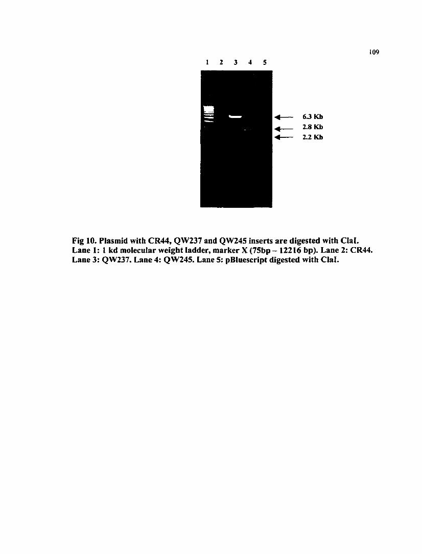
109
63 Kb 2.8 Kb 2.2 Kb
Fig 10. Plasmid with CR44, QW237 and QW245 inserts are digested with Clal. Lane 1: 1 kd molecular weight ladder, marker X (75bp - 12216 bp). Lane 2: CR44. Lane 3: QW237. Lane 4: QW245. Lane 5: pBluescript digested with Clal.
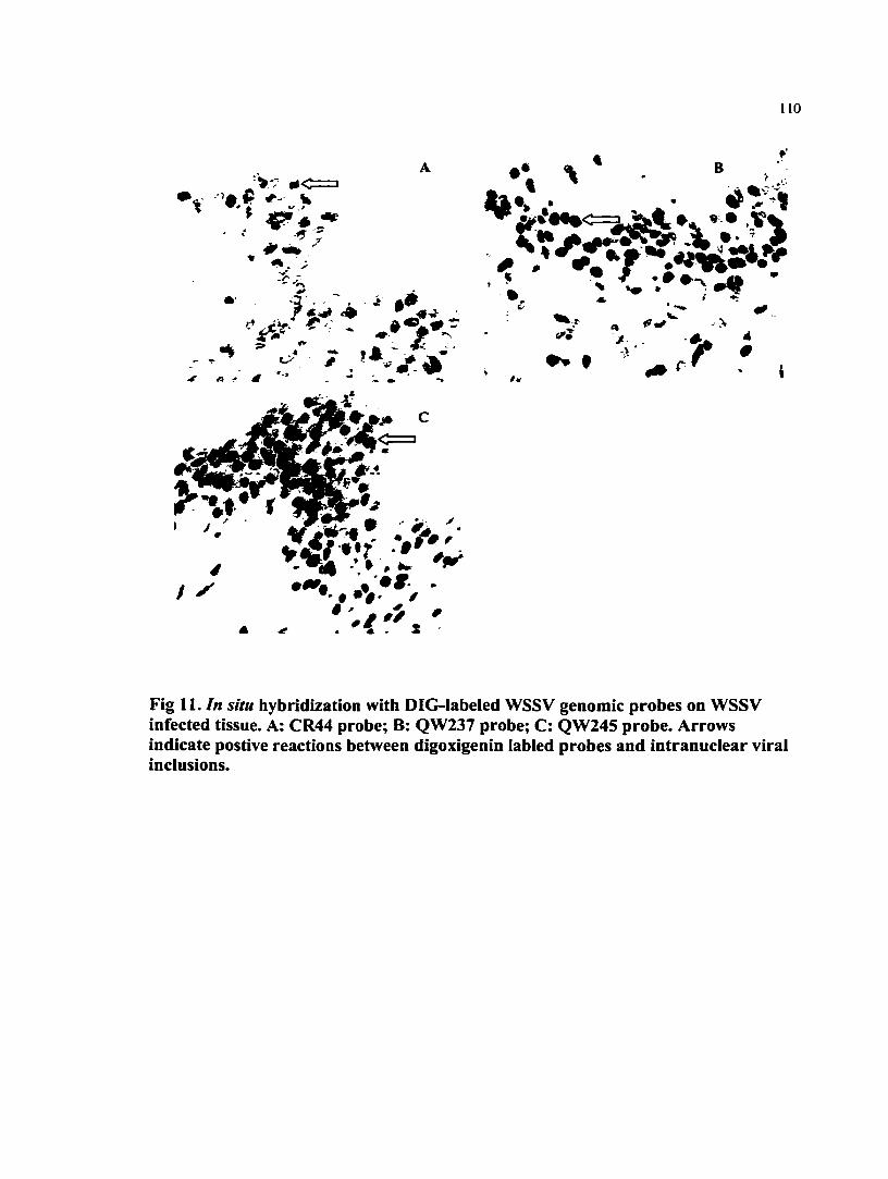
no
jV-7 #<=1
4
- - ,*V-
• y
/ • ^ r . » • ^ • •• • •#• /
<• . *\ . X
•* % B .
;• ̂«, <»-» > > - ^
-•V •
Fig 11. In situ hybridization with DIG-labeled WSSV genomic probes on WSSV infected tissue. A: CR44 probe; B: QW237 probe; C: QW245 probe. Arrows indicate postive reactions between digoxigenin labled probes and intranuclear viral inclusions.

I l l
5 • ATCGATGCTATATCGGTAAAGGTACCATTGATTCGCCCCCTAAAATACTACCCATACTTAACAGTTTCT TTTCGACTTTGTTGTTGGTTATCCCACAACCCATCAATAAATCAGCAGTTACAATTATGTACTTGCAGTTG TGAATTGCTGCATTGAATAGTCTATCAGTGAGAATTCTATATAGAGGGGAGAAAGTGAAAGACTTGTCATG GGGTAAAGCTTCTCTTTTCACTATATTGTTGATATTCCTCAAATCTTCTGGTAGTCTTTTGAGCCAATCAG ATTCAGATTCAGCACCTTTCCTTGTTAGAAATTCCTCATTTGTGAATGATGTTGAAGAAATAATGACGTTT TTTCCAAGCAGGGGTTGCCTGCTGAGAGGCGTATTTTATTCACAATTTCATCCACCTCATGTTTTGGAGTA TTTTTATGCAAAGAGACGATATATACACCTTCAGCGAGTTCGCTTCTCCGTATACTTTTGATAGACTCGAT AATGGACGGTATTCTTCTATAGTTTTTAACGATTCCCTTGATAGCAAACCCGGACGCCATCTTTCATGACT CATTTTGGAGGGTGAGATCATGCAATGAAAAAAACTTATAAGTGTGTGTGTTAACGCACGTAAATCCAGCC ATTGACGTCATATCCGCCTAGTTTTTATCCTCAACTTCTGAGAATTGCTCAACGTACTCATCTCTTTTCCT GGTACATGACGTCAATGGAAATGCCGATGACGTCATGTTAGGGGTATAAAACGACTGGCTCGAGCCCTCGG ATTCACACTTTGTCTGTCCTGAGCAGTAGAGAAGGACCCAGTGAAGAAAATACTCCTCAACTTCCAACAAG AATGGGCTCTGCTAACTTGAACGACCGAACTCTACTCGTGGAGGAAGGGCAAGTAAACGACTACGGGTCTA CCAACAAGAAACAACCAGTGTGCTGTCACATTTGCTATCTAGGAAGAGGAGGAAGAATTGCATGTGTCATC GCATCTGTTCTGGGCTGCGTCCTCCTGCTGGTGACTGTCATGACTTTGTTGATTGTGGTACTGGGAACTGC ACCAGTTAATTGTGATGTGAGCCCACAGAGCTACTCGCCGCCGCCGCAGCCGCCGGTGCAGTTTCATCCTT ACCATTCTTCTTCCACAACCACCACTACTTCCACTACTACTACTACTACACCAACTCCACCAGATACTAAA AAAGTTGACGACGACTATGATGACGACGTCAATATTGGAGGGCAATCAGTTACTGTGAATAATGGAGGTGT TTTCATCAATGGAAGAAAACTCTCAAAAGAAGAAGAAAAAGCAATGGGTATCAATACAGATAATGGAGGAT TTGTTTGGAAGAATGGTGTTTTTTCTCAATATGGCAAAAAATAAAATATGGTATATTAAATATGTGTTGGT
AATAGAATGAGTAAAAGGTACTGTGCCATTACATGTGGAGATCTTGTATTAGCAACATTAGAGAAATGGGG GCAATAAAGATTATGAAACCAAGACTGATTCAAAGAATTAATGATTTGGAATCAGAAATTGAAAATAAAAC TGAATTATGTGAAAAAATCAATGAGCAGATGAAAAATACACAACTAAAATATGATAAATGTTTCGTAGAGG AGGAGACGGAAAAATTCCGCAAGATGGAGGAAAGAGTTAAATACCTCAAAGAGCAGGGAATCCCTCTAGAC CCAGAAGAAAGACGTACAATGTTGGCTGAAATTGACAAGAGTAACAAAGAGTTAGATGCCCTTCTTGAGGA AAATGAACGTATAATAAAGCTCATTGATGAAGAGTTGGAAAGTATGAAATAAAATAAAAGAGTAGGAGACT TTTCAGTTGGTGTTATAAATATATCCCTGCAAGGAGATACGGTCTCAAAAAATCCATGACTTGTCAGTAGC CACGGAAAACACACTTGAAAAATACACCAGAATGATGAGCAGACAGGAGAGATGCAAAAAAAATATGATAA ATTATTTGAAGATGATAAAAGGTTCCGAGAAATAGAGGAACGAATCCTTCAACAAAAAGAGAAGGGAAACC CTCTAGACCCAGAAGAAAGACTTGTATTGTCGGCTGATATTGATAGGAGTATGAAAGAGATTGATGATTGT CTCGAGGAAATAAACCATATAGAATTATCCATTGATACATTATTGGATGAATGTGAAAACTTGCATTATGG TCTTCAAACAACTAAATAACTCTTTATCGGGCAAGTGACTGTATTTGTACGTATGCAATTAGGTCCATATT GGTTTGCAAATAAATAATCCACACAAAACGTGTTGTTTTTCTATATCCTATAAATTCTTGGAACGGAAGTT ATGACTCACATAGAGTGTATAAAAGGAATTGGATGTTCCTATGTCCGCATCACTTCTAACTGAGACATCGT GGAAGAGGAACAGAGAAGAGAGGTTAACAACAACACCAGAAGGAAAATGATACAATGGAAAAAAAGACTGA GACGGCTGCAACACAGAAAAAGACCCAGAACCGTCTGTCAGTAAAAGGTCCAGAAATAAAGAACCCAAAAC AACTTCTACTGTTTACACTTCTGTAAAGTGTTACCTTTCTTCCATAATCAAGAGTGAAAGTAGTAGAAGTA ATGTCACCTCAACCAAAGAAAGGTTTGAGGAGAGGTGTAAATCCGTAAGCAAGATGATGGTCAAAGGTTCA CTGTTTTTGAGGTTAGTAGTGGACGAGTGTCTGAGACGTTACAACCATCTAGAAGACGAAATCGATCGATA AATGGCCAGATATGACGAAGGATAACTTTTACGTCCAATTGTTGAGGAAGGGTTTAAGACAAGAAGAAATT GAAAGAAGGATCTACACATCCTGTTGTAGAAGATGTTTGGAATTCCCCCATCGTCCAAGAAACATTCCTAT CCCAGCAAGGAGAAGGAAATAATCCCGTAAAGAGACATCTCATGGATTTCAATACCATCACCTACGCCGCC AAACAACTAAAAACTTGCTTCGAAACAAACCTACGCACCCATTTCCGGACACGACAACAGAGGGCCATATC TGGATGGTTAGCTGAAAACGGGTTCGATAAAAAGTATACCGAAACTCGTACAACACTGGATAATTGGATGT ACCTACAAGAGTGATTGGGTGGACAAATTT 3 '
Fig 12. The combined sequence of CR44 and LN4. The mutant regions was located at approximately 3 kb downstream (3* direction) of this DNA strand.
1 1 2
APPENDIX 2.
SODIUM DODECYL SULFATE POLYACRYLAMINE GEL
ELECTROPHORESIS OF THE STRUCTURAL PROTEINS OF SIX
GEOGRAPHIC ISOLATES OF THE WHITE SPOT SYNDROME VIRUS AND
PARTIAL AMINO ACID SEQUENCING OF THREE OF THE MAJOR
STRUCTURAL POLYPEPTIDES
Qiong Wang*, Bonnie T. Poulos and Donald V. Lightner
Department of Veterinary Science and Microbiology, University of Arizona, Tucson,
Arizona 85721, USA
Correspondence should be addressed to:
Qiong Wang, Department of Veterinary Science and Microbiology, University of
Arizona, Tucson, Arizona 85721, USA
E-mail: [email protected]
Fax: (520) 6214899
Tel: (520) 6214438

1 1 3
Summary. Six geographic isolates of the white spot syndrome virus (WSSV), from
China, India, Thailand, South Carolina, Texas, as well as from craj^sh kept at the US
National Zoo in Washington D. C, were compared by electron microscopy and sodium
sulfate polyacrylamine gel electrophoresis (SDS-PAGE). Amino acid composition of four
of the major structural polypeptides of the South Carolina WSSV was analyzed, and three
of the four polypeptides were partially sequenced from their NHi terminals. The
morphologies of purified virions of the six geographic isolates of WSSV were
indistinguishable by transmission electron microscopy. By SDS-PAGE, the protein
profiles of the six isolates were very similar, but not identical. They all contained three
major polypeptides with sizes of approximately 25, 23 and 19 kDa. A fourth major
polypeptide at the 14.5 kDa position was observed in four of the geographic isolates. The
WSSV isolated from crayfish presented a slightly different structural protein profile,
particularly with regard to the protein in the 19 kDa range that appeared larger in size than
the other isolates. The NH2 terminal amino acids of the 25, 23 and 14.5 kDa polypeptides
of the South Carolina WSSV were sequenced as MDLSFTLSVVTA,
MEFGNLTNLDVA, and VARGGKTKGRRG, respectively. The information presented
in this paper is important for classification of this newly emerged virus.
KEY WORDS: white spot syndrome virus, SDS-PAGE, structural polypeptide, protein
sequence, geographic isolates.
RUNNING TITLE: Structural proteins of geographic isolates of WSSV.

1 1 4
Introduction
The white spot syndrome virus (WSSV) is one of the most devastating viruses to infect
penaeid shrimp [21]. First discovered in Taiwan in 1992 [8], it has quickly spread to most
shrimp-growing countries in Asia [5, 11, 15, 27, 30, 39] and to the Gulf of Mexico and
Southeastern regions of the United States [22], The name of white spot syndrome virus
(WSSV) or white spot baculovirus (WSBV) [8, 17, 21] was given because of the
distinctive clinical signs of the disease. However, other names such as systemic
ectodermal and mesodermal baculovirus (SEMBV) [43], rod-shaped virus of Penaeiis
japonicus (RV-PJ) [15], penaeid rod-shaped DNA virus (PEIDV) [16, 19] and
hypodermal and hematopoietic necrosis baculo-like virus of P. chinensis (HHNBV) [13]
are all thought to be the same or very closely related viruses.
In addition to the many species of penaeid shrimp which can be infected with WSSV [18,
24, 25, 34, 42], crabs, spiny lobsters, and fresh water shrimp can be naturally or
experimentally infected with the virus [7, 14, 25, 35, 42], and a number of other
arthropods can harbor the virus [14, 25, 42]. Severe mortalities were seldom seen in non-
penaeid species, with the exception of the fresh water shrimp Macrobrachium
rosenbergii and two species of freshwater crayfish (Orconectes punctimanus and
Procambarus sp.\ in which high mortalities occurred from the viral infection [35, 37].
Virulence studies have shown that, in less resistant hosts such as Litopenaeus vannamei,
all isolates of WSSV were extremely virulent, with the cumulative mortalities reaching
100% [24, 41]. However, in the more resistant host, Litopenaeus duorarum, a difference

1 1 5
in virulence among the geographic isolates became evident, with the crayfish isolate of
WSSV causing less and slower developing mortality (Wang et al., 1999).
The taxonomy of WSSV is unsettled. According to morphological characteristics [10, 13,
15, 27, 39, 40, 43], WSSV was previously assigned to the subfamily Nudibaculovirinae,
family Baculoviridae [3, 12]. Subsequent DNA sequence analysis of WSSV, however,
showed it to have a low degree of homology with other baculoviruses [33, unpublished
data]. Furthermore, in marked contrast to other baculoviruses which are highly host
specific [3], WSSV has a distinctively wide host range. In the sixth report of the
International Committee of Taxonomy of Virus (I. C. T. V.) [29], the non-occluded
baculoviruses (which theoritically includes WSSV) were excluded from the family
Baculoviridae and were assigned to the non-classified virus groups.
Because of the large size of the genome and the complex of the protein profiles, WSSV
has not yet been fully characterized, although diagnostic tools, such as gene probes, PCR,
and immunological methods have been developed [6, 9, 14, 18, 19, 23, 33, 38].
Preliminary studies to characterize structural proteins of some of the geographic isolates
of WSSV have been done [14, 31, 32, 38]. Currently, genomic sequencing is being
conducted in several laboratories around the world, but to date, very little sequence data
has been published. Protein sequence information derived from the structural proteins of
WSSV has not been previously published.

116
The purpose of the work reported in this paper was to characterize and partially sequence
the major structural proteins of WSSV. The resultant data on the amino acid composition
and the NHi-terminal amino acid sequences of the major structural polypeptides will be
applied to identify the coding regions in the genome.
Materials and Methods
Viruses: The viral materials used in the SDS-PAGE were the same as the ones used in a
previously published virulence study [41], The original virus samples were from China,
India, Thailand, South Carolina, Texas and Washington D. C. (Table I). The viruses were
amplified by feeding, or intramuscularly injecting, specific pathogen free (SPF) penaeid
shrimp Litopenaeus vannamei [44] with infected shrimp tissue homogenates or
hemolymph. The South Carolina WSSV isolate was used for protein sequencing. Severe
WSSV infections in inoculated shrimp were confirmed using Davidson's AFA fixed
tissue followed by routine histological processing and hematoxylin and eosin-phloxine
(H&E) staining [2, 21]. The taxonomy of shrimp used in this paper follows current
nomenclature [36].
Virus puriflcation: A Renografin gradient purification procedure was modified from the
method of Bonami et al. [4]. For each isolate, approximately 2 g WSSV infected tissue
was collected from the epidermis, stomach, appendages and gills of severely infected
shrimp. To prevent cross contamination, micro-centrifiige tubes and matched disposable
sticks were used to homogenize shrimp tissues. A tris-NaCl buffer (TN; 20 mM tris-Cl,

117
400 mM NaCl, pH 7.4) was used throughout the purification procedure. Each of the
homogenates was clarified at 500 X g for 10 min to pellet cellular debris. The resulting
supernatant fluids were further clarified by centrifligation at 3000 X g for 15 min (4°C)
twice. The viruses in the resultant supernatant fluids were pelleted for I h, at 70,000 X g
(4°C). The virus pellets were resuspended in 2 ml I X TN and layered carefully onto the
top of 15-45% (w/w, in I X TN) linear Renografin gradients. The gradients were
centrifuged at 153,200 X g for 1.5 hr at 4°C. The visible bands were harvested, mixed
with 4-5 volumes of I X TN, and then centrifuged at 124,100 X g for 1.5 hr at 4°C to
pellet the viruses. Each viral pellet was then resuspended in 500^1 distilled H2O and
refrigerated at 4°C.
Electron microscopy: The samples for transmission electron microscopic (TEM)
observation were prepared on carbon-coated copper grids by negative staining with 2%
phosphotungstic acid (PTA), pH 7.0, according to the method of Bonami et al. [4]. The
grids were examined with a Philips CM-12 TEM at a constant voltage of 75 volts and
photographed.
SDS-PAGE: A procedure of discontinuous, denaturing gel electrophoresis was
performed in a Hoefer mini-gel system (Pharmacia, San Francisco, CA). The laemmli
Tris-glycine buffer system [20] was used to prepare the 12% resolving and 4% stacking
gels prior to electroblotting. The purified virus samples were mixed with Laemmli
sample buffer, heated in a boiling water bath for 3 min, and electrophoresed at a constant
118
current of 25 mA for approximately 1 h until the bromophenol blue dye reached within I
cm of the end of the gel. The gels were stained with Coomassie brilliant blue (0.1%
Coomassie Blue R-250 in 1% acetic acid and 40% methanol). A premixed protein
molecular weight marker (Boehringer Maimheim, Indianapolis, IN), with proteins
ranging from 14.4 to 97.4 kDa, was co-electrophoresed to determine the molecular
weights of the WSSV proteins. A prestained SDS-PAGE standard low range molecular
weight marker (BIO-RAD, Hercules, CA), with proteins ranging from 18 to 106 kDa,
was also run with the samples to provide a reference during electrophoresis and after
U^sfer. For the protein sequencing, the gels were not stained prior to transfer.
Electroblotting: The electroblotting procedure was performed in a tank system using a
Hoefer TE22 MINI transphor apparatus. The proteins were electroblotted onto an
Immobilon™ -PSQ membrane with 0.2 |am pore size (Millipore, Bedford, MA), which is
a microporous polyvinylidene fluoride (PVDF) membrane with a modified structure for
increased protein binding. The electroblotting procedure was carried out in CAPS buffer
[28] with 0.1% SDS added at a constant voltage of 10 volts for approximately 18 hr at
4°C. After transfer was complete, the PVDF membrane was rinsed with distilled H2O.
Amino acid analysis and protein sequencing: Protein samples transferred to PVDF
membrane were stained with Coomassie Brilliant Blue. The membrane was sent to the
Laboratory for Protein Sequencing and Analyses, at the University of Arizona for amino
acid composition analysis and sequencing. Amino acid analysis was completed using an
119
Applied Biosystem (ABI) 420A/H amino acid analyzer (Foster city, CA). The
polypeptide was hydrolyzed by vapor-phase using 6 N HCI at 155°C for 1.25 h, then
derivatized with phenylisothiocyanate (PITC) to form phenyithiocarbamyl amino acid
derivatives (PTC-AA) which were extracted and transferred to an online HPLC for
analysis at 254 nm. Protein sequencing was completed using an ABI 477A
Protein/Peptide sequencer interfaced with a 120 A HPLC (C-18 PTH column, reverse-
phase chromatography) analyzer to determine phenylthiohydantoin (PTH) amino acid.
Results
The shrimp tissues were confirmed to have severe WSSV infections by routine H and E
histopathology (Fig. 1). By electron microscopy, the morphology of virions and
nucleocapsids of the six geographic isolates appeared indistinguishable (Fig. 2, 3). The
mean sizes of virions and nucleocapsids were approximately 320 x 120 nm, and 350 x 80
nm, respectively (Table 2).
After SDS-PAGE, more than 20 bands, with three to four major ones predominating,
were observed in most of the isolates (Fig. 4). The isolates shared three major bands of
approximately 25, 23 and 19 kDa. The crayfish isolate of WSSV displayed a slightly
different protein profile from the other isolates: the two bands at 25 and 23 kDa were not
as dominant as in other isolates, and the 19 kDa band appeared larger in size than that of
other isolates. In addition, a 14.5 kDa major band was observed from the China, Thailand

120
and crayfish isolates. When the concentration of virus was increased for the protein
sequencing, the 14.5 kDa major band was also evident in the South Carolina WSSV
isolate (not shown).
The amino acid compositions of the four major polypeptides at 25, 23, 19, and 14.5 kDa
were analyzed (Table 3), using the South Carolina WSSV isolate. The sequences of the
NH2- terminal amino acids of the polypeptides at 25, 23 and 14.5 kDa were:
MDLSFTLSWTA, MEFGNLTNLDVA, and VARGGKTKGRRG, respectively. These
three bands generated good signals, however, some of the amino acids were uncertain: T
(in position 11 of the 25 kDa polypeptide); M or S (in position 1 of the 23 kDa
polypeptide); and V or S (in position 1 of the 14.5 kDa polypeptide).
Discussion
The results of SDS-PAGE analysis indicated that WSSV has a complex protein
composition, more than 20 polypeptides were visualized in the gels. The complicated
structural protein profile agrees with the data firom other baculoviruses where the
genomes can be composed of 150 or more open reading frames [1]. Previous publications
on the characterization of the structural proteins of WSSV [14, 31, 32, 38] also show that
WSSV consists of a complex structural protein profile, and that at least three dominant
polypeptides, 25 kDa, 23 kDa, and 19 kDa, are present in the WSSV virion. Some of the
protein seen in the polyacrylamine gel may be derived from shrimp. However, when the
121
tissue from a SPF shrimp was run through the same viral purification procedure as the
infected tissue, and the materials were pelleted from identical positions in Renografin
gradients for electrophoresis, no protein bands were observed in the SDS-PAGE gel (lane
3 of Fig. 4). Therefore, the bands in the viral preparation are derived from the viral
structural proteins of WSSV although some of them may be host-derived proteins which
arise during a WSSV infection and which are co-purified with the virus particles.
The virions and nucleocapsids of the six geographic isolates of WSSV appeared to be
indistinguishable in the electron micrographs (Fig. 2, 3). The size of the virion and the
characteristic appearance of the nucleocapsid agree with the descriptions of WSSV type
virus in previous publications [10, 13, 15, 27, 39,40,43].
The slight differences in the structural polypeptides of different geographic isolates were
evident in the SDS-PAGE gel. Five of the isolates (China, India, Thailand, South
Carolina, Texas) shared the same three major bands at 25, 23, and 19 kDa. The crayfish
isolate also demonstrated these bands at 25 and 23 kDa, but they were less dominant,
while the band at 19 kDa appeared more dominant and slightly larger in size than in the
other isolates. The China, Thailand, South Carolina and crayfish isolates shared another
major band at the 14.5 kDa position, while the isolates from India and Texas lacked this
band. Interestingly, neither of the previous publications [14, 31, 32, 38] noted a 14.5 kDa
major protein in the viral structural protein profiles. This fiorther suggests that there are
differences among geographic isolates of WSSV. Because WSSV is a large, enveloped

122
virus, it is unstable. For this reason, freshly prepared purified viruses were used in the
SDS-PAGE in order to obtain reliable and comparable protein profiles. Since the protein
concentrations of viral isolates varied, it was difficult to compare the minor bands present
in each isolate. For instance, when a concentrated South Carolina WSSV sample was
electrophoresed for sequencing, the previously non-detectable 14.5 kDa band was
detected. Differences seen within the major protein bands provided more conclusive
evidence of protein variations. The crayfish WSSV differs firom other isolates in the
position of the major band at approximately the 19 kDa position. That this difference
takes place in crayfish isolate WSSV was supported by virulence and DNA analysis [41,
unpublished data]. The difference of the 19 kDa major band can be confirmed by
sequencing, and it may be used to distinguish the less virulent crayfish WSSV.
The major proteins 25, 23 and 14.5 kDa transferred efficiently onto the PVDF membrane
and they provided good signals in the sequencing process. The major 19 kDa polypeptide
transferred with poor efficiency and may due to its physiochemical properties. This
observation suggests that the 19 kDa protein could be a glycoprotein which does not
always transfer efficiently out of SDS-PAGE gels. However, this hypothesis is
contradicted by a previous publication [31] that concluded that none of the four major
polypeptides is glycosylated.
Comparing our results with the previously published paper [31], it is suggested that the
25 and 19 kDa polypeptides are located on the lipid envelope of the virion, whereas the
123
23 kDa polypeptide is part of the nucleocapsid and the 14.5 kDa polypeptide is associated
with the nucleoprotein core.
The work reported here represents the first time that the major structural polypeptides of
the WSSV were analyzed and sequenced. It is anticipated that when the genomic
sequencing is finahzed, these amino acid sequence data can be used to locate the coding
regions of the structural proteins in the genome. The protein sequence information will be
critical for determining the taxonomic classification of WSSV, and will be useful for in
vitro transcription and translation of these structural proteins.
Acknowledgement
This research was funded by the Gulf Coast Research laboratory Consortium Marine
Shrimp Farming Program, CREES, USDA under Grant No. 95-38808-1420, the National
Sea Grant Program, USDC under the Grant No. NA56RG0617, USDC under Grant No.
NA56FD0621, and by a grant from the U. S. National Fisheries institute. The original
isolates of WSSV were kindly provided by Dr. Paul Frelier (Department of Veterinary
Pathology, College of Veterinary Medicine, Texas A & M University, College Station,
Texas), Dr. Craig Browdy (South Carolina Department of Natural Resources, Waddell
Mariculture Center, Blufflon, South Carolina), Dr. Laura K. Richman (Department of
Pathology, National Zoological Park, Washington D. C.), Dr. Eileen Reddington
(Diagxotics, Ltd.), Dr. Ramana Murty (Andhra University, Visakhapatnam, India), and
Mr. Jie Huang (Yellow Sea Fishery Research Institute, Qingdao, Shandong, China). We

124
thank Wallace Clark for amino acid analysis and protein sequencing, and the Imaging
Facility Laboratory at the University of Arizona for access to the electron microscope.
References
1. Ayres MD, Howard SC, Kuzio J, Lopez-Ferber M, Possee RD (1994) The complete
DNA sequence of Autographa califomica nuclear polyhedrosis virus. Virology 202: 586-
605
2. Bell TA, Lightner DV (1988) A handbook of normal shrimp histology. Special
Publication No. 1. World Aquaculture Society, Baton Rouge LA USA
3. Blissard GW, Rohrmann GF (1990) Baculovirus diversity and molecular biology.
Annu Rev Entomol 35:127-155
4. Bonami JR, Brehelin M, Mari J, Trumper B, Lightner DV (1990) Purification and
characterization of IHHN virus of penaeid shrimps. J Gen Virol 71: 2657-2664
5. Cai SL, Huang J, Wang CM, Song XL, Sun X, Yu J, Zhang Y, Yang CH (1995)
Epidemiological studies on the explosive epidemic disease of prawn in 1993-1994. J
China Fish 19: 112-117

125
6. Chang PS, Lo CF, Wang YC, Kou GH (1996) Identification of white spot syndrome
associated baculovirus (WSBV) target organs in the shrimp Penaeus monodon by in situ
hybridization. Dis Aquat Org 27: 131-139
7. Chang PS, Chen HC, Wang YC (1998) Detection of white spot syndrome associated
baculovirus in experimentally infected wild shrimp, crab and lobsters by in situ
hybridization. Aquaculture 164: 233-242
8. Chen SN (1995) Current status of shrimp aquaculture in Taiwan. In: Browdy CL,
Hopkins JS (eds) Swimming through troubled water. Proceedings of special session on
shrimp farming. Aquaculture '95. World Aquaculture Society, Baton Rouge LA USA, pp
29-34
9. Chou HY, Huang CY, Lo CF, Kou GH (1998) Studies on transmission of white spot
syndrome associated baculovirus (WSBV) in Penaeus monodon and P. japoniciis via
waterbome contact and oral ingestion. Aquaculture 164: 263-276
10. Durand S, Lighmer DV, Nunan LM, Redman RM, Mari J, Bonami JR (1996)
Application of gene probes as diagnostic tools for White Spot Baculovirus (WSBV) of
penaeid shrimp. Dis Aquat Org 27:59-66

126
11. Flegel TW, Fegan DF, Sriurairatana S (1995) Environmental control of infectious
disease in Thailand. In: Browdy CL, Hopkins JS (eds) Swimming through troubled water.
Proceedings of the special session on shrimp farming, Aquaculture '95. World
Aquaculture Society, Baton Rouge LA USA, pp 65-79
12. Francki RJB, Fauquet CM, Knudson DL, Brown F (1991) Classification and
nomenclature of viruses. Arch Virol (suppl) 2: 1-450
13. Huang J, Song XL,Yu J, Yang CH (1995) Baculoviral hypodermal and hematopoietic
necrosis, study on the pathogen and pathology of the shrimp explosive epidemic disease
of shrimp. Mar Fish Res 16: 1-10
14. Huang J, Yu J, Wang XH, Song XL, Ma CS, Zhao FX, Yang CH (1995) Survey on
the pathogen and route of transmission of baculoviral hypodermal and hematopoietic
necrosis in shrimp by ELISA of monoclonal antibody. Mar Fish Res 16: 40-50
15. Inouye K, Miwa S, Oseko N, Nakano H, Kimura T, Momoyama K, Hiraoka M (1994)
Mass mortalities of cultured Kuruma shrimp Penaeus japonicus in Japan in 1993:
electron microscopic evidence of the causative virus. Fish Pathol 29: 149-158

127
16. Inouye K, Yamano K, Ikeda N, Kimura T, Nakano H, Momoyama K, Kobayashi J,
Miyajima S (1996) The penaeid rod-shaped DNA virus (PEIDV), which causes penaeid
acute viremia (PAV). Fish Padiol 31: 39-45
17. Kasomchandra J, Boonyaratpalin S (1996) Red disease with white patches or white
spot disease in cultured penaeid shrimp in Asia. Asian Shrimp News 273: 4
18. Kasomchandra J, Boonyaratpalin S, Itami T (1998) Detection of white-spot syndrome
in cultured penaeid shrimp in Asia: microscopic observation and polymerase chain
reaction. Aquaculture 164: 243-251
19. Kimura T, Yamano K, Nakano H, Momoyama K, Hiraoka M, Inouye K (1996)
Detection of penaeid rod-shaped DNA virus (PRDV) by PCR. Fish Pathol 31: 93-98
20. Laemmli UK (1970) Cleavage of structural proteins during the assembly of the head
of bacteriophage T4. Nature 227: 680-685
21. Lightner DV (1996) A handbook of shrimp pathology and diagnostic procedures for
diseases of cultured penaeid shrimp. World Aquaculture Society, Baton Rouge LA USA
22. Lightner DV, Redman RM, Poulos BT, Nunan LM, Mari JL, Hasson KW (1997) Risk
of spread of penaeid shrimp viruses in the Americas by the international movement of

128
live shrimp for aquacuiture and frozen shrimp for commodity markets. Revue
Scientifique et technique Office International des Epizooties 16: 146-160
23. Lightner DV, Redman RM (1998) Shrimp diseases and current diagnostic methods.
Aquacuiture 164: 201 -220
24. Lightner DV, Hasson KW, White BL, Redman RM (1998) Experimental infection of
Western Hemisphere penaeid shrimp with Asian white spot syndrome virus and Asian
yellow head virus. J Aquat Anim Health 10: 271-281
25. Lo CF, Ho CH, Peng SE, Chen CH, Hsu HC, Chiu YL, Chang CF, Liu KF, Su MS,
Wang CH, Kou GH (1996) White spot syndrome baculovirus (WSBV) detected in
cultured and captured shrimp, crabs, and other arthropods. Dis Aquat Org 27: 215-225
26. Lo CF, Lightner DV, Ho CH, Hsu HC (In Press) Specific genomic fragment analysis
of different geographic isolates of white spot syndrome associated virus. Dis Aquat Org
27. Momoyama K, Hiraoka M, Nakano H, Koube H, Inouye K, Oseka N (1994) Mass
mortalities of cultured Kuruma shrimp, Penaeus japonicits, in Japan in 1993:
histopathological study. Fish Pathol 29: 141-148

129
28. Madsudaira, P. (1987) Sequence from picomole quantities of proteins electroblotting
onto polyvinylidene difluoride membranes. J. Biol. Chem. 262: 10035-10038.
29. Murphy FA, Fauquet CM, Bishop DHL, Ghabrial SA, Jarvis AW, Martelli GP, Mayo
MA, Summers MD (1995) Virus taxonomy. Classification and nomenclature of viruses.
Sixth report of the international committee on taxonomy of viruses. Arch Virol (suppi)
10: 1-586
30. Nakano H, Koube H, Umezawa S, Momoyama K, Hiraoka M, Inouye K., Oseko N
(1994) Mass mortalities of cultured Kuruma shrimp, Penaeus japonicus, in Japan in
1993: epizootiological survey and infection trials. Fish Pathol 29: 135-139
31. Nadala-Jr. ECB, Tapay LM, Loh PC (1998) Characterization of a non-occluded
baculovirus-Iike agent pathogenic to penaeid shrimp. Dis Aquat Org 33: 221-229.
32. Nadala-Jr. ECB, Loh PC (1998) A comparative study of three different isolates of
white spot virus. Dis Aquat Org 33: 231-234.
33. Nunan LN, Lightner DV (1997) Development of a non-radioactive gene probe by
PCR for detection of white spot syndrome virus (WSSV). J Virol Meth 63: 193-201
130
34. Nunan LM, Poulos BT, Lightner DV (1998) The detection of white spot syndrome
virus (WSSV) and yellow head virus (YHV) in imported commodity shrimp. Aquaculture
160:19-30
35. Peng SE, Lo CF, Ho CH, Chang CF, Kou GH (1998) Detection of white spot
baculovirus (WSBV) in giant freshwater prawn, Macrobrachium rosenbergii, using
polymerase chain reaction. Aquaculture 164: 253-262.
36. Perez-Farfante 1, Kensley BF (1997) Penaeoid and sergestoid shrimps and prawns of
the world: keys and diagnoses for the families and genera. Memoires du Museum
National D'Histoire Naturelle 175: 1-233
37. Richman LK, Montali RJ, Nichols DK, Lightner DV (1997) A newly recognized fatal
baculovirus infection in freshwater crayfish. Proc Am Assoc Zoo Veter.
38. Sahul-Hameed AS, Anikumar M, Stephen-Raj ML, Jayaraman IC (1998) Studies on
the pathogenicity of systemic ectodermal and mesodermal baculovirus and its detection
in shrimp by immunological methods. Aquaculture 160:31-45
39. Takahashi Y, Itami T, Kondo M, Maeda M, Fujii R, Tomonaga S, Supamattaya K.,
Booyaratpalin S (1994) Electron microscopic evidence of bacilliform virus infection in
Kuruma shrimp {Penaeus japonicus). Fish Pathol 29: 121-125

131
40. Wang CH, Lo CF, Leu JH, Chou CM, Yeh PY, Chou HY, Tung MC, Chang CF, Su
MS, K.OU GH (1995) Purification and genomic analysis of baculovirus associated with
white spot syndrome (WSBV) of Penaeits monodon. Dis Aquat Org 23: 239-242
41. Wang Q, White BL, Redman RM, Lightner DV (1999) Per os challenge of
Litopenaeus vannamei postlarvae and Farfantepenaeiis duoranim juveniles with six
geographic isolates of White Spot Syndrome Virus (WSSV). Aquacultiure 170:179-194
42. Wang YC, Lo CF, Chang PS, Kou GH (1998) Experimental infection of white spot
baculovirus in some cultured and wild decapods in Taiwan. Aquaculture 164: 221-231
43. Wongteerasupaya C, Vickers JE, Sriurairatana S, Nash GL, Akarajamom A,
Boonsaeng V, Panyim S, Tassanakajon A, Withyachumnamkul B, Flegel TW (1995) A
nonoccluded, systemic baculovirus that occurs in cells of ectodermal and mesodermal
origin and causes high mortality in the black tiger prawn, Penaeus monodon. Dis Aquat
Org 21: 69-77
44. Wyban JA, Swingle JS, Sweeny JN, Pruder GD (1992) Development and commercial
performance of high health shrimp using specific pathogen free (SPF) broodstock
Penaeus vannamei. In: Wyban JA (ed) Proceedings of the Specific Session on Shrimp
Farming. World Aquaculture Society. Baton Rouge LA USA, pp 254-260

Table 1. Description of the original geographic isolates of WSSV.
Viral isolate Identification
number
Collection location Host species Life stage of
shrimp
Wild or
cultured
Date of
collection
WSSV China UAZ95-II6A Shandong, China Fenneropenaeus chinensis Adult Cultured May I99S
WSSV India UAZ95-3I4 VIsakhapatnam, India Penaeus monodon Juvenile Cultured 28/8/95
WSSV Thailand UAZ95-38A Thailand Penaeus monodon Adult Cultured 27/2/95
WSSV Texas UAZ97-85 Texas, in Guir of Mexico Litopenaeus seti/erus Adult Wild 13/5/97
WSSV South UAZ97-5 BlufRon, South Carolina Litopenaeus seti/erus Adult Captive -Wild, in over 8/1/97
Carolina wintering facility
WSSV crayfish UAZ96-52 National Zoological Park, Orconecies punctimanus Subadult Captive - Wild 8/3/96
Washington D, C.
(Acquired from Arkansas)

133
Table 2. Morphometry of intact virions and nucleocapsids of the six geographic
isolates of WSSV after negative staining.
WSSV isolate Size of intact virions
[length (nm) X width(nm)]*
Size of nucleocapsids
[length (nm) X width(nm)]*
China 324±16 X 120±6 366±25 X71±10
India 312±13 X 124±5 356±15 X 75±7
Thailand 325±20 X 125±8 358±17X83±3
Texas 305±17X I38±8 357±I7 X 82±5
South Carolina 300±9X 138±13 356±29 X 77±I4
Crayfish from 297±23 X 132±7 343±I5X77±9
US National Zoo
* The sizes of viruses were expressed as: mean ± standard deviation.

134
Table 3. Amino acid composition of four of the major polypeptides from the South
Carolina isolate of WSSV.
25 kDa polypeptide 23 kDa polypeptide 19 kDa polypeptide 14.5 kDa polypeptide
AA residue Mol % AA residue Mol % AA residue Mol % AA residue Mol %
B 6.95 B 6.61 B 6.08 B 6.44
Z 15.94 Z 14.58 Z 16.72 Z 11.94
s 5.68 s 6.68 S 3.81 S 9.19
G 23.31 G 25.65 G 21.9 G 24.26
H 1.61 H 2.45 H 1.04 R 6.86
R 3.13 R 3.00 R 2.53 T 2.94
T 4.19 T 3.52 T 2.84 A 9.58
A 8.21 A 9.00 A 11.20 P 3.95
P 0.57 Y 1.24 P 5.02 Y 2.53
Y 1.67 V 7.11 Y 1.17 V 4.51
V 8.25 C 1.08 V 7.49 I 3.62
C 0.45 1 6.78 1 4.14 L 7.09
I 5.31 L 7.32 L 11.40 F 3.11
L 7.89 F 2.52 F 2.58 K 3.98
F 3.69 K 2.45 K 2.08
K 3.17

135
Figure 1. Histological section of cuticular epithelium of the stomach region of a WSSV-infected Litopenaeus vannamei showing typical WSSV lesions which were observed among all the shrimp samples used for SDS-PAGE and protein sequencing. The arrows indicate hypertrophied nuclei filled with intranuclear WSSV inclusions. H and E stain. Bar = 20 ^m.
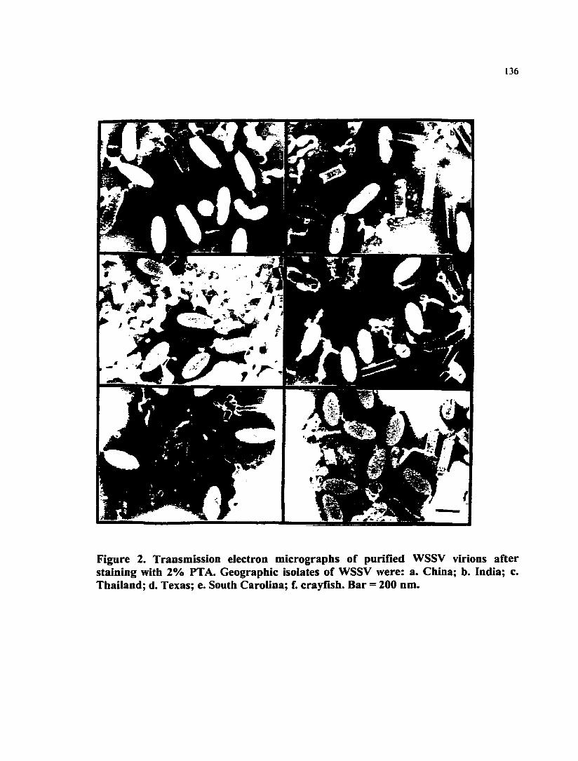
136
Figure 2. Transmission electron micrographs of purified WSSV virions after staining with 2% PTA. Geographic isolates of WSSV were: a. China; b. India; c. Thailand; d. Texas; e. South Carolina; f. crayfish. Bar = 200 nm.

137
Figure 3. Transmission electron micrographs of WSSV nucleocapsids after staining with 2% PTA. Geographic isolates of WSSV were: a. China; b. India; c. Thailand; d. Texas; e. South Carolina; f. crayfish. Bar = 200 nm.

138
1 2 3 4 T -̂ 1
8 10
97.4 KDa
66.2 KDa
39.2 KDa
26.6 KDa
21.5 KDa
14.4 KDa
Figure 4. The structural protein profiles of six geographic isolates of WSSV separated by SDS-PAGE in a 12% poiyacrylamide gel. Lane 1: Pre-stained protein molecular weight marker (16-106 kDa); Lane 2. Pre-mixed protein molecular weight marker (14.4-97.4 kDa) ; Lane 3. Extracts from the SPF shrimp; Lane 4. WSSV China; Lane 5. WSSV India; Lane 6. WSSV Thailand; Lane 7. WSSV Texas. Lane 8. WSSV South Carolina; Lane 9. WSSV crayfish. Lane 10. Pre-mixed protein molecular weight marker (14.4 -97.4 kDa).

APPENDIX 3.
IDENTIFICATION OF GENOMIC VARIATIONS AMONG GEOGRAPHIC
ISOLATES OF WHITE SPOT SYNDROME VIRUS USING RESTRICTION
ANALYSIS AND SOUTHERN BLOT HYBRIDIZATION
Qiong Wang*, Linda M. Nunan, Donald V. Lightner
Department of Veterinary Science and Microbiology, University of Arizona, Tucson,
Arizona 85721, USA
Correspondence should be addressed to:
Qiong Wang, Department of Veterinary Science and Microbiology, University of
Arizona, Tucson, Arizona 85721, USA
E-mail: [email protected]
Fax: (520)6214899
Tel: (520) 6214438

140
ABSTRACT: White spot syndrome virus (WSSV) is widely distributed in most of the
Asian countries where penaeid shrimp are cultured, as well as in some regions of the
USA. Six geographic isolates of WSSV, from China, India, Thailand, Texas and South
Carolina, and one crayfish isolate from the National Zoological Park in Washington D.
C., were compared by combining the methods of restriction analysis and Southern blot
hybridization. Viral and host total genomic DNA were extracted by first treating the
infected shrimp tissue with proteinase K and cetyltrimethylarmnonium bromide (CTAB),
followed by a phenol-chloroform extraction and an ethanol precipitation. The resultant
DNA was digested with each of the nine endonucleases: AccI, Bglll, Clal, BemiHI,
EcoRJ, Hindll, Hael, Sad and Xhol. DNA restriction fragments were electrophoresed,
transferred to nylon membranes, and detection was visualized using three different
digoxigenin-l l-dUTP labeled WSSV genomic probes: LN4, C42 and A6. No distinctive
difference among five WSSV isolates from penaeid shrimp was detected, however,
differences in the crayfish isolate of WSSV were observed. The results indicate that some
genomic components of WSSV from different geographic regions share a high degree of
homology. Nonetheless, this method can be used to differentiate the crayfish isolate of
WSSV from the penaeid WSSV isolates.
KEY WORDS: White spot syndrome virus, WSSV, penaeid shrimp, restriction analysis.
Southern blot hybridization, genomic variation.
RUNNING TITLE: Genomic variation of white spot syndrome virus.

141
INTRODUCTION
White spot syndrome disease of penaeid shrimp emerged during the early 1990's and it
has caused a serious ongoing epizootic in shrimp-growing countries of Asia, including
China, India, Thailand, Japan, Taiwan, Korea, Indonesia, Malaysia, and Vietnam (Chen,
1995; Flegel et al., 1995; Huang et al., 1995a; Inouye et al., 1994, 1996; Kimura et al.,
1996; Mohan et al., 1998; Momoyama et al., 1994; Nakano et al., 1994; Takahashi et al.,
1994; Wang et al., 1995; Wongteerasupaya et al., 1995). Several disastrous outbreaks of
white spot syndrome disease have also occurred in the Gulf of Mexico and on the
Southeastern coast of the United States (Lightner et al., 1997; Lo et al., 1999; Wang et
al., 1999).
Almost all the species of penaeid shrimp are susceptible to WSSV infection. When listed
according to the new taxonomy of penaeid shrimp by Perez-Farfante and Kensley (1997),
the major species naturally infected by the virus in Asian countries include Penaeiis
monodon, Fenneropenaeus chinensis, Fe. indicus, Fe. penicillatus, and Marsupenaeus
japonicus (Chang et al., 1996; Chou et al., 1995, 1998; Flegel et al., 1995; Huang et al.,
1995a; Inouye et al., 1994, 1996; Kasomchandra et al., 1998; Kimura et al., 1996; Lo et
al., 1996a; Mohan et al., 1998; Nakano et al., 1994; Nunan et al., 1998; Park et al., 1998;
Takahashi et al., 1994; Wang et al., 1995; Wongteerasupaya et al., 1995). Severe WSSV-
induced mortalities have been observed in Litopenaeus setifenis stocks from the states of
Texas and South Carolina in USA (Lightner et al., 1997; Lo et al., 1999; Wang et al..

142
1999). Additional penaeid species infected by WSSV include; Metapenaeus ensis.
Farfantepenaeus aztecus. Fa. duorarum, Fe. merguiensis, P. semisulcatus, L
stylirostris, L. vannamei, Trachypenaeus curvirostris (Cai et al., 1995; Chang et al.,
1998; Lightner et al., 1997, 1998; Nunan and Lightner, 1997; Nunan et al., 1998; Tapay
et al., 1997; Wang et al., 1998; Wang et al., 1999). Among these penaeid species, the
cumulative mortality caused by this disease can reach as high as 100%. Severe mortalities
among several non-penaeid species, including Exopalaemon orientalis. Macrobrachium
rosenbergii (a caridean shrimp), Orconectes punctimanus and Procambanis sp.
(crayfish), were also reported (Chang et al., 1998; Peng et al., 1998; Elichman et al., 1997;
Wang et al., 1998). WSSV has also been detected by PCR, in situ hybridization or
monoclonal antibody assays in wild crabs {Calappa lophos, Portunus sanguinolentus.
Charybdis sp., Helice tridens), wild lobsters {Paniilims sp.), palaemonidian pest shrimp,
planktonic copepods. Anemia spp. and pupae of an ephydridian insect (Chang et al.,
1998; Kanchanaphum et al., 1998; Lo et al., 1996b; Huang et al., 1995b; Wang et al.,
1998).
Since WSSV has a wide geographic distribution and host range, efforts have been
directed to compare morphology, virulence, genomic composition and protein
composition among WSSV isolates (Kasomchandra et al., 1998; Lo et al., 1999; Nadala-
Jr. et al., 1998a, 1998b; Park et al., 1998; Wang et al., 1999; Wongteerasupaya et al.,
1996). The results have demonstrated that WSSV isolated from different locations are
almost identical, although slight differences may exist among some of the isolates.
143
The objective of the present study was to compare the genomic composition of six
geographic isolates of WSSV in order to develop genetic markers that could be used to
distinguish different WSSV strains.
MATERIALS AND METHODS
Viruses: The six WSSV isolates were collected from China, India, Thailand, Texas,
South Carolina, and crayfish kept at the US National Zoological Park (Table 1). To
obtain working quantities of these viruses, these viruses were amplified in specific
pathogen free (SPF) L. vannamei (Wyban et al., 1992). WSSV infection among these
samples were confirmed by standard procedures using Davidson's alcohol formalin acetic
acid (AFA) fixed tissue followed by routine histological processing, and hematoxylin and
eosin-phloxine (H and E) staining (Bell and Lightner, 1988; Lightner 1996). The severity
of infection was rated according to Lightner (1996).
Extraction of DNA from normal and infected shrimp tissue: The procedure used to
extract DNA from normal (SPF) viral infected tissue was modified from the method of
Lo (I996a,b). For each viral isolate, approximately 100 mg of shrimp tissue was collected
from the appendages and the stomach, and placed into a microflige tube containing 1.2 ml
digestion buffer (100 mM NaCl; 10 mM Tris-HCl, pH 8; 25 mM EDTA, pH 8; 0.5 % N-
lauryl sarcosine; 0.5% mg/ml proteinase K) and crushed with a disposable stick. After I h
144
incubation at 65°C, 600 jil of the supernatant fluid of the DNA/digestion buffer mixture
was collected. A volume of 100 jil of 5M NaCl and 70 ^il of CTAB/NaCl stock solution
(10% N-cetyl-N, N, N-trimethylammonium bromide in 0.7 M NaCl) was then added and
incubated at 65°C for another 10 min. The DNA was extracted with an equal volume of
chloroform/isoamyl alcohol (49:1) once, followed by an equal volume of phenol three
times, and a double volume of chloroform/isoamyl alcohol once. A 1/10 volume of
sodium acetic acid (NaAc) and a double volume of 100% ethanol (-20°C) were added to
precipitate the DNA by centrifiigation at 33,000 x g (4°C) for 15 min. After a wash with
70% cold ethanol (-20°C), the DNA was dried and resuspended in 30 |il of HPLC grade
H2O and incubated at 65°C for 30 min. The DNA solution was stored at 4°C until used
for endonuclease digestion.
Endonuclease digestion of DNA: Nine endonucleases: AccI, BamHI, Bglll, Clal,
EcoRI, Haelll, Hindll, Sac I and Xhol, were used to digest the extracted viral DNA
according to the manufacture's protocol (Boehringer Mannheim, Indianapolis, IN). A
volume of 0.5 ^m of endonuclease was applied to digest 3 ̂ il extracted viral DNA in a 10
|il reaction, at 37°C for 1 to 2 h.
Gel electrophoresis: After endonuclease digestion, 1 |a1 10 x RNase Plus™ gel loading
buffer (5 primer-3 prime. Inc., Boulder, CO) was added into each of the 10 |il digestion
reactions, then loaded into the wells of a 1 % agarose gel in 0.5 X TBE (Tris-Borate-
EDTA) buffer (Sambrook et al. 1989) for electrophoresis. A volume of 2 |il digoxigenin-

145
labeled DNA molecular Marker III (Boehringer Mamiheim, Indianapolis, IN) was co-
electrophoresed to indicate the molecular weight of the DNA samples. Ethidium bromide
(0.5 (ig/ml) was incorporated into the gel, and was used to visualize DNA. The
electrophoresis was conducted at 100 volts for about I h. The resultant gel was
transferred to a nylon membrane for Southern blot hybridization.
DNA probe labeling: Three DNA inserts, LN4 (Nunan and Lightner, 1997), C42, and
A6 (unpublished data), were labeled with digoxigenin DIG-ll-dUTP by PGR method
using Genius™ non-radioactive DNA labeling and detection kit (Boehringer Mannheim,
Indianapolis, IN). Clones with plasmids carrying LN4 and C42 inserts were developed in
the Shrimp Pathology Laboratory at the University of Arizona. A Thailand WSSV isolate
was used to generate the clone containing LN4 insert, and the C42 came from a Chinese
isolate of WSSV. Likewise, the clone carrying A6 was developed at J. R. Bonami's
laboratory in France using a Thailand isolate of WSSV. Three pairs of primers (Table 2)
corresponding to each of the inserts were designed for the PCR labeling procedure
according to the sequence information of LN4 (Nunan and Lightner, 1997), C42 and A6
(unpublished data). For each insert, a 100 nl labeling reaction solution containing 10 |il
10 X PCR Buffer II, 10 pmol of each dNTP, 10 ^il DIG label mix, 200 pmol MgCh, 1 ^1
AmpliTaq Gold DNA polymerase (PE Applied Biosystem, Foster City, CA), 200-280 ng
plasmid with desired insert, 250 ng of each primer for C42 and LN4; or 100 ng of each
primer for A6, and 63 |al H2O, was combined in a microcentrifuge tube and reacted in a
PCR machine (PE Applied Biosystem, Foster city, CA). The PCR procedure was
146
conducted by initiating the reaction with a hot start at 94°C for 10 min; continuing with
50 cycles of 94°C for I min, 55°C for I min, and 72°C for 2 min; ending with an
extension at 72°C for 7 min. After PCR labeling, the probes were precipitated by adding
10 jil 200 mM EDTA (PH 8.0), 11 jil 4 M LiCl, 1 ^il 20mg/ml glycogen, and 360 |al
absolute ethanol to the 100 (il PCR reaction, at -70°C for 1 h. The precipitated DNA
probes were then centrifliged at 13,000 g, 4°C, for 30 min. After a wash with 70%
ethanol, the probe pellets were centrifliged and resuspended in 100 jil distilled H2O. The
concentration of resultant probes was estimated in electrophoresis by comparison with a
low molecular mass ladder (GIBCO BRL, Grand Island, NY). The newly labeled probes
were further tested by direct detection using the DIG-labeled standard provided in the
Genius kit for comparison. The concentration of each of the three probes was adjusted to
50 ng/|il using distilled H2O. The specificity of these probes was tested by in situ
hybridization method according to Bruce et al. (1993) and dot blot hybridization
according to the application manual of Boehringer Mannheim.
Southern blot hybridization: The Southern blot hybridization procedure was modified
from Sambrook (1989), and is described briefly here. After electrophoresis, 0.25 M HCl
was used to depurinate DNA in the agarose gel for 10-15 min. The gel was then placed
into 1.5 M NaCl / 0.5 M NaCl solution for 20 min at RT with gentle agitation to denature
the DNA. After repeating the denaturing step once, the gel was soaked in a neutralization
solution (1 M Tris, pH 7.5 / 1.5 M NaCl) for 20 min at RT with gentle agitation, and this
neutralization step was repeated once by using fresh solution. A positively-charged nylon
147
membrane (Boehringer Marmheim, Indianapolis, IN) was cut to the size of the gel,
soaked in 10 X SSC (saline sodium citrate, 1.5 M NaCl, 0.15 M Na citrate) for 15 min
just prior to use. The Southern blotting was set up in a pan. Two pieces of 3 mm gel
blotting paper (Schleicher & Schuell, Keene, NH) cut to the size of the gel were put on
the bottom of the assembly. On top of the filter paper, the gel, the nylon membrane, two
more sheets of 3 mm paper and then a stack of brown paper towels measuring 3 inches in
height, were assembled. On top of all this, a 1 kg weight was placed to secure the
assembly. After transfer overnight, the nylon membrane in the Southern blot was washed
with 5 X SSC for 5 min at RT with gentle agitation. The membrane was air dried for 30
min, and cross-linked with UV light for 4 min. And then the membrane was hybridized
with one of the specific probes (LN4, C42 and A6) under stringent conditions. The dried
nylon membrane was placed into a hybridization bag with 5-10 ml prehybridization
solution (1% blocking agent, 0.1% sarkosyl, 0.02% SDS, in 5 X SSC buffer, pH 7.0), and
incubated in a 65°C water bath for 1 h. DNA probes were denatured by boiling for 10
min, then quenching on ice for 2 min. A volume of 2 ml fresh prehybridization solution
and a DIG-labeled probe (1^1/1ml pre-hybridization solution) were added into the bag
with the nylon membrane. The solution was mixed well, the bag was sealed and placed in
the 65°C water bath to incubate overnight (12-16 h). After the hybridization, the
membrane was washed for 5 min in 2 X SSC / 0.1% SDS two times at RT. A second
wash followed with 0.1 X SSC / 0.1% SDS two times, for 15 min each, at 65°C. The
membrane was then treated with buffer I (100 mM Tris-Cl, 150 mM NaCl, pH 7.5) for 1
min at RT, buffer II (0.5% blocking agent in 100 mM Tris-Cl, 150 mM NaCl, pH 7.5i for
148
30 min at RT, and buffer I again for I min at RT. After the washes, the nylon membrane
was reacted for 30 min at RT with 2.5 ml buffer I containing 375 mU of anti-DIG-AP-
conjugate on a shaker platform. Then, the membrane was washed with buffer I two times,
15 min each, at RT, and with buffer III (100 mM Tris-Cl, 100 mM NaCl, 50 mM MgCh,
pH 9.5) for 2 min at RT. A chemofluorescent system using the kit CSPD® (Boehringer
Mannheim, Indianapolis, IN) was applied to visualize the probe signal. A volume of 1 ml
CSPD ready to use solution {disodium 3- (4-methoxyspiro [I,2-dioxetane-3, 2'-(5'-
chloro) tricyclo [3.3.1^"^] decan)-4-yl) phenyl phosphate, Ci8H2oCI07PNa2} was spread
evenly on the nylon membrane. After a 5 min incubation at RT, excess liquid was
squeezed out. The edges of the hybridization bag were sealed, and the damp membrane
was incubated for 5-15 min at 37°C to enhance the luminescent reaction. The membrane
was then exposed to a X-ray film for 15-25 min at RT to visualize the signal. To reuse the
same membrane for other hybridization analyses with different probes, the blot was
rinsed in dH20 for 1 min, treated with an alkaline solution (0.2N NaOH, 1% SDS) at
37°C for 30 min, and then rinsed in 2XSSC. The probe-stripped blot was ready for use in
the prehybridization step for another probe hybridization.
RESULTS
Viruses and probes: All shrimp samples used were confirmed to be heavily infected
with WSSV by histological examination (Fig. 1). The size of labeled probe LN4, C42,
and A6 were 720 bp, 450 bp and 1600 bp, respectively (Fig. 2). Every probe showed
149
comparable concentration and high specificity in the dot blot and in situ hybridization
assays (data not shown).
Endonuclease digestion of extracted virai DNA: DNA extracted fi'om virus infected
tissue were completely digested by endonucleases AccI, BamHI, Bglll, Clal, EcoRI,
Haelll, Hindll, Sad, Xhol. A DNA smear was observed in each well of the agarose gel
(Fig. 3, lane 2-13) after the digested DNA was electrophoresed, while the undigested
DNA samples only showed bands at the high molecular weight position in the gel (Fig. 3,
Lane 14 and 15).
Southern blot hybridization: Band patterns were observed on the transblotted
membrane after the Southern blot hybridization with probes (Fig. 4-7). When probe LN4
was used to hybridize with DNA bands resulting from Xhol digestion, a band at 4.5 Kb
position was detected from the crayfish WSSV (Fig. 4, Lane 7); while a 15 Kb band was
detected from each of the other WSSV isolate (Fig. 4, Lane 2-6). Likewise, for the
crayfish WSSV, the LN4 probe detected a 6 Kb band after Bglll digestion (Fig. 5, Lane
7), and bands at 2.1 Kb and 4 Kb positions after Hindll digestion (Fig. 5, Lane 13); while
for each of the other WSSV isolates, the LN4 probe detected a 15 Kb band after Bglll
digestion (Fig. 5, Lane 2-6), and bands at 2.1 Kb, 2.8Kb and 5 Kb after Hindll digestion
(Fig. 5, Lane 8-12). More distinctively, after the Clal digestion, the LN4 probe illustrated
bands at 3 Kb and 7 Kb position for the crayfish WSSV (Fig. 6, Lane 9); while bands at 3
Kb and 3.5 Kb for each of the other WSSV isolates (Fig. 6, Lane 1, 3, 5, 7, 11). The C42

150
probe also illustrated a distinctive difference between the crayfish WSSV and other
WSSV isolates after the Clal digestion: a band of 7 K.b for the crayfish WSSV (Fig. 7,
Lane 9), a band of 5.2 Kb for each of the others (Fig. 7, Lane 1, 3, 5, 7, II). Restriction
fragments detected by probe LN4, C42, and A6 are summarized in Table 3. Several
combinations of endonuclease and probe can differentiate the crayfish WSSV from other
isolates of WSSV (Table 4).
DISCUSSION
The six geographic isolates of WSSV shared a high degree of homology of their DNA
composition in the areas of the genome studied. All three probes, LN4, C42, and A6,
react with each of the six geographic isolates of WSSV. No apparent difference has been
detected among geographic isolates of WSSV from China, India, Thailand, South
Carolina and Texas in the Southern blot analysis investigated here. These five WSSV
isolates presented very similar band pattems when their DNA was digested with
endonucleases and hybridized with the three specific genomic probes (Fig. 4-7, Table 3,
4). Our findings are consistent with the results reported in a previous publication (Lo et
al., 1999) in which several PCR products were analyzed using the restriction firagment
length polymorphism (RFLP) assay, and little or no genomic differences among WSSV
geographic isolates were found. However, the genomic fragments studied in this paper,
including LN4, C42, A6, only account for about 1% of the total viral genome (200 Kb-

151
300 Kb), and it is possible that genomic differences may occur in other parts of the
genome.
Slight differences of DNA composition between the crayfish isolate of WSSV and other
WSSV isolates have been detected through several combinations of restriction analysis
and Southern blot assay (Fig. 4-7, Table3, 4). Since the differences occurred when
several endonucleases were used, it is not likely that the difference is a point mutation
randomly arising from a viral population. However, the species of host may play a role in
selection of a mutant in a viral population. Further studies are needed to conclude
whether the difference came from geographic isolation or from host selection. That the
crayfish isolate of WSSV is slightly different from other WSSV isolates is supported by
previous virulence and protein composition studies (Wang et al., 1999). However, more
research is needed to correlate the information of genomic sequence, protein data and
virulence. The difference illustrated in this paper can be utilized as a genetic marker to
distinguish the crayfish WSSV fi-om other WSSV geographic isolates. The Southern blot
results can provide a very useful reference when a new WSSV isolate is to be analyzed.
The Southern hybridization results showed that the regions flanking the C42 and A6
fragments have fewer variations than the region flanking the LN4 fragment. However, the
LN4 firagment itself appeared identical among all WSSV geographic isolates when this
firagment was amplified from different WSSV isolates, sequenced, and compared (data
not shown). The 3 Kb Clal fragment (Fig. 7) from the crayfish was later cloned and
152
sequenced (Fig. 8). Depending on the Southern blot results (Fig. 7) and the sequence
information of LN4 (Nunan and Lightner, 1997) and the 3 Kb Clal fragment, the
variation of the crayfish WSSV genome was located at about 3 Kb to the end the 3 FCb
Clal fragment (Fig. 8). However, the significance of this region to pathogenicity is yet to
be determined. When the full sequence of the flanking region of the LN4 fiiagment is
known, a PCR assay may be developed to differentiate different WSSV strains.
ACKNOWLEDGEMENT
This research was funded by the Gulf Coast Research Laboratory Consortium Marine
Shrimp Farming Program, CSREES, USDA under Grant No. 95-38808-1420, the
National Sea Grant Program, USDC under the Grant No. NA56RG0617, USDC under
Grant No. NA56FD062I, and by a grant from the U. S. National Fisheries institute. The
original isolates of WSSV were kindly provided by Dr. Paul Frelier (Department of
Veterinary Pathology, College of Veterinary Medicine, Texas A & M University, College
Station, Texas), Dr. Craig Browdy (South Carolina Department of Natural Resources,
Waddell Mariculture Center, Bluffton, South Carolina), Dr. Laura K.. Richman
(Department of Pathology, National Zoological Park, Washington D. C.), Dr. Eileen
Reddington (Diagxotics, Ltd.), Dr. Ramana Murty (Andhra University, Visakhapatnam,
India), and Mr. Jie Huang (Yellow Sea Fishery Research Institute, Qingdao, Shandong,
China).

153
REFERENCES
Bell TA, Lightner DV (1988) A handbook of normal shrimp histology. Special
publication No. I. World Aquaculture Society, Baton Rouge, LA, USA, 114 pp.
Bruce LD, Redman RM, Lightner DV, Bonami JR (1993) Application of gene probes to
detect a penaeid shrimp baculovirus in fixed tissue using in situ hybridization. Dis Aquat
Org 17:215-221
Cai S, Huang J, Wang CM, Song XL, Sun X, Yu J, Zhang Y, Yang CH (1995)
Epidemiological studies on the explosive epidemic disease of prawn in 1993-1994. J Fish
China 19: 112-117 (in Chinese)
Chang PS, Lo CF, Wang YC, Kou GH (1996) Identification of white spot syndrome
associated baculovirus (WSBV) target organs in the shrimp Penaeus monodon by in situ
hybridization. Dis Aquat Org 27: 131-139
Chang PS, Chen HC, Wang YC (1998) Detection of white spot syndrome associated
baculovirus in experimentally infected wild shrimp, crab and lobsters by in situ
hybridization. Aquaculture 164: 233-242

154
Chen SN (1995) Current status of shrimp aquaculture in Taiwan. In: Browdy CL,
Hopkins JS (eds) Swimming through troubled water. Proceedings of special session on
shrimp farming, Aquaculture '95. World Aquaculture Society, Baton Rouge, LA, USA,
pp. 29-34
Chou HY, Huang CY, Wang CH, Lo CF (1995) Pathogenicity of a baculovirus infection
causing white spot syndrome in cultured penaeid shrimp in Taiwan. Dis Aquat Org
23:165-173
Chou HY, Huang CY, Lo CF, Kou GH (1998) Studies on transmission of white spot
syndrome associated baculovirus (WSBV) in Penaeus monodon and P. japonicus via
waterbome contact and oral ingestion. Aquaculture 164: 262)-216
Flegel TW, Fegan DF, Sriurairatana S (1995) Environmental control of infectious disease
in Thailand. In: Browdy CL, Hopkins JS (eds) Swimming through troubled water.
Proceedings of the special session on shrimp farming, Aquaculture '95. World
Aquaculture Society, Baton Rouge, LA, USA, pp. 65-79
Huang J, Song XL, Yu J, Yang CH (1995a) Baculoviral hypodermal and hematopoietic
necrosis, study on the pathogen and pathology of the shrimp explosive epidemic disease
of shrimp. Mar Fish Res 16: I-10 (in Chinese)

155
Huang J, Yu J, Wang XH, Song XL, Ma CS, Zhao FX, Yang CH (1995b) Survey on the
pathogen and route of transmission of baculoviral hypodermal and hematopoietic
necrosis in shrimp by ELISA of monoclonal antibody. Mar Fish Res 16: 40-50 (in
Chinese)
Inouye K, Miwa S, Oseko N, Nakano H, Kimura T, Momoyama K., Hiraoka M (1994)
Mass mortalities of cultured Kuruma shrimp Penaeus japonicus in Japan in 1993:
electron microscopic evidence of the causative virus. Fish Pathol. 29: 149-158
Inouye K, Yamano K, Ikeda N, Kimura T, Nakano H, Momoyama K, Kobayashi J,
Miyajima S (1996) The penaeid rod-shaped DNA virus (PRDV), which causes penaeid
acute viremia (PAV). Fish Pathol 31: 39-45 (in Japanese)
Kanchanaphum P, Wongteerasupaya C, Sitidilokratana N, Boonsaeng V, Panyim S,
Tassanakajon A, Withyachumnamkul B, Flegel TW (1998) Experimental transmission of
White Spot Syndrome Virus (WSSV) from crabs to shrimp Penaeus monodon. Dis Aquat
Org 34: 1-7
Kasomchandra J, Boonyaratpalin S, Itami T (1998) Detection of white-spot syndrome in
cultured penaeid shrimp in Asia: microscopic observation and polymerase chain reaction.
Aquaculture 164: 243-251

156
Kimura T, Yamano K, Nakano H, Momoyama K, Hiraoka M, Inouye K (1996) Detection
of penaeid rod-shaped DNA virus (PRDV) by PCR. Fish Pathol 31: 93-98 (in Japanese).
Lightner DV (Ed) (1996) A handbook of shrimp pathology and diagnostic procedures for
diseases of cultured penaeid shrimp. World Aquaculture Society, Baton Rouge,
Louisiana, USA, 305 pp.
Lightner DV, Redman RM, Poulos BT, Nunan LM, Mari JL, Hasson KW (1997) Risk of
spread of penaeid shrimp viruses in the Americas by the international movement of live
shrimp for aquaculture and frozen shrimp for commodity markets. Revue Scientifique et
technique Office International des Epizootics 16: 146-160
Lightner DV, Hasson KW, White BL, Redman RM (1998) Experimental infection of
Western Hemisphere penaeid shrimp with Asian white spot syndrome virus and Asian
yellow head virus. J Aquat Anim Health 10: 271-281
Lo CF, Leu JH, Ho CH, Chen CH, Peng SE, Chen YT, Chou CM, Yeh PY, Huang CJ,
Chou HY, Wang CH, Kou GH (1996a) Detection of baculovirus associated with white
spot syndrome (WSBV) in penaeid shrimps using polymerase chain reaction. Dis Aquat
Org 25:133-141
157
Lo CF, Ho CH, Peng SE, Chen CH, Hsu HC, Chiu YL, Chang CF, Liu KF, Su MS,
Wang CH, Kou GH (1996b) White spot syndrome bacuiovirus (WSBV) detected in
cultured and captured shrimp, crabs and other arthropods. Dis Aquat Org 27:215-225
Lo CF, Lightner DV, Ho CH, Hsu HC (1999) Specific genomic fragment analysis of
different geographic isolates of white spot syndrome associated virus. Dis Aquat Org
•••_*»•
Mohan CV, Shankar KM, Kulkami S, Sudha PM (1998) Histopathology of cultured
shrimp showing gross signs of yellow head syndrome and white spot syndrome during
1994 India epizootics. Dis Aquat Org 34: 9-12
Momoyama K, Hiraoka M, Nakano H, Koube H, Inouye K, Oseka N (1994) Mass
mortalities of cultured Kuruma shrimp, Penaeus japonicus, in Japan in 1993:
histopathological study. Fish Pathol 29: 141-148
Nadala-Jr ECB, Tapay LM, Loh PC (1998a) Characterization of a non-occluded
baculovirus-Iike agent pathogenic to penaeid shrimp. Dis Aquat Org 33:221-229
Nadala-Jr ECB, Loh PC (1998b) A comparative study of three different isolates of white
spot virus. Dis Aquat Org 33: 231-234
158
Nakano H, Koube H, Umezawa S, Momoyama K, Hiraoka M, Inouye K, Oseko N (1994)
Mass mortalities of cultured Kuruma shrimp, Penaeus japonicus, in Japan in 1993:
epizootiological survey and infection trials. Fish Pathol 29: 135-139
Nunan LM, Lightner DV. (1997) Development of a non-radioactive gene probe by PCR
for detection of white spot syndrome virus (WSSV). J Virol Meth 63: 193-201
Nunan LM, Poulos BT, Lightner DV (1998) The detection of white spot syndrome virus
(WSSV) and yellow head virus (YHV) in imported commodity shrimp. Aquaculture
160:19-30
Park JH, Lee YS, Lee S, Lee Y (1998) An infectious viral disease of penaeid shrimp
newly found in Korea. Dis Aquat Org 34: 71-75
Peng SE, Lo CF, Ho CH, Chang CF, Kou GH (1998) Detection of white spot baculovirus
(WSBV) in giant freshwater prawn, Macrobrachium rosenbergii, using polymerase chain
reaction. Aquaculture 164: 253-262
Perez-Farfante, I., Kensley, B. F., 1997. Penaeoid and sergestoid shrimps and prawns of
the world: Keys and diagnoses for the families and genera. Memoires du Museum
National D'Histoire Naturelle 175, 1-233.

159
Richman LK, Montali RJ, Nichols DK, Lightner DV (1997) A newly recognized fatal
baculovirus infection in freshwater crayfish. Proc Am Assoc Zoo Veter
Sambrook J, Fritsch EF, Maniatis T (1989) Molecular cloning. A laboratory manual, 2 nd
ed. Vol 1-3. Cold Spring Harbor Laboratory, Cold Spring Harbor, NY.
Takahashi Y, Itami T, Kondo M, Maeda M, Fujii R, Tomonaga S, Supamattaya K,
Booyaratpalin S (1994) Electron microscopic evidence of bacilliform virus infection in
Kuruma shrimp {Penaeus japonicus). Fish Pathol 29: 121-125
Tapay LM, Lu Y, Gose RB, Brock J A, Loh PC (1997) Infection of white-spot
baculovirus-like virus (WSBV) in two species of penaeid shrimp, Penaeus stylirostris
(Stimpson) and P. vannamei (Boone). In: Flegel TW, MacRae IH (eds) Diseases in Asian
aquaculture III. Fish Health Section, Asian Fisheries Society, Manila, p 297-302
Wang CH, Lo CF, Leu JH, Chou CM, Yeh PY, Chou HY, Tung MC, Chang CF, Su MS,
Kou GH (1995) Purification and genomic analysis of baculo virus associated with white
spot syndrome (WSBV) of Penaeus monodon. Dis Aquat Org 23: 239-242
Wang Q, White BL, Redman RM, Lightaer DV (1999) Per os challenge of Litopenaeus
vannamei postlarvae and Farfantepenaeus duorarum juveniles with six geographic
isolates of White Spot Syndrome Virus (WSSV). Aquaculture 170: 179-194

160
Wang YC, Lo CF, Chang PS, Kou GH (1998) Experimental infection of white spot
baculovirus in some cultured and wild decapods in Taiwan. Aquaculture 164: 221-231
Wongteerasupaya C, Vickers JE, Sriurairatana S, Nash GL, Akarajamom A, Boonsaeng
V, Panyim S, Tassanakajon A, Withyachumnamkul B, Flegel TW (1995) A nonoccluded,
systemic baculovirus that occurs in cells of ectodermal and mesodermal origin and causes
high mortality in the black tiger prawn, Penaeus monodon. Dis Aquat Org 21: 69-77
Wongteerasupaya C, Wongwisansri S, Boonsaeng V, Panyim S, Pratanpipat P, Nash G,
Whithyachumnamkul B, Flegel TW (1996) DNA fragment of Penaeus monodon
baculovirus PmNOBlI gives positive in situ hybridization with white-spot viral infections
in six penaeid shrimp species. Aquaculture 143:23-32
Wyban, J. A., Swingle, J. S., Sweeny J. N., Pruder, G. D., 1992. Development and
commercial performance of high health shrimp using specific pathogen free (SPF)
broodstock Penaeus vannamei. In: Wyban, J. (Editor), Proceedings of the Specific
Session on Shrimp Farming. World Aquaculture Society. Baton Rouge, LA, pp. 254-260.

Table 1. Description of the WSSV samples collected directly from the original geographic locations.
WSSV isolates Identification Collection location Host species Life stage Wild or Collection
number of shrimp cultured time
WSSV China UAZ95-II6A Shandong, China Fenneropenaeus chinensis Adult Cultured May I99S
WSSV India UAZ95-314 Visak, India Penaeus monodon Juvenile Cultured 28/8/9S
WSSV Thailand UAZ95-38A Thailand Penaeus monodon Adult Cultured 27/2/95
WSSV Texas UAZ97-85 Texas, Gulf of Mexico Utopenaeus setiferus Adult Wild 13/5/97
WSSV South Carolina UAZ97-5 South Carolina Litopenaeus setiferus Adult Captive Wild 8/1/97
WSSV crayfish UAZ96-52 National 2^ological Park Orconecles puncUmanus Subadult Captive Wild 8/3/96

162
Table 3. Lengths of viral DNA fragments detected by the WSSV genomic probes: LN4, C42, A6 in the Southern Blot analysis. The vir ̂DNA were extracted from shrimp tissues infected with each of the six geographic isolate of WSSV from Thailand, India, China, Texas, South Carolina, and crayfish in Washington DC, respectively. Extracted viral DNA were then digested with endonucleases AccI, BamHI, Bglll, Clal, EcoRI, HaelH, Hindll, Sad and Xhol before electrophoresis and Sonthem blot anafysis.
WSSV Probes Eadodtm Mcd lo ditnt txtraeted rtral DNA Aecl BamHI BgUl Chi EcoRI HkUI Hindu Sad Xhol
LN4 I.Tkb >21kb ISkb 3kbJJkb 3kb 3kb.0Jkb 2.lkbJ.2kb.4kbJJ kb >2Ikb ISkb
1" C42 4.5kb >21kb t00bp,18kb 5.2kb I.lkb.I.6kb IJkb.l.6kb I.S UU.5UOkb 200bp IOkb
A6 l.licb 8kb iOkb 2akb Ukb.4kb^ I.tkb.i.9fcbZlkb >2ifcb 3.7kb.i5kb
3* LN4 I.Tkb >21kb ISkb 3kbJJkb Skb 3kb,a2kb 2.IkbJ.2kbk4kb4Jkb >2Ikb I5kb
a C42 4.jkb >2lkb I0«ip.l8kb S.2kb l.Ikb.l.6kb l.3kb.l.6kb ISkbJJkb^ 200bp IOkb
A6 I.lkb 8kb lOkb 2akb tJkb.4kb^ l.lkb.l.9kbj.lkb >21kb 37kb.tSkb
r» LN4 I.Tkb >2lkb ISkb 3kbJJkb T 7lrli7 « Irh < Ith 3kb.0.2kb 2.IkbJ.2kb.4kb^Jkb >21kb ISkb
g C42 4Jkb >21kb 100l)p.llkb S.2kb l.lkb,l.<kb IJkb.t.6kb LSUUJkbJkb 200bp IOkb
A6 t.tkb 8kb IOkb 2akb Ukb.4kb4kb I.lkb.I.9kb:2.Ikb >2Ikb 3.7kb.lSkb
5* LN4 I.Tkb >21kb ISkb 3kbJJkb 77H. •> «M. < Ich 3kb.0Jkb 2.tkbJJkb.4kbJJkb >2Ikb ISkb
M B C42 4.Skb >21kb I0ab|),ltkb 5.2kb l.lkb.I.fiU> IJkb.I.6kb I.SkbJJUOkb 200bp IOkb
A6 l.Ikb 8kb IOkb 2akb IJkh.4kb^ l.Ikb.l.9fcbaikb >21kb 3.7kb.I5kb
cn LN4 I.Tkb >2ikb ISkb 3kbJJkb 7 TkfaJ IUrt>.ftb 3kb^.2kb 2.IkbJJkb.4kb4-3kb >2lkb ISkb
5-rt
C42 4.5kb >2lkb I00b|i.lSkb 5.2kb I.lkb.I.6U> IJkb.t.6kb I.SkbJ.Skb^ 200bp IOkb S
5' m A6 l.Ikb 8kb IOkb 2akb IJkb.4id>^ I.lkb.l.9kbaikb >2Ikb 3.7kb.lS kb
LN4 1.7kb >2lkb 6kb 3kb.7ldi 2^Jlcb^Jkb 3kb,0^kb 2.1kb.4kb I5kb 4.SI(b
a C42 4.5kb >2tlcb lOObp^ Tkb l.liEb.l.6icb IJIcb.l.6lEb 3.5Ui^ 200bp lOkb
A6 l.licb 8kb tSkfa lOdi Ukfa.4icb^ l.lkb.l.9icbj.lkb >2tkb 3.7kb.lSicb

163
Table 4. Different lengths of viral DNA fragments detected in the crayfish WSSV
and other geographic isolates of WSSV by Southern blot analysis when using the
following combinations of endonucleases probes (Bglll, Clal, EcoRI, Hindll, Sad,
and Xhol) and genomic probes (LN4, C42 and A6).
Bglll Clal EcoRI Hindi! Sad Xhol
o 3
•j% LN4 6kb 3kb.7 kb 2.2kbJ kbj.2 kb 2.1kb.4kb 15kb 4.5kb
V5 Vi < Ui
C42 I00bp,5kb 7kb 3.5kb.5kb V5 Vi < Ui A6 ISkb
o y n <
LN4 ISkb 3kbJ.5 kb 2.2kba.8kb^kb 2.1kbJ.2kb.4kb,5.3kb >21kb ISkb
C/1 < C42 lOObp.lSkb 5.2kb 1.5kbJ.5kb.5kb us
A6 lOkb

164
Fig 1. Histological section in the stomach region of one of the representative L vannamei used for this experiment. Characteristic WSSV infection such as hypertrophied nuclei fllled with viral particles (arrows) was observed at a severity of G4 (according to Lightner 1996).
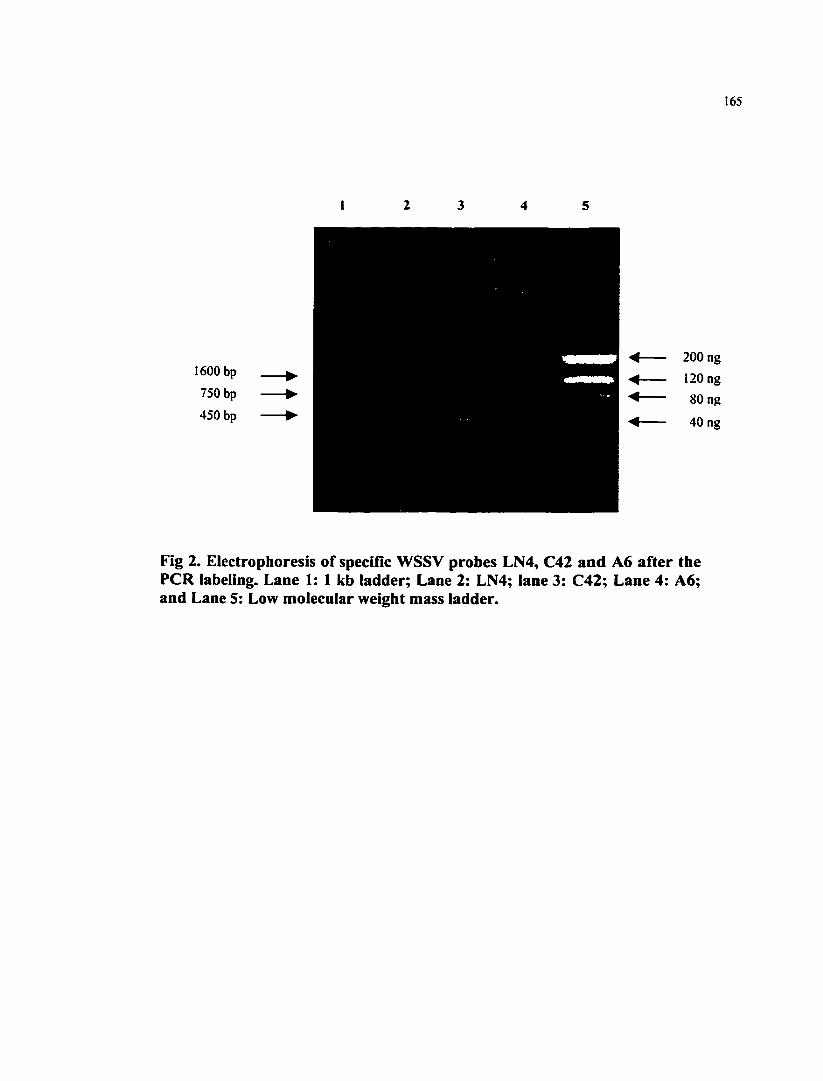
165
1600 bp
750 bp
450 bp
200 ng
120 ng
80 ng
40 ng
Fig 2. Electrophoresis of specific WSSV probes LN4, C42 and A6 after the PCR labeling. Lane 1: 1 kb ladder; Lane 2: LN4; lane 3: C42; Lane 4: A6; and Lane 5: Low molecular weight mass ladder.

166
Fig 3. Electrophoresis of DNA samples undigested or digested with two of the representative endonucleases EcoRI and Clal. Lane 1: DIG-labeled molecular weight marker III; Lane 2- Lane 7: DNA of WSSV from the following geographic regions digested with EcoRI: 2: Thailand; 3: India; 4: China; 5: Texas; 6: South Carolina; 7: crayfish from Washington D. C.; Lane 8-13: DNA of WSSV from following geographic regions digested with Clal: 8:Thailand; 9: India; 10: China; 11: Texas; 12: South Carolina; 13: crayfish from Washington D. C.; Lane 14: Undigested DNA from Thailand WSSV; and Lane 15: Undigested DNA from Specific Pathogen Free (SPF) shrimp.
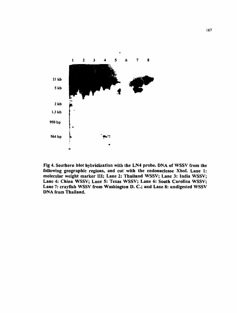
167
1 2 3 4 5 6 7 8
2 1 k b
5 k b
2 k b
I J k b
950 bp
564 bp
Fig 4. Southern biot hybridization with the LN4 probe. DNA of WSSV from the following geographic regions, and cut with the endonuclease Xhol. Lane 1: molecular weight marker III; Lane 2: Thailand WSSV; Lane 3: India WSSV; Lane 4: China WSSV; Lane 5: Texas WSSV; Lane 6: South Carolina WSSV; Lane 7: crayfish WSSV from Washington D. C.; and Lane 8: undigested WSSV DNA from Thailand.

168
Fig 5. Southern blot hybridization with the LN4 probe. Lane 1: molecular weight marker III; Lane 2-7: DNA of WSSV from the following geographic regions digested with Bglll: 2: Thailand; 3: India; 4: China; 5: Texas; 6: South Carolina; 7: crayfish from Washington D. C.; Lane 8-13: DNA of WSSV from the following geographic regions digested with Hindll: 8: Thailand; 9: India; 10: China; 11: Texas; 12: South Carolina; 13: crayfish from Washington D. C.; and 14: undigested WSSV DNA from Thailand.

169
1 2 3 4 5 6 7 8 9 1 0 1 1
7KB
3.5 KB
3 KB
k
Fig 6. Southern blot hybridization with the LN4 probe. Lane 1, 3, 5, 7, 9,11: DNA of WSSV from the following geographic regions digested with Clal: 1: China; 3: India; 5: Thailand; 7: South Carolina; 9: crayfish from Washington D. C.; 11: Texas; Lane 2, 4, 6, 8,10: undigested DNA of WSSV from the following regions: 2: India; 4: Thailand; 6: South Carolina; and 8: crayfish from Washington D. C.; 10: Texas.

170
Fig 7. Southern blot hybridization with the C42 probe. Lane I: Lane 1, 3, 5, 7, 9,11: DNA of WSSV from the following geographic regions digested with Clal: 1: China; 3: India; 5: Thailand; 7: South Carolina; 9: crayfish from Washington D. C.; 11: Texas; Lane 2, 4, 6, 8,10: undigested DNA of WSSV from the following regions: 2: India; 4: Thailand; 6: South Carolina; and 8: crayfish from Washington D. C.; and 10: Texas.
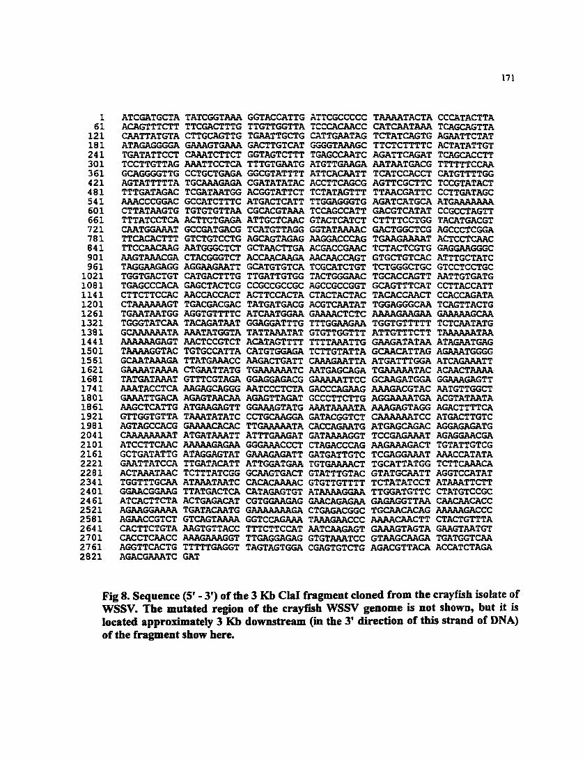
171
1 ATCGATGCTA TATCGGTAAA GGTACCATTG ATTCGCCCCC TAAAATACTA CCCATACTTA 61 ACAGTTTCTT TTCGACTTTG TTGTTGGTTA TCCCACAACC CATCAATAAA TCAGCAGTTA 121 CAATTATGTA CTTGCAGTTG TGAATTGCTG CATTGAATAG TCTATCAGTG AGAATTCTAT 181 ATAGAGGGGA GAAAGTGAAA GACTTGTCAT GGGGTAAAGC TTCTCTTTTC ACTATATTGT 241 TGATATTCCT CAAATCTTCT GGTAGTCTTT TGAGCCAATC AGATTCAGAT TCAGCACCTT 301 TCCTTGTTAG AAATTCCTCA TTTGTGAATG ATGTTGAAGA AATAATGACG TTTTTTCCAA 361 GCAGGGGTTG CCTGCTGAGA GGCGTATTTT ATTCACAATT TCATCCACCT CATGTTTTGG 421 AGTATTTTTA TGCAAAGAGA CGATATATAC ACCTTCAGCG AGTTCGCTTC TCCGTATACT 481 TTTGATAGAC TCGATAATGG ACGGTATTCT TCTATAGTTT TTAACGATTC CCTTGATAGC 541 AAACCCGGAC GCCATCTTTC ATGACTCATT TTGGAGGGTG AGATCATGCA ATGAAAAAAA 601 CTTATAAGTG TGTGTGTTAA CGCACGTAAA TCCAGCCATT GACGTCATAT CCGCCTAGTT 661 TTTATCCTCA ACTTCTGAGA ATTGCTCAAC GTACTCATCT CTTTTCCTGG TACATGACGT 721 CAATGGAAAT GCCGATGACG TCATGTTAGG GGTATAAAAC GACTGGCTCG AGCCCTCGGA 781 TTCACACTTT GTCTGTCCTG AGCAGTAGAG AAGGACCCAG TGAAGAAAAT ACTCCTCAAC 841 TTCCAACAAG AATGGGCTCT GCTAACTTGA ACGACCGAAC TCTACTCGTG GAGGAAGGGC 901 AAGTAAACGA CTACGGGTCT ACCAACAAGA AACAACCAGT GTGCTGTCAC ATTTGCTATC 961 TAGGAAGAGG AGGAAGAATT GCATGTGTCA TCGCATCTGT TCTGGGCTGC GTCCTCCTGC 1021 TGGTGACTGT CATGACTTTG TTGATTGTGG TACTGGGAAC TGCACCAGTT AATTGTGATG 1081 TGAGCCCACA GAGCTACTCG CCGCCGCCGC AGCCGCCGGT GCAGTTTCAT CCTTACCATT 1141 CTTCTTCCAC AACCACCACT ACTTCCACTA CTACTACTAC TACACCAACT CCACCAGATA 1201 CTAAAAAAGT TGACGACGAC TATGATGACG ACGTCAATAT TGGAGGGCAA TCAGTTACTG 1261 TGAATAATGG AGGTGTTTTC ATCAATGGAA GAAAACTCTC AAAAGAAGAA GAAAAAGCAA 1321 TGGGTATCAA TACAGATAAT GGAGGATTTG TTTGGAAGAA TGGTGTTTTT TCTCAATATG 1381 GCAAAAAATA AAATATGGTA TATTAAATAT GTGTTGGTTT ATTGTTTCTT TAAAAAATAA 1441 AAAAAAGAGT AACTCCGTCT ACATAGTTTT TTTTAAATTG GAAGATATAA ATAGAATGAG 1501 TAAAAGGTAC TGTGCCATTA CATGTGGAGA TCTTGTATTA GCAACATTAG AGAAATGGGG 1561 GCAATAAAGA TTATGAAACC AAGACTGATT CAAAGAATTA ATGATTTGGA ATCAGAAATT 1621 GAAAATAAAA CTGAATTATG TGAAAAAATC AATGAGCAGA TGAAAAATAC ACAACTAAAA 1681 TATGATAAAT GTTTCGTAGA GGAGGAGACG GAAAAATTCC GCAAGATGGA GGAAAGAGTT 1741 AAATACCTCA AAGAGCAGGG AATCCCTCTA GACCCAGAAG AAAGACGTAC AATGTTGGCT 1801 GAAATTGACA AGAGTAACAA AGAGTTAGAT GCCCTTCTTG AGGAAAATGA ACGTATAATA 1861 AAGCTCATTG ATGAAGAGTT GGAAAGTATG AAATAAAATA AAAGAGTAGG AGACTTTTCA 1921 GTTGGTGTTA TAAATATATC CCTGCAAGGA GATACGGTCT CAAAAAATCC ATGACTTGTC 1981 AGTAGCCACG GAAAACACAC TTGAAAAATA CACCAGAATG ATGAGCAGAC AGGAGAGATG 2041 CAAAAAAAAT ATGATAAATT ATTTGAAGAT GATAAAAGGT TCCGAGAAAT AGAGGAACGA 2101 ATCCTTCAAC AAAAAGAGAA GGGAAACCCT CTAGACCCAG AAGAAAGACT TGTATTGTCG 2161 GCTGATATTG ATAGGAGTAT GAAAGAGATT GATGATTGTC TCGAGGAAAT AAACCATATA 2221 GAATTATCCA TTGATACATT ATTGGATGAA TGTGAAAACT TGCATTATGG TCTTCAAACA 2281 ACTAAATAAC TCTTTATCGG GCAAGTGACT GTATTTGTAC GTATGCAATT AGGTCCATAT 2341 TGGTTTGCAA ATAAATAATC CACACAAAAC GTGTTGTTTT TCTATATCCT ATAAATTCTT 2401 GGAACGGAAG TTATGACTCA CATAGAGTGT ATAAAAGGAA TTGGATGTTC CTATGTCCGC 2461 ATCACTTCTA ACTGAGACAT CGTGGAAGAG GAACAGAGAA GAGAGGTTAA CAACAACACC 2521 AGAAGGAAAA TGATACAATG GAAAAAAAGA CTGAGACGGC TGCAACACAG AAAAAGACCC 2581 AGAACCGTCT GTCAGTAAAA GGTCCAGAAA TAAAGAACCC AAAACAACTT CTACTGTTTA 2641 CACTTCTGTA AAGTGTTACC TTTCTTCCAT AATCAAGAGT GAAAGTAGTA GAAGTAATGT 2701 CACCTCAACC AAAGAAAGGT TTGAGGAGAG GTGTAAATCC GTAAGCAAGA TGATGGTCAA 2761 AGGTTCACTG TTTTTGAGGT TAGTAGTGGA CGAGTGTCTG AGACGTTACA ACCATCTAGA 2821 AGACGAAATC GAT
Fig 8. Sequence (5' - 3') of the 3 Kb Clal fragment cloned from the crayfish isolate of WSSV. The mutated region of the crayfish WSSV genome is not shown, but it is located approximately 3 Kb downstream (in the 3' direction of this strand of DNA) of the fragment show here.

172
APPENDIX 4.
Per OS CHALLENGE OF Litopenaeus vannamei POSTLARVAE AND
Farfantepenaeus duorarum JUVENILES WITH SIX GEOGRAPHIC ISOLATES
OF WHITE SPOT SYNDROME VIRUS
Qiong Wang*, Brenda L.White, Elita M. Redman, Donald V. Lightner
Department of Veterinary Science and Microbiology, University of Arizona, Tucson,
Arizona 85721, USA
Correspondence should be addressed to:
Qiong Wang, Department of Veterinary Science and Microbiology, University of
Arizona, Tucson, Arizona 85721, USA
E-mail: qwang(^aruba.u.arizona.edu
Tel: 1-520-621-4438
Fax: 1-520-621-4899

Reprinted from
Aquaculture
Aqiucultuie 170(1999) 179-194
Per OS challenge of Litopenaeus vannamei postlarvae and Farfantepenaeus duoramm
juveniles with six geographic isolates of white spot syndrome virus
Qiong Wang *, Brenda L. White, Rita M. Redman, Donald V. Lightner
Department of Veterinary Science and Microbiology. Uniuernty (̂ Arizona. Tucson. AZ S572I, USA
Accepted 26 October 1998
ELSEVIER
174
ELSEVIER SCIENCE B.V. TRANSFER OF COPYRIGHT AGREEMENT
Scientific pubiisheis and autiiors share a common interest in tiie protection of copyright: authors principally because they want their creative works to be protected fiom plagiarism and other unlawful uses, publhhers because ttey need to protect their work and investment in the producdon. marketing and distribution of the article written by the author. order to do so effectively, publishers need a formal written transfer of copyright from the author(s) for each article published. Publishers and authors are also concerned that the integrity of an article (once refereed and accepted for publication) be maintained, and in order to protect that reference value and validation process, we ask that authors recognize that distribution (including throu ̂ the IniernctAVWW or other on-line means) of the version of the article as accepted for publication is best administered by the publisher. Please complete and retum this form promptly.
Article entitled: Per os challenge of Litopenaeus vannamei posdarvae and farfantepenarus duorarum juveniles with six geograpgic isolates of white spot syndrome virus (WSSV)
Author(s): Dr. Q. Wang
To be published in the journal: Aquaculture
Effective upon acceptance for publication, copyright (including all rights thereunder and die right to obtain copyright registration in the name of the publisher, whether separately or as a pan of a journal issue or otherwise) in the above article and any modifications of it by the author(s) is hereby transferred throu^out the world and for die full term and all extensions and renewals thereof, to: Elsevier Science B.V.
This transfer includes the right to adapt the article for use in conjunction with computer systems and programs, including reproduction or publication in machine-readable form and incorporation in retrieval systems.
Rights of authors Elsevier Science B.V.
recognizes the retention of the following rights by the authoi(s):
1. Patent and trademark rights and rights to any process or procedure described in the article.
• 2. The right to photocopy or make single electronic copies of the article for dieir own personal use, including for their own classroom use, or for the personal use of colleagues, provided the copies are not offered for sale and are not distributed in a systematic way outside of their employing institution (e.g. via an e-mail list or public file server). Posting of the article on a secure network (not accessible to the public) within the author's instimtion is permitted. However, if a prior version of this work (normally a preprint) has been posted to an electronic public server, the author(s) agree not to update and/or replace this prior version on the server in order to make it idratical in content to die final published version, and fiirther diat posting of the article as published on a public server can only be done with Elsevier's written permission.
3. The right, subsequent to publication, to use the article or any pan diereof firee of charge in a printed compilation of works of their own, such as collected writings or lecture notes, in a thesis, or to expand the article into book-length form for publication
Note All copies, paper or electronic, or other use of the information must include an indication of the Elsevier Science B.V.
copyright and a fiiU citation of the journal source. Please refer requests for all uses not included above, including the authorization of third parties to reproduce or otherwise use all or part of the article (including figures and tables), to:
Elsevier ScierKe, Rights & Permissions Department, P.O. Box 800, O^^ord, 0X5IDX, UK

ELSEVIER Aquacultuie 170(1999) 179—194
Per OS challenge of Litopenaeus vannamei postlarvae and Farfantepenaeus duorarum
juveniles with six geographic isolates of white spot s)^cirome virus
Qiong Wang *, Brenda L. White, Jlita M. Redman, Donald V. Lightner
Department irf Veterinary Science and Microbiology. University of Arizona, Tucson. AZ 8S72I. USA
Accqxed 26 October 1998
Abstract
White spot syndrome vims (WSSV) is one of the most important pathogens of penaeid shrimp. It is widely distributed in most Asian countries where penaeid shrimp are cultured, as well as in the Gulf of Mexico and SB USA. The virulence of six geographic isolates of WSSV was compared using Litopenaeus vannamei postlarvae and Farfantepenaeus duorarum juveniles. The six geographic isolates of WSSV originated from (Thina. India. Thailand, Texas. South Carolina, as well as from crayfish maintained at the USA National Zoo. For challenge smdies. virus infected tissues were given per os to L. vannamei postlarvae and Fa. duorarum juveniles. Resultant WSSV infections were confirmed by histological examination. The ciunulative mortality of L. vannamei postlarvae reached 100% after challenge with each of the six geographic isolates of WSSV. However, the Texas isolate caused mortalities more rapidly than did the other shrimp isolates; the crayfish WSSV isolate was the slowest. In marked contrast, cumulative mortalities of juvenile Fa. duorarum reached only 35-60%. and varied among the geographic isolates of WSSV. Interestingly, in Fa. duorarum, the Texas WSSV isolate was also the most virulent, while the crayfish WSSV was the least virulent The findings suggest that slight differences in virulence exist among geographic isolates of WSSV. and that susceptibility may vary with species and lifestages of the host. O 1999 Elsevier Science B.V. All rights reserved.
Keywords: White spot syndioine vinis: WSSV; Penaeid shrimp; Geographic isolates; Virulence; Susceptibility
Corresponding author. Tel.: + 1-520-621-4438; Fax: + 1-520-621-4899; E-mail: qwang®aruba.u,ari7nna edu
0044-8486/99/$ - see fitont matter © 1999 Elsevier Science B.V. All rights reserved. PH: 50044-8486(98)00425-6

iso Q. Wang etaL/Aquaaitatre 170(1999) 179-J94
1. Introdaction
Hist recognized in 1992, white spot syndrome vims (WSSV) causes the most serious disease of cultured penaeid shrimp in the Eastern Hemisphere (Chen. 199S; Flegel et al., 1995; Huang et al., 1995a; Inouye et al., 1994, 1996; Kimuia et al., 1996; Nakano et al., 1994; Takahashi et al., 1994; Wang et al., 1995; Wongteerasupaya et al., 1995). Penaeus monodon, Fenneropenaeus chinensis , Fe. indicus, Fe. penicillatus, and Marsupenaeus japonicus are the major species naturally infected by the virus in Asian countries. Some infected shrimp show white spots on the inner surface of the cuticle (Chen, 1995; Kasomchandra and BoonyaratpaUn, 1996), and the cumulative mortality can reach as high as 100% after the appearance of the clinical signs. In North America, WSSV has been found every year since 1995 in wild shrimp or in shrimp growing facilities in Texas and South Carolina (Lightner et al., 1997; Lo et al., in press: Nunan and Lightner, 1997; Nunan et al., 1998). Severe WSSV-induced mortalities have been dociunented in Litopenaeus setiferus stocks from both of these states. In December 1995, a serious disease outbreak was investigated in crayfish, Orconectes punctimanus and Procambarus sp., at the USA National Zoo in Washington O.C (Richman et al., 1997). Histological analysis and transmission electron microscopy (TEM) showed that the pathogen closely resembled WSSV from shrimp. That the isolate from the crayfish was a strain of WSSV was confirmed by dot blot with specific gene probes, PCR methods, and bioassays with shrimp (Richman et al., 1997). Additional hosts reported for WSSV include: Metapenaeus ensis, Farfantepenaeus aztecus. Fa. duorarum, Fe. merguiensis, P. semisulcattts, L. stylirostris, L vannamei, Trachypenaeus curvirostris (all penaeid shrimp), Exopalaemon orientalis, Macrobrachium rosenbergii (caridean shrimp) and Procambarus clarfdi (a crayfish). All of the species showed high susceptibilities to WSSV under natural or experimental conditions (Cai et al., 1995; Chang et al., 1998; Lighmer et al., 1998; Wang et al.. 1998). WSSV has also been detected in wild crabs iCalappa lophos, Portunus sanguinolentus, Charybdis sp., Helice tridens), wild lobsters (Panulirus sp.), Palaemonidae pest shrimp, Copepoda plankton. Anemia and pupae of an Ephydridae insect, by PCR, in situ hybridization or monoclonal antibody assays (Chang et al., 1998; Lo et al., 1996; Huang et al., 1995b; Wang et al., 1998).
The objectives of the present study were to compare the identities and virulence of six geographic isolates of WSSV, and to investigate the susceptibility of two representative American penaeid shrimp species to the geographic isolates of WSSV.
2. Materials and methods
2.1. Experimental shrimp
'Specific pathogen firee (SPF)' Pacific white shrimp, L uammaei, were obtained as postlarvae (PL) fivm the breeding program of the Oceanic Instimte (OI), Oahu, HI. These SPF L vannamei were from OI's 'Kona' stock, which originated from Mexico in 1989 (Wyban et al., 1992). The population number of the batch of shrimp used in the present experiment was designated as UAZ 10-97. Fa. duorarum, pink shrimp, were

Q. (Vang et aL / Aquaculmre 170(1999) 179-194 181
obtained as hatchery reared posdarvae (derived from wild broodstock) from Harbor Branch, Fort EHerce, FL, and reared to juveniles at the University of Arizona's Aquaculture Pathology Center (UAZ) in Tucson, untU the time when they were used in the experiments. The designated population number of this batch of Fa. duorarum was UAZ 22-96. The taxonomy in this paper followed Perez-Farfante and Kensley (1997).
2.2. Viruses
The white spot syndrome virus (WSSV) samples used in this smdy were obtained from China, India, Thailand. South Carolina, Texas and the National Zoo in Washington, DC (Table 1). To amplify the quandty of these viruses, SPF (WSSV-free) L. vannamei or L, settferus were fed or injected with viral inocula. Representative samples of moribund shrimp were sampled for histological confirmaQon of WSSV infection, while other moribund shrimp were collected and stored at — 70°C until used in this present smdy (Table 2).
2.3. Challenge studies
The experimental design for WSSV challenge followed closely that of Hasson et al. (1995). In the first experiment, the six geographic WSSV isolates (fit>m China, India. Thailand, Texas, South Carolina and the National Zoo) were compared for differences in virulence using 700 SPF PL30 L vannamei, with average weight of 0.05 g. The PLs were stocked in seven. 40 I aquaria (100 PL/aquaria). Six aquaria were prepared for use as treatment groups and the seventh was designated as a negative control. Each aquarium was individually outfitted with previously activated oyster shell biofilter and with an air diffiiser. Water recirculadon to the biofrlters was provided by an airlift pipe. Each aquarium was covered with a separate plasdc sheet to contain aerosols, and the negative control aquarium was set up in a separate room to prevent possible contamination. Each treatment group received two feedings of WSSV (one geographic WSSV in each tank) infected minced tissue (10% of body weight/feeding) on day 0. All control and treatment groups were otherwise fed ad lib Rangen No. 2 (Buhl, ID) pellets twice daily. All feedings were conducted at 0800 and I6(X). Observations for clinical signs of WSSV infection and mortalities were conducted daily at 0700, 1100, 1500 and 2000 on each day of the study. Dead and moribund animals were counted and removed when observed. To confirm the infection status, moribund aniTnals from each of the treatment tanks were preserved in Davidson's AFA fixative and sectioned for histological observation using routine methods (Bell and Lighmer, 1988). Surviving shrimp and shrimp frt)ni the negative control group were also sampled for histological examination. Nitrite and ammotiia levels of the control and treatment tanks were monitored throughout the course of the study, and did not exceed 0.1 and 0.25 ppm, respectively. Water temperature in each of the aquaria was checked daily and adjusted with immersion heaters to maintain 26 ± 2°C. The salinity was set at 26 ppt.
In the second experiment, four geographic isolates of WSSV (from Thailand, Texas, South Carolina and the National Zoo) were compared using 75 Fa. duorarum and
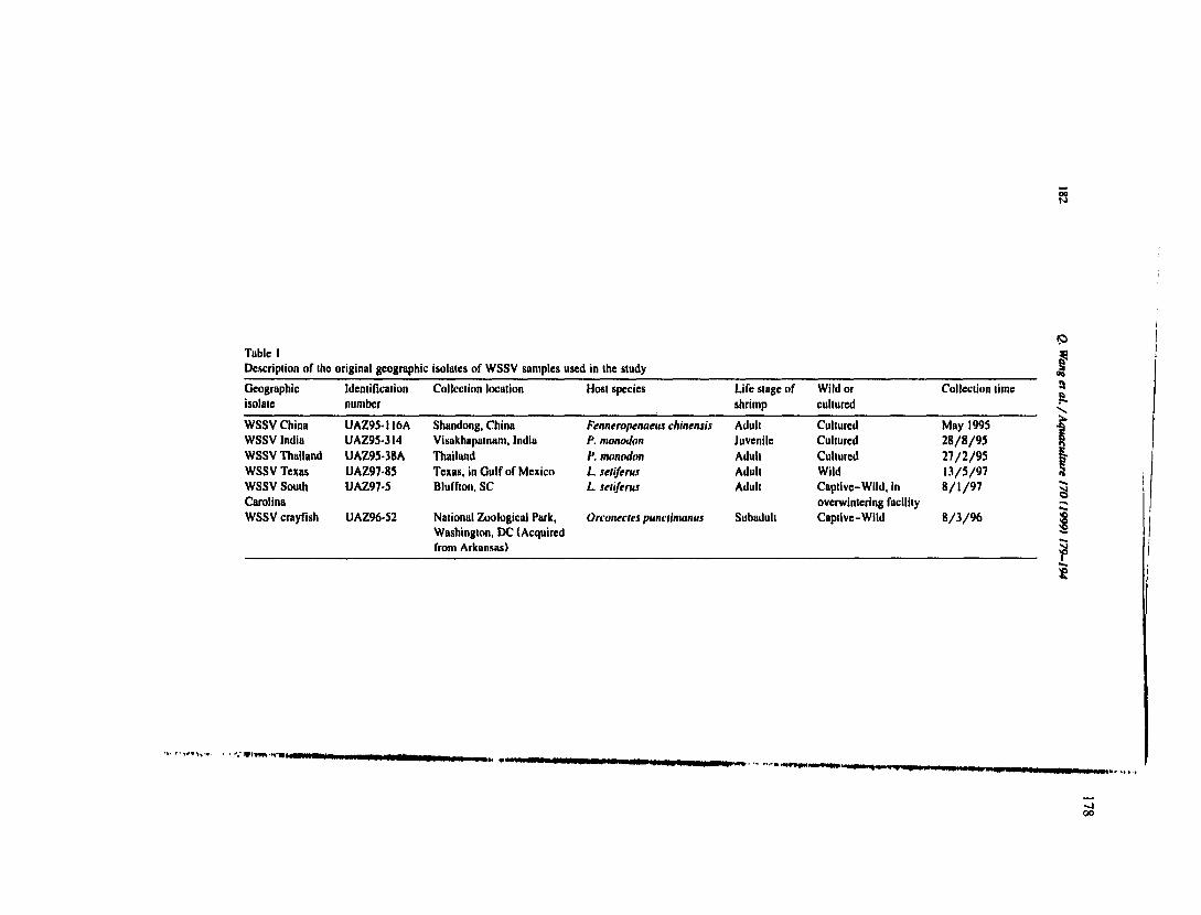
Table I
Description of the original geographic isolates of WSSV samples used in the study
Geographic Identiflcation Collection location Host species Life stage of Wild or Collection lime
isolate number shrimp cultured
WSSV China UAZ95-II6A Shandong, China Fenneropenaeus chinensis Adult Cultured May I99S
WSSV India UAZ95-314 Visakhapatnam, India P. monodan Juvenile Cultured 28/8/95
WSSV Thailand UAZ95-38A Thailand P. monodim Aduh Cultured 27/2/95
WSSV Texas UAZ97-85 Texas, in Gulf of Mexico L setiferus Adult Wild 13/5/97
WSSV South UAZ97-5 Bluffton, SC L setiferus Adult Captive-Wild, in 8/1/97
Carolina overwintering facility
WSSV croyflsh UAZ96.52 National Zoological Park,
Washington, DC (Acquired
from Arkansas)
Orconectes punciimanus Subadult Caplive-Wild 8/3/96

Table 2
Description of the WSSV inocula propagated at the University of Arizona and used in the present study
Geographic ID number Host species Average Host ID number of ID number of the Propagating Propagating Histological
isolates of the inocula size host population original viral source time methods findings*'
WSSV China UAZ97-54 L vannamei 8g UAZ17-96 UAZ95-II6A April 1997 I.M.* Injecting 04 WSSV
WSSV India UAZ97-56 L rannamei 8.6 g UAZI7-96 UAZ95-3I4 April 1997 Feeding 04 WSSV
WSSV Thuilond UAZ97-86 L raimumei 4g UAZ27-96 UAZ95-38A May 1997 I.M.' Injecting G4 WSSV
WSSV Texas UAZ97-I06 L rannamei 4g UAZ27-96 UAZ97-85 June 1997 Feeding 04 WSSV
WSSV South Carolina UAZ97-64 L vannamei l l g UAZ04-% UAZ97-5 April 1997 I.M.* Injecting 04 WSSV
WSSV crayfish UAZ96-2II L seiiferus l O g UAZ 13-95 UAZ96-52 November 1996 Feeding 04 WSSV
'I.M.—intramuscular.
''Routine H and E histological Hndings of moribund shrimp samples. Lesion severity was graded on a scale from G0-Q4: GO indicates no lesions detected, and 04
denotes severe multifocal to diffuse WSSV lesions (Lightner, 1996).

184 Q. Wang etaL/Aquaculmre 170 (1999) 179-194
15 SPF L vannamei, with average body weights of 6.4 g and 5.0 g. respectively. The shrimp were stocked in six, 90 1 aquaria (15 shrimp/aquaria). Four aquaria were used as treatment tanks, the fifth aquarium served as a negative control, and the sixth aquarium was stocked with L vannamei to serve as a positive control, in which the Thailand WSSV isolate was used as the inoculum. Each treatment group on day 0 received one feeding of WSSV (one geographic isolate in each tank) infected minced tissue ( ~ 5% of body weight/feeding). For maintenance, all negative control shrimp and treatment group shrimp were fed ad lib 35% Rangen pellets twice daily. Otherwise, this experiment was conducted in the same manner as that used in the Z. vannamei bioassay.
2.4. Histological procedures
Samples were processed according to standard procedures using Davidson's AFA fixed tissue followed by routine histological processing, and hematoxylin and eosin-phloxine (H and E) staining (Bell and Lightner, 1988; Lighmer, 1996). All anatomical, histological and pathological nomenclatures for normal and WSSV-infected shrimp followed Bell and Lighmer (1988), and Lighmer (1996). Severity was rated following the grading scheme of Lighmer (1996).
3. Results
Postlarval L vannamei were highly susceptible by per os challenge to all six geographic WSSVs. Cumulative mortalities of 100% were reached within 14 d after initial exposure to all six WSSV isolates (Fig. 1).
Juvenile Fa. duorarum were found to be less susceptible to WSSV infection and to suffer cumulative mortality rates of 35% to 60%, by 18 d after challenge, with the mortality rate varying among the WSSV isolates (Fig. 2).
WSSV infection of all moribund animals was confirmed by histological examination. The lesions were typical and severe (grade 4). Prominent intranuclear eosinophilic to basophilic viral inclusions were observed in cells of the cuticular epidermis of the appendages, gills and body surface, foregut and hindgut; various coimective tissues; the antennal gland; lymphoid organ; hematopoietic tissues; and in a variety of other cell types (Figs. 3 and 4). The negative control L vannamei and Fa. duorarum appeared to be normal by histological examination. Significant histological changes were observed among Fa. duorarum survivors. Tissues conunonly targeted by WSSV (e.g., the subcuticuiar epithelial and coimective tissues) appeared relatively normal with only a few cells presenting WSSV eosinophilic intranuclear inclusions (Hg. 5). Cells displaying viral inclusions appeared to induce an inflammatory response characterized by hemocytic nodule formation (Fig. 6). The lymphoid organs of surviving Fa. duorarum were hypertrophied and edematous. Large numbers of vacuoles were observed in the connective tissue among the lymphoid organ mbules (Hg. 7). Spherical masses of cells, presumed to be metastasized lymphoid organ spheroids, were present in the coimective tissue capsules of the hepatopancreas (Fig. 8). La the antennal gland, hemocytic nodules formed in the lumen or on the tubule waU (Fig. 9), and sometimes these cells appeared
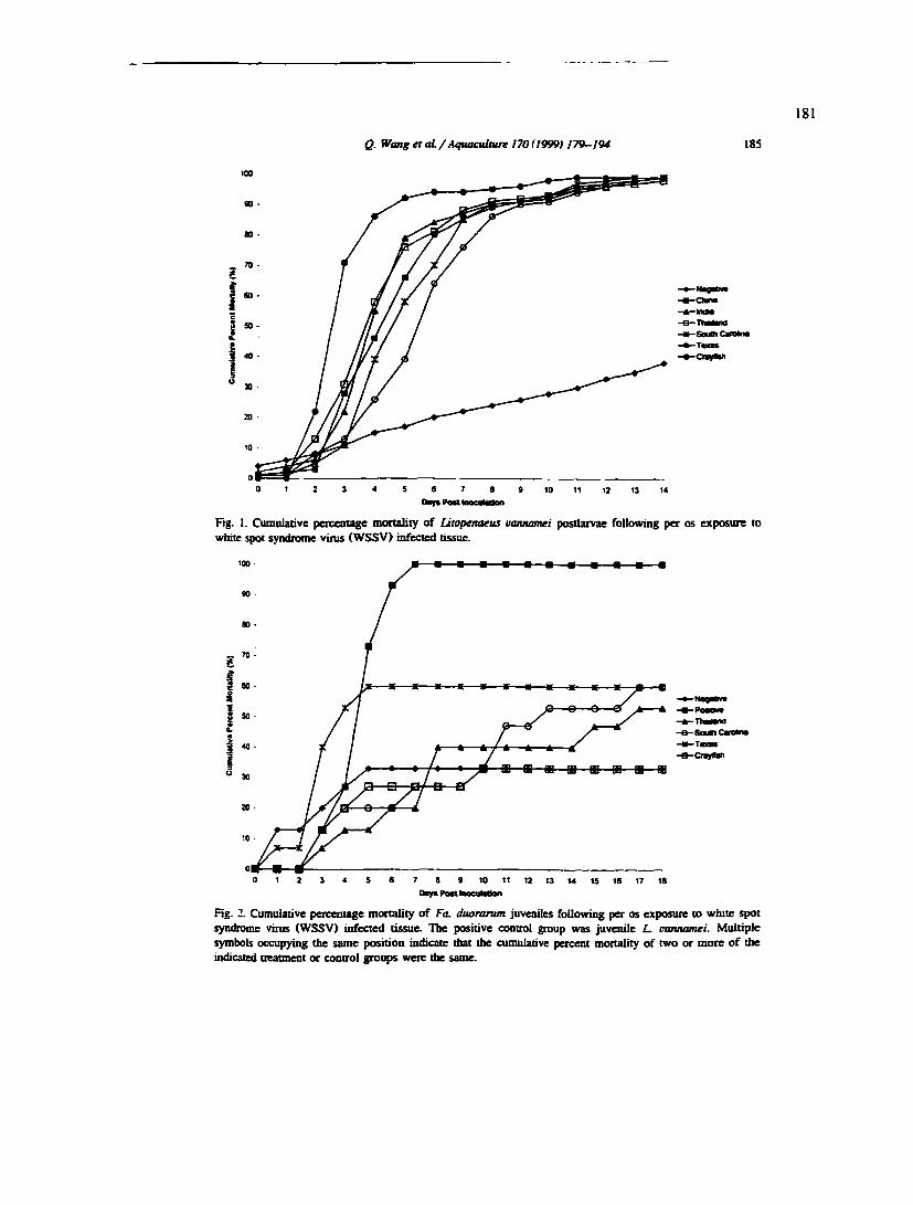
Q. Wangetat/Axfuaculture 170(1999} 179-194 185
-SauOiCmna
-OayMi
10 11 12 13 14
Fig. 1. Cumulative percentage mortality of Litopenaeus vamamei postlarvae following per os exposure to white spot syndrome vinis (WSSV) infected tissue.
100
9—B—B—a -CnyMi
Fig. 2. Cumulative percentage mortality of Fa. duoramm juveniles following per os exposure to while spot syndrome virus (WSSV) infected tissue. The positive control group was juvenile L. vannamei. Multiple symbols occupying the same position indicate that the cumulative percent mortality of two or more of tiie indicated treatment or control groups were the same.

)86 Q. Wang et aL / Aquacubure 170(I999> 179—194
Fig. 3. Chaiacteristic white spot syndrome virus (WSSV) lesions in the cuticular epithelium of the stomach of an experimentally infected penaeid shrimp (L. utmnamei). Arrows indicate hypertrophied nuclei with
intranuclear WSSV inclusions. H and E stain. Bar 20 |i.m.
Ftg. 4. Characteristic white spot syndrome virus (WSSV) lesions in the cuticular epithelium of the stomach of an experimentally infected penaeid shrimp iFa. duorantm). Arrows indicate hypertrophied nuclei with
intranuclear WSSV inclusions. H and E stain. Bar — 20 ̂ .m.

Q. Wang etaL/Aquacubure 170(1999) 179-194 187
Fig. S. HUcological abnonnalities observed within a Fa. duoramm juvenile ifaal survived experimental infection with white spot syndrome virus (WSSV). WSSV infected subcuticular connective tissue cells (small arrows) within the stomach region. Necrotic cells with pyloiotic nuclei (large arrows) are also visible. H and E stain. Bar » 20 p.m.
Fig. 6. Histological abnormalities observed within a Fa. duomrum juvenile that survived experimental infection with white spot syndrome. Hemocyte encapsulation (arrows) of presumed WSSV-infected cells within the cuticular epithelium of the stomach. H and E stain. Bar » 20 |i.m.

188 Q. Wmg et aL/Aquacuiture 170(1999) 179-194
Fig. 7. Histological abnormalities observed within a Fa. duorarum juvenile that survived experimental infection with white spot syndrome. Numerous vacuoles (Va) within the intertubular connective tissue of (he lymphoid organ. H and E stain. Bar •• 20 M.m.
Ftg. 8. Histological abnormalities observed within a Fa. duorarum juvenile that survived experimental infection with white spot syndrome. An ectopic spheroid within the coiuiective tissue capsule of the
bepatopancreas. H and E stain. Bar " 20 (im.

Q. Wang et oL/AquacuUurt 170 < I999t 179—194 1S9
Fig. 9. Histological abnonnaiities obsoved within a Fa. duorarum juvenile that survived experimental infection with white spot syndrome. Hemocytic nodule (auows) formation within the antennal gland. H and E stain. Bar — 20 pun.
Fig. 10. Histological abnormalities observed within a Fa. duorarum juvenile that survived experimental infection with white spot syndrome. A longitudinal section throagb a portion of the subgastric artery illustrating WSSV inclusions (large arrows) and nuclear pyknosis (small arrows). H and E stain. Bar ~ 20 (im.

190 Q. Wang et aL/Aquaculture 170 (1999) 179-194
Fig. 11. Histological abnormalities observed within a Fa. duorarum juvenile that survived experimental infection with white spot syndrome. WSSV intranuclear inclusions within the gonad (arrows). H and E stain. Bar "»20 (im.
Fig. IX Histological abnormalities observed within a Fa. duoramm juvenile that survived experimental infection with white spot syndrome. Pyknotic and karyotrhectic nuclei (arrows) within hematopoietic tissue. H
and E stain. Bar -• 20 M.m.

Q. Wang et aL / Aquaculnire 170 (19991179-194 191
to fiise to form multinucleated giant cells. Interestingly, the subgastric artery, gonad and hematopoietic tissues appeared to be the tissue most affected by WSSV infection in surviving Fa. duorarum. WSSV inclusions were found in the sheath cells of the subgastric anery and in germinal and connective tissue cells in gonad (Figs. 10 and 11). Multiple foci of pyknotic nuclei were present in the hematopoietic tissue (Hg. 12).
4. Discussion
Two lifestages of two species of American penaeid shrimp. L vannamei PLs, and L vannamei and Fa. duorarum juveniles, were tested for their susceptibility to six geographic isolates of WSSV. The L, vannamei (PLs and juveniles) tested were highly susceptible to all six geographic WSSVs, and the ctunulative mortalities, caused by each WSSV isolate, reached 100% in 14 days (Figs. 1 and 2). In contrast the Fa. duorarum showed moderate resistance to WSSV infection, with 40% to 65% shrimp surviving to day 18 (termination) of the experiment (Hg. 2). These findings confirm the results of a previous study (Lighmer et al.. 1998) in which juvenile Fa. duorarum. but not juvenile L vannamei. displayed some resistance to "per os' challenge by a WSSV isolate from China. However, in a preliminary study. PL Fa. duorarum. ftom the same batch of shrimp used in the present experiment, were challenged with the China isolate of WSSV. the mortality reached 100% (unpublished data). Therefore, it appears that juvenile Fa. duorarum are more resistant to WSSV than PLs. and that L varummei PLs (Lightner et al.. 1998) and juveniles (ibid and this study) show little resistance to WSSV.
The Texas isolate of WSSV was more virulent than the other isolates: the crayfish isolate was the least virulent. Although no apparent difference in cumulative mortality was observed in the L. vannamei study, the Texas WSSV isolate caused mortalities more rapidly than did the other geographic WSSV isolates, while the crayfish WSSV isolate was the slowest (Fig. I). In the Fa. duorarum study, the differences among geographic isolates of WSSV were more obvious. The mortalities caused by the Texas isolate were higher and occurred earlier than the other WSSV isolates. In contrast, the crayfish WSSV isolate was the least virulent for Fa. duorarum, causing the lowest cumulative mortalities which occurred later than those caused by other WSSV isolates (Fig. 2).
All moribund shrimp samples collected for histological analysis displayed WSSV infections (Figs. 3 and 4). Hemocytic encapsulation and phagocytosis of infected and necrotic cells were observed eimong surviving Fa. duorarum (Figs. 5-12), which suggests that a protective response could have been stimulated among these shrimp. Relatively severe WSSV lesions in the lymphoid organ, subgastric artery and hematopoietic tissue (Hgs. 7, 8, 10 and 12) suggested that hemocytes are important in the shrimp's defense response. The presence of WSSV inclusions observed within the gonads of Fa. duorarum survivors (Fig. 11) suggests a possible mechanism for vertical transmission.
Because of the lack of shrimp cell lines for culturing WSSV. it is difficult to measure the concentration of shrimp viruses accurately. In the experiments reported here, each challenged group was given the same percentage of body weight of infected tissue as the
192 Q. Wang et at / Aquacubure 170 (1999) 179—194
challenge inoculum; the dosages of viruses were evaluated by histological observation and grading, dot blot assay, DNA and protein visualization (unpublished data). Because of the storage time and biological nature of shrimp tissue (the size of the animals, the distribution of viruses in the tissue), the viral concentration in the inocula that we used in the experiment was very likely not equal among treatments. However, since WSSV is a rapidly replicating and extremely virulent shrimp pathogen, differences of mortality patterns detected must have been due to genetic differences rather than to dosage differences. This possibility is supported by the presence of genetic differences among WSSV isolates detected in RFLP assays (unpublished data).
In summary, the results of the present study demonstrated that all six geographic WSSV isolates are infectious and highly virulent to the PL stages of L vannamei, while Fa. duorarum juvenUes showed moderate resistance to WSSV infection. Slight differences of virulence among WSSV geographic isolates were detected, with the Texas WSSV isolate being the most viitilent and the crayfish WSSV isolate the least virulent. Future research should be directed towards development of genetic markers that can distinguish different geographic isolates of WSSV. as well as on the development of markers for differences in virulence.
Acknowledgements
This research was funded by the Gulf Coast Research laboratory Consortium Marine Shrimp Farming I*rogram, GSRS, USDA under Grant No. 88-388083320, the National Sea Grant Program. USDC under the Grant No. NA56RG0617, USDC under Grant No. NA56FD0621. and a grant from the National Hshery instimte. The original isolates of WSSV were kindly provided by Dr. Paul Frelier (Department of Veterinary Pathology. College of Veterinary Medicine. Texas A&M University. College Station. TX). Dr. Craig Browdy (South Carolina Department of Natural Resources. Waddell Mariculture Center. Bluffton. South Carolina), Dr. Laura K. Richman (Department of Pathology. National Zoological Park. Washington DC), Dr. Eileen Reddington (Diagxotics). Dr. Ramana Murty (Andhra University. Visakhapatnam. India), and Mr. Jie Huang (Yellow Sea Fishery Research Instimte. C^gdao. Shangdong. China). Mr. Jim Sweeny and Dr. Will Carr (The Oceanic Institute. Makapuu Point. Hawaii) provided the SPF Utope-naeus varmamei and Dr. David Vaughan (Harbor Branch Oceanographic Instimte, Fort Pierce, FL) provided the stocks of Farfantepenaeus duorarum. Jeffery R. Garza is thanked for assistance with computer graphics.
References
Bell. TA.. Ugboier. D.V.. 1988. A handbook of normal shrimp histology. Special publicatioo No. 1. Worid Aquaculnue Society. Baton Rouge. LA. USA. 114 pp.
Cai. SJ_. Huang. Wang, CM.. Song. Xi.. Sun. X.. Yu. J., Zhang. Y.. Yang. CJt. 1995. ̂ idemiological studies on the explosive epidemic disease of prawn in 1993-1994. J. Fish. Chiiui 19. 112-117. in Chinese.

Q. Wang et at /Aquacuiture 170 (I999i 179-194 193
Chang. P.S.. Chen. H.C.. Wang. Y.C.. 1998. Detection of white spot syndrome associated baculovinis in experimentally infected wild shrimp, crab and lobsters by in sim byhiidizanon. Aquacuiture 164. 233-242.
Chen. S.N.. 199S. Current status of sfaiinip aquacuiture in TaiwaiL In; Browdy. C.L.. Hopkins. J.S. (Eds.). Swimming through troubled water. Proceedings of special session on shrimp farming. Aquaculnirc '95. World Aquacuiture Society. Baton Rouge. LA. USA, pp. 29—34.
FlegeL T.W.. Fegan. DJ .̂. Sriurairatana. S.. 199S. Enviroimiental control of infectious disease in Thailand. In: Browdy. CX.. Hopkins. J.S. (Eds.). Swimming through troubled water. Proceedings of the special session on shrimp farming. Aquacuiture '93. World Aquacuiture Society. Baton Rouge. LA. USA. pp. 65-79.
Hasson. K.W.. Lighmer. D.V.. Poulos, B.T.. Redman. R.M.. White. BX.. Brock. JA.. Bonami. JiL. 1995. Taura Syndronse in Penaeus vamamei: demonstration of a viral etiology. Dis. Aquat. Org. 23. 115—126.
Huang. Song. XX.. Yu. J.. Yang. CJL. 199Sa. Baculoviral hypodermal and hematopoietic necrosis, study on the pathogen and pathology of the shrimp explosive epidemic disease of shrimp. Mar. Bsh. Res. 16. 1-10. in Chinese.
Huang. J.. Yu. J.. Wang. XJL. Song. XX.. Ma. Zhao. FJC.. Yang. CJL. 1995b. Survey on the pathogen and route of transmission of baculoviral hypodermal and hematopoietic necrosis in shrimp by ELISA of monoclonal antibody. Mar. Bsh. Res. 16. 40-50. in Chinese.
Inouye. fC. Miwa. S.. Oseko, N.. Nakano. H.. Kimura. T.. Momoyama. K.. Hiraoka. M.. 1994. Mass mortalities of cultured kuruma shrimp Penaeus japonicus in Japan in 1993; electron microscopic evidence of the causative virus. Fish PathoL 29. 149—158. in Japanese.
Inouye. K.. Yamano. K.. Dceda. N.. Kimura. T.. Nakano. H.. Momoyama. K.. Kobayashi. J.. Miyajima. S.. 1996. The penaeid rodshaped DNA virus (PRDV). which causes penaeid acute vuemia (PAV). Fish Pathol. 31. 39—45. in Japanese.
Kasomchandra. J.. Boonyaratpalin, S.. 1996. Red disease with white patches or white spot disease in cultured penaeid shrimp in Asia. Asian Shrimp News 273.4.
Kimura. T.. Yamano. K.. Nakano. H.. Momoyama. K.. Hiraoka. M.. Inouye. K.. 1996. Detection of penaeid rod-shaped DNA virus (PRDV) by PCR. Fish PathoL 31. 93-98. in Japanese.
Lighmer. D.V.. (Ed.). 1996. A handbook of shrimp pathology and diagnostic procedures for diseases of cultured penaeid shrimp. World Aquacuiture Society. Baton Rouge. LA. USA. 305 pp.
Lighmer. D.V.. Redman. R.M.. Poulos. B.T.. Nunan. L.M.. Mari. JX.. Hasson. K.W.. 1997. Risk of spread of penaeid shrimp viruses in the Americas by the intemationai movement of live shrimp for aquacuiture and frozen shrimp for commodity markets. Revue Scientifique et technique OfBcelntematioaal des Epizooties 16. 146-160.
Lightner. D.V.. Hasson. K.W.. White. BX. Redman. R.M.. 1998. Experimental infection of Western Hemisphere penaeid shrimp with Asian white spot syndrome virus and Asian yellow head viru. J. Aquat Anim. Health 10. 271-281.
Lo, C.F.. Ho. CJL. Peng. S£.. Chen. CJI.. Hsu. H.C.. Chiu. YX.. Chang. CJ=.. Uu. K.F.. Su. M.S.. Wang. C.H.. Kou. G.H.. 1996. White spot syndrome baculovirus (WSBV) detected in cultured and captured shrimp, crabs, and other arthropods. Dis. Aquat. Org. 27. 215-225.
Lo. C.F.. Lighmer. D.V.. Ho. C.H.. Hsu. H.C.. in press. Specific genomic fragment analysis of different geographic isolates of white spot syndrome associated virus. Dis. Aquat. Org.
Nakano. H.. Koube. H.. Umezawa. S.. Momoyama. K.. Hiraoka. M.. Inouye. K.. Oseko. N.. 1994. Mass mortalities of cultured Kuruma shrimp. Penaeus japonicus, in Japan in 1993: epizootiological survey and infection triab. Fish PathoL 29. 135-139.
Nunan. L.N.. Lighmer. D.V.. 1997. Development of a non-radioactive gene probe by PCR for detection of white spot syndrome virus (WSSV). J. ViroL Meth. 63. 193-201.
Nunan. L.M.. Poulos. B.T.. Lightner. D.V.. 1998. The detection of white spot syndrome virus (WSSV) and yellow bead virus (YHV) in imported commodity shrimp. Aquacuiture 160. 19-30.
Perez-Far&nte, L. Kensley. B J .̂. 1997. Penaeoid and sergestoid shrimps and prawns of the world: keys and diagnoses for the families and genera. Memoires du Museimi National DHistoire Naturelle 175. 1-233.
Richman. L.K.. Montali. RJ.. Nichols. D.K.. Lightner. D.V.. 1997. A newly recognized fatal baculovirus infection in freshwater crayfish. Proc. Artu Assoc. Zoo Veter.
Takahashi, Y.. Itami. T.. Kondo. M.. Maeda. M.. Fujii. R.. Tomonaga. S.. Supamattaya. K.. Booyaratpalin. S.. 1994. Electron microscopic evidence of bacilliform virtis infection in Kunuia shrimp (Penaeus japonicus). Fish PathoL 29. 121-125.

194 Q. Wong er aL / Aquacubure 170 (1999) 179-194
Wang. CJi. Lo. Leu. JJL. Chou. CM.. Yeh. P.Y ̂Chou. H.Y.. Tung, M.C„ Chang. CF.. Su. M.S.. Kou. GJL. 199S. Puiificaiion and genomic analysis of baculovinis associated with white spot syndrome (WSBV) of Penaeus numodon. Dis. Aquat. Org. 23. 239-24Z
Wang. Y.C„ Lo. CJ .̂. Chang. P.S.. Kou. GJL. 1998. Expenmemal infecoon of white spot baculovinis in some cultured and wild decapods in Taiwan. Aquacultine 164.221-231.
Wongteerasupaya. C. Vicicers. J.E.. Sriuzaizatana. S.. Nash. GX.. Akarajamoni. A.. Boonsaeng, V.. Panyim. S.. Tassanakajon. A.. WitfayachumnamkuL B,. FlegeU T.W,. 199S. A nonoccluded. systemic baculovinis that occurs in cells of ectodermal and mesodermal origin and causes high mortality in the black tiger pnwn Penaeus numodon. Dis. AquaL Org. 21. 69—TJ.
Wyban. J.A.. Swingle. J,S.. Sweeny J,N.. Pnider. GJ3.. 1992. Development and commercial performance of high health shrimp using specific pathogen fiee (SPF) broostock Penaeus vannamei. In; Wyban. J. (Ed.). Proceedings of the Specific Session on Shrimp Farming. World Aquaculture Society. Baton Rouge. LA. pp. 254-260.

191
REFERENCES
Adams. J. R., and J. R. Bonami (Eds). 1991. Altas of invertebrate viruses. CRC Press, Boca Raton, FL, 684 pp.
Arif, B., and W. Doerfler. 1984. Identification and localization of reiterated sequences in the Choristoneiira fiimiferana MNPV genome. EMBO J. 3:525-529.
Arimoto, M., T. Yamazaki, Y. Mizuta, and I. Furusawa. 1995. Characterization and partial cloning of the genomic DNA of a baculovirus from Penaeus japoniciis (PjNOB=BMNV). Aquaculture 132:213-220.
Ayres, M. D., S. C. Howard, J. Kuzio, M. Lopez-Ferber, and R. D. Possee. 1994. The complete DNA sequence oi Autographa californica nuclear polyhedrosis virus. Virology 202: 586-605.
Beames, B., and M. D. Summers. 1988. Comparisons of host cell DNA insertions and altered transcription at the site of insertion in few polyhedra baculovirus mutants. Virology 162:206-220.
Beams, B., and M. D. Summers. 1989. Location and nucleotide sequence of 25k protein missing from baculovirus few polyhedra (FP) mutants. Virology 168:344-353.
Bell, T. A., and D. V. Lightner. 1984. IHHN virus: infectivity and pathogenicity studies in Penaeus stylirostris and Penaeus vannamei. Aquaculture 38: 185-194.
Bell, T. A., and D. V. Lightner. 1988. A handbook of normal shrimp histology. Special publication No. 1. World Aquaculture Society, Baton Rouge, LA, USA, 114 pp.
Bicknell, J. N., D. J. Leisy, G. F. Rohrmann, and G. S. Beaudreau. 1987.Comparison of the p26 gene region of two baculoviruses. Virology 161:589-592.
Blissard, G. W., R. L. Quant-Russell, G. F. Rohrmann, G. S. Beaudreau. 1989. Nucleotide sequence, transcriptional mapping, and temporal expression of the gene encoding p39, a major structural protein of the multicapsid nuclear polyhedrosis virus of Orgyia pseudotsugata. Virology 168:354-362.
Blissard, G. W., G. F. Rohrmann. 1989. Location, sequence, transcriptional mapping, and temporal expression of the gp64 gene of the Orgyia pseudotsugata multicapsid nuclear polyhedrosis virus. Virology 170:537-555.
Blissard, G. W., and G. F. Rohrmarm. 1990. Baculovirus diversity and molecular biology. Armu. Rev. Entomol. 35:127-155.

192
Bonami, J. R., M. Brehelin, J. Mari, B. Trumper, and D. V. Lightoer. 1990. Purification and characterization of IHHN virus of penaeid shrimps. J. Gen. Virol. 71: 2657-2664.
Bonami, J. R., K. W. Hasson, J. Mari, B. T. Poulos, and D. V. Lightner. 1997. Taura syndrome of marine penaeid shrimp: characterization of the viral agent. J. Gen. Virol. 78:313-319.
Bonami, J. R., D. V. Lightaer, R. M. Redman, and B. T. Poulos. 1992. Partial characterization of a togavirus (LOW) associated with histopathological changes of the lymphoid organ of penaeid shrimps. Dis. Aquat. Org. 14: 145-152.
Boonyaratpalin, S., K. Supamattaya, J. Kasomchandra, S. Direkbusaracom, and U. Aekpanithanpong, C. Chantanachooklin. 1993. Non-occluded baculo-like virus, the causative agent of Yellow Head Disease in the black tiger shrimp {Penaeiis monodon). Fish pathol. 28: 103-109.
Brock, J. A., R. B.Gose, D. V. Lightner, and K. W. Hasson. 1997. Recent developments and overview of Taura Syndrome of farmed shrimp in the Americas. In T. W. Flegel, I. H. MacRae (eds.). Diseases in Asian Aquaculture IIL Fish health section, Asian Fisheries Society, Manila, pp 275-283.
Brock, J. A., R. B. Gose, D. V. Lightner, and K. W. Hasson. 1995. An overview on Taura syndrome, an important disease of farmed Penaeiis vannamei. In: Browdy, C. L., J. S. Hopkins (Eds.), Swimming through troubled water. Proceedings of the special session on shrimp farming, Aquaculture '95. World Aquaculture Society, Baton Rouge, LA, USA, pp.84-94.
Brock, J. A., and D. V. Lightner. 1990. Diseases of crustacea. Diseases caused by microorganisms. In Kinne, O (Ed.), Disease of Marine Animals, Vol. III. Biologische Anstalt Helgoland, Hamburg, Germany, pp. 245-349.
Brock, J. A., L. K. Nakagawa, H. Van-Campen, T. Hayashi, and S. Teruya. 1986. A record of Baculovinis penaei form Penaeiis marginatiis Randall in Hawaii. J. Fish Dis. 9:353-355.
Bruce, L. D., R. M. Redman, D. V. Lightner, and J. R. Bonami. 1993. Application of gene probes to detect a penaeid shrimp baculovirus in fixed tissue using in situ hybridization. Dis. Aquat. Org. 17:215-221.
Burgess, S. 1977. Molecular weights of lepidopteran baculovirus DNAs: derivation by electron microscopy. J. Gen. ViroL 37:501-510.

193
Cai, S., J. Huang, C. M. Wang, X. L. Song, X. Sun, J. Yu, Y. Zhang, and C. H. Yang. 1995. Epidemiological studies on the explosive epidemic disease of prawn in 1993-1994. J. Fish. China 19: 112-117 (in Chinese with English abstract).
Carstens, E. B. 1987. Identification and nucleotide sequence of the regions o^Autographa califomica nuclear polyhedrosis virus genome carrying insertion elements derived from Spodopterafnigiperda. Virology 161:8-17.
Chang, P. S., C. F. Lo, Y. C. Wang, and G. H. Kou. 1996. Identification of white spot syndrome associated baculovirus (WSBV) target organs in the shrimp Penaeus monodon by in situ hybridization. Dis. Aquat. Org. 27: 131-139.
Chang, P. S., H. C. Chen, and Y. C. Wang. 1998. Detection of white spot syndrome associated baculovirus in experimentally infected wild shrimp, crab and lobsters by in situ hybridization. Aquaculture 164: 233-242.
Chen, S. N. 1995. Current status of shrimp aquaculture in Taiwan. In: Browdy, C. L., and J. S. Hopkins (ed.). Swimming through troubled water. Proceedings of special session on shrimp farming, Aquaculture '95. World Aquaculture Society, Baton Rouge, LA, USA, pp. 29-34.
Chou, H. Y., C. Y. Huang, C. H. Wang, and C. F. Lo. 1995. Pathogenicity of a baculovirus infection causing white spot syndrome in cultured penaeid shrimp in Taiwan. Dis. Aquat. Org. 23:165-173.
Chou, H. Y., C. Y. Huang, C. F. Lo, and G. H. Kou. 1998. Studies on transmission of white spot syndrome associated baculovirus (WSBV) in Penaeus monodon and P. japonicus via waterbome contact and oral ingestion. Aquaculture 164: 263-276.
Cochran, M. A., and P. Faulkner. 1983. Location of homologous DNA sequences interspersed at five regions in the baculovirus AcMNPV genome. J. Virol. 45:961-970.
Couch, J. A. 1974a. Free and occluded virus similar to Baculovirus in hepatopancreas of pink shrimp. Nature 247:229-231.
Couch, J. A. 1974b. An enzootic nuclear polyhedrosis virus of pink shrimp: ultrastructure, prevalence, and enhancement. J. Invertebr. Pathol. 24:311-331.
Couch, J. A. 1989. The membrane labyrinth in baculovirus-infected crustacean cells: possible roles in viral reproduction. Dis. Aquat. Org. 7:39-53.
Cowley, J. A., C. M. Dimmock, C. Wongteerasupaya, V. Boonsaeng, S. Panyim, and P. J. Walker. In press. Yellow head virus from Thailand and gill-associated virus from Australia are closely related but distinct prawn viruses. Dis. Aquat. Org.
194
Crawford, A. M., and L. K. Miller. 1988. Characterization of an early gene accelerating expression of late genes of the baculovirus Autographa califomica nuclear polyhedrosis virus. J. Virol. 62:2773-2781.
Durand, S., D. V. Lightner, L. M. Nunan, R. M. Redman, J. Mari, and J. R. Bonami. 1996. Application of gene probes as diagnostic tools for White Spot Baculovirus (WSBV) of penaeid shrimp. Dis. Aquat. Org. 27:59-66.
Flegel, T. W., D. F. Fegan, and S. Sriurairatana. 1995. Environmental control of infectious disease in Thailand. In: Browdy, C. L., and J. S. Hopkins (ed.). Swimming through troubled water. Proceedings of the special session on shrimp farming, Aquacuiture '95. World Aquacuiture Society, Baton Rouge, LA, USA, pp. 65-79.
Flegel, T. W., S. Sriurairtana, C. Wongteerasupaya, V. Boonsaeng, S. Panyim, and B. Withyachumnamkul. 1995. Progress in characterization and control of yellow-head virus of Penaeiis monodon. In: Browdy, C. L., J. S. Hopkins (eds.) Swimming through troubled water. Proceedings of the special session on shrimp farming, Aquacuiture '95. World Aquacuiture Society, Baton Rouge, LA, pp. 76-83.
Francki, R. I. B., C. M. Fauquet, D. L. Knudson, and F. Brown. 1991. Classification and nomenclature of viruses. Arch. Virol, (suppl) 2: 1-450.
Fraser, C. A., and L. Owens. 1996. Spawner-isolated mortality virus from Australian Penaeus monodon. Dis. Aquat. Org. 27: 141-148.
Fraser, M. J., J. S. Brusca, G. E. Smith, and M. D. Summers. 1985. Transposon mediated mutagenesis of a baculovirus. Virology 145:356-361.
Fraser, M. J., G. E. Smith, and M. D. Summers. 1983. Acquisition of host cell DNA sequences by Baculoviruses; Relationship between host DNA insertions and FP mutants of Autographa califomica and Galleria mellonella nuclear polyhedrosis virus. J. Virol. 47:287-300.
Friesen, P. D., and L. K. Miller. 1986. The regulation of baculovirus gene expression. Curr. Top. Microbiol. Immunol. 131:31-49.
Gombart, A. F., G. W. Blissard, and G. F. Rohrmarm. 1989. Characterization of the genetic organization of the Hindlll M region of the multicapsid nuclear polyhedrosis virus of Orgyia pseudotsugata reveals major differences among baculoviruses. J. Gen. Virol. 70:1815-1828.
Granados, R. R. 1980. Infectivity and mode of action of baculoviruses. Biotechnol. Bioeng. 22:1377-1405.
195
Hasson, K. W., D. V. Lightner, B. T. Poulos, R. M. Redman, B. L. White, J. A. Brock, and J. R. Bonami. 1995. Taura Syndrome in Penaeus vannamei: demonstration of a viral etiology. Dis. Aquat. Org. 23: 115-126.
Huang, J., X. L. Song, J. Yu, and C. H. Yang. 1995a. Baculoviral hypodermal and hematopoietic necrosis, study on the pathogen and pathology of the shrimp explosive epidemic disease of shrimp. Mar. Fish. Res. 16: 1-10 (in Chinese with English abstract).
Huang, J., C. H. Yang, J. Yu, and X. L. Song. 1995b. Rapid on-site diagnosis of the explosive epidemic disease of shrimp (SEEDS) by T-E staining. Mar. Fish. Res. 16: 29-34 (in Chinese with English abstract).
Huang, J., J. Yu, X. H. Wang, X. L. Song, C. S. Ma, F. X. Zhao, and C. H. Yang. 1995c. Survey on the pathogen and route of transmission of baculoviral hypodermal and hematopoietic necrosis in shrimp by ELISA of monoclonal antibody. Mar. Fish. Res. 16: 40-50 (in Chinese with English abstract).
Inouye, K., S. Miwa, N. Oseko, H. Nakano, T. Kimura, K.. Momoyama, and M. Hiraoka. 1994. Mass mortalities of cultured Kuruma shrimp Penaeus japoniciis in Japan in 1993: electron microscopic evidence of the causative virus. Fish Pathol. 29: 149-158.
Inouye, K., K.. Yamano, N. Ikeda, T. Kimura, H. Nakano, K.. Momoyama, J. Kobayashi, and S. Miyajima. 1996. Ihc penaeid rod-shaped DNA virus (PRDV), which causes penaeid acute viremia (PAV). Fish Pathol. 31: 39-45 (in Japanese).
Itami, T., M. Asano, K. Tokushige, K. Kubono, A. Nakagawa, N. Takeno, H. Nishimura, M. Maeda, M. Kondo, and Y. Takahashi. 1998. Enhancement of disease resistance of Kuruma shrimp, Penaeus japonicus, after oral administration of peptidoglycan derived from Bifidobacterium thermophilum. Aquaculture 164:277-288.
Jasny, B. R. 1988. Insect viruses invade biotechnology. Science 238:1653.
Kanchanaphum, P., C. Wongteerasupaya, N. Sitidilokratana, V. Boonsaeng, S. Panyim, A. Tassanakajon, B. Withyachumnamkul, and T. W. Flegel. 1998. Experimental transmission of White Spot Syndrome Virus (WSSV) from crabs to shrimp Penaeus monodon. Dis. Aquat. Org. 34: 1-7.
Kasomchandra, J., and S. Boonyaratpalin. 1996. Red disease with white patches or white spot disease in cultured penaeid shrimp in Asia. Asian Shrimp News 273: 4.
Kasomchandra J, S. Boonyaratpalin, and T. Itami. 1998. Detection of white-spot syndrome in cultured penaeid shrimp in Asia: microscopic observation and polymerase chain reaction. Aquaculture 164: 243-251.

196
Keddie, A., and L. E. Volkman. 1985. Infectivity difference between the two phenotypes of Autograpfia califomica nuclear polyhedrosis virus: Importance of the 64k envelope glycoprotein. J. Gen. Virol. 66:1195-1200.
Kelly, D. C. 1981. Baculovirus replication: stimulation of thymidine kinase and DNA polymerase activities in Spodoptera frugiperda cells infected with Trichoplusia ni nuclear polyhedrosis virus. J. Gen. Virol. 52:313-319.
Kelly, D. C. 1982. Baculovirus replication. J. Gen. Virol. 63:1-13.
Kimura, T., K. Yamano, H. Nakano, K. Momoyama, M. Hiraoka, and K. Inouye. 1996. Detection of penaeid rod-shaped DNA virus (PRDV) by PCR. Fish Pathol. 31: 93-98 (in Japanese with English abstract).
Kobayashi, J. 1997. Nucleotide sequence analysis of the penaeid rod-shaped DNA virus: taxonomic implications. In: NRJA International Workshop, New Approaches to Viral Diseases of Aquatic Animals. Kyoto, Japan, pp 242-245.
Kuzio, J. D., and P. Faulkner. 1984. Regions of repeated DNA in the genome of Choristoneura fumiferana nuclear polyhedrosis virus. Virology 139:185-188.
Lester, R. J. G., A. Doubrovsky, J. L. Paynter, S. K. Sambhi, J. G. Atherton. 1987. Light and electron microscope evidence of baculovirus infection in the prawn Penaeiis plebejiis. Dis. Aquat. Org. 3:217-219.
Levy, J. A., H. Fraenkel-Conrat, R. A. Owens (eds.). 1994. Virology. The third edition. Prentice Hall, Inc. Englewood Cliffs, New Jersey, pp 447.
Lewin, B. 1987. Genes III. New York:Wiley.
Lewis, D. H. 1986. An enzyme-linked immunosorbent assay (ELISA) for detecting penaeid baculovirus. J. Fish Dis. 9:519-522.
Lightner, D. V. (Ed.) 1996. A handbook of shrimp pathology and diagnostic procedures for diseases of cultured penaeid shrimp. World Aquaculture Society, Baton Rouge, Louisiana, USA, 305 pp.
Lightaer, D. V., K. W. Hasson, B. L. White, and R. M. Redman. 1998. Experimental infection of Western Hemisphere penaeid shrimp with Asian white spot syndrome virus and Asian yellow head virus. J. Aquat. Anim. Health 10: 271-281.
Lightner, D. V., and R. M. Redman. 1985. A parvo-like virus disease of penaeid shrimp. J. Invertebr. Pathol. 45:47-53.

197
Lightner, D. V., and R. M. Redman. 1992. Geographic distribution, hosts, and diagnostic procedures for the penaeid virus diseases of concern to shrimp culturists in the Americas, pp. 573-592. In: Fast A. W., L. J. Lester (eds.) Culture of Marine Shrimp: principles and practices. Elsevier.
Lightner, D. V., and R. M. Redman. 1993. A putative iridovirus from the penaeid shrimp Protrachypene precipua Burkenroad (Crustacea: Decapoda). J. Invertebr. Pathol. 62: 107-109.
Lightner, D. V., and R. M. Redman. 1998. Shrimp diseases and current diagnostic methods. Aquaculture 164: 201-220.
Lightner, D. V., R. M. Redman, and T. A. Bell. 1983a. Infectious hypodermal and hematopoietic necrosis (IHHN), a newly recognized virus disease of penaeid shrimp. J. Invertebr. Pathol. 42:62-70.
Lightner, D. V., R. M. Redman, and T. A. Bell. 1983b. Observations on the geographic distribution, pathogenesis and morphology of the baculovirus from Penaeus monodon Fabricius. Aquaculture 32: 209-233.
Lightner, D. V., R. M. Redman, T. A. Bell, and J. A. Brock. 1983c. Detection of IHHN virus in Penaeus stylirostris and P. vannamei imported into Hawaii. J. World Maricult. Soc. 14:212-225.
Lightner, D. V., R. M. Redman, K. W. Hasson, and C. R. Pantoja. 1995. Taura syndrome in Penaeus vannamei (Crustacea: Decapod): gross signs, histopathology and ultrastructure. Dis. Aquat. Org. 21: 53-59.
Lightner, D. V., R. M. Redman, B. T. Poulos, L. M. Nunan, J. L. Mari, and K. W. Hasson. 1997a. Risk of spread of penaeid shrimp viruses in the Americas by the international movement of live shrimp for aquaculture and frozen shrimp for commodity markets. Revue Scientifique et technique Office International des Epizootics 16: 146-160.
Lightner, D. V., R. M. Redman, B. T. Poulos, L. M. Nunan, J. L. Mari, and K. W. Hasson, J. R. Bonami. 1997b. Taura syndrome: etiology, pathology, hosts and geographic distribution, and detection methods. In : New approaches to viral diseases of aquatic animals. Proceedings of the NRIA workshop, Jan. 21-24, Kyoto, Japan, pp. 190-205.
Lightner, D. V., R. M. Redman, R. R. Williams, L. L. Mohney, J. P. M. Clerx, T. A. Bell, and J. A. Brock. 1985. Recent advances in penaeid virus disease investigations. J. World Maricult. Soc. 16:267-274.

198
Lo, C. F., J. H. Leu, C. H. Ho, C. H. Chen, S. E. Peng, Y. T. Chen, C. M. Chou, P. Y. Yeh, C. J. Huang, H. Y. Chou, C. H. Wang, and G. H. Kou. 1996a. Detection of baculovims associated with white spot syndrome (WSBV) in penaeid shrimps using polymerase chain reaction. Dis. Aquat. Org. 25:133-141.
Lo, C. F., C. H. Ho, S. E. Peng, C. H. Chen, H. C. Hsu, Y. L. Chiu, C. F. Chang, K. F. Liu, M. S. Su, C. H. Wang, and G. H. Kou. 1996b. White spot syndrome baculovirus (WSBV) detected in cultured and captured shrimp, crabs and other arthropods. Dis. Aquat. Org. 27:215-225.
Lo, C. F., D. V. Lightner, C. H. Ho, and H. C. Hsu. 1999. Specific genomic fragment analysis of different geographic isolates of white spot syndrome associated virus. Dis. Aquat. Org. *•:
Mari, J., J. R. Bonami, and D. V. Lightner. 1998. Taura syndrome of penaeid shrimp: cloning of viral genomic fragments and development of specific gene probes. Dis. Aquat. Org. 33: 11-17.
Martignoni, M. E., and P. J. Iwai. 1986. A catalog of viral diseases of insects, mites, and ticks. USDA Forest Service PNW-195. Washington, DC:USGPO.
Miller, L. K., J. E. Jewell, and D. Browne. 1981. Baculovirus induction of a DNA polymerase. J. Virol. 40:305-308.
Mohan, C. V., K. M. Shankar, S. Kulkarai, and P. M. Sudha. 1998. Histopathology of cultured shrimp showing gross signs of yellow head syndrome and white spot syndrome during 1994 Indian epizootics. Dis. Aquat. Org. 34: 9-12.
Momoyama, K., M. Hiraoka, H. Nakano, H. Koube, K. Inouye, and N. Oseka. 1994. Mass mortalities of cultured Kuruma shrimp, Penaeiis japonicus, in Japan in 1993: histopathological study. Fish Pathol. 29: 141-148.
Murphy, F. A., C. M. Fauquet, D. H. L. Bishop, S. A. Ghabrial, A. W. Jarvis, G. P. Martelli, M. A. Mayo, and M. D. Summers. 1995. Virus taxonomy. Classification and nomenclature of viruses. Sixth report of the international committee on taxonomy of viruses. Arch. Virol, (suppi) 10: 1-586.
Nadala, E. C. B., Jr., and P. C. Loh. 1998. A comparative study of three different isolates of white spot virus. Dis. Aquat. Org. 33: 231-234.
Nadala, E. C. B., Jr., Y. Lu, P. C. Loh, and J. A. Brock. 1992. Infection of Penaeiis stylirostris (Boone) with a rhabdovirus from Penaeus spp. Fish Pathol. 27: 143-147.

199
Nadala, E. C. B., Jr., L. M. Tapay, S. Cao, and P. C. Loh. 1997. Detection of yellowhead virus and Chinese baculovirus in penaeid shrimp by the western blot technique. J. Virol. Meth. 69: 39-44.
Nadala, E. C. B., Jr., L. M. Tapay, and P. C. Loh. 1998. Characterization of a non-occluded baculovirus-like agent pathogenic to penaeid shrimp. Dis. Aquat. Org. 33:221-229.
Nakano, H., H. Koube, S. Umezawa, K. Momoyama, M. Hiraoka, K. Inouye, and N. Oseko. 1994. Mass mortalities of cultured Kuruma shrimp, Penaeus japonicus, in Japan in 1993: epizootiological survey and infection trials. Fish Pathol. 29: 135-139.
Nissen, M. S., and P. D. Friesen. 1989. Molecular analysis of the transcriptional regulatory region of an early baculovirus gene. J. Virol. 63:493-503.
Nunan, L. M., and D. V. Lightner. 1997. Development of a non-radioactive gene probe by PCR for detection of white spot syndrome virus (WSSV). J. Virol. Meth. 63: 193-201.
Nunan, L. M., B. T. Poulos, and D. V. Lightner. 1998. The detection of white spot syndrome virus (WSSV) and yellow head virus (YHV) in imported commodity shrimp. Aquaculture 160:19-30.
Oellig, C., B. Happ, T. Muller, and W. Doerfler. 1987. Overlapping sets of viral RNAs reflect the array of polypeptides in the EcoRI J and N fragments (map positions 81.2 to 85.0) of the Autographa califomica nuclear polyhedrosis virus. J. Virol. 61:3048-3057.
Overstreet, R. M., D. V. Lightner, K. W. Hasson, S. Mcllwain, and J. Lotz. 1997. Susceptibility to TSV of some penaeid shrimp native to the Gulf of Mexico and southeast Atlantic Ocean. J. Invert. Pathology, 69: 165-176.
Owens, L. 1993. Description of the first haemocytic rod-shaped virus from a penaeid prawn. Dis. Aquat. Org. 16:217-221.
Owens, L., S. De-Beer, and J. Smith. 1991. Lymphoidal parvovirus-like particles in Australian penaeid prawns. Dis. Aquat. Org. 11: 129-134.
Owens, L., I. G. Anderson, M. Kenway, L. Trott, and J. A. H. Benzie. 1992. Infectious hypodermal and haematopoietic necrosis virus (IHHNV) in a hybrid penaeid prawn from tropical Australia. Dis. Aquat. Org. 14: 219-228.
Park, J. H., Y. S. Lee, S. Lee, and Y. Lee. 1998. An infectious viral disease of penaeid shrimp newly found in Korea. Dis. Aquat. Org. 34: 71-75.

200
Peng, S. E., C. F. Lo, C. H. Ho, C. F. Chang, and G. H. Kou. 1998. Detection of white spot baculovirus (WSBV) in giant freshwater prawn, Macrobrachiiim rosenbergii, using polymerase chain reaction. Aquaculture 164: 253-262.
Perez-Farfante, I., and B. F. Kensley. 1997. Penaeoid and sergestoid shrimps and prawns of the world: Keys and diagnoses for the families and genera. Memoires du Museum National D'Histoire Naturelle 175, 1-233.
Quant-Russell, R. L., M. N. Pearson, G. F. Rohrmann, and G. S. Beaudreau. 1987. Characterization of baculovirus plO synthesis using monoclonal antibodies. Virology 160:9-19.
Richman, L. K., R. J. Montali, D. K. Nichols, and D. V. Lightner. 1997. A newly recognized fatal baculovirus infection in freshwater crayfish. Proc. Am. Assoc. Zoo Veter.
Rohrmann, G. F. 1986. Polyhedrin structure. J. Gen. Virol. 67:1499-1513.
Rosenberry, B. (ed.). 1996. World shrimp farming, 1996, and annual report published by Shrimp News International, San Diego, CA, pp 164.
Rosenberry, B. (ed.). 1997. World shrimp farming, 1997, an annual report published by Shrimp News International, San Diego, CA, pp.
Sahul-Hameed, A. S., M. Anikumar, M. L. Stephen-Raj, and BC. Jayaraman. 1998. Studies on the pathogenicity of systemic ectodermal and mesodermal baculovirus and its detection in shrimp by inmiunological methods. Aquaculture 160:31-45.
Sano, T., T. Nishimura, K. Oguma, K. Momoyama, and N. Takeno. 1981. Baculovirus infection of cultured Kuruma shrimp Penaeiis japoniciis in Japan. Fish. Pathol. 15:185-191.
Schafer, M. P., G. Rohrmann, U. Heine, and G. S. Beaudreau. 1979. DNA from two Orgyia pseudotsugata baculoviruses: Molecular weight determination by means of electron microscopy and restriction endonuclease analysis. Virology 95:176-184.
Shapiro, J. A. 1983. Mobile genetic elements. New York: Academic press.
Smith, G. E., and M. D. Summers. 1982. DNA homology among subgroup A, B, and C baculoviruses. Virology 123:393-406.
Smith, G. E., M. D.Summers, and M. J. Eraser. 1983. Production of human beta interferon in insect cells infected with a baculovirus expression vector. Mol. Cell. Biol. 3:2156-2165.
201
Stoltz, D. B., C. Pavan, and A. B. Dacunha. 1973. Nuclear polyhedrosis virus, a possible example of de novo intranuclear membrane morphogenesis. J. Gen. Virol. 19:145-150.
Summers, M. D., and L. E. Volkman. 1976. Comparison of biophysical and morphological properties of occluded baculovirus from in vivo and in vitro host systems. J. Virol. 17:962-972.
Takahashi, Y., T. Itami, M. Kondo, M. Maeda, R. Fujii, S. Tomonaga, K. Supamattaya, and S. Booyaratpalin. 1994. Electron microscopic evidence of bacilliform virus infection in Kuruma shrimp {Penaeus japonicus). Fish Pathol. 29: 121-125.
Tapay, L. M., Y. Lu, R. B. Gose, J. A. Brock, and P. C. Loh. 1997. Infection of white-spot baculovirus-Iike virus (WSBV) in two species of penaeid shrimp, Penaeus stylirostris (Stimpson) and P. vannamei (Boone). In: Flegel, T. W., I. H. MacRae (eds) Diseases in Asian aquaculture III. Fish Health Section, Asian Fisheries Society, Manila, p 297-302.
Thiem, S. M., and L. K. Miller. 1989. Identification, sequence, and transcriptional mapping of the major capsid protein gene of the baculovirus Autographa califomica nuclear polyhedrosis virus. J. Virol. 63:2008-2018.
Tsing, A., and J. R. Bonami. 1987. A new virus disease of the tiger shrimp Penaeus Japonicus (Bate) J. Fish. Dis. 10: 139-141.
Tweeten, K. A., L. A. Bulla, and R. A. Consigli. 1980. Characterization of an extremely basic protein derived from granulosis virus nucleocapsid. J. Virol. 33:866-876.
Volkman, L. E. 1986. The 64k envelope protein of budded Autographa califomica nuclear polyhedrosis virus. Curr. Top. Microbiol. Immunol. 131:103-118.
Volkman, L. E., and P. A. Goldsmith. 1984. Budded Autographa califomica NPV 64k protein: further biochemical analysis and effects of post-immunoprecipitation sample preparation conditions. Virology 139:295-302.
Volkman, L. E., and P. A. Goldsmith. 1985. Mechanism of neutralization of budded Autographa califomica nuclear polyhedrosis virus by a monoclonal antibody: inhibition of entry by adsorptive endocytosis. Virology 143:185-195.
Wang, C. H., C. F. Lo, J. H. Leu, C. M. Chou, P. Y. Yeh, H. Y. Chou, M. C. Tung, C. F. Chang, M. S. Su, and G. H. Kou. 1995. Purification and genomic analysis of baculovirus associated with white spot syndrome (WSBV) of Penaeus monodon. Dis. Aquat. Org. 23: 239-242.

202
Wang, Q., B. L. White, R. M. Redman, and D. V. Lightner. 1999. Per os challenge of Litopenaeiis vannamei postlarvae and Farfantepenaeiis duoranim juveniles with six geographic isolates of White Spot Syndrome Virus (WSSV). Aquaculture 170: 179-194.
Wang, Q., B. T. Poulos, and D. V. Lightner. In press. Sodium Dodecyl Sulfate Polyacrylamine gel electrophoresis of the structural proteins of six geographic isolates of the white spot syndrome virus and partial amino acid sequencing of three of the major structural polypeptides. Arch. Virol.
Wang, Y. C., C. F. Lo, P. S. Chang, and G. H. Kou. 1998. E.xperimental infection of white spot baculovirus in some cultured and wild decapods in Taiwan. Aquaculture 164: 221-231.
Whitford, M., S. Stewart, J. Kuzio, and P. Faulkner. 1989. Identification and sequence analysis of a gene encoding gp67 an abundant envelope glycoprotein of the baculovirus, Autographa califomica nuclear polyhedrosis virus. J. Virol. 63:1393-1399.
Whitt, M. A., and J. S. Manning. 1988. A phosphorylated 34-kDa protein and a subpopulation of polyhedrin are thiol linked to the carbohydrate layer surrounding a baculovirus occlusion body. Virology 163: 33-42.
Williams, G. V., D. Z. Rohel, J. Kuzio, and P. Faulkner. 1989. A cytopathological investigation of Autographa califomica nuclear polyhedrosis virus plO gene function using insertion/deletion mutants. J. Gen. Virol. 70:187-202.
Wilson, M. E. 1988. A synthetic peptide to the predicted 6.9k translation product of the Hindlll-H/EcoRI-D region of the AcNPV genome induces antibodies to the basic DNA-binding protein. Virus Res. 9:21-31.
Wilson, M. E., and R. A. Consigli. 1985. Functions of a protein kinase activity associated with purified capsids of the granulosis virus infecting Plodia interpunctella. Virology 143:526-535.
Wongteerasupaya, C., J. E. Vickers, S. Sriurairatana, G. L. Nash, A. Akarajamom, V. Boonsaeng, S. Panyim, A. Tassanakajon, B. Withyachumnamkul, and T. W. Flegel. 1995. A nonoccluded, systemic baculovirus that occurs in cells of ectodermal and mesodermal origin and causes high mortality in the black tiger prawn, Penaeits monodon. Dis. Aquat. Org. 21: 69-11.
Wongteerasupaya, C., S. Wongwisansri, V. Boonsaeng, S. Panyim, P. Pratanpipat, G. Nash, B. Whithyachumnamkul, and T. W. Flegel. 1996. DNA fragment of Penaeus monodon baculovirus PmNOBII gives positive in situ hybridization with white-spot viral infections in six penaeid shrimp species. Aquaculture 143:23-32.
203
Wyban, J. A., J. S. Swingle, J. N. Sweeny, and G. D. Pruder. 1992. Development and commercial performance of high health shrimp using specific pathogen free (SPF) broodstock Penaeus vannamei. In: Wyban, J. (ed.). Proceedings of the Specific Session on Shrimp Farming. World Aquaculture Society. Baton Rouge, LA, pp. 254-260.

IMAGE EVALUATION TEST TARGET (QA-3)
1.0
I.I
1.25
|2^
1.4
22
2£
!£
1.6
150mm
•m o ^
V
;> ... > 4^
V /;
(9 /
/APPLIED ̂ IM/4GE . Inc 1653 East Main Street
-̂ ="• Rochester. NY 14609 USA Plione: 716/482-0300 Fax; 716/288-5989
01993, ApfiOed Image. Inc.. Ml Rights Rasefved