inhibition of anthracycline alcohol metabolites formation
TRANSCRIPT

DMD # 65110
1
Inhibition of Anthracycline Alcohol Metabolites Formation in Human Heart
Cytosol: a Potential Role for Several Promising Drugs
Alvaro Mordente, Andrea Silvestrini, Giuseppe Ettore Martorana, Daniela Tavian, and
Elisabetta Meucci
Institute of Biochemistry and Clinical Biochemistry, School of Medicine, Catholic
University, Largo F. Vito 1, 00168, Roma, Italy (A.M., A.S., G.E.M., E.M.); Laboratory
of Cellular Biochemistry and Molecular Biology, CriBeNS, Catholic University, Largo
A. Gemelli, 1, 20123, Milan, Italy (D.T.).
This article has not been copyedited and formatted. The final version may differ from this version.DMD Fast Forward. Published on August 11, 2015 as DOI: 10.1124/dmd.115.065110
at ASPE
T Journals on N
ovember 12, 2021
dmd.aspetjournals.org
Dow
nloaded from

DMD # 65110
2
Running title: ANTHRACYCLINE REDUCTIVE METABOLISM IN HUMAN HEART
Address correspondence to:
Prof. Alvaro Mordente, Institute of Biochemistry and Clinical Biochemistry, School of
Medicine, Catholic University, Largo F. Vito 1, 00168, Rome, Italy.
E-mail: [email protected]
Andrea Silvestrini, Institute of Biochemistry and Clinical Biochemistry, School of
Medicine, Catholic University, Largo F. Vito 1, 00168, Rome, Italy.
E-mail: [email protected]
Text pages = 23
Tables = 2
Figures = 10
References = 74
Abstract = 200 words
Introduction = 742 words
Discussion = 1568 words
ABBREVIATIONS: AKR, aldo-keto reductase; CBR, carbonyl reductase; CoQ,
coenzyme Q; cyPG, cyclopentenone prostaglandin; DB, decylubiquinone; DEANO,
diethylamine NONOate; DNR, daunorubicin; DNRol, daunorubicinol; DOX,
doxorubicin; DOXol, doxorubicinol; DTT, dithiothreitol; GSNO, S-nitrosoglutathione;
HNE, 4-hydroxy-2-nonenal; IDB, idebenone; NEM, N-ethylmaleimide; NO, nitric oxide;
PBN, N-t-butyl-α-phenyl-nitrone; PG, prostaglandin; p-HMB para-hydroxymercuric
benzoic acid; POBN, α-(4-pyridyl, 1-oxide)-N-tert-butylnitrone; ROS, reactive oxygen
species; SDR, short-chain reductase\dehydrogenase; SNAP, (±)-S-nitroso-N-
acetylpenicillamine.
This article has not been copyedited and formatted. The final version may differ from this version.DMD Fast Forward. Published on August 11, 2015 as DOI: 10.1124/dmd.115.065110
at ASPE
T Journals on N
ovember 12, 2021
dmd.aspetjournals.org
Dow
nloaded from

DMD # 65110
3
ABSTRACT
The clinical efficacy of anthracyclines (e.g. doxorubicin and daunorubicin) in cancer
therapy is limited by their severe cardiotoxicity, the aetiology of which is still not fully
understood. The development of anthracycline-induced cardiomyopathy has been found
to correlate with myocardial formation and accumulation of anthracycline secondary
alcohol metabolites (e.g. doxorubicinol and daunorubicinol) that are produced by distinct
cytosolic NADPH-dependent reductases. The aim of the current study is to identify
chemical compounds capable of inhibiting myocardial reductases implied in
anthracycline reductive metabolism in the attempt to decrease the production of
cardiotoxic C-13 alcohol metabolites. Among the variety of tested compounds (metal
chelators, radical scavengers, antioxidants, β-blockers, nitrone spin traps and lipid-
lowering drugs), ebselen, cyclopentenone prostaglandins, nitric oxide donors and short-
chain coenzyme Q analogs resulted effective inhibitors of both doxorubicinol and
daunorubicinol formation. In particular, ebselen (as well as ebselen diselenide, its storage
form in the cells) was the most potent inhibitor of cardiotoxic anthracycline alcohol
metabolites with a 50 % inhibition of doxorubicinol formation at 0.2 molar equivalents of
ebselen in respect to doxorubicin concentration. The high efficacy, together with its
favorable pharmacological profile (low toxicity, lack of adverse effects, metabolic
stability) portends ebselen as a promising cardioprotective agent against anthracycline-
induced cardiotoxicity.
This article has not been copyedited and formatted. The final version may differ from this version.DMD Fast Forward. Published on August 11, 2015 as DOI: 10.1124/dmd.115.065110
at ASPE
T Journals on N
ovember 12, 2021
dmd.aspetjournals.org
Dow
nloaded from

DMD # 65110
4
Introduction
The anthracycline antibiotics doxorubicin (DOX) and daunorubicin (DNR) are among the
most potent anticancer drugs ever developed and, in spite of half a century of clinical use,
they continue to play, either individually or in combination with other chemotherapeutics,
an undisputed role in the treatment of a variety of haematological neoplasms and solid
tumors (Minotti et al., 2004; Gianni et al., 2008; Blanco et al., 2012). Anthracyclines,
moreover, are an essential component of childhood cancer therapy, as evidenced by their
incorporation into more than 50% of front-line therapeutic regimens (Blanco et al., 2012).
Unfortunately, the clinical utility of anthracyclines is severely limited by their selective
toxicity for myocardial tissue, leading to a progressive cardiomyopathy that irreversibly
evolves to congestive heart failure (Minotti et al., 2004; Gianni et al., 2008; Menna et al.,
2011; Blanco et al., 2012).
Although the aetiopathogenesis of anthracycline-related cardiomyopathy has been not yet
completely elucidated (Minotti et al., 2004; Gianni et al., 2008; Mordente et al., 2009;
Menna et al., 2011), the cardiotoxicity is thought to be due to a multifactorial process that
correlates with myocardial accumulation of anthracyclines, their by-products and/or their
metabolites (Minotti et al., 2004; Salvatorelli et al., 2006; Gianni et al., 2008; Menna et
al., 2008; Mordente et al., 2009; Menna et al., 2011; Octavia et al., 2012). Current
thinking is that anthracyclines are cardiotoxic per se, but develop further toxicity after
intracellular activation by reductive metabolism (Salvatorelli et al., 2006; Menna et al.,
2008; Mordente et al., 2009; Menna et al., 2011). One-electron reduction of the quinone
moiety of anthracyclines and the subsequent semiquinone redox-cycling result in reactive
oxygen species (ROS) overproduction that causes oxidative stress and energy depletion in
cardiomyocytes (Minotti et al., 2004; Tokarska-Schlattner et al., 2006; Gianni et al.,
2008; Mordente et al., 2009; Octavia et al., 2012). Alternatively, two-electron reduction
This article has not been copyedited and formatted. The final version may differ from this version.DMD Fast Forward. Published on August 11, 2015 as DOI: 10.1124/dmd.115.065110
at ASPE
T Journals on N
ovember 12, 2021
dmd.aspetjournals.org
Dow
nloaded from

DMD # 65110
5
of the side-chain C-13 carbonyl group converts anthracyclines to their secondary alcohol
metabolites, doxorubicinol (DOXol) or daunorubicinol (DNRol), that are much less
effective in killing cancer cells (Heibein et al., 2012) but remarkably more potent than
their parent compounds at impairing myocardial cells calcium (Menna et al., 2011;
Octavia et al., 2012) and iron homeostasis (Minotti et al., 1998; Minotti et al., 2004).
Oxidative stress (Minotti et al., 2004; Salvatorelli et al., 2006; Gianni et al., 2008; Menna
et al., 2011; Octavia et al., 2012), mitochondrial dysfunction (Tokarska-Schlattner et al.,
2006; Mordente et al., 2012), energy depletion (Minotti et al., 2004; Tokarska-Schlattner
et al., 2006), ions dysregulation (Minotti et al., 2004; Menna et al., 2011) and
concomitant alterations of the cardiospecific signalling pathways (Mordente et al., 2012)
can be also assumed to be part of the multifactorial process that eventually leads to
cardiomyopathy.
The involvement of secondary alcohol metabolites in anthracycline-induced
cardiomyopathy is indicated by several biochemical, pharmacokinetic, and genetic
evidence (Minotti et al., 2004; Salvatorelli et al., 2006; Gianni et al., 2008; Mordente et
al., 2009; Ferguson et al., 2015). Anthracycline alcohol metabolites formation is
catalyzed mainly by distinct cytosolic NADPH-dependent oxidoreductases (Mordente et
al., 2003; Jin and Penning, 2007; Oppermann, 2007; Bains et al., 2010; Malatkova et al.,
2010; Blanco et al., 2012) that metabolize a broad range of endogenous and exogenous
carbonyl-containing compounds, including steroids, eicosanoids, cofactors,
neurotransmitters and polyols (Jin and Penning, 2007; Oppermann, 2007; Bains et al.,
2010; Malatkova et al., 2010). Moreover, the conversion of C-13 carbonyl moiety into an
alcohol group renders anthracycline secondary alcohol metabolites appreciably more
polar than the parent drugs (Salvatorelli et al., 2007; Gianni et al., 2008; Menna et al.,
2008). Accordingly, anthracycline alcohol metabolites, due to their lowered clearances,
This article has not been copyedited and formatted. The final version may differ from this version.DMD Fast Forward. Published on August 11, 2015 as DOI: 10.1124/dmd.115.065110
at ASPE
T Journals on N
ovember 12, 2021
dmd.aspetjournals.org
Dow
nloaded from

DMD # 65110
6
tend to accumulate in cardiomyocytes forming a long-lived drug reservoir that eventually
represents the only or prevailing remnant of an anthracycline treatment (Gianni et al.,
2008; Menna et al., 2008). Therefore, the unique pharmacokinetic characteristics of
secondary alcohol metabolites might explain how anthracycline regimens foreshadow a
lifelong risk of cardiotoxicity (Gianni et al., 2008; Menna et al., 2008).
The aforesaid reasoning indicates that inhibitors of myocardial reductases might be useful
in mitigating cardiotoxicity and improving the therapeutic index of these anticancer
drugs. Although this attractive strategy has been repeatedly prompted, the data are still
scarce and often disappointing (Tanaka et al., 2005; Silvestrini et al., 2006).
In the present study we have, therefore, characterized several compounds capable of
inhibiting human heart cytosolic reductases involved in anthracycline carbonyl reduction
in order to decrease the production of toxic C-13 alcohol metabolites responsible for
anthracycline-induced cardiomyopathy.
This article has not been copyedited and formatted. The final version may differ from this version.DMD Fast Forward. Published on August 11, 2015 as DOI: 10.1124/dmd.115.065110
at ASPE
T Journals on N
ovember 12, 2021
dmd.aspetjournals.org
Dow
nloaded from

DMD # 65110
7
Materials and Methods
Chemicals. Doxorubicin, doxorubicinol, daunorubicin and daunorubicinol were kindly
provided by Nerviano Medical Sciences (Milan, Italy). Anthracycline stock solutions
were prepared in 18.2 MΩ.cm double-distilled deionized water (Milli-Q, Millipore,
Bedford, MA) and shown to be stable for at least 1 month if stored at + 4 °C in the dark.
Melatonin, 2-cyclopenten-1-one, cyclopentene, cyclopentanone, 1-octenen-3-ol,
oenanthic acid, sodium dihydrogen phosphate monohydrate (NaH2PO4·H2O), 85% ortho-
phosphoric acid, NADPH (tetrasodium salt), HEPES, ammonium sulfate, 2-phenyl-1,2-
benzisoselenazol-3[2H]-one (Ebselen), sodium chloride, bovine serum albumin, EDTA
(disodium salt), dicumarol, para-hydroxymercuric benzoic acid (p-HMB), N-
ethylmaleimide (NEM), dimethyl sulfoxide, all trans-Retinal (Vitamin A aldehyde), N-t-
butyl-α-phenyl-nitrone (PBN), dithiothreitol (DTT), 2-(3-Aminopropyl)
aminoethylphosphorothioate (amifostine), DL-α-Tocopherol, (±)-6-Hydroxy-2,5,7,8-
tetramethylchromane-2-carboxylic acid (Trolox), mevinolin (Lovastatin), mevastatin; α-
(4-pyridyl, 1-oxide)-N-tert-butylnitrone (POBN), adenosine, 4-hydroxy-2-nonenal,
ethacrynic acid, acrolein, decylubiquinone (DB) and GSH were purchased from Sigma-
Aldrich Co. (St. Louis, MO); L-sepiapterin, diethylamine NONOate (DEANO),
prostaglandin A1 (PGA1), A2 (PGA2), B1 (PGB1), B2 (PGB2), D1 (PGD1), D2 (PGD2), E1
(PGE1), E2 (PGE2), J2 (PGJ2), 15-deoxy-Δ12,14-PGJ2 (15d-PGJ2) and 9,10-dihydro-15-
deoxy-Δ12,14-PGJ2 and Δ12-PGJ2 were from Cayman Chemicals (Ann Arbor, MI); S-
nitrosoglutathione (GSNO) and (±)-S-nitroso-N-acetylpenicillamine (SNAP) were from
Calbiochem (Darmstadt, Germany); HPLC-grade acetonitrile and chloroform and di-
sodium hydrogen phosphate 12-hydrate (Na2HPO4 · 12 H2O) were from Merck; ethyl
alcohol absolute for spectrophotometry was from Carlo Erba (Milan, Italy); 1-heptanol
was from BDH (Poole, UK); Bicinchoninic acid protein assay reagent kit was purchased
This article has not been copyedited and formatted. The final version may differ from this version.DMD Fast Forward. Published on August 11, 2015 as DOI: 10.1124/dmd.115.065110
at ASPE
T Journals on N
ovember 12, 2021
dmd.aspetjournals.org
Dow
nloaded from
DMD # 65110
8
from Pierce (Rockford, IL). 2,3-dimethoxy-5-methyl-6-(10-hydroxydecyl)-1,4-
benzoquinone, Idebenone (IDB) was kindly provided by Takeda Pharmaceutical Co.
(Tokyo, Japan). Coenzyme Q10 (CoQ10), Q0, Q1, Q2, Q4, Q6 (short chain coenzyme Q
analogs) and Carvedilol were a generous gift from Hoffmann-La Roche (Basel,
Switzerland), and Dexrazoxane (Cardioxane) from Chiron (Milan, Italy). Ebselen
diselenide was a kind gift from Arne Holmgren, (Department of Medical Biochemistry
and Biophysics, Karolinska Institute, Stockholm, Sweden).
Reduced form of coenzyme Q was prepared as described by Mordente et al (Mordente et
al., 1994).
Preparation of cytosolic fractions. Human heart ventricular samples (10-20 g) were
obtained during authorized autopsies at the Department of Forensic Medicine of the
Catholic University School of Medicine. Tissue removal and examination were in
accordance to the Institutional Ethical Guidelines for the use of human tissues for teaching
and research purposes. The study has been carried out in accordance with The Declaration
of Helsinki. Samples derived from 20-40 yr male (n = 3) or female (n = 2) individuals with
morphologically normal myocardium and no clinical history of acute myocardial
infarction, severe cardiosclerosis or other cardiomyopathies. All samples were collected 24
h after death and stored at -80°C until use. Heart samples were carefully rinsed in ice-cold
saline and homogenized in 4 volumes of ice-cold 10 mM HEPES buffer (pH 7.4),
containing 0.3 M NaCl and 0.5 mM EDTA, using an Ultra Turrax and a glass-teflon
Potter-Elvehjem homogenizer. Cytosolic fractions were prepared by sequential
centrifugation, 20 min at 8,500 g and 23,000 g, and 90 min ultracentrifugation at 140,000
g, all in 0.3 M NaCl-10 mM HEPES, pH 7.4 (standard buffer). Next, 140,000 g
supernatants were stirred overnight with 65% ammonium sulfate and centrifuged at 10,000
g for 20 min. Protein precipitates were suspended in 5-6 ml of homogenization buffer,
This article has not been copyedited and formatted. The final version may differ from this version.DMD Fast Forward. Published on August 11, 2015 as DOI: 10.1124/dmd.115.065110
at ASPE
T Journals on N
ovember 12, 2021
dmd.aspetjournals.org
Dow
nloaded from

DMD # 65110
9
dialyzed against three one-liter changes of the same buffer added with 1 mM EDTA (to
remove adventitious iron) and then against three one-liter changes of EDTA-free buffer (to
remove EDTA and EDTA-iron complexes) (Mordente et al., 2003). After low speed
centrifugation to remove any insoluble material, cytosolic proteins were assayed by the
bicinchoninic acid method against bovin serum albumin standard curves and stored in
aliquots at -80 °C until use.
Effect of tested compounds on anthracycline secondary alcohol metabolites
formation. The effect of xenobiotics on anthracycline metabolism was characterized in
incubation mixtures that contained human heart cytosol (1.0 mg protein/ml) and xenobiotic
(or vehicle) in standard buffer, at 37°C. The incubation time and the concentration of each
tested compound are specified in the legends of Figures or Tables.
At the indicated time intervals, 50 μM (final concentration) DOX or DNR was added into
the incubation mixture and the reaction was started by adding 250 μM (final concentration)
NADPH.
After 240 min at 37°C, aliquots (500 μl) were withdrawn from the incubation mixture and
assayed for anthracycline secondary alcohol metabolites as described below.
Where indicated, the IC50 values (the concentration of the inhibitor required to produce
50% inhibition of anthracycline alcohol metabolite formation) were determined by
nonlinear regression analysis of the dose-inhibition curves. Each curve was obtained
using at least eight concentrations of xenobiotic. All values were the means ± S.E. of
three separate experiments performed in triplicate.
To identify the inhibition mechanism of ebselen and ebselen diselenide and to calculate
the inhibition constants, the formation of anthracycline alcohol metabolites was measured
in the absence or in the presence of different concentrations of drug by varying DOX
concentration (25-500 μM) at a fixed concentration of NADPH (250 μM). Results were
This article has not been copyedited and formatted. The final version may differ from this version.DMD Fast Forward. Published on August 11, 2015 as DOI: 10.1124/dmd.115.065110
at ASPE
T Journals on N
ovember 12, 2021
dmd.aspetjournals.org
Dow
nloaded from

DMD # 65110
10
presented as double-reciprocal Lineweaver-Burk plots. Inhibition constants (Ki) were
determined by simultaneously fitting the untransformed data (i.e., control and inhibition
data set) to competitive, uncompetitive, noncompetitive, and mixed enzyme inhibition
equations using a nonlinear regression program (GraphPad Prism 4.0, Graphpad
Software, San Diego, CA).
Other experimental conditions are given in the legends of the Figures or Tables.
Effect of nitric oxide donors on anthracycline secondary alcohol metabolites
formation. Human heart cytosolic fractions (1.0 mg protein/ml) were incubated at 37°C
in the absence (vehicle) or in the presence of varying concentrations of nitric oxide
donors. Aliquots were withdrawn from the incubation mixture and nitric oxide donor was
removed by 3 cycles of concentration-dilution into standard buffer using a
microcentrifugal concentration device with a 10 kDa cutoff membrane (Centricon,
Millipore, Bedford, MA). After re-adjusting protein concentration (1 mg/ml, final
concentration), 50 μM (final concentration) either DOX or DNR was added into the
mixture and the reaction was started by adding 250 μM (final concentration) NADPH.
Incubation conditions and metabolite extraction were the same as described under assay
for anthracycline secondary alcohol metabolites.
The concentrations of GSNO and SNAP were determined by absorption using
ε330nm = 767 and 717 M-1 cm-1, respectively (Tao and English, 2004).
Fresh GSNO solutions were prepared in MilliQ water just before being used and kept on
ice and always protected from light. Decomposed GSNO was prepared by storing
aqueous solutions at room temperature in the dark for 72h (72h-decomposed GSNO)
(Tao and English, 2004).
HPLC assay for anthracycline secondary alcohol metabolites. Unless otherwise
indicated, the reaction mixture (500 μl) was stopped by adding an equal volume of 0.2 M
This article has not been copyedited and formatted. The final version may differ from this version.DMD Fast Forward. Published on August 11, 2015 as DOI: 10.1124/dmd.115.065110
at ASPE
T Journals on N
ovember 12, 2021
dmd.aspetjournals.org
Dow
nloaded from

DMD # 65110
11
Na2HPO4, pH 8.4, and samples were extracted with 4 ml of a 9:1 (v/v) chloroform/1-
heptanol mixture. After vigorous shaking (15 min), samples were centrifuged at 4000
rpm for 10 minutes at 20 °C to separate an upper aqueous phase and a lower organic
phase. The aqueous phase was recovered and assayed spectrophotometrically for the
determination of NADPH content. The organic phase was re-extracted with 250 μl of 0.1
M ortophosphoric acid and vortexed vigorously for 1 min at room temperature to obtain
an upper aqueous layer from which 50 μl were eventually removed and used for HPLC
analysis as previously described (Mordente et al., 2003; Silvestrini et al., 2006). The
chromatographic apparatus consisted of an Agilent 1200 system (Agilent Technologies,
Santa Clara, CA) equipped with diode array and fluorescence detectors. Reverse-phase
chromatography was performed with an Agilent ZORBAX CN column (250 x 4.6 mm, 5
μm) protected by a ZORBAX CN analytical guard column (12.5 x 4.6 mm, 5 μm).
Isocratic elution was performed at 1 ml/min with a daily prepared mobile phase
consisting of a 75:25 (v:v) mixture of 50 mM sodium dihydrogen phosphate:acetonitrile,
adjusted to pH 4.0 with ortophosphoric acid and filtered through a 0.22 μm membrane
(Millipore, Bedford, MA). C-13 alcohol metabolites were detected fluorimetrically with
excitation at 480 nm and emission at 560 nm and quantified against appropriate standard
curves. Retention times were as follow: DOX 10 min, DOXol 6 min, DNR 18 min,
DNRol 9 min. Incubation mixtures lacking cytosol or NADPH were also included as
blanks. Whatever the experimental conditions employed, the formation of anthracycline
alcohol metabolites linearly increased during the incubation period (240 min).
Statistical Analysis. All values were the means ± S.E. of three separate experiments
performed in triplicate. Data were analyzed by unpaired Student’s t test, and statistical
probability (P) was expressed as follows: * P < 0.05, ** P < 0.01, and ***P < 0.001.
This article has not been copyedited and formatted. The final version may differ from this version.DMD Fast Forward. Published on August 11, 2015 as DOI: 10.1124/dmd.115.065110
at ASPE
T Journals on N
ovember 12, 2021
dmd.aspetjournals.org
Dow
nloaded from

DMD # 65110
12
Results
Effect of various drugs on the formation of anthracycline secondary alcohol
metabolites. We have initially screened a variety of drugs (metal chelators, radical
scavengers, antioxidants, β-blockers, nitrone spin traps and lipid-lowering drugs), known
to mitigate anthracycline-induced cardiotoxicity in animal models and/or in humans
(Minotti et al., 2004; van Dalen et al., 2011; Cardinale et al., 2013). These studies,
however, are still few and are often reporting conflicting results between preclinical
models and clinical settings (Minotti et al., 2004; Menna et al., 2011; van Dalen et al.,
2011; Cardinale et al., 2013).
With the only exception of all-trans retinal, which slightly inhibited DNRol but not
DOXol formation, none of the compounds reported in Table 1 significantly affected
anthracycline secondary alcohol metabolites formation in our experimental model.
Conversely, ebselen, cyclopentenone prostaglandins, nitric oxide donors, and short-chain
coenzyme Q analogs resulted efficient inhibitors of both DOXol and DNRol formation
(see below).
Effect of ebselen. Ebselen is a synthetic organo-selenium compound (see structure in
Figure 1) that exhibits antioxidant, anti-inflammatory and cytoprotective properties in a
variety of animal models (Sakurai et al., 2006; Sarma and Mugesh, 2008). Moreover,
unlike other selenium compounds, ebselen displays a very low toxicity due to its great
structural stability that does not allow the selenium moiety to be released during drug
transformation and does not impair selenium metabolism (Sies and Masumoto, 1997;
Zhao and Holmgren, 2002). Accordingly, ebselen is well tolerated in humans and has
been successfully used in phase III clinical trials for the treatment of patients with acute
ischemic stroke (Yamaguchi et al., 1998) or delayed neurological deficits after
aneurismal subarachnoid hemorrhage (Saito et al., 1998).
This article has not been copyedited and formatted. The final version may differ from this version.DMD Fast Forward. Published on August 11, 2015 as DOI: 10.1124/dmd.115.065110
at ASPE
T Journals on N
ovember 12, 2021
dmd.aspetjournals.org
Dow
nloaded from

DMD # 65110
13
Interestingly, ebselen significantly ameliorated anthracycline-induced cardiomyopathy in
animal models, but the molecular mechanism of this cardioprotective effect is still
unclear (Pritsos et al., 1992; Saad et al., 2006).
As shown in Figure 2A, ebselen inhibited anthracycline alcohol metabolites formation in
a dose-dependent manner, displaying a higher efficacy on DOXol (IC50 = 9.8 ± 0.7 μM)
than on DNRol (IC50 = 32.7 ± 5.6 μM) production.
In our experimental conditions (i.e. 50 μM anthracycline), about 50% inhibition of
DOXol formation was achieved with 0.2 molar equivalents of ebselen in respect to DOX
concentration. Accordingly, since during chemotherapy DOX concentration reaches an
intracellular level of approximately 1 μM, speculatively 0.2 μM ebselen might thus be
enough to inhibit significantly DOXol formation in vivo.
The ebselen-induced inhibition occurred quickly and the extent of inhibition did not
depend on the duration of the pre-incubation period (from 5 to 60 min) as well as on the
presence of anthracycline (see just below and later on in the discussion section), NADPH
or both in the reaction mixture (data not shown). Accordingly, a double-reciprocal
Lineweaver-Burk plot (Figure 3A) showed that ebselen inhibited DOXol formation by a
noncompetitive mechanism (Ki = 5.586 ± 0.254 μM). The same inhibition behavior has
been already observed for other enzymes treated with ebselen (Schewe et al., 1994;
Semianrio-Vidal et al., 2010). Moreover, the inhibition was irreversible because
ultrafiltration or long-term dialysis of ebselen-treated heart cytosolic fractions failed to
restore anthracycline reductase activity (activity recovery was less than 5 %).
In addition, when the ebselen-inactivated cytosolic fractions were treated with 5 mM
DTT or 5 mM GSH for 4 h at 37° C, only a partial recovery of the original anthracycline
reductase activity was observed (28 ± 6 % with DOX and 23 ± 5 % with DNR after DTT
treatment and 25 ± 3 % with DOX and 24 ± 5 % with DNR after GSH treatment).
This article has not been copyedited and formatted. The final version may differ from this version.DMD Fast Forward. Published on August 11, 2015 as DOI: 10.1124/dmd.115.065110
at ASPE
T Journals on N
ovember 12, 2021
dmd.aspetjournals.org
Dow
nloaded from

DMD # 65110
14
The chemistry of ebselen is considered complex and still controversial under several
viewpoints (Sakurai et al., 2006; Sarma and Mugesh, 2008). In fact, although, most of the
biological activities of ebselen seem to be related to its ability to quickly react with
protein and nonprotein thiol groups, GSH included (Sakurai et al., 2006; Sarma and
Mugesh, 2008), the details of these reactions remain obscure, mainly for the lack of
undoubted identification of the reaction intermediates. According to the revised catalytic
mechanism, ebselen rapidly reacts with GSH to produce the corresponding selenenyl
sulfide derivative that undergoes a disproportionation reaction to produce ebselen
diselenide, which is now considered the storage form of ebselen in the cells (Sarma and
Mugesh, 2008). We have therefore investigated the effect of ebselen diselenide on
anthracycline alcohol metabolite formation by human heart cytosolic fractions. As shown
in Figure 2B, ebselen diselenide (which, due to its low water solubility, cannot be used at
concentrations higher than 10 μM) was a powerful inhibitor of both DOXol (IC50 = 5.3 ±
1.1 μM) and DNRol (IC50 = 11.6 ± 4.1 μM) formation with higher efficiency (about
double) than that of ebselen. Similarly to ebselen, ebselen diselenide behaves as a
noncompetitive inhibitor (Ki = 3.782 ± 0.189 μM) of doxorubicin reductase activity
(Figure 3B).
To confirm the importance of cysteine residues for the catalytic activity of the reductases
involved in anthracycline metabolism, human heart cytosolic fractions were treated with
the thiol alkylating agent N-ethylmaleimide (NEM) and with the thiol-specific blocker
para-hydroxymercuric benzoic acid (p-HMB). Both NEM (50 μM) and p-HMB (50 μM)
strongly inhibited DOXol formation (90 ± 6% and 88 ± 5%, respectively) and DNRol
formation (91 ± 5% and 87 ± 6 %, respectively).
Effect of cyclopentenone prostaglandins. Prostaglandins are a family of biologically
active eicosanoids that are involved in the regulation of numerous physiopathological
This article has not been copyedited and formatted. The final version may differ from this version.DMD Fast Forward. Published on August 11, 2015 as DOI: 10.1124/dmd.115.065110
at ASPE
T Journals on N
ovember 12, 2021
dmd.aspetjournals.org
Dow
nloaded from

DMD # 65110
15
processes, including inflammation, cellular growth and differentiation (Sanchez-Gomez
et al., 2010; Garzon et al., 2011; Diez-Dacal and Perez-Sala, 2012). Within PGs family,
the A and J series, known as cyclopentenone prostaglandins (cyPGs), possess an α,β-
unsaturated carbonyl group in the cyclopentene ring which confers them an high
reactivity toward nucleophiles, such as thiol groups, and can lead to the formation of
covalent adducts by Michael addition reactions (Sanchez-Gomez et al., 2010; Garzon et
al., 2011; Diez-Dacal and Perez-Sala, 2012).
Post-translational modification of proteins by cyPGs (referred as prostanylation or
eicosanylation) affects the function of specific transcription factors, tumor suppressors
and antioxidant enzymes and seems therefore responsible for several of the biological
effects of cyPGs (Garzon et al., 2011; Diez-Dacal and Perez-Sala, 2012). These findings
have led to the proposal of employing cyPGs as potential therapeutic agents in the
treatment of pathological conditions with inflammatory or proliferative components like
cancer (Diez-Dacal et al., 2011; Diez-Dacal and Perez-Sala, 2012).
In this work, we studied the effects of different classes of PGs on anthracycline alcohol
metabolites formation in human heart cytosolic fractions, also in view of findings that
raise the possibility of protecting the heart of patients undergoing DOX treatment by PGs
co-administration (Dowd et al., 2001; Neilan et al., 2006).
As shown in Figure 4, PGs of the A and J series (PGA1, PGA2, PGJ2, 15d-PGJ2 and
Δ12-PGJ2) with the exception of 9,10-dihydro-15d-PGJ2 (see below), strongly decreased
DOXol formation (Fig. 4A), whereas a lower effect was observed on DNRol production
for PGA1 and PGA2 (Fig. 4B). Conversely, PGs of the B, D and E series were completely
ineffective on both DOXol (Fig. 4A) and DNRol (Fig. 4B) formation. For the sake of
clarity, only PGB1, PGD1 and PGE1 are shown in Figure 4, whereas PGB2, PGD2, and
PGE2 are not shown.
This article has not been copyedited and formatted. The final version may differ from this version.DMD Fast Forward. Published on August 11, 2015 as DOI: 10.1124/dmd.115.065110
at ASPE
T Journals on N
ovember 12, 2021
dmd.aspetjournals.org
Dow
nloaded from

DMD # 65110
16
The inhibitory effects of cyPGs on DOXol (Fig. 5A) and DNRol (Fig. 5B) formation
were dose-dependent, showing saturation kinetics, except for Δ12-PGJ2 (see again Fig.
5B). It is noteworthy, however, that cyPGs, even at the highest concentrations employed,
did not completely inhibit DNRol formation (Fig. 5B).
As evidenced by IC50 values (Fig. 6), the inhibitory potency of cyPGs on DOXol
formation decreased in the following order: PGA1 > PGA2 > PGJ2 >15d-PGJ2 > Δ12-PGJ2.
To further characterize the structural determinants responsible for cyPGs inhibitory
activity, we treated heart cytosolic fractions with 2-cyclopenten-1-one, cyclopentanone
and cyclopentene, three chemical compounds containing different five-member ring
moieties, but all lacking aliphatic side chains typical of the eicosanoid structure. Again,
cytosolic fractions were also treated with 1-octen-3-ol and oenanthic acid, whose
chemical structure resembles that of the two aliphatic side chains of PGA1.
2-cyclopenten-1-one actually inhibited both DOXol (IC50 = 8.8 ± 0.45 mM) and DNRol
(IC50 = 9.1 ± 0.44 mM) formation but at much higher concentrations (about 150 fold)
than PGA1 or PGA2. Similar results were obtained by Strauss et al. (Straus et al., 2000),
who observed that 100-200 fold higher concentrations of 2-cyclopenten-1-one in
comparison to 15d-PGJ2 were needed to inhibit NF-κb activation. Conversely, neither
cyclopentanone (a compound similar to 2-cyclopenten-1-one, but containing a saturated
five member ring) nor cyclopentene (a five-member ring system containing a double
bond, but not a carbonyl group) affected anthracycline alcohol metabolites formation.
Similarly, neither 1-octen-3-ol nor oenanthic acid were able to decrease DOXol or
DNRol production (less than 3 % inhibition at 250 μM, final concentration).
Effect of nitric oxide donors. Nitric oxide (NO) is an endogenous cell-signaling
molecule essential for the integrity of the cardiovascular system, and decreased
production and/or bioavailability of NO lead to the development of cardiovascular
This article has not been copyedited and formatted. The final version may differ from this version.DMD Fast Forward. Published on August 11, 2015 as DOI: 10.1124/dmd.115.065110
at ASPE
T Journals on N
ovember 12, 2021
dmd.aspetjournals.org
Dow
nloaded from

DMD # 65110
17
diseases and heart failure (Zhu et al., 2011). Moreover, NO sensitizes tumor cells to
ionizing radiation and photodynamic therapy and increases anticancer activity of several
chemotherapic agents, anthracyclines included (Matthews et al., 2001; Frederiksen et al.,
2003). Accordingly, NO donors could be promising therapeutic agents against
anthracycline-induced cardiotoxicity (Zhu et al., 2011) and multidrug resistance in tumor
cells (de Luca et al., 2011).
In addition to the classic cGMP-dependent pathway, NO also regulates cell function
through protein S-nitrosylation (also referred to as S-nitrosation), a reversible, redox-
dependent, posttranslational protein modification that involves attachment of an NO
group to a nucleophilic protein sulfhydryl group (Tao and English, 2004; Hartmanova et
al., 2013).
In particular, NO donors modulate AKR activity leading to either activation or inhibition,
depending on the chemical properties of NO derivatives and on the reaction conditions
alike (Srivastava et al., 2001; Baba et al., 2009). Furthermore, S-nitrosoglutathione
(GSNO), the major intracellular storage and transport form of NO in vivo, has recently
been identified as a carbonyl reductase 1 (CBR1) substrate (Bateman et al., 2008; Staab
et al., 2011), whereas, at higher concentrations, GSNO inactivates human CBR1 by
covalent modification of cysteine residues (Staab et al., 2011).
We have therefore studied the effect of two biologically important nitrosothiols, GSNO
and SNAP, and a non-thiol-based NO donor, DEANO, on anthracycline alcohol
metabolites formation.
Incubation of human heart cytosolic fractions at 37° C with different concentrations of
GSNO, SNAP or DEANO led to a time- (data not shown) and dose-dependent inhibition
of DOXol and DNRol formation (Fig. 7). GSNO, SNAP and DEANO, unlike ebselen and
cyPGs, were more effective in inhibiting DNRol than DOXol formation (compare Fig.
This article has not been copyedited and formatted. The final version may differ from this version.DMD Fast Forward. Published on August 11, 2015 as DOI: 10.1124/dmd.115.065110
at ASPE
T Journals on N
ovember 12, 2021
dmd.aspetjournals.org
Dow
nloaded from

DMD # 65110
18
7A with Fig. 7B). In fact, the inhibition of DOXol formation remains incomplete even
upon increasing GSNO, SNAP or DEANO concentration and/or the incubation time, with
an Imax of about 76 % for GSNO and SNAP and about 65% for DEANO (Fig. 7A).
As evidenced by IC50 values (Fig. 7A and 7B), the inhibitory potency of GSNO and
SNAP was practically similar whereas DEANO, whose action can be attributed solely to
the released NO group and its subsequent chemistry (Yang et al., 2002), was considerably
less effective in inhibiting anthracycline alcohol metabolites formation.
As suggested by Tao and English (Tao and English, 2004), GSNO is capable of both S-
nitrosylating and S-glutathiolating reactive cysteines, and the degree of S-nitrosylation/S-
glutathiolation depends upon both the protein structure and the chemistry of GSNO.
Freshly prepared GSNO was more effective in S-nitrosylation of proteins through
transnitrosylation reactions, whereas decomposed GSNO was more effective in S-
glutathiolation of proteins (Tao and English, 2004). Therefore, we compared the effects
of fresh and 72 hours-decomposed GSNO on anthracycline alcohol metabolites formation
by human heart cytosol. As shown in Figure 8, decomposed GSNO was much less
effective than fresh GSNO in inhibiting both DOXol and DNRol formation. Conversely,
GSSG, the main GSNO dismutation product (the stoichiometry of GSSG formation by
GSNO is 1:2, i.e. 1 mol of GSSG per 2 mol of GSNO), only slightly inhibited DOXol and
DNRol formation save for the highest concentrations employed (see again Fig. 8) that,
however, should be ruled out in our experimental conditions. It is noteworthy, indeed,
that in fresh GSNO solutions, the amount of GSSG is negligible and its concentration
increases minimally during the incubation period.
Also supporting the S-nitrosylation mechanism, SNAP, known to modify protein thiols
exclusively by S-nitrosylation because of a S-nitroso group more sterically hindered than
GSNO (Konorev et al., 2000), inhibited DOXol and DNRol formation with an efficiency
This article has not been copyedited and formatted. The final version may differ from this version.DMD Fast Forward. Published on August 11, 2015 as DOI: 10.1124/dmd.115.065110
at ASPE
T Journals on N
ovember 12, 2021
dmd.aspetjournals.org
Dow
nloaded from

DMD # 65110
19
practically similar to that of GSNO (see again Fig. 7).
Very remarkably, moreover, the inhibitory effect of S-nitrosothiols on anthracycline
alcohol metabolites formation was reversible. When the GSNO- or SNAP-inactivated
heart cytosolic fractions were treated with DTT (10 mM) for 60 min at 37°C, most of
anthracycline reductase activity was recovered (80 ± 6 % and 82 ± 5 % using DOX and
DNR as substrate, respectively).
Effect of short-chain coenzyme Q analogs. Coenzyme Q10 (CoQ10) or ubiquinone is an
essential component of the mitochondrial electron transport chain, playing a key role in
cellular energy production (Genova and Lenaz, 2011; Orsucci et al., 2011). In its reduced
hydroquinone form, coenzyme Q10 is a powerful antioxidant protecting cells both
directly, by preventing membrane lipid peroxidation, and/or indirectly by regenerating
other antioxidants such as ascorbate and α-tocopherol (Genova and Lenaz, 2011; Orsucci
et al., 2011).
Preclinical and clinical studies (Conklin, 2005; van Dalen et al., 2011; Cardinale et al.,
2013), albeit controversial (Greenlee et al., 2012), suggested that CoQ10 might be useful
in preventing or mitigating anthracycline-induced cardiotoxicity without interfering with
anthracycline anti-cancer activity. Many mechanisms can be evoked for the
cardioprotective action of CoQ10, but the current hypothesis suggests that CoQ10 may
limit anthracycline semiquinone formation and then ROS overproduction by competing
with anthracyclines for the active site of mitochondrial NADH:coenzyme Q
oxidoreductase (Complex I) (Conklin, 2005). Interestingly, one of the subunits of
mitochondrial NADH:coenzyme Q oxidoreductase displays a strong homology with
several members of short-chain dehydrogenase/reductase (SDR), a cytosolic reductase
superfamily involved in anthracycline metabolism (Baker et al., 1999).
The extreme hydrophobicity of CoQ10, that precludes its use in our experimental
This article has not been copyedited and formatted. The final version may differ from this version.DMD Fast Forward. Published on August 11, 2015 as DOI: 10.1124/dmd.115.065110
at ASPE
T Journals on N
ovember 12, 2021
dmd.aspetjournals.org
Dow
nloaded from
DMD # 65110
20
conditions, and the potentiality of short-chain CoQ10 analogs to act as substrates and/or
inhibitors of mitochondrial NADH:coenzyme Q oxidoreductase (King et al., 2009) and as
substrates for several different cytosolic NADPH oxidoreductases (Jin and Penning,
2007; Malatkova et al., 2010) induced us to investigate about the eventual effect of
several water soluble CoQ10 analogs on anthracycline alcohol metabolites formation.
As shown in Figure 9, CoQ0, CoQ1, CoQ2, DB and IDB inhibited significantly DOXol
and even more DNRol formation, with CoQ0 displaying the highest efficiency, whereas
CoQ4, and CoQ6 were almost totally ineffective. The IC50 values for short-chain CoQ10
analogs are reported in Table 2.
Unlike the quinone form, the hydroquinone form of any short-chain CoQ10 analog was
almost totally ineffective in inhibiting (less than 5%) either DOXol or DNRol formation.
Moreover, the addition of 20 μM dicumarol into the reaction mixture together with IDB
significantly enhanced quinone-dependent inhibition of both DOXol and DNRol
formation (Fig. 10A, Fig. 10B and Table 2). Dicumarol, indeed, is the most potent
inhibitor of NADPH-quinone oxidoreductase 1, a cytosolic flavoenzyme that catalyzes
the obligatory two-electron reduction of various quinones to their hydroquinone forms
(Haefeli et al., 2011; Erb et al., 2012). Dicumarol alone, instead, very slightly affected
anthracycline alcohol metabolites formation (less than 5% of inhibition).
This article has not been copyedited and formatted. The final version may differ from this version.DMD Fast Forward. Published on August 11, 2015 as DOI: 10.1124/dmd.115.065110
at ASPE
T Journals on N
ovember 12, 2021
dmd.aspetjournals.org
Dow
nloaded from

DMD # 65110
21
Discussion
A pharmacological strategy aimed at inhibiting the conversion of anthracyclines into their
cardiotoxic metabolites could provide a twofold advantage thwarting anthracycline-
induced cardiomyopathy as well as overcoming tumor resistance towards these anticancer
drugs. Since the evidence at this regard is still scarce and unsettled (Tanaka et al., 2005;
Hintzpeter et al., 2015), we have initially evaluated the effects on anthracycline reductive
metabolism of a variety of compounds (metal chelators, radical scavengers, antioxidants,
β-blockers, nitrone spin traps and lipid-lowering drugs) employed as protective agents
against anthracycline-induced cardiotoxicity. Among the compounds here examined
many have failed to significantly inhibit anthracycline alcohol metabolites formation (see
Table 1), thus confirming that their cardioprotective effects against anthracycline-induced
cardiotoxicity should be ascribed to their antioxidant and/or antiapoptotic properties
(Gianni et al., 2008; van Dalen et al., 2011; Octavia et al., 2012; Cardinale et al., 2013).
Other drugs (i.e. ebselen, cyPGs, NO donors and short-chain CoQ10 analogs) have been
here characterized for the first time as effective inhibitors of the enzymatic conversion of
anthracyclines into their cardiotoxic metabolites.
As can be evinced from IC50 values, ebselen and its derivative ebselen diselenide are the
most potent inhibitors of both DOXol and DNRol formation.
Interestingly, the inhibitory potency of ebselen and, even better, ebselen diselenide is
comparable to that of curcumin, one of the more potent tight-binding inhibitor of human
carbonyl reductase 1 (Hintzpeter et al., 2015).
The inhibition pattern of ebselen as well as of ebselen diselenide is noncompetitive (Ki
values of 5.586 ± 0.254 and 3.782 ± 0.189 μM, respectively), suggesting that the two
compounds are capable of inhibiting NADPH-dependent reductases by binding equally
well to the free enzyme or to the enzyme-substrate complex.
This article has not been copyedited and formatted. The final version may differ from this version.DMD Fast Forward. Published on August 11, 2015 as DOI: 10.1124/dmd.115.065110
at ASPE
T Journals on N
ovember 12, 2021
dmd.aspetjournals.org
Dow
nloaded from

DMD # 65110
22
Ebselen exhibits a strong electrophilic activity and is therefore capable of forming
selenyl-sulfide bonds with cysteines of a variety of proteins (Zembowicz et al., 1993;
Terentis et al., 2010).
In human myocardium, two different cytosolic NADPH-dependent oxidoreductases,
namely carbonyl reductase 1 (CBR1, belonging to the SDR superfamily) and aldehyde
reductase (AKR1A1, belonging to the AKR superfamily) are by far the most potent
carbonyl reducing enzymes involved in anthracycline metabolism (Mordente et al., 2003;
Salvatorelli et al., 2007; Barski et al., 2008; Bateman et al., 2008; Kassner et al., 2008).
CBR1 primarily reduces DNR to DNRol whereas AKR1A1 prevalently converts DOX
into DOXol (Mordente et al., 2003; Salvatorelli et al., 2007; Kassner et al., 2008).
Human CBR1 contains five cysteines located in, or close to, the active site and among
them Cys227 has been identified as the residue involved in the binding of both substrate
and GSH (Tinguely and Wermuth, 1999; Hartmanova et al., 2013). AKR1A1 contains six
cysteines, none of which appears to be implicated in the catalytic mechanism of the
enzyme (Barski et al., 2008).
It can be then inferred that ebselen is capable of inhibiting human heart anthracycline
reductases by forming covalent adducts with catalytically essential (in CBR1) or non-
essential cysteine residues (in AKR1A1, i.e. Cys4 and Cys259), either by increasing local
hydrophobicity and/or steric hindrance with its bulky, hydrophobic aromatic groups,
thereby leading to dose-dependent alterations in the protein native structure with
perturbation of anthracycline binding site and inhibition of enzyme activity.
Moreover, the ebselen-protein adduct looks stable, well sheltered at or near the enzyme
active site so that DTT or GSH result practically unable to rescue the native anthracycline
reductase activity.
The high efficiency of ebselen (and of its intracellular storage form ebselen diselenide)
This article has not been copyedited and formatted. The final version may differ from this version.DMD Fast Forward. Published on August 11, 2015 as DOI: 10.1124/dmd.115.065110
at ASPE
T Journals on N
ovember 12, 2021
dmd.aspetjournals.org
Dow
nloaded from

DMD # 65110
23
in decreasing toxic anthracycline reductive metabolism, together with unique biochemical
properties and favorable pharmacological profile (low toxicity, lack of adverse effects,
metabolic stability), makes ebselen a most promising cardioprotective agent against
anthracycline-induced cardiotoxicity (see Figure 1).
At pharmacological concentrations (Straus and Glass, 2001), also cyPGs have been found
to be potent inhibitors of anthracycline alcohol metabolites formation in isolated human
heart cytosol. Structure-activity studies evidenced that the eicosanoid structure is not a
prerequisite for cyPGs-induced inhibition of anthracycline alcohol metabolites formation.
Conversely, the presence of a reactive α, β-unsaturated carbonyl group in the
cyclopentenone ring moiety (endocyclic α, β-unsaturated ketone) is an absolute
requirement for the PGs inhibitory activity. Indeed, PGs containing an endocyclic α, β-
unsaturated ketone (i.e. PGA1, PGA2, PGJ2, 15d-PGJ2 and Δ12-PGJ2) were effective
inhibitors of anthracycline alcohol metabolites formation, whereas PGs, either lacking
(i.e. PGD1, PGD2, PGE1 and PGE2 as well as 9,10-dihydro-15d-PGJ2) or containing a
sterically hindered α, β-unsaturated carbonyl group (i.e. PGB1 and PGB2), were
ineffective. Moreover, the presence of a second, potentially reactive, β-carbon located on
one of the two aliphatic side chains (exocyclic α, β-unsaturated ketone) does not actually
increase but instead diminishes cyPGs-mediated inhibition of DOXol formation. In fact,
dienone cyPGs like 15d-PGJ2 and Δ12-PGJ2 (with an endocyclic β-carbon at C9 and an
exocyclic one at C13 position) were much less effective than single enone structure
cyPGs (like PGA1 and PGA2 with an endocyclic β-carbon at C11 or PGJ2 with an
endocyclic β-carbon at C9) in inhibiting DOXol formation (Fig. 6). Interestingly, the
finding that cyPGs (mainly PGA1 and PGA2) were much more potent inhibitors of DOXol
and DNRol formation than 2-cyclopenten-1-one highlights the importance for cyPGs
inhibitory activity of the aliphatic side chains (cyPGs differs from 2-cyclopenten-1-one
This article has not been copyedited and formatted. The final version may differ from this version.DMD Fast Forward. Published on August 11, 2015 as DOI: 10.1124/dmd.115.065110
at ASPE
T Journals on N
ovember 12, 2021
dmd.aspetjournals.org
Dow
nloaded from

DMD # 65110
24
only for the presence of the aliphatic side chains). Furthermore, the comparison of IC50
values of cyPGs of the A series with those of the J series evidences that the position of
the ring structure in relation to the side chain configuration is fundamental in modulating
the inhibitory potency of this class of eicosanoids. The highest inhibitory efficiency is
indeed achieved when the carbonyl group and the α-side chain (containing a carboxyl
group) are on the same side of the molecule. Although the molecular mechanisms
underlying the cyPGs inhibitory activity remain to be verified, it is nevertheless
reasonable to postulate that cyPGs, due to their high reactivity towards protein
nucleophilic residues (Michael addition) like cysteine (Renedo et al., 2007; Garzon et al.,
2011) or histidine (Yamaguchi et al., 2010), may decrease the conversion of
anthracyclines into their respective alcohol metabolites by covalently binding to, and
irreversibly inhibiting, cytosolic reductases (e.g. AKR1A1 and/or CBR1) involved in
anthracycline metabolism. At this regard, our data are in agreement with already reported
findings (Diez-Dacal et al., 2011) which have identified AKR1B10, a member of the
AKR superfamily involved in tumor development and cancer chemoresistance, as a
selective target for PGA1 modification. PGA1 inhibited AKR1B10 and increased the
accumulation of DOX in lung cancer cells, thus potentiating anthracycline anticancer
effects and helping in counteracting multidrug chemoresistance.
Also NO donors are capable of regulating myocardial reductive metabolism of
anthracyclines. GSNO, SNAP and, albeit less efficiently, DEANO inhibited significantly
both DOXol and DNRol formation. Experiments with DTT, fresh or decomposed
GSNO, and GSSG indicate that S-nitrosylation of functionally important cysteine
residues of cytosolic reductases might be the prevalent mechanism accounting for
nitrosothiol-dependent inhibition of cytosolic reductases.
A recent study, moreover, shows that GSNO-dependent S-glutathiolation of cysteines of
This article has not been copyedited and formatted. The final version may differ from this version.DMD Fast Forward. Published on August 11, 2015 as DOI: 10.1124/dmd.115.065110
at ASPE
T Journals on N
ovember 12, 2021
dmd.aspetjournals.org
Dow
nloaded from

DMD # 65110
25
CBR1, along with the formation of a disulfide bridge between Cys226 and Cys227
(Hartmanova et al., 2013) may be also an important mechanism of enzyme regulation.
Apart from the precise molecular mechanism (S-nitrosylation and/or S-glutathiolation)
underlying their inhibitory effects on human heart cytosolic reductases, NO donors
prevent the development of the hypoxia-induced drug resistance (Matthews et al., 2001;
Frederiksen et al., 2003).
Finally, the present study shows, for the first time, that the quinone form of short-chain
CoQ10 analogs is an effective inhibitor of anthracycline alcohol metabolite formation,
whereas the respective hydroquinone form is completely ineffective. CoQ0, IDB and
CoQ1 are the most potent inhibitors of both DOXol and DNRol formation whereas
quinones with a slightly longer isoprenoid side chain were much less effective (see CoQ2)
or almost completely ineffective (see CoQ4 and CoQ6). It is noteworthy that all the short-
chain CoQ10 analogs are more powerful in inhibiting DRNol than DOXol formation,
probably reflecting quinones greater affinity for CBR1 than for AKR1A1.
The experiments with dicumarol confirm that: a) the quinone form of the short-chain
CoQ10 analogs is the functionally active form; b) IDB is a good substrate for NADPH-
quinone oxidoreductase 1; c) NADPH-quinone oxidoreductase 1 is not involved in
anthracycline reductive metabolism in human heart cytosol.
Mechanistically, as already suggested for mitochondrial NADH:coenzyme Q
oxidoreductase, short-chain quinones might decrease anthracycline alcohol metabolite
formation by competing with the anticancer drug for the active site of NADPH-dependent
cytosolic reductases.
Some short-chain quinones were investigated as potential therapeutic molecules in many
mitochondrial diseases (Becker et al., 2010; Erb et al., 2012; Koopman et al., 2012) but,
because of their severe cytotoxic effects, extreme caution must be warranted in
This article has not been copyedited and formatted. The final version may differ from this version.DMD Fast Forward. Published on August 11, 2015 as DOI: 10.1124/dmd.115.065110
at ASPE
T Journals on N
ovember 12, 2021
dmd.aspetjournals.org
Dow
nloaded from

DMD # 65110
26
therapeutic applications of these quinones (Haefeli et al., 2011).
Clinical trials have, instead, evidenced that IDB is safe (only mild adverse effects were
observed) and well tolerated in single oral dose and in repeated daily doses (Kutz et al.,
2009; Becker et al., 2010). IDB supplementation consistently improved cardiomyopathy
commonly associated with Friedreich ataxia (Kearney et al., 2012) and, although a
positive phase III study is still lacking, IDB therapy is nonetheless temporarily authorized
for treating cardiomyopathy in these patients (Becker et al., 2010). Therefore, like
ebselen, IDB also might be considered putatively beneficial for the treatment of
anthracycline-induced cardiotoxicity.
In conclusion, this study identifies novel compounds able to significantly inhibit
reductive anthracycline metabolism in reconstituted human heart cytosolic fractions as
potential candidates in association therapy for the prevention or attenuation of
anthracycline-induced cardiomyopathy and we hope that the results reported herein open
the way, with due careful attention, to their clinical testing.
This article has not been copyedited and formatted. The final version may differ from this version.DMD Fast Forward. Published on August 11, 2015 as DOI: 10.1124/dmd.115.065110
at ASPE
T Journals on N
ovember 12, 2021
dmd.aspetjournals.org
Dow
nloaded from

DMD # 65110
27
Authorship Contributions
Participated in research design: Mordente, Silvestrini, and Meucci.
Conducted experiments: Mordente, Silvestrini, and Meucci.
Contributed new reagents or analytic tools: Mordente, Tavian, Martorana and Meucci.
Performed data analysis: Mordente, Tavian.
Wrote or contributed to the writing of the manuscript: Mordente, Silvestrini, Martorana
and Meucci.
This article has not been copyedited and formatted. The final version may differ from this version.DMD Fast Forward. Published on August 11, 2015 as DOI: 10.1124/dmd.115.065110
at ASPE
T Journals on N
ovember 12, 2021
dmd.aspetjournals.org
Dow
nloaded from

DMD # 65110
28
References
Baba SP, Wetzelberger K, Hoetker JD, and Bhatnagar A (2009) Posttranslational glutathiolation of aldose reductase (AKR1B1): a possible mechanism of protein recovery from S-nitrosylation. Chemico-biological interactions 178:250-258.
Bains OS, Grigliatti TA, Reid RE, and Riggs KW (2010) Naturally occurring variants of human aldo-keto reductases with reduced in vitro metabolism of daunorubicin and doxorubicin. J Pharmacol Exp Ther 335:533-545.
Baker ME, Grundy WN, and Elkan CP (1999) A common ancestor for a subunit in the mitochondrial proton-translocating NADH:ubiquinone oxidoreductase (complex I) and short-chain dehydrogenases/reductases. Cell Mol Life Sci 55:450-455.
Barski OA, Tipparaju SM, and Bhatnagar A (2008) The aldo-keto reductase superfamily and its role in drug metabolism and detoxification. Drug Metab Rev 40:553-624.
Bateman RL, Rauh D, Tavshanjian B, and Shokat KM (2008) Human carbonyl reductase 1 is an S-nitrosoglutathione reductase. J Biol Chem 283:35756-35762.
Becker C, Bray-French K, and Drewe J (2010) Pharmacokinetic evaluation of idebenone. Expert Opin Drug Metab Toxicol 6:1437-1444.
Blanco JG, Sun CL, Landier W, Chen L, Esparza-Duran D, Leisenring W, Mays A, Friedman DL, Ginsberg JP, Hudson MM, Neglia JP, Oeffinger KC, Ritchey AK, Villaluna D, Relling MV, and Bhatia S (2012) Anthracycline-related cardiomyopathy after childhood cancer: role of polymorphisms in carbonyl reductase genes--a report from the Children's Oncology Group. J Clin Oncol 30:1415-1421.
Cardinale D, Bacchiani G, Beggiato M, Colombo A, and Cipolla CM (2013) Strategies to prevent and treat cardiovascular risk in cancer patients. Semin Oncol 40:186-198.
Conklin KA (2005) Coenzyme q10 for prevention of anthracycline-induced cardiotoxicity. Integr Cancer Ther 4:110-130.
de Luca A, Moroni N, Serafino A, Primavera A, Pastore A, Pedersen JZ, Petruzzelli R, Farrace MG, Pierimarchi P, Moroni G, Federici G, Sinibaldi Vallebona P, and Lo Bello M (2011) Treatment of doxorubicin-resistant MCF7/Dx cells with nitric oxide causes histone glutathionylation and reversal of drug resistance. The Biochemical journal 440:175-183.
Diez-Dacal B, Gayarre J, Gharbi S, Timms JF, Coderch C, Gago F, and Perez-Sala D (2011) Identification of aldo-keto reductase AKR1B10 as a selective target for modification and inhibition by prostaglandin A(1): implications for antitumoral activity. Cancer Res 71:4161-4171.
Diez-Dacal B and Perez-Sala D (2012) A-class prostaglandins: early findings and new perspectives for overcoming tumor chemoresistance. Cancer Lett 320:150-157.
Dowd NP, Scully M, Adderley SR, Cunningham AJ, and Fitzgerald DJ (2001) Inhibition of cyclooxygenase-2 aggravates doxorubicin-mediated cardiac injury in vivo. J Clin Invest 108:585-590.
This article has not been copyedited and formatted. The final version may differ from this version.DMD Fast Forward. Published on August 11, 2015 as DOI: 10.1124/dmd.115.065110
at ASPE
T Journals on N
ovember 12, 2021
dmd.aspetjournals.org
Dow
nloaded from
DMD # 65110
29
Erb M, Hoffmann-Enger B, Deppe H, Soeberdt M, Haefeli RH, Rummey C, Feurer A, and Gueven N (2012) Features of idebenone and related short-chain quinones that rescue ATP levels under conditions of impaired mitochondrial complex I. PLoS One 7:e36153.
Ferguson DC, Cheng Q, and Blanco JG (2015) Characterization of the Canine Anthracycline Metabolizing Enzyme Carbonyl Reductase 1 (cbr1) and the Functional Isoform cbr1 V218. Drug Metab Dispos.
Frederiksen LJ, Siemens DR, Heaton JP, Maxwell LR, Adams MA, and Graham CH (2003) Hypoxia induced resistance to doxorubicin in prostate cancer cells is inhibited by low concentrations of glyceryl trinitrate. J Urol 170:1003-1007.
Garzon B, Oeste CL, Diez-Dacal B, and Perez-Sala D (2011) Proteomic studies on protein modification by cyclopentenone prostaglandins: expanding our view on electrophile actions. J Proteomics 74:2243-2263.
Genova ML and Lenaz G (2011) New developments on the functions of coenzyme Q in mitochondria. Biofactors 37:330-354.
Gianni L, Herman EH, Lipshultz SE, Minotti G, Sarvazyan N, and Sawyer DB (2008) Anthracycline cardiotoxicity: from bench to bedside. J Clin Oncol 26:3777-3784.
Greenlee H, Shaw J, Lau YK, Naini A, and Maurer M (2012) Lack of effect of coenzyme q10 on Doxorubicin cytotoxicity in breast cancer cell cultures. Integr Cancer Ther 11:243-250.
Haefeli RH, Erb M, Gemperli AC, Robay D, Courdier Fruh I, Anklin C, Dallmann R, and Gueven N (2011) NQO1-dependent redox cycling of idebenone: effects on cellular redox potential and energy levels. PLoS One 6:e17963.
Hartmanova T, Tambor V, Lenco J, Staab-Weijnitz CA, Maser E, and Wsol V (2013) S-nitrosoglutathione covalently modifies cysteine residues of human carbonyl reductase 1 and affects its activity. Chemico-biological interactions 202:136-145.
Heibein AD, Guo B, Sprowl JA, Maclean DA, and Parissenti AM (2012) Role of aldo-keto reductases and other doxorubicin pharmacokinetic genes in doxorubicin resistance, DNA binding, and subcellular localization. BMC Cancer 12:381.
Hintzpeter J, Hornung J, Ebert B, Martin HJ, and Maser E (2015) Curcumin is a tight-binding inhibitor of the most efficient human daunorubicin reductase - Carbonyl reductase 1. Chemico-biological interactions 234:162-168.
Jin Y and Penning TM (2007) Aldo-keto reductases and bioactivation/detoxication. Annu Rev Pharmacol Toxicol 47:263-292.
Kassner N, Huse K, Martin HJ, Godtel-Armbrust U, Metzger A, Meineke I, Brockmoller J, Klein K, Zanger UM, Maser E, and Wojnowski L (2008) Carbonyl reductase 1 is a predominant doxorubicin reductase in the human liver. Drug Metab Dispos 36:2113-2120.
This article has not been copyedited and formatted. The final version may differ from this version.DMD Fast Forward. Published on August 11, 2015 as DOI: 10.1124/dmd.115.065110
at ASPE
T Journals on N
ovember 12, 2021
dmd.aspetjournals.org
Dow
nloaded from

DMD # 65110
30
Kearney M, Orrell RW, Fahey M, and Pandolfo M (2012) Antioxidants and other pharmacological treatments for Friedreich ataxia. Cochrane Database Syst Rev 4:CD007791.
King MS, Sharpley MS, and Hirst J (2009) Reduction of hydrophilic ubiquinones by the flavin in mitochondrial NADH:ubiquinone oxidoreductase (Complex I) and production of reactive oxygen species. Biochemistry 48:2053-2062.
Konorev EA, Kalyanaraman B, and Hogg N (2000) Modification of creatine kinase by S-nitrosothiols: S-nitrosation vs. S-thiolation. Free Radic Biol Med 28:1671-1678.
Koopman WJ, Willems PH, and Smeitink JA (2012) Monogenic mitochondrial disorders. N Engl J Med 366:1132-1141.
Kutz K, Drewe J, and Vankan P (2009) Pharmacokinetic properties and metabolism of idebenone. J Neurol 256 Suppl 1:31-35.
Malatkova P, Maser E, and Wsol V (2010) Human carbonyl reductases. Curr Drug Metab 11:639-658.
Matthews NE, Adams MA, Maxwell LR, Gofton TE, and Graham CH (2001) Nitric oxide-mediated regulation of chemosensitivity in cancer cells. J Natl Cancer Inst 93:1879-1885.
Menna P, Gonzalez Paz O, Chello M, Covino E, Salvatorelli E, and Minotti G (2011) Anthracycline cardiotoxicity. Expert Opin Drug Saf.
Menna P, Salvatorelli E, and Minotti G (2008) Cardiotoxicity of antitumor drugs. Chem Res Toxicol 21:978-989.
Minotti G, Menna P, Salvatorelli E, Cairo G, and Gianni L (2004) Anthracyclines: molecular advances and pharmacologic developments in antitumor activity and cardiotoxicity. Pharmacol Rev 56:185-229.
Minotti G, Recalcati S, Mordente A, Liberi G, Calafiore AM, Mancuso C, Preziosi P, and Cairo G (1998) The secondary alcohol metabolite of doxorubicin irreversibly inactivates aconitase/iron regulatory protein-1 in cytosolic fractions from human myocardium. FASEB J 12:541-552.
Mordente A, Meucci E, Silvestrini A, Martorana GE, and Giardina B (2009) New developments in anthracycline-induced cardiotoxicity. Curr Med Chem 16:1656-1672.
Mordente A, Meucci E, Silvestrini A, Martorana GE, and Giardina B (2012) Anthracyclines and mitochondria. Adv Exp Med Biol 942:385-419.
Mordente A, Minotti G, Martorana GE, Silvestrini A, Giardina B, and Meucci E (2003) Anthracycline secondary alcohol metabolite formation in human or rabbit heart: biochemical aspects and pharmacologic implications. Biochem Pharmacol 66:989-998.
Mordente A, Santini SA, Miggiano AG, Martorana GE, Petiti T, Minotti G, and Giardina B (1994) The interaction of short chain coenzyme Q analogs with different redox states of myoglobin. J Biol Chem 269:27394-27400.
This article has not been copyedited and formatted. The final version may differ from this version.DMD Fast Forward. Published on August 11, 2015 as DOI: 10.1124/dmd.115.065110
at ASPE
T Journals on N
ovember 12, 2021
dmd.aspetjournals.org
Dow
nloaded from

DMD # 65110
31
Neilan TG, Jassal DS, Scully MF, Chen G, Deflandre C, McAllister H, Kay E, Austin SC, Halpern EF, Harmey JH, and Fitzgerald DJ (2006) Iloprost attenuates doxorubicin-induced cardiac injury in a murine model without compromising tumour suppression. Eur Heart J 27:1251-1256.
Octavia Y, Tocchetti CG, Gabrielson KL, Janssens S, Crijns HJ, and Moens AL (2012) Doxorubicin-induced cardiomyopathy: from molecular mechanisms to therapeutic strategies. J Mol Cell Cardiol 52:1213-1225.
Oppermann U (2007) Carbonyl reductases: the complex relationships of mammalian carbonyl- and quinone-reducing enzymes and their role in physiology. Annu Rev Pharmacol Toxicol 47:293-322.
Orsucci D, Mancuso M, Ienco EC, LoGerfo A, and Siciliano G (2011) Targeting mitochondrial dysfunction and neurodegeneration by means of coenzyme Q10 and its analogues. Curr Med Chem 18:4053-4064.
Pritsos CA, Sokoloff M, and Gustafson DL (1992) PZ-51 (Ebselen) in vivo protection against adriamycin-induced mouse cardiac and hepatic lipid peroxidation and toxicity. Biochem Pharmacol 44:839-841.
Renedo M, Gayarre J, Garcia-Dominguez CA, Perez-Rodriguez A, Prieto A, Canada FJ, Rojas JM, and Perez-Sala D (2007) Modification and activation of Ras proteins by electrophilic prostanoids with different structure are site-selective. Biochemistry 46:6607-6616.
Saad SY, Najjar TA, and Arafah MM (2006) Cardioprotective effects of subcutaneous ebselen against daunorubicin-induced cardiomyopathy in rats. Basic Clin Pharmacol Toxicol 99:412-417.
Saito I, Asano T, Sano K, Takakura K, Abe H, Yoshimoto T, Kikuchi H, Ohta T, and Ishibashi S (1998) Neuroprotective effect of an antioxidant, ebselen, in patients with delayed neurological deficits after aneurysmal subarachnoid hemorrhage. Neurosurgery 42:269-277; discussion 277-268.
Sakurai T, Kanayama M, Shibata T, Itoh K, Kobayashi A, Yamamoto M, and Uchida K (2006) Ebselen, a seleno-organic antioxidant, as an electrophile. Chem Res Toxicol 19:1196-1204.
Salvatorelli E, Guarnieri S, Menna P, Liberi G, Calafiore AM, Mariggio MA, Mordente A, Gianni L, and Minotti G (2006) Defective one- or two-electron reduction of the anticancer anthracycline epirubicin in human heart. Relative importance of vesicular sequestration and impaired efficiency of electron addition. J Biol Chem 281:10990-11001.
Salvatorelli E, Menna P, Gianni L, and Minotti G (2007) Defective taxane stimulation of epirubicinol formation in the human heart: insight into the cardiac tolerability of epirubicin-taxane chemotherapies. J Pharmacol Exp Ther 320:790-800.
Sanchez-Gomez FJ, Diez-Dacal B, Pajares MA, Llorca O, and Perez-Sala D (2010) Cyclopentenone prostaglandins with dienone structure promote cross-linking of the
This article has not been copyedited and formatted. The final version may differ from this version.DMD Fast Forward. Published on August 11, 2015 as DOI: 10.1124/dmd.115.065110
at ASPE
T Journals on N
ovember 12, 2021
dmd.aspetjournals.org
Dow
nloaded from

DMD # 65110
32
chemoresistance-inducing enzyme glutathione transferase P1-1. Molecular pharmacology 78:723-733.
Sarma BK and Mugesh G (2008) Antioxidant activity of the anti-inflammatory compound ebselen: a reversible cyclization pathway via selenenic and seleninic acid intermediates. Chemistry 14:10603-10614.
Schewe C, Schewe T, and Wendel A (1994) Strong inhibition of mammalian lipoxygenases by the antiinflammatory seleno-organic compound ebselen in the absence of glutathione. Biochem Pharmacol 48:65-74.
Semianrio-Vidal L, van Hesuden C, Mugesh G, and Lazarowski ER (2010) Ebselen is a potent non-competitive inhibitor of extracellular nucleoside diphosphokinase. Purinergic Signal 6:383-391.
Sies H and Masumoto H (1997) Ebselen as a glutathione peroxidase mimic and as a scavenger of peroxynitrite. Adv Pharmacol 38:229-246.
Silvestrini A, Meucci E, Vitali A, Giardina B, and Mordente A (2006) Chalcone inhibition of anthracycline secondary alcohol metabolite formation in rabbit and human heart cytosol. Chem Res Toxicol 19:1518-1524.
Srivastava S, Dixit BL, Ramana KV, Chandra A, Chandra D, Zacarias A, Petrash JM, Bhatnagar A, and Srivastava SK (2001) Structural and kinetic modifications of aldose reductase by S-nitrosothiols. The Biochemical journal 358:111-118.
Staab CA, Hartmanova T, El-Hawari Y, Ebert B, Kisiela M, Wsol V, Martin HJ, and Maser E (2011) Studies on reduction of S-nitrosoglutathione by human carbonyl reductases 1 and 3. Chemico-biological interactions 191:95-103.
Straus DS and Glass CK (2001) Cyclopentenone prostaglandins: new insights on biological activities and cellular targets. Medicinal research reviews 21:185-210.
Straus DS, Pascual G, Li M, Welch JS, Ricote M, Hsiang CH, Sengchanthalangsy LL, Ghosh G, and Glass CK (2000) 15-deoxy-delta 12,14-prostaglandin J2 inhibits multiple steps in the NF-kappa B signaling pathway. Proc Natl Acad Sci U S A 97:4844-4849.
Tanaka M, Bateman R, Rauh D, Vaisberg E, Ramachandani S, Zhang C, Hansen KC, Burlingame AL, Trautman JK, Shokat KM, and Adams CL (2005) An unbiased cell morphology-based screen for new, biologically active small molecules. PLoS Biol 3:e128.
Tao L and English AM (2004) Protein S-glutathiolation triggered by decomposed S-nitrosoglutathione. Biochemistry 43:4028-4038.
Terentis AC, Freewan M, Sempertegui Plaza TS, Raftery MJ, Stocker R, and Thomas SR (2010) The selenazal drug ebselen potently inhibits indoleamine 2,3-dioxygenase by targeting enzyme cysteine residues. Biochemistry 49:591-600.
Tinguely JN and Wermuth B (1999) Identification of the reactive cysteine residue (Cys227) in human carbonyl reductase. Eur J Biochem 260:9-14.
This article has not been copyedited and formatted. The final version may differ from this version.DMD Fast Forward. Published on August 11, 2015 as DOI: 10.1124/dmd.115.065110
at ASPE
T Journals on N
ovember 12, 2021
dmd.aspetjournals.org
Dow
nloaded from

DMD # 65110
33
Tokarska-Schlattner M, Zaugg M, Zuppinger C, Wallimann T, and Schlattner U (2006) New insights into doxorubicin-induced cardiotoxicity: the critical role of cellular energetics. J Mol Cell Cardiol 41:389-405.
van Dalen EC, Caron HN, Dickinson HO, and Kremer LC (2011) Cardioprotective interventions for cancer patients receiving anthracyclines. Cochrane Database Syst Rev:CD003917.
Yamaguchi S, Aldini G, Ito S, Morishita N, Shibata T, Vistoli G, Carini M, and Uchida K (2010) Delta12-prostaglandin J2 as a product and ligand of human serum albumin: formation of an unusual covalent adduct at His146. J Am Chem Soc 132:824-832.
Yamaguchi T, Sano K, Takakura K, Saito I, Shinohara Y, Asano T, and Yasuhara H (1998) Ebselen in acute ischemic stroke: a placebo-controlled, double-blind clinical trial. Ebselen Study Group. Stroke 29:12-17.
Yang ES, Richter C, Chun JS, Huh TL, Kang SS, and Park JW (2002) Inactivation of NADP(+)-dependent isocitrate dehydrogenase by nitric oxide. Free Radic Biol Med 33:927-937.
Zembowicz A, Hatchett RJ, Radziszewski W, and Gryglewski RJ (1993) Inhibition of endothelial nitric oxide synthase by ebselen. Prevention by thiols suggests the inactivation by ebselen of a critical thiol essential for the catalytic activity of nitric oxide synthase. J Pharmacol Exp Ther 267:1112-1118.
Zhao R and Holmgren A (2002) A novel antioxidant mechanism of ebselen involving ebselen diselenide, a substrate of mammalian thioredoxin and thioredoxin reductase. J Biol Chem 277:39456-39462.
Zhu SG, Kukreja RC, Das A, Chen Q, Lesnefsky EJ, and Xi L (2011) Dietary nitrate supplementation protects against Doxorubicin-induced cardiomyopathy by improving mitochondrial function. J Am Coll Cardiol 57:2181-2189.
This article has not been copyedited and formatted. The final version may differ from this version.DMD Fast Forward. Published on August 11, 2015 as DOI: 10.1124/dmd.115.065110
at ASPE
T Journals on N
ovember 12, 2021
dmd.aspetjournals.org
Dow
nloaded from

DMD # 65110
34
Funding
This work was partially supported by Grants from Catholic University, Linea D1 Roma
[Grant: 70200375]
This article has not been copyedited and formatted. The final version may differ from this version.DMD Fast Forward. Published on August 11, 2015 as DOI: 10.1124/dmd.115.065110
at ASPE
T Journals on N
ovember 12, 2021
dmd.aspetjournals.org
Dow
nloaded from

DMD # 65110
35
Figure Legends
Fig. 1. Scheme of ebselen inhibition of myocardial cytosolic reductases responsible for the formation of antracycline cardiotoxic metabolites. Fig. 2. Effect of ebselen and ebselen diselenide on anthracycline secondary alcohol metabolites formation. Experiments were performed by incubating human heart cytosolic fractions (1 mg protein/ml) in standard buffer, at 37 C° with or without (vehicle) different concentrations of ebselen (A) or ebselen diselenide (B) added 5 min before 50 μM DOX or DNR. Reactions were started by adding 250 μM NADPH. DOXol () or DNRol () were assayed after 240 min at 37 °C as described in the Materials and Methods section. Values are the means ± S.E. of three separate experiments performed in triplicate. The IC50 values are determined as described in the Materials and Methods section. Fig. 3. Lineweaver-Burk plots for the inhibition of DOXol formation by ebselen (A) or ebselen diselenide (B). Experiments were performed by incubating human heart cytosolic fractions (1 mg protein/ml) in standard buffer, at 37 C° with 0 μM (), 10 μM (), 17 μM (), and 25 μM () of ebselen (panel A) or with 0 μM (), 2.5 μM (), 5 μM (), and 10 μM () of ebselen diselenide (panel B) in presence of increasing concentrations of DOX (25-500 μM). Reactions were started by adding 250 μM NADPH. Alcohol metabolites were assayed after 240 min at 37 °C as described in the Materials and Methods section. Values are the means ± S.E. of three separate experiments performed in triplicate. Fig. 4. Effect of different classes of prostaglandins on anthracycline secondary alcohol metabolites formation. Experiments were performed by incubating human heart cytosolic fractions (1 mg protein/ml) in standard buffer, at 37 °C with or without (vehicle) of 100 μM of each prostaglandin added 5 min before 50 μM DOX (A) or DNR (B). Reactions were started by adding 250 μM NADPH. Alcohol metabolites were assayed after 240 min at 37 °C as described in the Materials and Methods section. Values are the means ± S.E. of at least three separate experiments performed in triplicate. Statistical probability (P) is expressed as follows: * P < 0.05, ** P < 0.01, and ***P < 0.001. Fig. 5. Effect of cyclopentenone prostaglandins on anthracycline secondary alcohol metabolites formation. Experiments were performed by incubating human heart cytosolic fractions (1 mg protein/ml) in standard buffer, at 37 °C with or without (vehicle) different concentrations of PGA1 (), PGA2 (), PGJ2 (), 15d-PGJ2 (), and Δ12-PGJ2 () added 5 min before 50 μM DOX (A) or DNR (B). Reactions were started by adding 250 μM NADPH. Alcohol metabolites were assayed after 240 min at 37 °C as described in the Materials and Methods section. Values are the means ± S.E. of at least three separate experiments performed in triplicate. Fig. 6. Structures and IC50 values of the most active cyPGs employed in the study. Experimental details are described in Fig. 5. The IC50 values are determined as described in the Materials and Methods section. Fig. 7. Effect of nitric oxide donors on anthracycline secondary alcohol metabolites formation. Experiments were performed by incubating human heart cytosolic fractions (1 mg protein/ml) in standard buffer, for 120 min at 37 °C with or without (vehicle)
This article has not been copyedited and formatted. The final version may differ from this version.DMD Fast Forward. Published on August 11, 2015 as DOI: 10.1124/dmd.115.065110
at ASPE
T Journals on N
ovember 12, 2021
dmd.aspetjournals.org
Dow
nloaded from

DMD # 65110
36
different concentrations of GSNO (), SNAP () and DEANO (). After removal of tested compound (see Materials and Methods), 50 μM DOX (A) or DNR (B) was added into the mixture and the reaction was started by adding 250 μM NADPH. Alcohol metabolites were assayed after 240 min at 37 °C as described in the Materials and Methods section. Values are the means ± S.E. of at least three separate experiments performed in triplicate. The IC50 values are determined as described in the Materials and Methods section. Fig. 8. Effect of fresh or 72h-decomposed GSNO and GSSG on anthracycline secondary alcohol metabolites formation. Experiments were performed by incubating human heart cytosolic fractions (1 mg protein/ml) in standard buffer for 60 min at 37 °C with or without (vehicle) different concentrations of fresh or 72h-decomposed GSNO and GSSG. After removal of tested compound (see Materials and Methods section.), 50 μM DOX (A) or DNR (B) was added into the mixture and the reaction was started by adding 250 μM NADPH. Alcohol metabolites were assayed after 240 min at 37 °C as described in the Materials and Methods section. Values are the means ± S.E. of at least three separate experiments performed in triplicate. Statistical probability (P) is expressed as follows: * P < 0.05, ** P < 0.01, and ***P < 0.001. Fig. 9. Effect of short-chain CoQ analogs on anthracycline secondary alcohol metabolites formation. Experiments were performed by incubating human heart cytosolic fractions (1 mg protein/ml) in standard buffer, at 37 °C with or without (vehicle) 50 μM of each short-chain CoQ analog added 5 min before 50 μM DOX (A) or DNR (B). Reactions were started by adding 250 μM NADPH. Alcohol metabolites were assayed after 240 min at 37 °C as described in the Materials and Methods section. Values are the means ± S.E. of at least three separate experiments performed in triplicate. Statistical probability (P) is expressed as follows: * P < 0.05, ** P < 0.01, and ***P < 0.001. Fig. 10. Effect of CoQ0 and IDB on anthracycline secondary alcohol metabolites formation. Experiments were performed by incubating human heart cytosolic fractions (1 mg protein/ml) in standard buffer, at 37 °C with or without (vehicle) different concentrations of CoQ0 (), IDB () or IDB plus 20 μM dicumarol () added 5 min before 50 μM DOX (A) or DNR (B). Reactions were started by adding 250 μM NADPH. Alcohol metabolites were assayed after 240 min at 37 °C as described in the Materials and Methods section. Values are the means ± S.E. of at least three separate experiments performed in triplicate.
This article has not been copyedited and formatted. The final version may differ from this version.DMD Fast Forward. Published on August 11, 2015 as DOI: 10.1124/dmd.115.065110
at ASPE
T Journals on N
ovember 12, 2021
dmd.aspetjournals.org
Dow
nloaded from

DMD # 65110
37
TABLE 1. Effect of metal chelators, radical scavengers, antioxidants, β-blockers, nitrone spin traps and lipid-lowering drugs on the formation of anthracycline secondary alcohol metabolites by human heart cytosolic fractionsa.
% of inhibition
Compound DOXol
formation DNRol
formation
Dexrazoxane 4 ± 5 5 ± 5 Melatonin 3 ± 4 8 ± 5 α-tocopherol 5 ± 2 6 ± 3 Trolox 4 ± 2 8 ± 4 Amifostine 4 ± 2 7 ± 4 Carvedilol 8 ± 4 6 ± 4 Adenosine 3 ± 2 5 ± 3 PBN 3 ± 2 7 ± 4 POBN 4 ± 3 5 ± 3 Lovastatin 7 ± 4 5 ± 4 Mevastatin 2 ± 2 3 ± 2 All-trans-retinal 2 ± 4 29 ± 4 aExperiments were performed by incubating cytosolic fractions (1 mg protein/ml) in standard buffer, at 37 °C with or without (vehicle) 100 μM of each compound added 5 min before DOX or DNR addition (50 μM, final concentration). Reactions were started by adding NADPH (250 μM, final concentration). Alcohol metabolites were assayed after 240 min at 37 °C as described in the Materials and Methods section. Xenobiotic stock solutions were prepared just before use as follows: melatonin, all-trans-retinal and α-tocopherol, 5 mM, carvedilol, lovastatin and mevastatin, 10 mM in absolute ethanol; trolox, adenosine and amifostine, 5 mM, dexrazoxane, 12.5 mM in standard buffer; POBN and PBN, 100 mM in dimethyl sulfoxide. Values are the means ± S.E. of at least three separate experiments performed in triplicate.
This article has not been copyedited and formatted. The final version may differ from this version.DMD Fast Forward. Published on August 11, 2015 as DOI: 10.1124/dmd.115.065110
at ASPE
T Journals on N
ovember 12, 2021
dmd.aspetjournals.org
Dow
nloaded from

DMD # 65110
38
TABLE 2. Inhibitory parameters of short-chain coenzyme Q analogs on DOXol and DNRol formation by human heart cytosolic fractionsa.
DOX
DNR Compound
(IC50) (μM) (Imax) (%) (IC50) (μM) (Imax) (%)
CoQ0 11.5 ± 0.6 103.6 ± 1.7 10.1 ± 1.2 100.9 ± 0.6 CoQ1 151.5 ± 9.3 98.7 ± 3.8 77.8 ± 5.1 102.1 ± 3.0 CoQ2 n.d.b n.d.b 93.5 ± 7.4 103.0 ± 4.1 DB 198.9 ± 8.6 102.3 ± 5.4 150.7 ± 9.0 101.1 ± 4.2 IDB 94.3 ± 6.7 99.4 ± 4.1 58.5 ± 4.4 98.3 ± 2.8 IDB plus Dicumarol (20 μM) 27.0 ± 1.7 98.7 ± 3.3 21.6 ± 3.2 101.7 ± 0.9
aExperiments were performed by incubating cytosolic fractions (1 mg protein/ml) in standard buffer, at 37 °C with or without (vehicle) different concentrations of each short-chain coenzyme Q analog added 5 min before DOX or DNR (50 μM, final concentration). Reactions were started by adding NADPH (250 μM, final concentration). Alcohol metabolites were assayed after 240 min at 37 °C as described in the Materials and Methods section. Values are the means ± S.E. of three separate experiments performed in triplicate. The IC50 values are determined as described in the Materials and Methods section. bNot determinable.
This article has not been copyedited and formatted. The final version may differ from this version.DMD Fast Forward. Published on August 11, 2015 as DOI: 10.1124/dmd.115.065110
at ASPE
T Journals on N
ovember 12, 2021
dmd.aspetjournals.org
Dow
nloaded from

Figure 1
This article has not been copyedited and formatted. The final version may differ from this version.DMD Fast Forward. Published on August 11, 2015 as DOI: 10.1124/dmd.115.065110
at ASPE
T Journals on N
ovember 12, 2021
dmd.aspetjournals.org
Dow
nloaded from

Figure 2
This article has not been copyedited and formatted. The final version may differ from this version.DMD Fast Forward. Published on August 11, 2015 as DOI: 10.1124/dmd.115.065110
at ASPE
T Journals on N
ovember 12, 2021
dmd.aspetjournals.org
Dow
nloaded from

Figure 3
This article has not been copyedited and formatted. The final version may differ from this version.DMD Fast Forward. Published on August 11, 2015 as DOI: 10.1124/dmd.115.065110
at ASPE
T Journals on N
ovember 12, 2021
dmd.aspetjournals.org
Dow
nloaded from

Figure 4
This article has not been copyedited and formatted. The final version may differ from this version.DMD Fast Forward. Published on August 11, 2015 as DOI: 10.1124/dmd.115.065110
at ASPE
T Journals on N
ovember 12, 2021
dmd.aspetjournals.org
Dow
nloaded from

Figure 5
This article has not been copyedited and formatted. The final version may differ from this version.DMD Fast Forward. Published on August 11, 2015 as DOI: 10.1124/dmd.115.065110
at ASPE
T Journals on N
ovember 12, 2021
dmd.aspetjournals.org
Dow
nloaded from

Figure 6
Structures of cyPGs
IC50 (µM)
46.9 ± 2.4
PGA1
69.0 ± 3.3
PGA2
113.1 ± 17.4
PGJ2
167.6 ± 14.7
15-d-PGJ2
339.1 ± 62.3
Δ12-PGJ2
O
COOH
OH
O
OH
COOH
OHO
COOH
O
COOH
OHO
COOH
This article has not been copyedited and formatted. The final version may differ from this version.DMD Fast Forward. Published on August 11, 2015 as DOI: 10.1124/dmd.115.065110
at ASPE
T Journals on N
ovember 12, 2021
dmd.aspetjournals.org
Dow
nloaded from

Figure 7
This article has not been copyedited and formatted. The final version may differ from this version.DMD Fast Forward. Published on August 11, 2015 as DOI: 10.1124/dmd.115.065110
at ASPE
T Journals on N
ovember 12, 2021
dmd.aspetjournals.org
Dow
nloaded from
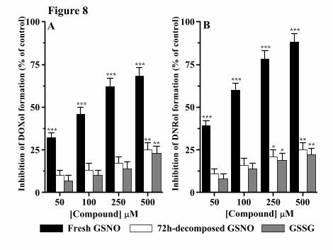
Figure 8
This article has not been copyedited and formatted. The final version may differ from this version.DMD Fast Forward. Published on August 11, 2015 as DOI: 10.1124/dmd.115.065110
at ASPE
T Journals on N
ovember 12, 2021
dmd.aspetjournals.org
Dow
nloaded from
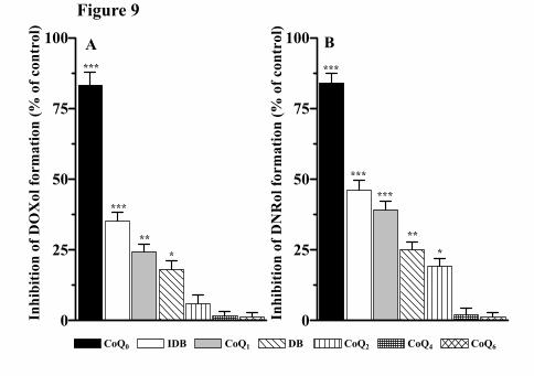
Figure 9
This article has not been copyedited and formatted. The final version may differ from this version.DMD Fast Forward. Published on August 11, 2015 as DOI: 10.1124/dmd.115.065110
at ASPE
T Journals on N
ovember 12, 2021
dmd.aspetjournals.org
Dow
nloaded from

Figure 10
This article has not been copyedited and formatted. The final version may differ from this version.DMD Fast Forward. Published on August 11, 2015 as DOI: 10.1124/dmd.115.065110
at ASPE
T Journals on N
ovember 12, 2021
dmd.aspetjournals.org
Dow
nloaded from