inter-cellular variation in dna content of entamoeba ... · inter-cellular variation in dna content...
TRANSCRIPT

Inter-Cellular Variation in DNA Content of Entamoebahistolytica Originates from Temporal and SpatialUncoupling of Cytokinesis from the Nuclear CycleChandrama Mukherjee.¤a, Shubhra Majumder.¤b, Anuradha Lohia*
Department of Biochemistry, Bose Institute, Kolkata, India
Abstract
Accumulation of multiple copies of the genome in a single nucleus and several nuclei in a single cell has previously beennoted in Entamoeba histolytica, contributing to the genetic heterogeneity of this unicellular eukaryote. In this study, wedemonstrate that this genetic heterogeneity is an inherent feature of the cell cycle of this organism. Chromosomesegregation occurs on a variety of novel microtubular assemblies including multi-polar spindles. Cytokinesis in E. histolyticais completed by the mechanical severing of a thin cytoplasmic bridge, either independently or with the help of neighboringcells. Importantly, cytokinesis is uncoupled from the nuclear division cycle, both temporally and spatially, leading to theformation of unequal daughter cells. Sorting of euploid and polyploid cells showed that each of these sub-populationsacquired heterogeneous DNA content upon further growth. Our study conclusively demonstrates that geneticheterogeneity originates from the unique mode of cell division events in this protist.
Citation: Mukherjee C, Majumder S, Lohia A (2009) Inter-Cellular Variation in DNA Content of Entamoeba histolytica Originates from Temporal and SpatialUncoupling of Cytokinesis from the Nuclear Cycle. PLoS Negl Trop Dis 3(4): e409. doi:10.1371/journal.pntd.0000409
Editor: Daniel Eichinger, New York University School of Medicine, United States of America
Received November 12, 2008; Accepted March 11, 2009; Published April 7, 2009
Copyright: � 2009 Mukherjee et al. This is an open-access article distributed under the terms of the Creative Commons Attribution License, which permitsunrestricted use, distribution, and reproduction in any medium, provided the original author and source are credited.
Funding: This study was supported by National Institutes of Health sponsored FIRCA sub-grant from Stanford University (Sub-award number 16846170-33918-A;Primary award number TW007421). The funders had no role in study design, data collection and analysis, decision to publish, or preparation of the manuscript.
Competing Interests: The authors have declared that no competing interests exist.
* E-mail: [email protected]
¤a Current address: Department of Molecular and Cellular Biochemistry, Ohio State University, Columbus, Ohio, United States of America¤b Current address: Department of Molecular Genetics, Ohio State University, Columbus, Ohio, United States of America
. These authors contributed equally to this work.
Introduction
Eukaryotic cells undergo two major events during proliferation-
the nuclear cycle, when the whole genome is duplicated and
segregated equally into two nuclei and cytokinesis, the physical
separation of a mother cell into two daughter cells. Sequential
progression of events during the cell cycle is enforced by
checkpoint proteins [1] that also ensure the spatial and temporal
coordination of mitosis with cytokinesis [2–4]. While this paradigm
is true for most organisms that have been studied, it is becoming
increasingly clear that diverse groups of eukaryotes, including
plants, ciliates, Drosophila, and protists, show striking differences in
regulating the transmission of genetic information during prolif-
eration [5]. Many of these organisms tolerate large variations in
DNA content during different life cycle stages such that typical cell
cycle checkpoints are altered. Entamoeba histolytica is one such
protist parasite that proliferates in the intestinal lumen of human
beings often causing severe disease in the host. Indeed this parasite
is responsible for millions of cases of dysentery and liver abscess
world wide especially in developing countries [6].
Axenically grown E. histolytica cells exhibit variations in DNA
content, which may vary several folds within a population [7].
Multiple genome complements may be present in a single nucleus
or distributed over multiple nuclei in a single cell [8,9]. Re-
duplication of the genome and de-linking of S-phase from
cytokinesis were identified as two of the reasons for the generation
of polyploidy in these trophozoites [8]. A significant number of E.
histolytica trophozoites with multiple nuclei were observed in
infected intestinal tissue suggesting this is an intrinsic property of
this parasite and not due to in vitro culture conditions [10].
Remarkably, under different growth conditions such as switching
from xenic to axenic growth leads to a significant increase in the
nuclear DNA content of E. histolytica trophozoites [10]. Similar
variation in nuclear DNA content was also observed in the related
reptilian parasite E. invadens during conversion of cysts to
trophozoites and vice versa [10]. These observations clearly
indicate the inherent plasticity of the Entamoeba genome [10] and
the ability of this protist to survive in the absence of strict
regulatory mechanisms that are a hallmark of the eukaryotic cell
cycle.
Most eukaryotes segregate their genomes once duplication is
complete. While more than 2n genome content accumulates in
some E. histolytica cells due to multiple rounds of DNA duplication,
it has not been clear how polyploid amoebae partition their
genomes. Bipolar microtubular spindles are not frequently visible
in E. histolytica [11]. Rather, several electron microscopic studies
have reported atypical organization of microtubules (MTs) during
nuclear division [12–14]. Thus, the possible cell-cycle mechanism
underlying the dynamic variation in DNA content of E. histolytica
cells during their axenic growth include- a) multiple cycles of
replication of the genome without nuclear or cell division, b)
nuclear division without cytokinesis and c) polyploidization from
www.plosntds.org 1 April 2009 | Volume 3 | Issue 4 | e409

over-replication of the genome followed by segregation on
multipolar mitotic spindles.
In this study, we demonstrate: a) the formation of atypical
microtubular structures during genome segregation; b) multiple
microtubule organizing centers (MTOCs) and multi-polar spindles
are formed in polyploid nuclei; c) cell division is spatially and
temporally uncoupled from the nuclear cycle; d) cell division can
be asymmetric, thereby producing either uni-nucleate, multi-
nucleate or anucleate daughter cells. In summary, asynchrony
between nuclear division and cytokinesis in a fraction of the
population combined with segregation of the polyploid genome on
multi-polar spindles accounts for the extreme variation of genomic
DNA content in individual E. histolytica cells in axenic culture.
Materials and Methods
Cell culture and maintenanceE. histolytica HM-1:IMSS trophozoites were maintained axeni-
cally and routinely sub-cultured every 48–72 h in TYI-S-33
medium containing 10% adult bovine serum at 37uC [15].
Cell synchronization by serum starvationE. histolytica HM-1:IMSS cells were sub-cultured every 24 h for
3–4 days followed by serum starvation [8]. Adult bovine serum
was added (10%) after 12–13 h of serum starvation. Cells were
then withdrawn at different times as indicated and fixed.
Scanning cytometry to determine nuclear DNA contentEthanol fixed cells were stained with 49-6-Diamidino-2-
phenylindole (DAPI, 0.1 mg/ml, Sigma) for 10 min, washed once
with 16 PBS and then scanned for DNA content of individual
nuclei [10] under a 406oil objective (numerical aperture 1.3) of a
Zeiss Axiovert 200 M fluorescence microscope fitted with the
MetaCyte scanning cytometer (Zeiss, Germany). A minimum of
2000 nuclei was scanned for each sample and analyzed by
Metafer4 software (Zeiss, Germany). The DAPI fluorescence
values (x-axis) were represented as histograms. The scan yields
varying numbers of nuclei with different fluorescence values. The
number of nuclei on the y-axis was represented as a fraction of the
highest number of nuclei obtained in any one sub-class of each
scan.
Immunofluorescence and confocal microscopyE. histolytica HM-1:IMSS cells were grown on coverslips in 24-
well plates at 37uC, fixed directly with warm 3.7% formaldehyde
for 15 min and permeabilized with 0.1% Triton X-100 for
10 min. Fixed cells were stained with polyclonal anti-Eh b-tubulin
antibody [11] followed by tetramethyl rhodamine isothiocyanate
(TRITC) conjugated anti-rabbit secondary antibody (1:200;
Jackson Laboratories, USA). For visualization of actin filaments,
cells were incubated with Alexa Fluor 488 conjugated phalloidin
for 30 min (Molecular Probes, Invitrogen, USA). Images were
acquired with (i) 636 Plan-Apochromat 1.4 oil differential
interference contrast (DIC) objective (numerical aperture 1.4) in
a Zeiss LSM 510 Meta confocal microscope equipped with a
488 nm argon laser and a 543-nm He/Ne laser and were analyzed
with the LSM Meta 510 software package (Zeiss, Germany) or (ii)
a 406oil objective (numerical aperture 1.3) in an Axiovert 200 M
fluorescence microscope using Z-stacking and analyzed by
deconvolution (Axiovision v4.6). DNA was stained with DAPI
(0.2 mg/ml, Sigma) for 30 min.
Real-time microscopyE. histolytica HM-1:IMSS trophozoites were plated on 35-mm
plastic culture dishes filled with growth medium at 37uC. After the
cells adhered, the medium was replaced with fresh growth
medium. The dish was kept inside an incubator (Tempcontrol
37-2 digital, Zeiss, Germany) at 37uC and under 5% CO2 flow
system (PeCon GmbH, Erbach, Germany) which was fitted to the
Axiovert 200 M fluorescence microscope (Zeiss, Germany). Cells
were visualized under a 206 phase contrast objective. The time-
lapse images were captured with 1 sec interval for the indicated
time, then analyzed and further processed by Axiovision v4.6
software (Zeiss, Germany).
Cell sortingAsynchronously growing E. histolytica HM-1:IMSS cells (16108
cells) were harvested 48 h after sub-culture, washed with 16PBS
and incubated with 1 mM DNA binding dye- Vybrant DyeCycle
Orange for 1 h (Molecular Probes, Invitrogen, USA). Cells were
passed through a 40 mm nylon mesh (Becton Dickinson, USA) to
remove aggregates and debris before sorting the cells. Vybrant
DyeCycle Orange stained cells were excited at 488 nm in a flow
cytometer (FACSAria, Becton Dickinson, USA) and emission was
measured through a 585/42 Band Pass filter for analyzing the
DNA content. On the basis of DNA content analysis (FACSDIVA
6.0 software, Becton Dickinson, USA), cells were demarcated in
four different electronic gates and sorted at 4uC. The sorted cells
were washed with 16 PBS and resuspended in 2 ml TYI-S-33
medium. Half the cells were inoculated into growth medium and
harvested after 3 days. The remaining cells were fixed in 70%
ethanol for analysis of the nuclear DNA content and number of
nuclei in each cell.
Results
Endo-replication and uncoupling of the nuclear divisioncycle from cell division are both seen in synchronizedcells
DNA synthesis of E. histolytica is arrested when cells are
incubated in serum free media for at least 12 h, followed by re-
initiation within 2 h after addition of serum [7,8]. Endo-
replication of the genome in the population can be seen following
this synchronization protocol [8]. We have now examined the fates
of individual nuclei following this synchronization. Progression of
S-phase was monitored by estimating the nuclear DNA content in
DAPI stained cells. Nuclear division was estimated from the
increase in bi-nucleated cells while cell division was estimated from
counting cell numbers. Our data show that after 12 h of serum
Author Summary
Proliferating eukaryotic cells regulate their DNA synthesis,chromosome segregation, and cell division with greatprecision so that daughter cells are genetically identical.Our study demonstrates that in proliferating cells of theprotist pathogen Entamoeba histolytica re-duplication ofDNA followed by segregation on atypical and diversemicrotubular structures is frequently observed. In thisparasite, cell division is erratic, so that each daughter cellmay contain one or more nuclei and sometimes no nuclei.This uncoupling of cell cycle events and survival ofdaughter cells with unequal DNA contents leads to geneticheterogeneity in E. histolytica. Our study highlights theinherent plasticity of the Entamoeba genome and theability of this protist to survive in the absence of strictregulatory mechanisms that are a hallmark of theeukaryotic cell cycle.
Genetic Heterogeneity in Amoeba
www.plosntds.org 2 April 2009 | Volume 3 | Issue 4 | e409

starvation, E. histolytica nuclei contain heterogeneous amounts of
DNA. 1 h after addition of serum the nuclei show a dramatic
homogenization and reduction of DNA content (Figure 1A). This
early homogenization likely results from nuclear division in
polyploid nuclei. An earlier report identified two copies of selected
loci using fluorescent in situ hybridization and estimated amoeba
to be diploid [16] while other studies have demonstrated that the
DNA content can vary several fold in proliferating amoebae so
that single cells may contain up to 10n or 12n [7,10]. We assigned
1n genome content to the lowest nuclear DNA content after
homogenization. Compared to nuclei 1 h after the addition of
serum (1n–2n), the average DNA content of E. histolytica nuclei
increased 2 fold in 80–90% of the nuclei between 2 h and 8 h after
addition of serum (Figure 1A). Between 8 h and 10 h after
addition of serum, the nuclear DNA content again showed
reduction and homogenization to a level similar to that observed
at 1 h after serum addition. This suggests completion of
chromosome segregation and nuclear division within 10 h. The
homogenous euploid nuclear DNA content at 1 h and 10 h after
serum addition marks the temporal boundaries of a single nuclear
cycle. The nuclear DNA content increased again after 10 h
suggesting that another cycle had been initiated. In each of these
cycles, 10–20% of the nuclei accumulated greater than 4n DNA
content (Figure 1A) suggesting that some nuclei undergo endo-
replication without nuclear division. This polyploid population
was absent at 1 h and 10 h after serum addition, suggesting that
either chromosome segregation occurs more than once in a single
nuclear cycle in the polyploid nuclei or the polyploid nuclei can
segregate multiple copies of the genome simultaneously.
In order to assess whether chromosome segregation was coupled
with nuclear division, we scored the number of microtubular
assemblies and bi-nucleated cells in conjunction with changes in
DNA content. Earlier studies have demonstrated that microtu-
bules were mostly nuclear [11,12,17]. After 12 h of serum
starvation, tubulin was dispersed in the nucleus without any
obvious structure in most cells. Microtubular assemblies or
structures began to appear 2 h after serum addition, continued
to increase in number up to 8 h, and then decreased markedly at
10 h (Figure 1B). Bi-nucleated cells were highest (,20%) at 10 h
after serum addition in these cells (Figure 1B). Thus for 20% of the
cells nuclear DNA replication was immediately followed by
segregation of the chromosomes into daughter nuclei.
Importantly, cell numbers increased gradually rather than in a
step-wise fashion coinciding with increase in nuclear number
(Figure S1A). While cell density affected the rate of increase in cell
number (Figure S1B), the temporal progression of the nuclear
division cycle was independent of these factors. This suggests that
although nuclear division is coupled to DNA synthesis and
chromosome segregation, cell division is random and not
obligately linked to nuclear division in these synchronized E.
histolytica cells.
Genome segregation may occur on monopolar, bipolaror multi-polar microtubular spindles in E. histolytica
During the course of a mitotic cycle, we observed different MT
assemblies and structures in the amoeba nucleus. Commonly, anti-
Eh b-tubulin antibody showed a diffuse nuclear stain and MT
structures were not visible in most nuclei. In some nuclei, a single
pole-like body was observed at the center (Figure 2A). Confocal
microscopic analyses clearly showed a central ring-like arrange-
ment of MTs with radially disposed short MT fibers (Figure 2A1).
It has been shown that Eh c-tubulin formed a similar ring-like
arrangement at the center of E. histolytica nuclei thus defining the
pole-like body as ‘MTOC’ in these cells [17]. During progression
of the nuclear cycle we identified a variety of MT spindle-like
assemblies that likely originated from the MTOC and radial MTs
described above. The arrangement of chromosomes on these MT
structures suggested that these structures were intermediates
formed during genome segregation. Figure 2B–E (corresponding
confocal images Figure 2D1 and 2E1) show MT fibres emanating
from a single pole that may be an extension of the central pole
seen in Figure 2A. Chromosomal DNA appears to have segregated
at an early stage after attachment to the central pole and then been
subsequently pushed apart at one end of the uni-directionally
extending MT fibers (Figure 2C–E). These structures were
observed in 25–30% of the spindle intermediates.
Figure 1. S-phase and chromosome segregation are coupled to nuclear division but the nuclear cycle is uncoupled from celldivision. (A) E. histolytica HM-1:IMSS cells were fixed after 12–13 h serum starvation and after serum re-addition (1 h–12 h). The nuclear DNAcontent was deduced from DAPI fluorescence intensities and shown as individual histograms (X-axis). Y-axis represents the number of nuclei as afraction of the highest number of nuclei obtained in any sub-class of each scan. Representative data from three independent experiments are shown.The nuclear DNA content profiles between 2 h to 4 h and 5 h to 9 h were similar and therefore only 4 h and 8 h have been shown. (B) Number of bi-nucleated cells (line) and the frequency of distinct microtubular assemblies (bar) were scored after serum starvation (0 h) and serum re-addition (2 h–12 h). At least 150–200 cells were analysed at each time point and an average of three independent experiments are shown with error barsindicating6S.D.doi:10.1371/journal.pntd.0000409.g001
Genetic Heterogeneity in Amoeba
www.plosntds.org 3 April 2009 | Volume 3 | Issue 4 | e409
Genetic Heterogeneity in Amoeba
www.plosntds.org 4 April 2009 | Volume 3 | Issue 4 | e409

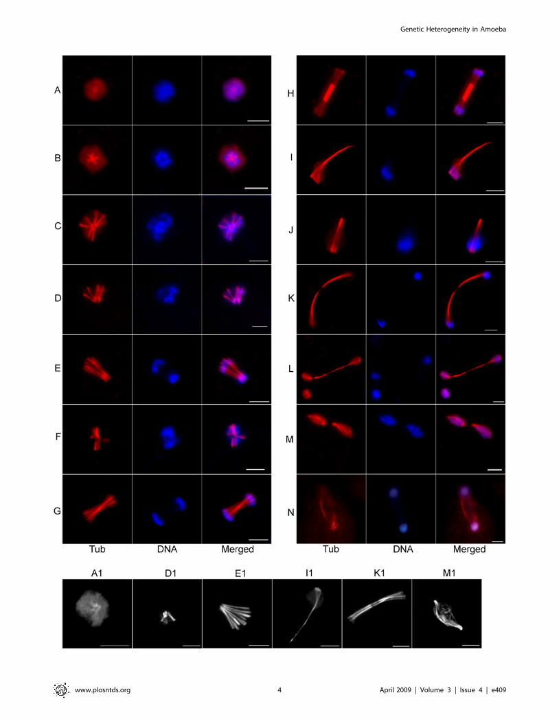
We also observed several non-conventional MT structures - a)
with DNA at the two ends of bi-directionally extending MT fibers
(Figure 2F–H) and b) with DNA bound at one end of several
radiating MT fibers that bundled together at the other end
(Figure 2I–J and 2I1). Segregation appears to be completed on
extended MT structures where the elongated MTs have disap-
peared from the centre (Figure 2K–L and 2K1). Bipolar spindle like
structures were also observed but at a low frequency (3–5% of the
MT structures) compared to the monopolar intermediates.
Chromosomal DNA was distributed longitudinally over the bipolar
spindle or at the two poles (Figure 2M–N and 2M1).
The most frequent structures visible at 2 h–4 h after serum
addition were the intermediates shown in Figure 2I–J (50–55% of
the MT structures). Based on the different MT structures observed
during progression of the nuclear cycle, it is likely that genome
segregation occurs either by extension of a radial monopolar
assembly (Figure 2C–E) or by lateral separation of MT fibers that
are joined at a distal end (Figure 2I and 2J). It is unclear whether
the MT structures shown in Figure 2F–H originated from
monopolar assemblies or duplication of a MTOC. Instead of
chromosomal segregation due to pole-ward contraction of MT
fibers, the MT structures in amoeba nuclei suggest either a)
separation of chromosomal DNA on the growing end of MTs, b)
lateral separation of duplicated chromosomes and MTs, or c)
longitudinal movement of chromosomes on elongated MTs.
Importantly, several nuclei with high DNA content were seen
that contained two, three or four MTOCs (Figure 3A). Bipolar
spindle intermediates likely originated from duplication of the
Figure 2. Diverse microtubular assemblies in E. histolytica. Synchronized E. histolytica HM-1:IMSS cells were stained with anti-Eh b-tubulinantibody and visualized under Axiovert 200 M flourescence microscope. Representative images of a radial assembly (A), variety of monopolar (B–L)and bipolar (M–N) microtubular assemblies are shown. Chromosomal DNA was stained with DAPI. In panel M, two bipolar MT spindle intermediateare seen in a single cell. Cells from this experiment were visualized under a laser scanning confocal microscope for finer details of the microtubularstructures. Comparable confocal images of the MT assemblies shown in panel A, D, E, I, K and M are shown as A1, D1, E1, I1, K1 and M1 respectively.Bar in each panel represents 5 mm.doi:10.1371/journal.pntd.0000409.g002
Figure 3. Multiple MTOCs in polyploid nuclei may lead to multi-polar spindles. (A) Anti-Eh b-tubulin antibody (red) and DAPI (blue) stainedE. histolytica HM-1:IMSS cells showing several nuclei with high DNA content (arrowhead) compared to other nuclei (a–d, ‘Merged’ panel,). Thepresence of two (a, b), three (c) and four (d) MTOC or poles (arrows) in these polyploid nuclei are shown by the staining with anti-Eh b-tubulinantibody with higher magnification (‘Tub’ panel). (B) Representative images of tri-polar (a, b), tetra-polar (c) and multi-polar (d) MT assemblies areshown. Bar represents 5 mm.doi:10.1371/journal.pntd.0000409.g003
Genetic Heterogeneity in Amoeba
www.plosntds.org 5 April 2009 | Volume 3 | Issue 4 | e409
central pole (Figure 3A-a, b). Interestingly, tri-polar, tetra-polar
and multi-polar spindles were also seen in several cells (Figure 3B).
It is possible that these multi-polar spindles originated from the
multiple MTOCs in polyploid nuclei and were used to segregate
multiple copies of the genome simultaneously. This could be one
of the mechanisms for the segregation of multiple genome copies
in polyploid nuclei during a single nuclear cycle.
In summary, E. histolytica uses novel MT structures for
chromosome segregation mostly originating from a single pole.
Multi-polar spindles are also observed and may account for the
rapid generation of DNA content heterogeneity from polyploid
cells. Another unique feature is the use of multiple modes of
segregation, as suggested by the diverse MT structural interme-
diates and inter-cellular variation in DNA content.
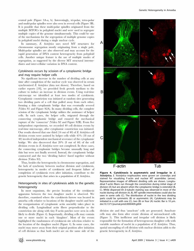
Cytokinesis occurs by scission of a cytoplasmic bridgeand may require helper cells
No significant increase in the number of dividing cells at any
time after completion of the nuclear cycle was observed in serum
synchronized E. histolytica (data not shown). Therefore, based on
earlier reports [18], we provided fresh growth medium to the
culture to induce an increase in division events. Using real-time
microscopy we identified at least two modes of cytokinesis.
Cytoplasmic constriction was initiated at random sites generating
two dividing parts of a cell that pulled away from each other,
forming a thin cytoplasmic bridge that was eventually severed
(Video S1 and Figure S2A). In many dividing cells, the complete
scission of the cytoplasmic bridge utilizes the assistance of helper
cells. In such cases, the helper cells, migrated through the
connecting cytoplasmic bridge and ensured the mechanical
rupture of the ‘connector’ (Video S2 and Figure S2B). From five
independent experiments, we recorded 40 cell division events by
real-time microscopy, after cytoplasmic constriction was initiated.
Our results showed that one third (14 out of 40) of E. histolytica cell
division events were assisted by helper cells while 45% (18 out of
40) involved independent mechanical severance of the cytoplasmic
bridge. We also discovered that 20% (8 out of 40) of the cell
division events in E. histolytica were not completed. In these cases,
the connecting cytoplasmic bridges became unusually long and
thin but were not finally severed. Instead, the cytoplasmic bridge
contracted and the two ‘dividing halves’ fused together without
division (Video S3).
Thus, besides the heterogeneity in chromosome segregation, and
the lack of synchrony between nuclear division and cytokinesis,
heterogeneity in modes of cytokinesis and frequent failure of
completion of cytokinesis even after initiation, contribute to the
genetic heterogeneity that arises in a population of E. histolytica.
Heterogeneity in sites of cytokinesis adds to the geneticheterogeneity
In most organisms, the precise location of the cytokinesis
apparatus between the two daughter nuclei is observed. We
therefore, examined where the cytokinesis furrow was located in
amoeba cells relative to locations of the daughter nuclei and how
the reorganization of cytoplasmic actin assembly takes place in
dividing cells. Longitudinal actin fibers perpendicular to the
constriction site along with tiny F-actin patches were seen in cells
likely to divide (Figure 4). Importantly, dividing cells may contain
one or more nuclei in each ‘daughter’. Most of the events
highlighted the randomness of cytokinesis site selection relative to
the location of the daughter nuclei (Figure 4). In some cases, the
nuclei may move away from their original position after initiation
of cell division so that both nuclei are on the same side of the
division site and thus ‘anucleate’ cells are generated. Anucleate
cells may also form after erratic division of uni-nucleated cells
(Figure 5). This inefficient and irregular cell division is likely
responsible for the formation of heterogeneous daughter cells with
variable numbers of nuclei in a population of E. histolytica. Thus,
spatial uncoupling of cell division with nuclear division adds to the
genetic heterogeneity in E. histolytica.
Figure 4. Cytokinesis is asymmetric and irregular in E.histolytica. E. histolytica trophozoites were grown on coverslips andstained for visualizing F-actin and nuclear MT assemblies (arrow).Representative images of different dividing cells are shown. Longitu-dinal F-actin fibers run through the constriction during initial stage ofdivision (A) but are absent when the cytoplasmic bridge is extended (B,C). While dispersed Eh b-tubulin staining was observed in most of thenuclei during cell division (A, B), MT spindle intermediates could also beseen in some cells (C). The distribution of nuclei in the two dividingparts could be symmetric (B) or asymmetric (A). Cytokinesis may beinitiated in a cell with one (C), two (B) or four (A) nuclei. Bar is 10 mm.doi:10.1371/journal.pntd.0000409.g004
Genetic Heterogeneity in Amoeba
www.plosntds.org 6 April 2009 | Volume 3 | Issue 4 | e409

Euploidy, polyploidy and variations in nuclear numberare inherent features of the E. histolytica cell cycle
In order to determine if the difference in DNA content was a
stable property of both euploid and polyploid cells, we separated
cells on the basis of their DNA content and followed the DNA
content of the progeny. Log phase E. histolytica trophozoites were
stained with the vital dye Vybrant orange and sub-populations of
live amoebae with varying DNA contents were isolated using a
fluorescence activated cell sorter. The fluorescence values
representing cellular DNA content profile of unsorted cells showed
a broad distribution (Figure 6A, a). Electronic gates (P1–P4) were
set (Figure 6A, a) and cells were separated according to differences
in DNA content. Figure 6A, b-e shows the cellular DNA content
profile of the different sub-populations that were recovered after
sorting. The sub-populations P1–P4 were analyzed for multi-
nucleated cells and nuclear DNA content and compared with the
unsorted cells. Table 1 shows that while the unsorted cells
consisted of approximately 92% uni-nucleated cells and 8% bi-
and multi-nucleated cells, sorted P1 and P2 cells were mostly uni-
nucleated (92–96%) with only a small percentage of bi- and multi-
nucleated cells (4–8%). On the other hand, P3 and P4 cells
contained a significantly higher number of bi- and multi-nucleated
cells (13–20%).
A comparison of the nuclear DNA content in the unsorted cells
showed a broad distribution of nuclear DNA content from 1n to 6n
while about 15–20% nuclei contained greater than 6n DNA content
(Figure 6B, left). P1 cells showed 1n–4n nuclear DNA content, P2
cells showed 4n–14n nuclear DNA content while P3 and P4 cells
showed 3n–14n nuclear DNA content. (Figure 6B, right). To
investigate whether the isolated sub-populations of P1–P4 cells
would retain their DNA content and nuclear number phenotype
after growth, the sorted cells were incubated in fresh medium at
37uC. Following an initial lag of 24–36 h (possibly due to the
physiological stress induced by cell sorting), the cell numbers
subsequently increased in all the tubes. After 72 h all the sorted sub-
populations had undergone at least 2 doublings (final cell density
,1–36105 cells per ml in each tube). It was observed that P1 cells
had a higher percentage of bi-and multi-nucleated cells while P4
cells showed a homogenous population of uni-nucleated cells after
growth (Table 1). Changes in the number of bi- and multi-nucleated
cells were observed in P2 and P3 cells also. After growth the P2–P4
populations showed a reduction in the number of 10n–14n nuclei
(Figure 6C). Therefore E. histolytica is programmed to continuously
generate genetic heterogeneity in both nuclear number per cell and
DNA content per nucleus, and this program is equally active in the
polyploid and the euploid cells.
Discussion
In this study, we have discovered that genetic heterogeneity in
E. histolytica cells results from multiple modes of genome
segregation during nuclear cycle, asynchrony between nuclear
Figure 5. Asymmetric and erratic cell division leads to the formation of ‘anucleate’ daughter cells. Time-lapse images recorded during acell division event in live E. histolytica labeled with the vital dye Hoechst 33342 are shown. Initiation of cytoplasmic bridge (arrowhead) in a uni-nucleated cell led to the formation of an anucleate daughter cell (arrow). Bar represents 20 mm.doi:10.1371/journal.pntd.0000409.g005
Genetic Heterogeneity in Amoeba
www.plosntds.org 7 April 2009 | Volume 3 | Issue 4 | e409

division and cytokinesis along with variation in nuclear DNA
content.
Different microtubular structures suggest variable andatypical modes of chromosome segregation in E.histolytica
Although recent studies have reported the infrequent occur-
rence of bipolar MT spindles [11,19] in E. histolytica cells,
metaphase-like equatorial alignment of condensed chromosomes
or kinetochores could not be identified in amoeba [12,14] while
anaphase and telophase were identified on the basis of nuclear
shape [13]. Indirect immuno-fluorescence showed nuclear micro-
tubular assemblies with fibers radiating from a central region in
most E. histolytica cells [17].
This study shows that MT structures in E. histolytica include
monopolar, bipolar and multi-polar spindles for the segregation of
chromosomal DNA (Figures 2 and 3). On the basis of our findings,
Figure 6. Euploidy, polyploidy and variations in nuclear number are inherent features of the E. histolytica cell cycle. (A) Cellular DNAcontent of 48 h grown live cells was analysed before sorting (a). Cells were sorted on the basis of increasing DNA content (P1–P4) and reanalyzed (b–e). (B) Ethanol fixed pre-sorted cells (left panel) and sorted sub-populations (P1–P4, right panel) were stained with DAPI to estimate the nuclear DNAcontent. The DAPI fluorescence intensities were normalized against the lowest value of P1 cells and are shown as individual histograms. Y-axisrepresents the number of nuclei as a fraction of the highest number of nuclei obtained in any sub-class of each scan. (C) Sorted sub-populations (P1–P4) of E. histolytica cells were grown in fresh TYI-S-33 medium. Fixed cells from the P1–P4 sub-populations were stained with DAPI and the nuclearDNA content measured. Fluorescence intensities for each set were normalized and shown as individual histograms. Y-axis represents the number ofnuclei as a fraction of the highest number of nuclei obtained in any sub-class of each scan. Representative data from three independent experimentsare shown.doi:10.1371/journal.pntd.0000409.g006
Genetic Heterogeneity in Amoeba
www.plosntds.org 8 April 2009 | Volume 3 | Issue 4 | e409

we propose a model (Figure 7) to explain possible modes of
chromosome partitioning on these different MT structures that
were identified during the nuclear cycle in axenically grown E.
histolytica cells. It is conceivable that some of the proposed
intermediate or observed MT structures are abortive. It is
important to note that a large number of proteins known to
regulate chromosome segregation and spindle assembly were not
encoded in the amoeba genome [20]. Absence of these regulatory
Table 1. Percentage of E. histolytica cells with one, two or greater than two nuclei in unsorted and sorted sub-populations of cells.
Sample % of uni-nucleated cells6S.D. % of bi-nucleated cells6S.D. % of multi-nucleated cells6S.D.
Unsorted 92.060.1 6.860.1 1.160.3
After sorting After growth After sorting After growth After sorting After growth
P1 95.560.5 85.061.0 3.061.0 12.560.5 0.460.3 2.260.8
P2 91.561.5 91.061.0 7.361.3 7.6560.6 0.8560.1 0.460.3
P3 87.060.1 91.560.5 10.760.6 7.760.3 1.760.4 0.560.1
P4 82.062.0 89.560.5 17.060.4 8.660.4 2.760.4 1.560.5
The number of nuclei per cell was counted from the ethanol-fixed and DAPI stained cells. At least, 500 cells were counted at each time to determine the percentage ofuni-nucleated, bi-nucleated and multi-nucleated (.2) cells. Average obtained from three independent experiments was shown with indicated standard deviation (S.D.).doi:10.1371/journal.pntd.0000409.t001
Figure 7. A model proposing multiple modes of chromosome segregation on diverse microtubular structures in E. histolytica. Basedon the organization of chromosomal DNA along the different MT assemblies visualized during the nuclear cycle of axenically grown E. histolyticatrophozoites (cell density ,1.5–36105 cells per ml), a model for multiple modes of chromosome segregation is proposed. The schematic diagramsare adapted from the microscopic images shown in Figures 2 and 3. During interphase (I) microtubular structures are not visible (a). At the earlieststage, a single MTOC or pole is formed at the centre of the nucleus (b). Duplicated chromosomes may attach to MT fibers emanating from this pole(c). As the MT fibers elongate they carry one set of chromosomes away from the pole to which the other set is attached (d, e). In another scenario, ifthe two sets of chromosomes are bound to the leading end of the MTs only, then segregation may occur by lateral movement of the MT fibers (f, g).A third possibility is visualized from the lateral segregation of bundles of MTs joined at one end (i). While (j) was not actually seen in our study, wepropose it as a likely intermediate between (i) and (l). Ultimately, the spindle intermediates arising from the monopolar assembly (h, k and l) maysegregate into two nuclei at the completion of mitosis (M). In the case of pole duplication (m), a bipolar spindle assembly may segregatechromosomes, except that unlike typical spindles, the chromosomes are arranged along the MT fibers rather than the equatorial plate at metaphase(n, o). In polyploid nuclei, tri-polar (r) or tetra-polar spindles (s) may originate from multiple spindle poles (p and q).doi:10.1371/journal.pntd.0000409.g007
Genetic Heterogeneity in Amoeba
www.plosntds.org 9 April 2009 | Volume 3 | Issue 4 | e409

functions may lead to the formation of unconventional MT
structures.
Cytokinesis is uncoupled from the nuclear duplicationcycle in E. histolytica
Cytokinetic processes in E. histolytica (this study) and E. invadens
[21] are similar. Both organisms depend upon mechanical rupture
of a cytoplasmic bridge that may occur independently or utilizes
assistance from helper cells. Motility and the consequent
mechanical force driven by actin polymerization in a polymorphic
cell may have been one of the earliest modes of cell division [22].
Assistance from helper cells likely results from ‘altruistic’ behaviour
of genetically related cells in a clonal population. The cytokinetic
process is imprecise and irregular in Entamoeba, unlike the finely
regulated cell division in bacteria, yeasts and higher eukaryotes.
While endogenous Entamoeba proteins may affect the rate of cell
division as deduced from increased multi-nucleation in different
mutants [23–26], this event appears to be largely controlled by
extra-cellular factors. Random selection of cell division sites
coupled to a poorly controlled mechanical separation can lead to
the formation of anucleate and multi-nucleated daughter cells.
Similar cytokinetic events are observed in the social amoeba D.
discoideum which shows variations in its mode of cell division. Multi-
nucleated Myosin II-null D. discoideum cells, when allowed to
adhere on substrate, undergo a cell cycle-uncoupled, inefficient
cytofission [27,28].
The nuclear DNA content is not strictly regulated in E.histolytica
The nuclear cycle was clearly defined between initiation of
DNA synthesis and completion of nuclear division. That a
significant number of nuclei continued accumulation of DNA
after doubling their DNA content emphasizes the absence of
stringent regulatory mechanisms preventing re-replication that are
seen in other eukaryotes. Heterogeneity of MT structures also
suggests a lack of strict control of genome segregation mechanisms.
It may be noted that even after serum synchronization, the
maximal number of nuclei with MT structures and bi-nucleated
cells were only around 20%. Treatment of E. histolytica cells with
aphidicolin or taxol did not lead to a significant increase of nuclei
in different mitotic stages (data not shown). Thus the entire nuclear
cycle appears to be poorly controlled, resulting in leaky
phenotypes that are apparent in polyploid nuclei. In spite of leaky
regulatory mechanisms, S-phase, genome segregation and nuclear
division are temporally coupled. Multiple MTOCs and multi-
polar MT spindles possibly facilitate a return towards euploidy for
polyploid nuclei.
Strict regulation of the cell division cycle has been considered to
be the very basis of survival for eukaryotes. Indeed an unregulated
cell cycle leads to growth arrest, aneuploidy and tumorigenesis in
yeasts and higher eukaryotes. While prokaryotes use overlapping
parallel processes of DNA synthesis, segregation and cell division,
eukaryotes ensure completion of the preceding stage before
initiation of the next, with tight surveillance to ensure the same.
Clearly Entamoeba and few other organisms can bypass these
surveillance mechanisms and continue their life cycles. How does
Entamoeba preserve its genetic composition in the absence of cell
cycle regulation? The answer possibly lies in its survival as a
parasite, dependant on its surroundings, where plasticity and
heterogeneity are favored over precision and homogeneity.
Supporting Information
Figure S1 Effect of cell density on the growth rate of E. histolytica
trophozoites. (A) Cell number increases continuously rather than
in a step-wise fashion. Cell number was counted at different time
points (0–12 h) after serum starvation and addition and plotted.
Error bars indicate6S.D. (n = 3). (B) Growth rate is dependant on
cell density. Log phase E. histolytica HM-1:IMSS cells were
inoculated in TYI-S-33 medium at different cell densities-
16104, 56104 and 106104 cells/ml. Subsequent growth of these
cells after 20 and 40 h is shown graphically. Error bars
indicate6S.D. (n = 3).
Found at: doi:10.1371/journal.pntd.0000409.s001 (0.30 MB TIF)
Figure S2 Two modes of cell division in E. histolytica. Log phase
E. histolytica HM-1:IMSS cells were incubated with in fresh
medium at 37uC to induce cell division events. Cytokinesis was
visualized under a 206 phase contrast objective of the Axiovert
200 M microscope and time-lapse images were captured at 1 sec
intervals. During cell division, cytoplasmic constriction led to the
extension of a cytoplasmic bridge (arrowhead) between two
dividing halves. This bridge could either (A) rupture independently
or (B) with the assistance of a helper or midwife cell (star). Arrows
show ends of the severed cytoplasmic bridge. Bar represents
20 mm.
Found at: doi:10.1371/journal.pntd.0000409.s002 (3.47 MB TIF)
Video S1 Mechanical rupture of cytoplasmic extension leads to
cell division in E. histolytica.
Found at: doi:10.1371/journal.pntd.0000409.s003 (3.75 MB
MOV)
Video S2 Helper cell assisted cell division in E. histolytica.
Found at: doi:10.1371/journal.pntd.0000409.s004 (0.39 MB
MOV)
Video S3 Aborted cytokinesis in E. histolytica.
Found at: doi:10.1371/journal.pntd.0000409.s005 (9.98 MB
MOV)
Acknowledgments
We thank Profs. Anindya Dutta (University of Virginia, USA) and Graham
Clark (LSHTM, UK) for critically reading the manuscript. Confocal
microscopy was carried out at the Facility for Genomics and Proteomics at
Bose Institute, sponsored by IRHPA, DST. The authors thank Mr. Dipak
Manna for his support in real time microscopy.
Author Contributions
Conceived and designed the experiments: CM SM AL. Performed the
experiments: CM SM. Analyzed the data: CM SM AL. Contributed
reagents/materials/analysis tools: CM SM AL. Wrote the paper: CM SM
AL.
References
1. Hartwell LH, Weinert TA (1989) Checkpoints: controls that ensure the order of
cell cycle events. Science 246: 629–634.2. Muhua L, Adames NR, Murphy MD, Shields CR, Cooper JA (1998) A
cytokinesis checkpoint requiring the yeast homologue of an APC-binding
protein. Nature 393: 487–491.
3. Balasubramanian MK, McCollum D, Surana U (2000) Tying the knot:
linking cytokinesis to the nuclear cycle. J Cell Sci 113: 1503–1513.
4. Liu J, Wang H, Balasubramanian MK (2000) A checkpoint that monitors
cytokinesis in Schizosaccharomyces pombe. J Cell Sci 113: 1223–1230.
Genetic Heterogeneity in Amoeba
www.plosntds.org 10 April 2009 | Volume 3 | Issue 4 | e409

5. Parfrey LW, Lahr DJ, Katz LA (2008) The dynamic nature of eukaryotic
genomes. Mol Biol Evol 25: 787–794.6. Anonymous (1997) WHO/PAHO/UNESCO report. A consultation with
experts on amoebiasis. Mexico City, Mexico, 28–29 January, 1997. Epidemiol
Bull 18: 13–14.7. Gangopadhyay SS, Ray SS, Kennady K, Pande G, Lohia A (1997)
Heterogeneity of DNA content and expression of cell cycle genes in axenicallygrowing Entamoeba histolytica HM1:IMSS clone A. Mol Biochem Parasitol 90:
9–20.
8. Das S, Lohia A (2002) De-linking of S phase and cytokinesis in the protozoanparasite Entamoeba histolytica. Cell Microbiol 4: 55–60.
9. Lohia A, Mukherjee C, Majumder S, Dastidar PG (2007) Genome re-duplication and irregular segregation occur during the cell cycle of Entamoeba
histolytica. Biosci Rep 27: 373–384.10. Mukherjee C, Clark CG, Lohia A (2008) Entamoeba shows reversible variation
in ploidy under different growth conditions and between life cycle phases. PLoS
Negl Trop Dis 2: e281. doi:10.1371/journal.pntd.0000281.11. Dastidar PG, Majumder S, Lohia A (2007) Eh Klp5 is a divergent member of
the kinesin 5 family that regulates genome content and microtubular assembly inEntamoeba histolytica. Cell Microbiol 9: 316–328.
12. Orozco E, Solis FJ, Dominguez J, Chavez B, Hernandez F (1988) Entamoeba
histolytica: cell cycle and nuclear division. Exp Parasitol 67: 85–95.13. Solis FJ, Barrios R (1991) Entamoeba histolytica: microtubule movement during
mitosis. Exp Parasitol 73: 276–284.14. Chavez-Munguia B, Tsutsumi V, Martinez-Palomo A (2006) Entamoeba histolytica:
ultrastructure of the chromosomes and the mitotic spindle. Exp Parasitol 114:235–239.
15. Diamond LS, Harlow DR, Cunnick CC (1978) A new medium for the axenic
cultivation of Entamoeba histolytica and other Entamoeba. Trans R Soc Trop MedHyg 72: 431–432.
16. Ghosh S, Frisardi M, Ramirez-Avila L, Descoteaux S, Sturm-Ramirez K, et al.(2000) Molecular epidemiology of Entamoeba spp.: Evidence of a bottleneck
(demographic sweep) and transcontinental spread of diploid parasites. J Clin
Microbiol 38: 3815–3821.
17. Vayssie L, Vargas M, Weber C, Guillen N (2004) Double-stranded RNA
mediates homology-dependent gene silencing of gamma-tubulin in the human
parasite Entamoeba histolytica. Mol Biochem Parasitol 138: 21–28.
18. Chavez-Munguia B, Talamas-Rohana P, Rios A, Gonzalez-Lazaro M, Marti-
nez-Palomo A (2008) Entamoeba histolytica: fibrilar aggregates in dividing
trophozoites. Exp Parasitol 118: 280–284.
19. Dastidar PG, Lohia A (2008) Bipolar spindle frequency and genome content are
inversely regulated by the activity of two N-type kinesins in Entamoeba histolytica.
Cell Microbiol 10: 1559–1571.
20. Clark CG, Alsmark UC, Tazreiter M, Saito-Nakano Y, Ali V, et al. (2007)
Structure and content of the Entamoeba histolytica genome. Adv Parasitol 65:
51–190.
21. Biron D, Libros P, Sagi D, Mirelman D, Moses E (2001) Asexual reproduction:
‘midwives’ assist dividing amoebae. Nature 410: 430.
22. Mitchison TJ (1995) Evolution of a dynamic cytoskeleton. Philos Trans R Soc
Lond B Biol Sci 349: 299–304.
23. Guillen N, Boquet P, Sansonetti P (1998) The small GTP-binding protein RacG
regulates uroid formation in the protozoan parasite Entamoeba histolytica. J Cell Sci
111: 1729–1739.
24. Welter BH, Powell RR, Leo M, Smith CM, Temesvari LA (2005) A unique Rab
GTPase, EhRabA, is involved in motility and polarization of Entamoeba histolytica
cells. Mol Biochem Parasitol 140: 161–173.
25. Arias-Romero LE, de Jesus Almaraz-Barrera M, Diaz-Valencia JD, Rojo-
Dominguez A, Hernandez-Rivas R, et al. (2006) EhPAK2, a novel p21-activated
kinase, is required for collagen invasion and capping in Entamoeba histolytica. Mol
Biochem Parasitol 149: 17–26.
26. Majumder S, Lohia A (2008) Entamoeba histolytica encodes unique formins, a
subset of which regulates DNA content and cell division. Infect Immun 76:
2368–2378.
27. Uyeda TQ, Nagasaki A (2004) Variations on a theme: the many modes of
cytokinesis. Curr Opin Cell Biol 16: 55–60.
28. Nagasaki A, Uyeda TQ (2008) Chemotaxis-mediated scission contributes to
efficient cytokinesis in Dictyostelium. Cell Motil Cytoskeleton 65: 896–903.
Genetic Heterogeneity in Amoeba
www.plosntds.org 11 April 2009 | Volume 3 | Issue 4 | e409