interstitial lung disease abnormal deposition of collagen ... · [email protected]...
TRANSCRIPT
INTERSTITIAL LUNG DISEASE
Abnormal deposition of collagen/elastic vascular fibres andprognostic significance in idiopathic interstitial pneumoniasEdwin Roger Parra, Ronaldo Adib Kairalla, Carlos Roberto Ribeiro de Carvalho, Vera LuizaCapelozzi. . . . . . . . . . . . . . . . . . . . . . . . . . . . . . . . . . . . . . . . . . . . . . . . . . . . . . . . . . . . . . . . . . . . . . . . . . . . . . . . . . . . . . . . . . . . . . . . . . . . . . . . . . . . . . . . . . . . . . . . . . . . . . . . . . .
See end of article forauthors’ affiliations. . . . . . . . . . . . . . . . . . . . . . . .
Correspondence to:V L Capelozzi, Departmentof Pathology, Sao PauloMedical School, Universityof Sao Paulo, Dr ArnaldoAvenue 455, Sao Paulo01246-903, Brazil;[email protected]
Received 20 March 2006Accepted 2 November 2006. . . . . . . . . . . . . . . . . . . . . . . .
Thorax 2007;62:428–437. doi: 10.1136/thx.2006.062687
Background: Vascular remodelling has recently been shown to be a promising pathogenetic indicator inidiopathic interstitial pneumonias (IIPs).Aim: To validate the importance of the collagen/elastic system in vascular remodelling and to study therelationships between the collagen/elastic system, survival and the major histological patterns of IIPs.Methods: Collagen/elastic system fibres were studied in 25 patients with acute interstitial pneumonia/diffusealveolar damage, 22 with non-specific interstitial pneumonia/non-specific interstitial pneumonia and 55 withidiopathic pulmonary fibrosis/usual interstitial pneumonia. The Picrosirius polarisation method and Weigert’sresorcin–fuchsin histochemistry and morphometric analysis were used to evaluate the amount of vascularcollagen/elastic system fibres and their association with the histological pattern of IIPs. The associationbetween vascular remodelling and the degree of parenchymal fibrosis in usual interstitial pneumonia (UIP)was also considered.Results: The vascular measurement of collagen/elastic fibres was significantly higher in UIP than in the lungsof controls, and in those with diffuse alveolar damage and those with non-specific interstitial pneumonia. Inaddition, the increment of collagen/elastic fibres in UIP varied according to the degree and activity of theparenchymal fibrosis. The most important predictors of survival in UIP were vascular remodellingclassification and vascular collagen deposition.Conclusion: A progressive vascular fibroelastosis occurs in IIP histological patterns, probably indicatingevolutionarily adapted responses to parenchymal injury. The vascular remodelling classification and theincrease in vascular collagen were related to survival in IIP and possibly play a role in its pathogenesis.Further studies are needed to determine whether this relationship is causal or consequential.
The vascular extracellular matrix (VECM) consists ofcollagens, elastin, fibrillins, proteoglycans and others.1
Collagen fibres are the most abundant component of thevascular pulmonary wall and are found in all three tunicae,especially around smooth muscle cells of the tunica mediawhere they provide the necessary mechanical strength andcontractility. Collagen fibres are also found in the outer layer(adventitia) where they form large bundles of fibrils whichincrease progressively in size from its innermost component,closest to the media, to its outermost aspect.2 3 The elasticsystem, a major component of the pulmonary arteries, plays animportant role in wall elasticity, facilitated by concentricfenestrated lamellae of elastic fibres in the tunica media layer.1
The accumulation of ECM is an important process inpulmonary vascular structural remodelling.4 5 Elastosis hasbeen well studied in animal models of pulmonary fibrosis andhas been shown to be transcriptionally regulated by theaugmentation of lytic enzymes.6 Studies previously conductedby our group showed that lung collagen and elastic fibrecontent are increased in both acute and chronic interstitial lungdiseases, suggesting that significant remodelling of alveolartissue occurs in both conditions.7–11 We also found evidence ofvascular remodelling in lung biopsy specimens from patientswith usual interstitial pneumonia (UIP); more specifically, adirect relationship between vascular regression—characterisedby a progressive reduction in the internal area and internalperimeter, as well as by an increase in the wall thickness ofmedium or large lung vessels—and parenchymal remodelling.12
This finding is in agreement with previous reports of vesselablation in areas of honeycomb lung, regardless of the cause ofthe pulmonary fibrosis.13–16 Experimental studies have shown
that collagen and elastic fibre deposition might be importantfor understanding the alterations in vascular remodelling,17 18
but there has been uncertainty about the changes in thecollagen/elastic system in lung vessels of patients withidiopathic interstitial pneumonias (IIPs).
We hypothesise that the collagen/elastic system in vascularremodelling differs according to the adaptive responses toinjury that occur in acute interstitial pneumonia/diffusealveolar damage (AIP/DAD), non-specific interstitial pneumo-nia (NSIP)/NSIP and idiopathic pulmonary fibrosis (IPF/)/UIP.
This study was designed to measure the vascular collagen/elastic system in different adaptive responses to injury thatoccur in AIP/DAD, NSIP/NSIP and IPF/UIP, as well as theirassociation with survival.
METHODSPatient selectionPulmonary specimens were obtained by surgical lung biopsyfrom 109 patients, 25 with AIP/DAD, 22 with NSIP/NSIP and 55with IPF/UIP, according to the criteria outlined in the AmericanThoracic Society/European Respiratory Society internationalmultidisciplinary consensus classification of the IIPs.19 Onlyspecimens from patients who fulfilled these consensus criteriawere included.
Abbreviations: AIP, acute interstitial pneumonia; DAD, diffuse alveolardamage; ECM, extracellular matrix; FEV1, forced expiratory volume in 1 s;FVC, forced vital capacity; HRCT, high-resolution computed tomography;IIP, idiopathic interstitial pneumonia; IPF, idiopathic pulmonary fibrosis;NSIP, non-specific interstitial pneumonia; UIP, usual interstitial pneumonia;VECM, vascular extracellular matrix
428
www.thoraxjnl.com
on April 20, 2020 by guest. P
rotected by copyright.http://thorax.bm
j.com/
Thorax: first published as 10.1136/thx.2006.062687 on 24 January 2007. D
ownloaded from

Specimens of any other possible aetiology (eg, pneumoco-niosis) and/or with histological features suggestive of analternative diagnosis (eg, eosinophilic pneumonia) wereexcluded, as were biopsy specimens obtained from patientswith a concomitant systemic disease (eg, collagen vasculardisease), extensive honeycomb changes (end-stage lung dis-ease), a dual histological pattern (two different patterns at twodifferent biopsy sites), and/or morphological features notconsistent with a specific histological pattern (eg, broncho-centric distribution in an otherwise classic case of UIP). Afterexcluding specimens with histological and clinical evidence ofdesquamative interstitial pneumonia, lymphoid interstitialpneumonia or respiratory bronchiolitis, all patients includedexhibited clinical, radiological and physiological changesconsistent with AIP, NSIP or IPF and had been given thedefinitive pathological diagnosis of DAD, NSIP or UIP. Two orthree biopsy specimens per patient were sampled and the tissuespecimens collected according to the high-resolution computedtomography (HRCT) pattern, which usually includes normal,intermediate and more affected areas in different parts of thelung. Thus, the diagnosis of our patients with IIPs was obtainedby clinical, radiological and histological consensus criteria.
Baseline characteristicsThe median age of patients with AIP (13 men, 12 women) was51.7 years (range 35–74); 50.8 years (range 39–76) for NSIP(10 men, 12 women) and 65.3 years (range 50–84) for IPF (35men, 20 women).
A baseline assessment of severity of dyspnoea was madeusing the Level of Dyspnoea Scale20 (table 1).
Physiological testingThe pulmonary function tests included forced expiratoryvolume in 1 s (FEV1), forced vital capacity (FVC), FEV1/FVC
ratio6100, total lung capacity (TLC), residual volume andcarbon monoxide transfer factor (TLCO). TLC, residual volumeand residual volume/TLC percentages were measured by thehelium-dilution method with a Master Screen Apparatus (ErichJaeger GmbH, Wiirzburg, Germany), TLCO and TLCO/alveolarvolume by the single breath-holding helium-dilution method.21
Lung function measurements (table 1) were expressed aspercentages of predicted values. In all patients, the arterial PaO2
and PaCO2 were also measured at rest. The cardiac parametersof these patients were normal.
High-resolution computed tomographyHRCT examinations were performed using 1 or 1.5 mm-thicksections taken at 1 cm intervals throughout the entire lungduring inspirations in the supine position and through thecaudal 10 cm of the lungs at 2–3 cm increments in the proneposition. Two thoracic clinical radiologists prospectively andindependently scored all lobes or HRCT scans for ground-glassopacity (CT alveolar) and interstitial opacity (CT interstitial) ona scale of 0–5; the mean score for each lobe and for the entirelung was calculated22 (table 1).
Clinical scoringOverall clinical severity was assessed using the previouslydeveloped Clinical and HRCT Examinations Composite Score.20
The total clinical and HRCT score ranges from 0 to 100 points(100 being the most severe disease) depending on variablesincluding the Level of Dyspnoea Scale, HRCT and pulmonaryfunction test results (table 1).
Pathological review of the specimensPulmonary specimens were reviewed by three pathologistsblinded to their clinical features. Each specimen was assigneda histological diagnosis according to the criteria outlined inthe American Thoracic Society/European Respiratory Societymultidisciplinary consensus classification of the IIPs.19 DADwas characterised by involvement and a uniform temporalappearance caused by alveolar collapse, obliterative fibrosis,neosepta formation and moderately organising fibrosis.19
NSIP was characterised by temporally homogenous septalinflammatory thickening and minimal organising fibrosis.19
UIP was defined by alternating areas of normal parenchyma,alveolar collapse, honeycombing and severe organising fib-rosis, defined as sites of active remodelling overlying fibrousairspace walls and thus showing temporal heterogeneity, oroverlying normal rigid pulmonary structures (eg, interlobularsepta) in the form of fibroblast foci and granulation tissue.19
Table 1 shows the demographic data and radiological char-acteristics.
As a control, normal lung tissues from non-pneumonia andnon-emphysematous areas were obtained from five individuals(mean (SD) age 60 (3.6) years) who had died from violentcauses. None of these controls met any of the histologicalcriteria for IIP.
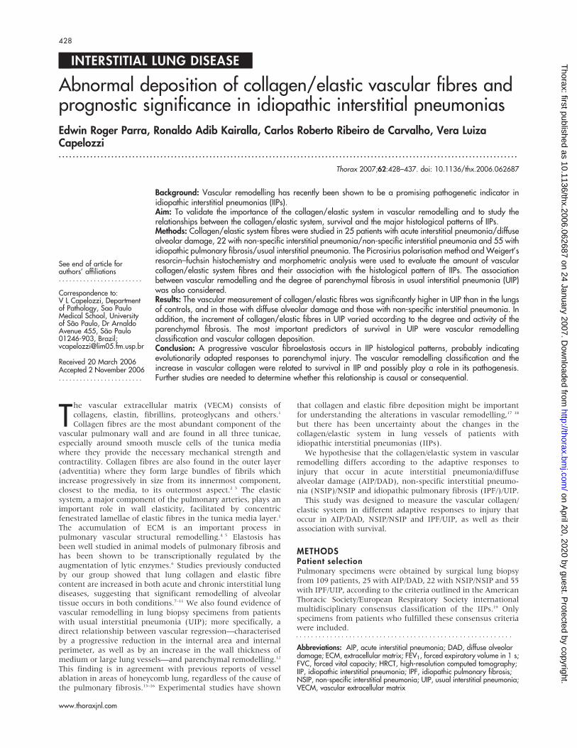
Morphological studyParenchymal remodelling definit ionParenchymal remodelling in UIP was evaluated by semiquanti-tative analysis for alternating areas of (1) minimal fibrosis(fig 1), defined as alveolar collapse with relatively unaffectedlung tissue or with mild interstitial thickening by fibrosis; (2)moderate fibrosis (fig 1), defined by intermediate organisingfibrosis of the wall with fibroblast foci; and (3) severe fibrosis(fig 1), defined as severe organising fibrosis of the wall withhoneycombing and foci of actively proliferating fibroblasts andmyofibroblasts.12
Table 1 Baseline characteristics of patients in the majorhistopathological categories
NSIP AIP IPF
Age at biopsy (years) 50.8 (16.6) 51.7 (14.9) 65.3 (7.4)Sex (F/M) 12/10 13/12 20/35
DyspnoeaLOD 5 (4) 14 (5) 10 (4)SpirometryFEV1 (% pr) 58 (6) 60 (3) 81 (5)FVC (% pr) 54 (5) 51 (3) 71 (4)FEV1/FVC 104 (7) 97 (81) 80 (6)TLC (% pr) 63 (5) 57 (8) 79 (4)RV (% pr) 94 (17) 64 (15) 93 (11)RV/TLC (% pr) 155 (16) 93 (23) 40 (3)TLCO (% pr) 43 (14) 46 (9) 60 (7)TLCO/VA (% pr) 97 (2) 83 (5) 63 (7)PaO2 (kPa) 8.5 (0.8) 10.4 (2.4) 7.9 (0.3)PaCO2 (kPa) 4.7 (0.1) 6.5 (0.7) 4.4 (1.1)
HRCT ScoreAlveolar 1.6 (1.1) 1.6 (0.9) 1.87 (0.9)Interstitial 0.87 (0.61) 0.73 (0.52) 1.9(0.56)Total 2.47 (0.85) 2.33 (0.71) 3.77 (0.73)
Total CS 33 (20) 42 (23) 53 (16)
AIP, acute interstitial pneumonia; CS, clinical score; F, female; FEV1, forcedexpiratory volume in 1 s; FVC, forced vital capacity; HRCT, high-resolutioncomputed tomography; IPF, idiopathic pulmonary fibrosis; LOD, level ofdyspnoea; M, male; NSIP, non-specific interstitial pneumonia; PaCO2,partial arterial pressure of carbon dioxide; PaO2, partial arterial pressure ofoxygen; RV, residual volume; pr, predicted; TLC, total lung capacity; TLCO,carbon monoxide transfer factor; VA, alveolar volume.Data are presented as means (SEM).
Collagen/elastic vascular fibres in IIPs 429
www.thoraxjnl.com
on April 20, 2020 by guest. P
rotected by copyright.http://thorax.bm
j.com/
Thorax: first published as 10.1136/thx.2006.062687 on 24 January 2007. D
ownloaded from
Vascular remodelling definit ionTo study the possibility of an important vascular contribution tothe fibrosis, we distinguished between vessels within thedifferent areas of fibrosis and compared vessels from severelyfibrotic areas with those in less fibrotic areas. In particular,vascular changes in relatively spared areas of patients withsevere fibrosis were compared with similar areas in patientswith less severe fibrosis to make a distinction betweenadventitia and surrounding parenchyma in patients withincreasing fibrosis.
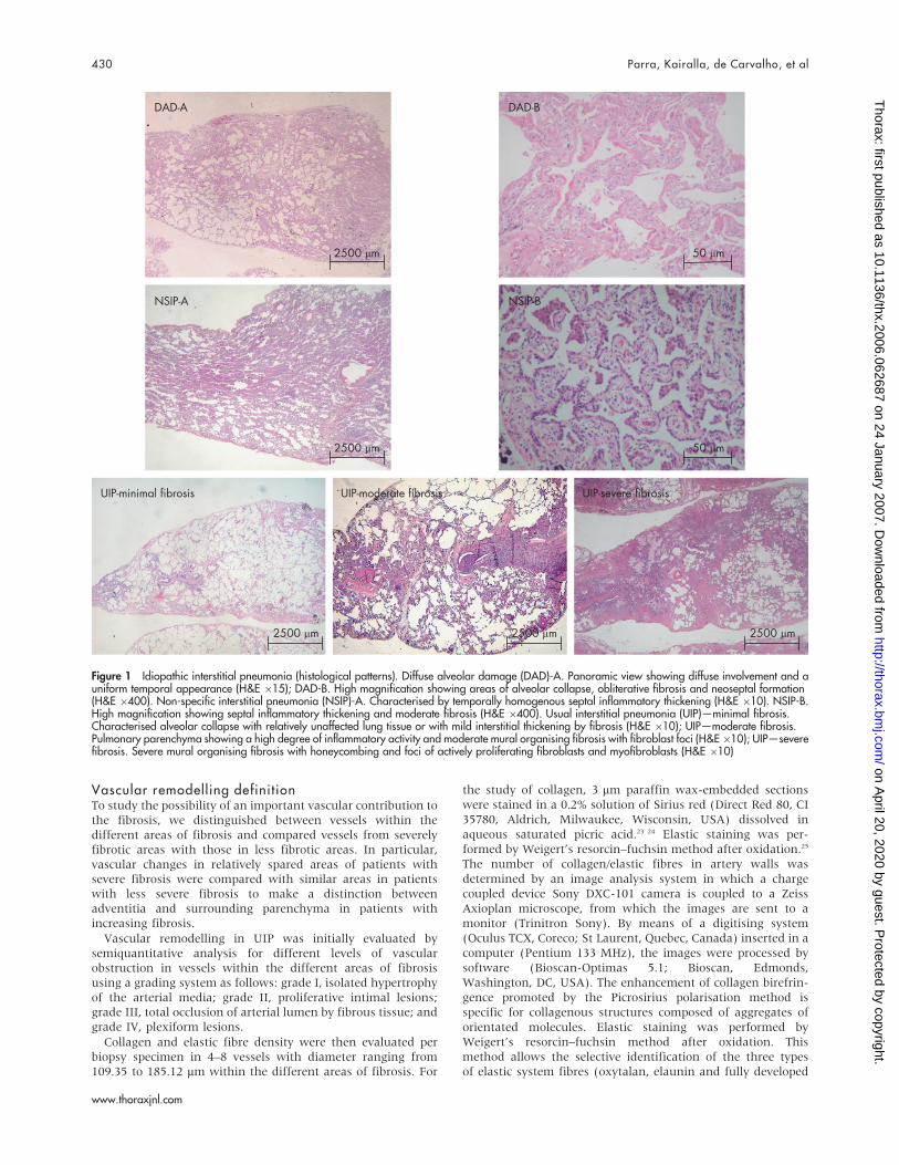
Vascular remodelling in UIP was initially evaluated bysemiquantitative analysis for different levels of vascularobstruction in vessels within the different areas of fibrosisusing a grading system as follows: grade I, isolated hypertrophyof the arterial media; grade II, proliferative intimal lesions;grade III, total occlusion of arterial lumen by fibrous tissue; andgrade IV, plexiform lesions.
Collagen and elastic fibre density were then evaluated perbiopsy specimen in 4–8 vessels with diameter ranging from109.35 to 185.12 mm within the different areas of fibrosis. For
the study of collagen, 3 mm paraffin wax-embedded sectionswere stained in a 0.2% solution of Sirius red (Direct Red 80, CI35780, Aldrich, Milwaukee, Wisconsin, USA) dissolved inaqueous saturated picric acid.23 24 Elastic staining was per-formed by Weigert’s resorcin–fuchsin method after oxidation.25
The number of collagen/elastic fibres in artery walls wasdetermined by an image analysis system in which a chargecoupled device Sony DXC-101 camera is coupled to a ZeissAxioplan microscope, from which the images are sent to amonitor (Trinitron Sony). By means of a digitising system(Oculus TCX, Coreco; St Laurent, Quebec, Canada) inserted in acomputer (Pentium 133 MHz), the images were processed bysoftware (Bioscan-Optimas 5.1; Bioscan, Edmonds,Washington, DC, USA). The enhancement of collagen birefrin-gence promoted by the Picrosirius polarisation method isspecific for collagenous structures composed of aggregates oforientated molecules. Elastic staining was performed byWeigert’s resorcin–fuchsin method after oxidation. Thismethod allows the selective identification of the three typesof elastic system fibres (oxytalan, elaunin and fully developed
Figure 1 Idiopathic interstitial pneumonia (histological patterns). Diffuse alveolar damage (DAD)-A. Panoramic view showing diffuse involvement and auniform temporal appearance (H&E 615); DAD-B. High magnification showing areas of alveolar collapse, obliterative fibrosis and neoseptal formation(H&E 6400). Non-specific interstitial pneumonia (NSIP)-A. Characterised by temporally homogenous septal inflammatory thickening (H&E 610). NSIP-B.High magnification showing septal inflammatory thickening and moderate fibrosis (H&E 6400). Usual interstitial pneumonia (UIP)—minimal fibrosis.Characterised alveolar collapse with relatively unaffected lung tissue or with mild interstitial thickening by fibrosis (H&E 610); UIP—moderate fibrosis.Pulmonary parenchyma showing a high degree of inflammatory activity and moderate mural organising fibrosis with fibroblast foci (H&E610); UIP—severefibrosis. Severe mural organising fibrosis with honeycombing and foci of actively proliferating fibroblasts and myofibroblasts (H&E 610)
430 Parra, Kairalla, de Carvalho, et al
www.thoraxjnl.com
on April 20, 2020 by guest. P
rotected by copyright.http://thorax.bm
j.com/
Thorax: first published as 10.1136/thx.2006.062687 on 24 January 2007. D
ownloaded from
elastic fibres). The thresholds for fibres of the collagenous andelastic systems were established for each slide after enhancingthe contrast up to a point at which the fibres were easilyidentified as black (elastic) or birefringent (collagen) bands.The area occupied by the fibres was determined by digitaldensitometric reconiting, by adjusting the threshold level ofmeasurement up to the grey density of the fibres of thecollagenous and elastic systems. The collagen of the mediallayer and the elastic fibre content were measured in eachvascular wall and expressed as a relationship between thequantity of collagen and elastic fibres divided by the totalvascular area studied. The vascular area of each artery analysedwas carefully measured in the image analysis system using acursor that allows the free determination of the area from theinternal elastic membrane to the external elastic membrane.The results express the amount of fibres of the collagenous andelastic systems (in area) per total area of vascular wallexpressed in fraction.
Statistical analysisOne-way analysis of variance was used to analyse the variancein means of collagen and elastic fibre and their distribution inthe histological pattern of IIPs (DAD, NSIP and UIP).Differences between the means were compared a priori byLevene’s test for homogeneity of variance and then by post hoctests using Bonferroni multiple comparisons for homogenousdistribution and Dunnet T3 for non-homogeneous distribution.Survival curves comparing the qualitative changes of thecollagen and elastic fibres in the vascular wall and morpholo-gical parameters in NSIP, AIP and IPF were initially tested in aunivariate model. The significant variables selected on the basisof a univariate model were considered in a Cox regressionmultivariate analysis using different model specifications. Thelevel of significance was established at 0.05. The data wereanalysed using the SPSS for Windows program, release 10.0.26
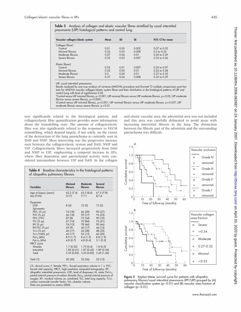
RESULTSThe different grades of vascular obstruction were significantlyrelated to parenchymal changes in UIP compared with thehistological patterns of DAD and NSIP. In other words, thealternating areas of minimal, moderate and severe fibrosisobserved in UIP (fig 1) were significantly related to the degreeof vascular occlusion (fig 2). High degrees of vascular occlusion(eg, grades III and IV) were correlated with a pulmonaryparenchyma heavily compromised by fibrosis. In all UIPsurgical lung biopsy specimens, alternating areas of moderatefibrosis were related to an intermediate degree of vascularremodelling or obstruction (grades II or III). In the remainingUIP lungs, categorised as alternating areas of minimal fibrosis,a significant relationship was found with a minimal degree ofvascular remodelling—that is, vascular remodelling was repre-sented by discrete intimal proliferation (grade I) with main-tenance of the vascular architecture.
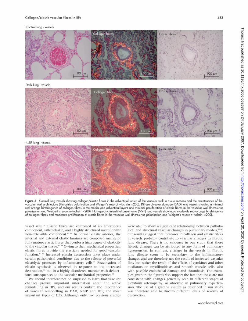
Figures 2 and 3 show the collagen/elastic fibre system incontrol and IIP lungs stained with Picrosirius polarised andWeigert’s resorcin–fuchsin. Control lungs had weak red–orangebirefringence in the adventitial tunica of the vascular wall intissue sections and maintenance of the vascular wall architec-ture (fig 3). By contrast, UIP lung had distortion of the vascularwall architecture and an increase in birefringence in thevascular medial layer in all four degrees of vascular obstruction(fig 2). This correlated with an increase in parenchymalremodelling activity, septal thickening and increase in vascularcollagen fibres (fig 1). DAD and NSIP had a moderate red–orange birefringence in the medial layers (fig 3). This increasein birefringence was greatest in NSIP lungs and correlated withmoderate parenchymal remodelling and moderate alveolar
septal thickening (fig 1). Equally important alterations of theelastic system were present. Figure 3 shows control groups inwhich the vascular pattern of the elastic component ispreserved in the internal and external elastic lamina. Figure 2shows major proliferation of elastic fibres in the internal andexternal elastic lamina of the vascular wall in UIP lungs, relatedto different degrees of vascular remodelling and different stagesof parenchymal remodelling activity (fig 1). A moderate degreeof elastic system proliferation was present in the vascular wallin the NSIP histological pattern whereas a minimal degree wasobserved in the DAD pattern (fig 3).
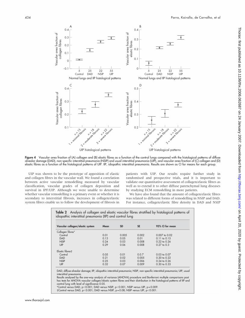
The qualitative changes in collagen and elastic fibres in thevascular wall correlated with differences in their quantificationin the four groups of patients (table 2). The density of thecollagen and elastic fibres was significantly higher in the arterywalls of UIP lungs than in control NSIP and DAD lungs(p = 0.001). UIP vascular walls had the greatest amount ofcollagen, followed by NSIP, DAD and controls (fig 4A). Asignificant difference in the amount of collagen was observedamong all groups (p = 0.001; fig 4A). Elastic fibres showed asimilar pattern which was significant for control versus DAD(p,0.001); and NSIP versus UIP (p,0.001; fig 4A).
Parenchymal remodelling in UIP ranged from minimal tomoderate and severe fibrosis. In this group, the greater theincrease in collagen and elastic fibres, the greater theparenchymal remodelling (fig 4C,D). The increase in fibre wasgreatest in lungs with parenchymal remodelling (severefibrosis), while an intermediate increase in fibre was presentin lungs with minimal and moderate fibrosis activity (p = 0.01;table 3) related to different degrees of vascular occlusion.
Fifty-two patients died during the follow-up period (5 NSIP,12 AIP, 35 IPF). All patients studied had a restrictive lungfunction pattern characterised by a decrease in TLC (meanvalues were NSIP (63%), AIP (57%) and IPF (72%) of predictedvalues) and an increase in FEV1/FVC ratio6100 (mean valueswere NSIP (104%), AIP (97%) and IPF (83%) of predictedvalues). The mean predicted values of TLCO were decreased inpatients with NSIP (43%), AIP (46%) and IPF (49%; table 1).Pulmonary function of patients with IPF with different degreesof fibrosis did not differ significantly between the groups(table 4). Pulmonary function tests were not significantlyrelated to vascular occlusion or collagen and elastic vasculardensity.
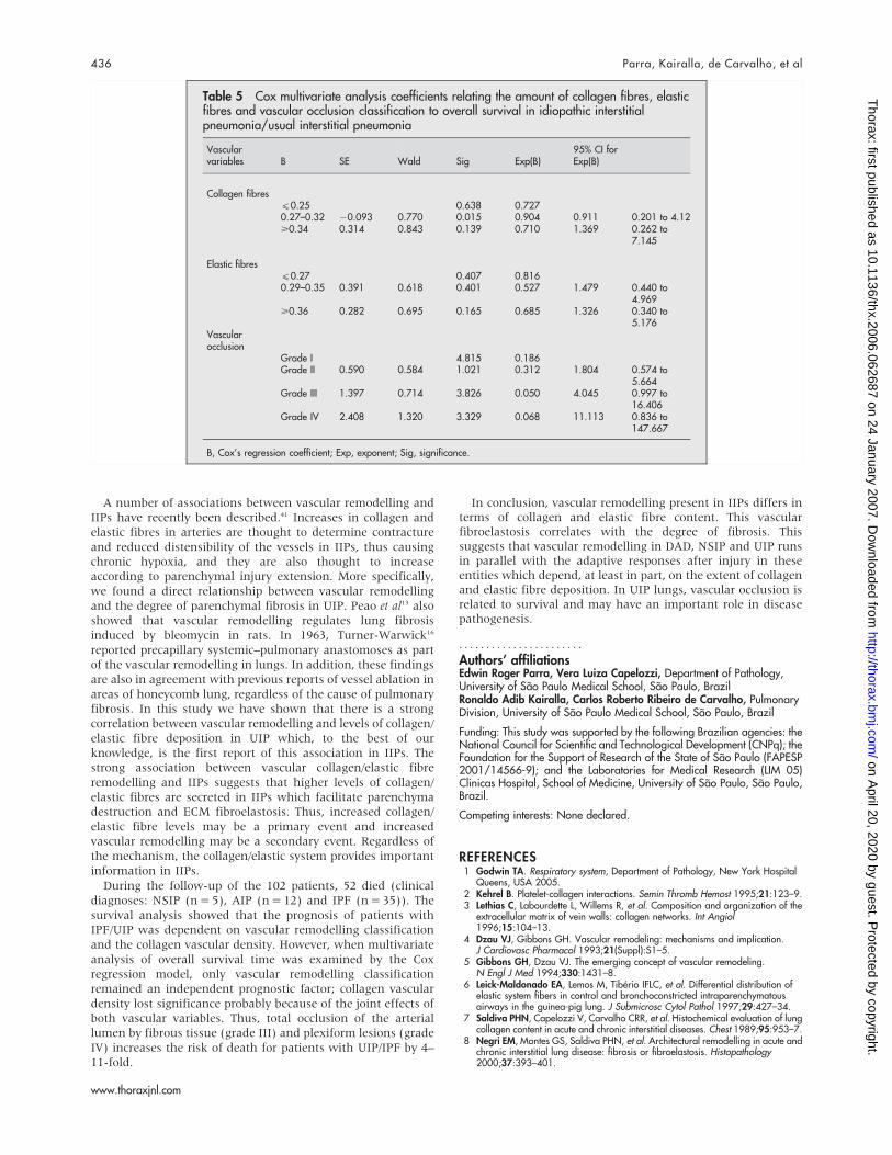
In the first statistical test, the individual effect of patientcharacteristics (age, sex, baseline data and physiological test),parenchymal remodelling (minimal fibrosis, moderate fibrosisand severe fibrosis) and vascular remodelling (grade I, grade II,grade III, grade IV, collagen and elastic fibre density) wereexamined to estimate the survival curve (fig 5A,B). The resultsof this analysis showed that the prognosis of patients with IPF/UIP was dependent on the vascular remodelling classification(log rank 18.24; p,0.001; fig 5A) and the collagen vasculardensity (log rank 10.57; p = 0.005; fig 5B). Multivariate analysisof overall survival time based on significant factors at univariateanalysis was examined by the Cox regression model. Initially, themodel was constructed with vascular remodelling classificationand collagen vascular density. In this situation, only the vascularremodelling classification was maintained as an independentprognostic factor; collagen vascular density lost significanceprobably owing to the joint effects of both vascular variables(table 5). Thus, total occlusion of the arterial lumen by fibroustissue (grade III) and plexiform lesions (grade IV) increases therisk of death in patients with UIP/IPF by 4–11-fold.
DISCUSSIONThe probable reason why patients with IIPs have differentoutcomes is uncontrolled fibrosis progression. The question of
Collagen/elastic vascular fibres in IIPs 431
www.thoraxjnl.com
on April 20, 2020 by guest. P
rotected by copyright.http://thorax.bm
j.com/
Thorax: first published as 10.1136/thx.2006.062687 on 24 January 2007. D
ownloaded from
interest is whether further information gathered from the lungparenchyma or interstitial or VECM active remodelling can helpus understand the heterogeneous development of IIPs. Theprocess of active remodelling comprises a series of complexsequential steps, of which ECM remodelling is thought to beimportant because this process facilitates scar formation whichoccurs by neovascularisation, myofibroblast migration, andcollagen and elastic fibre deposition. In IIPs, this process takes
place in an uncontrolled way, causing excessive fibrosisformation and progressive histoarchitectural and vascularchanges.27–33 VECM remodelling is a dynamic process thatinvolves changes in collagen and elastic fibres resulting indifferent degrees of vascular occlusion. Collagen fibres aredistributed diffusely throughout the media and adventitia ofvessels, being synthesised by fibroblasts, myofibroblasts andsmooth muscle cells, and providing tensile strength to the
Figure 2 Usual interstitial pneumonia (UIP) lung vessels. This groups shows distortion of the vascular wall architecture and an increase in red–orangebirefringence of collagen fibres in the vascular medial and adventitial layers in all four grades of vascular obstruction (grade I, isolated hypertrophy of thearterial media, grade II, proliferative intimal, grade III, total occlusion of the arterial lumen by fibrosis tissue and grade IV, plexiform lesion) and majorproliferation of elastic fibres in the internal and external elastic lamina of the vascular wall (H&E, Picrosirius polarisation and Weigert’s resorcin–fuchsin6200).
432 Parra, Kairalla, de Carvalho, et al
www.thoraxjnl.com
on April 20, 2020 by guest. P
rotected by copyright.http://thorax.bm
j.com/
Thorax: first published as 10.1136/thx.2006.062687 on 24 January 2007. D
ownloaded from
vessel wall.34 Elastic fibres are composed of an amorphouscomponent, called elastin, and a highly structured microfibrillarnon-extensible component.17 18 In normal elastic arteries, theinternal and external elastic laminas are composed mainly offully mature elastic fibres that confer a high degree of elasticityto the vascular tissue.17 18 Owing to their mechanical properties,elastic fibres provide the elasticity needed for good vascularfunction.17 18 Increased elastin destruction takes place undercertain pathological conditions due to the release of powerfulelastolytic proteases by inflammatory cells.35 Reactivation ofelastin synthesis is observed in response to the increaseddestruction,36 but in a highly disordered manner with deleter-ious consequences to the vascular mechanical properties.37
We should therefore not be surprised to learn that vascularchanges provide important information about the activeremodelling in IIPs, and our results confirm the importanceof vascular remodelling in DAD, NSIP and UIP, the mostimportant types of IIPs. Although only two previous studies
were able to show a significant relationship between patholo-gical and structural vascular changes in pulmonary models,37–40
our results suggest that increases in collagen and elastic fibresin vessels probably contribute to vascular changes in fibroticlung disease. There is no evidence in our study that thesefibrotic changes can be attributed to any form of pulmonaryhypertension. In contrast, changes in the vessels in fibroticlung disease seem to be secondary to the inflammatorychanges and are therefore not the result of increased vascularflow but rather the result of the effects of cytokines and othermediators on myofibroblasts and smooth muscle cells, alsowith possible endothelial damage and thrombosis. The exam-ples given in the figures also support the fact that these are notconsistent with changes generally seen in different stages ofplexiform arteriopathy, as observed in pulmonary hyperten-sion. The use of a grading system as described in our studywas therefore able to discern different levels of severity ofobstruction.
Figure 3 Control lung vessels showing collagen/elastic fibres in the adventitial tunica of the vascular wall in tissue sections and the maintenance of thevascular wall architecture (Picrosirius polarisation and Weigert’s resorcin–fuchsin 6200). Diffuse alveolar damage (DAD) lung vessels showing a minimalred–orange birefringence of collagen fibres in the medial and adventitial layers and minimal proliferation of elastic fibres in the vascular wall (Picrosiriuspolarisation and Weigert’s resorcin–fuchsin 6200). Non-specific interstitial pneumonia (NSIP) lung vessels showing a moderate red–orange birefringenceof collagen fibres and moderate proliferation of elastic fibres in the vascular wall (Picrosirius polarisation and Weigert’s resorcin–fuchsin 6200).
Collagen/elastic vascular fibres in IIPs 433
www.thoraxjnl.com
on April 20, 2020 by guest. P
rotected by copyright.http://thorax.bm
j.com/
Thorax: first published as 10.1136/thx.2006.062687 on 24 January 2007. D
ownloaded from
UIP was shown to be the prototype of apposition of elasticand collagen fibres in the vascular wall. We found a correlationbetween active vascular remodelling measured by vascularclassification, vascular grades of collagen deposition andsurvival in IPF/UIP. Although we were unable to determinewhether vascular remodelling is a primary event or whether it issecondary to interstitial fibrosis, increases in collagen/elasticsystem fibres enable us to follow the development of fibrosis in
patients with UIP. Our results require further study inrandomised and prospective trials, and it is important tovalidate our quantitative assessment of collagen/elastic fibres aswell as to extend it to other diffuse parenchymal lung diseasesby studying ECM remodelling in more patients.
We have also found that the amount of collagen/elastic fibreswas related to different forms of remodelling in NSIP and DAD.For instance, collagen/elastic fibre density in DAD and NSIP
Figure 4 Vascular area fraction of (A) collagen and (B) elastic fibres as a function of the control lungs compared with the histological patterns of diffusealveolar damage (DAD), non-specific interstitial pneumonia (NSIP) and usual interstitial pneumonia (UIP), and vascular area fraction of (C) collagen and (D)elastic fibres as a function of the histological patterns of UIP. IIP, idiopathic interstitial pneumonia. Results are shown as CI for means for each group.
Table 2 Analysis of collagen and elastic vascular fibres stratified by histological patterns ofidiopathic interstitial pneumonia (IIP) and control lung
Vascular collagen/elastic system Mean SD SE 95% CI for mean
Collagen fibres*Control 0.01 0.005 0.002 0.007 to 0.02DAD 0.13 0.05 0.01 0.11 to 0.15NSIP 0.24 0.03 0.008 0.22 to 0.26UIP 0.29 0.06 0.008 0.27 to 0.3
Elastic fibres�Control 0.05 0.01 0.0.7 0.03 to 0.07DAD 0.21 0.02 0.005 0.20 to 0.22NSIP 0.25 0.03 0.006 0.24 to 0.26UIP 0.32 0.07 0.009 0.30 to 0.33
DAD, diffuse alveolar damage; IIP, idiopathic interstitial pneumonia; NSIP, non-specific interstitial pneumonia; UIP, usualinterstitial pneumonia.Results analysed by the one-way analysis of variance (ANOVA) procedure and Bonferroni multiple comparisons posthoc tests for ANOVA vascular collagen/elastic system fibres and their distribution in the histological patterns of IIP andcontrol lung with level of significance 0.05.*Control versus DAD, p,0.001; DAD versus NSIP, p,0.001, NSIP versus UIP, p = 0.009.�Control versus DAD, p,0.001; DAD versus NSIP, p = 0.08; NSIP versus UIP, p,0.001.
434 Parra, Kairalla, de Carvalho, et al
www.thoraxjnl.com
on April 20, 2020 by guest. P
rotected by copyright.http://thorax.bm
j.com/
Thorax: first published as 10.1136/thx.2006.062687 on 24 January 2007. D
ownloaded from
was significantly related to the histological pattern, andcollagen/elastic fibre quantification provides more informationabout the remodelling state. The amount of collagen/elasticfibre was also significantly related to the responses to VECMremodelling, which depend largely, if not solely, on the extentof the destruction of the lung parenchyma as currently seen inDAD and NSIP. Most interesting was the progressive increaseseen between the collagen/elastic system and DAD, NSIP andUIP. Collagen/elastic fibres increased progressively from DADand NSIP to UIP, emphasising a temporal increase in IIPs,where fibre deposition and parenchymal activity were con-sidered intermediate between UIP and DAD. In the collagen
and elastic vascular area, the adventitial area was not includedand this area was carefully delineated to avoid areas withincreasing interstitial fibrosis in the lung. The distinctionbetween the fibrotic part of the adventitia and the surroundingparenchyma was difficult.
Table 3 Analysis of collagen and elastic vascular fibres stratified by usual interstitialpneumonia (UIP) histological patterns and control lung.
Vascular collagen/elastic system Mean SD SE 95% CI for mean
Collagen fibres*Control 0.01 0.05 0.002 0.07 to 0.02Minimal fibrosis 0.22 0.03 0.008 0.2 to 0.24Moderate fibrosis 0.27 0.04 0.01 0.25 to 0.29Severe fibrosis 0.35 0.03 0.007 0.33 to 0.36
Elastic fibres�Control 0.05 0.01 0.007 0.03 to 0.07Minimal fibrosis 0.25 0.05 0.01 0.22 to 0.28Moderate fibrosis 0.3 0.05 0.01 0.27 to 0.33Severe fibrosis 0.37 0.04 0.008 0.35 to 0.39
UIP, usual interstitial pneumonia.Results analysed by one-way analysis of variance (ANOVA) procedure and Dunnett T3 multiple comparisons post hoctests for ANOVA vascular collagen/elastic system fibres and their distribution in the histological patterns of UIP andcontrol lung with level of significance 0.05.*Control versus UIP minimal fibrosis, p,0.001; UIP minimal fibrosis versus UIP moderate fibrosis, p = 0.02, UIP moderatefibrosis versus severe fibrosis, p = 0.0001.�Control versus UIP minimal fibrosis, p = 0.001; UIP minimal fibrosis versus UIP moderate fibrosis, p = 0.037, UIPmoderate fibrosis versus severe fibrosis, p = 0.01.
Table 4 Baseline characteristics in the histological patternsof idiopathic pulmonary fibrosis
VariablesMinimalfibrosis
Moderatefibrosis
Severalfibrosis
Age at biopsy (years) 62.2 (7.4) 63.5 (8.6) 67.5 (7.8)Sex (F/M) 5/10 7/11 8/14
DyspnoeaLOD 8 (4) 10 (2) 12 (2)SpirometryFEV1 (% pr) 74 (18) 66 (20) 90 (27)FVC (% pr) 66 (18) 59 (17) 76 (23)FEV1/FVC 87 (8) 72 (34) 90 (10)TLC (% pr) 67 (14) 70 (84) 76 (21)RV (% pr) 74 (15) 78 (46) 90 (46)RV/TLC (% pr) 43 (9) 46 (17) 44 (14)TLCO (% pr) 44 (17) 54 (28) 48 (22)TLCO/VA(% pr) 45 (17) 52 (12) 60 (20)PaO2 (kPa) 8.9 (1.7) 8.4 (1.5) 8.8 (1.9)PaCO2 (kPa) 4.8 (0.7) 4.8 (0.4) 5.1 (0.5)
HRCT scoreAlveolar 1.7 (0.22) 1.75 (0.4) 1.8 (0.5)Interstitial 1.85 (0.61) 1.87 (0.42) 1.89 (0.56)Total 3.55 (0.83) 3.52 (0.82) 3.69 (1.06)
Total CS 50 (20) 52 (26) 53 (12)
CS, clinical score; F, female; FEV1, forced expiratory volume in 1 s; FVC,forced vital capacity; HRCT, high-resolution computed tomography; IPF,idiopathic interstitial pneumonia; LOD, level of dyspnoea; M, male; PaCO2,partial arterial pressure of carbon dioxide; PaO2, partial arterial pressure ofoxygen; RV, residual volume; pr, predicted; TLC, total lung capacity; TLCO,carbon monoxide transfer factor; VA, alveolar volume.Data are presented as means (SEM).
Figure 5 Kaplan–Meier survival curve for patients with idiopathicpulmonary fibrosis/usual interstitial pneumonia (IPF/UIP) grouped by (A)vascular classification system (p,0.01) and (B) vascular area fraction ofcollagen (p,0.01).
Collagen/elastic vascular fibres in IIPs 435
www.thoraxjnl.com
on April 20, 2020 by guest. P
rotected by copyright.http://thorax.bm
j.com/
Thorax: first published as 10.1136/thx.2006.062687 on 24 January 2007. D
ownloaded from
A number of associations between vascular remodelling andIIPs have recently been described.41 Increases in collagen andelastic fibres in arteries are thought to determine contractureand reduced distensibility of the vessels in IIPs, thus causingchronic hypoxia, and they are also thought to increaseaccording to parenchymal injury extension. More specifically,we found a direct relationship between vascular remodellingand the degree of parenchymal fibrosis in UIP. Peao et al13 alsoshowed that vascular remodelling regulates lung fibrosisinduced by bleomycin in rats. In 1963, Turner-Warwick16
reported precapillary systemic–pulmonary anastomoses as partof the vascular remodelling in lungs. In addition, these findingsare also in agreement with previous reports of vessel ablation inareas of honeycomb lung, regardless of the cause of pulmonaryfibrosis. In this study we have shown that there is a strongcorrelation between vascular remodelling and levels of collagen/elastic fibre deposition in UIP which, to the best of ourknowledge, is the first report of this association in IIPs. Thestrong association between vascular collagen/elastic fibreremodelling and IIPs suggests that higher levels of collagen/elastic fibres are secreted in IIPs which facilitate parenchymadestruction and ECM fibroelastosis. Thus, increased collagen/elastic fibre levels may be a primary event and increasedvascular remodelling may be a secondary event. Regardless ofthe mechanism, the collagen/elastic system provides importantinformation in IIPs.
During the follow-up of the 102 patients, 52 died (clinicaldiagnoses: NSIP (n = 5), AIP (n = 12) and IPF (n = 35)). Thesurvival analysis showed that the prognosis of patients withIPF/UIP was dependent on vascular remodelling classificationand the collagen vascular density. However, when multivariateanalysis of overall survival time was examined by the Coxregression model, only vascular remodelling classificationremained an independent prognostic factor; collagen vasculardensity lost significance probably because of the joint effects ofboth vascular variables. Thus, total occlusion of the arteriallumen by fibrous tissue (grade III) and plexiform lesions (gradeIV) increases the risk of death for patients with UIP/IPF by 4–11-fold.
In conclusion, vascular remodelling present in IIPs differs interms of collagen and elastic fibre content. This vascularfibroelastosis correlates with the degree of fibrosis. Thissuggests that vascular remodelling in DAD, NSIP and UIP runsin parallel with the adaptive responses after injury in theseentities which depend, at least in part, on the extent of collagenand elastic fibre deposition. In UIP lungs, vascular occlusion isrelated to survival and may have an important role in diseasepathogenesis.
Authors’ affiliations. . . . . . . . . . . . . . . . . . . . . . .
Edwin Roger Parra, Vera Luiza Capelozzi, Department of Pathology,University of Sao Paulo Medical School, Sao Paulo, BrazilRonaldo Adib Kairalla, Carlos Roberto Ribeiro de Carvalho, PulmonaryDivision, University of Sao Paulo Medical School, Sao Paulo, Brazil
Funding: This study was supported by the following Brazilian agencies: theNational Council for Scientific and Technological Development (CNPq); theFoundation for the Support of Research of the State of Sao Paulo (FAPESP2001/14566-9); and the Laboratories for Medical Research (LIM 05)Clinicas Hospital, School of Medicine, University of Sao Paulo, Sao Paulo,Brazil.
Competing interests: None declared.
REFERENCES1 Godwin TA. Respiratory system, Department of Pathology, New York Hospital
Queens, USA 2005.2 Kehrel B. Platelet-collagen interactions. Semin Thromb Hemost 1995;21:123–9.3 Lethias C, Labourdette L, Willems R, et al. Composition and organization of the
extracellular matrix of vein walls: collagen networks. Int Angiol1996;15:104–13.
4 Dzau VJ, Gibbons GH. Vascular remodeling: mechanisms and implication.J Cardiovasc Pharmacol 1993;21(Suppl):S1–5.
5 Gibbons GH, Dzau VJ. The emerging concept of vascular remodeling.N Engl J Med 1994;330:1431–8.
6 Leick-Maldonado EA, Lemos M, Tiberio IFLC, et al. Differential distribution ofelastic system fibers in control and bronchoconstricted intraparenchymatousairways in the guinea-pig lung. J Submicrosc Cytol Pathol 1997;29:427–34.
7 Saldiva PHN, Capelozzi V, Carvalho CRR, et al. Histochemical evaluation of lungcollagen content in acute and chronic interstitial diseases. Chest 1989;95:953–7.
8 Negri EM, Montes GS, Saldiva PHN, et al. Architectural remodelling in acute andchronic interstitial lung disease: fibrosis or fibroelastosis. Histopathology2000;37:393–401.
Table 5 Cox multivariate analysis coefficients relating the amount of collagen fibres, elasticfibres and vascular occlusion classification to overall survival in idiopathic interstitialpneumonia/usual interstitial pneumonia
Vascularvariables B SE Wald Sig Exp(B)
95% CI forExp(B)
Collagen fibres(0.25 0.638 0.7270.27–0.32 20.093 0.770 0.015 0.904 0.911 0.201 to 4.12>0.34 0.314 0.843 0.139 0.710 1.369 0.262 to
7.145
Elastic fibres(0.27 0.407 0.8160.29–0.35 0.391 0.618 0.401 0.527 1.479 0.440 to
4.969>0.36 0.282 0.695 0.165 0.685 1.326 0.340 to
5.176Vascularocclusion
Grade I 4.815 0.186Grade II 0.590 0.584 1.021 0.312 1.804 0.574 to
5.664Grade III 1.397 0.714 3.826 0.050 4.045 0.997 to
16.406Grade IV 2.408 1.320 3.329 0.068 11.113 0.836 to
147.667
B, Cox’s regression coefficient; Exp, exponent; Sig, significance.
436 Parra, Kairalla, de Carvalho, et al
www.thoraxjnl.com
on April 20, 2020 by guest. P
rotected by copyright.http://thorax.bm
j.com/
Thorax: first published as 10.1136/thx.2006.062687 on 24 January 2007. D
ownloaded from
9 Negri EM, Hoelz C, Barbas CSV, et al. Acute remodelling of parenchyma inpulmonary and extrapulmonary ARDS. An autopsy study of collagen-elasticsystem fibers. Pathol Res Pract 2002;198:355–61.
10 Faffe DS, Silva GH, Kurtz PM, et al. Lung tissue mechanics and extracellularmatrix composition in a murine model of silicosis. J Appl Physiol2001;90:1400–6.
11 Rocco PRM, Negri EM, Kurtz PM, et al. Lung tissue mechanics and extracellularmatrix remodelling in acute lung injury. Am J Respir Crit Care Med2001;164:1067–71.
12 Parra ER, David YR, da Costa LRS, et al. Heterogeneous remodeling of lungvessels in idiopathic pulmonary fibrosis. Lung 2005;183:291–300.
13 Peao MND, Aguas AP, DeSa CM, et al. Neoformation of blood vessels inassociation with rat lung fibrosis induced by bleomycin. Anat Rec1994;238:57–67.
14 Renzoni EA, Walsh DA, Salmon M, et al. Interstitial vascularity in fibrosingalveilitis. Am J Respir Crit Care Med 2003;167:438–43.
15 Salmon M, Lui YC, Mark JC, et al. Contribution of upregulation airwayendothelin –1 expression to airway smooth muscle and epithelial cell DNAsynthesis after repeated allergen exposure of sensitized Brown-Norway rats.Am J Respir Cell Mol Biol 2000;23:618–25.
16 Turner-Warwick M. Precapillary systemic-pulmonary anastomoses. Thorax1963;18:225–37.
17 Karnik SK, Brooke BS, Bayes-Genis A, et al. A critical role for elastin signaling invascular morphogenesis and disease. Development 2003;130:411–23.
18 Li, DY. Elastin is an essential determinant of arterial morphogenesis. Nature1998;393:276–80.
19 Demedts M, Costabel U. ATS/ERS international multidisciplinary consensusclassification of the idiopathic interstitial pneumonias. Eur Respir J 2002;19:794–6.
20 Watters LC, King TE, Schwarz MI, et al. A clinical, radiographic, and physiologicscoring system for the longitudinal assessment of patients with idiopathicpulmonary fibrosis. Am Rev Respir Dis 1986;133:97–103.
21 Quanjer PhH, Tammeling GJ, Cotes JE, et al. Lung volumes and forcedventilatory flows. Report working party, Standardization of lung function tests,European Community for steel and coal. Official Statement of the EuropeanRespiratory Society. Eur Respir J 1993;6(Suppl 16):5–40.
22 Kazerooni EA, Martinez FJ, Flint A, et al. Thinsection CT obtained at 10 mmincrements versus three-level thin-section CT for idiopathic pulmonary fibrosis:correlation with pathologic scoring. Am J Roentgenol 1997;169:977–83.
23 Montes GS, Junqueira LCU. Histochemical localization of collagen and ofproteoglycans in tissues. In: Nimni ME, ed. Collagen Vol 2. Boca Raton, FL: CRCPress, 1998:41–72.
24 Montes GS. Structural biology of the fibers of the collagenous and elastic systems.Cell Biol Intermed 1996;20:245–9.
25 Lemos M, Pozo RMK, Montes GS, et al. Organization of collagen and elasticfibers studied in stretch preparations of whole mounts of human visceral pleura.Ann Anat 1997;79:447–52.
26 Norusis MJ. SPSS for Windows. [10.0]. Chicago: SPSS, 2001.27 Basset F, Ferrans VJ, Soler P, et al. Intraluminal fibrosis in interstitial lung
disorders. Am J Pathol 1986;122:443–61.28 Karnik SK, Brooke BS, Bayes-Genis A, et al. A critical role for elastin signaling in
vascular morphogenesis and disease. Development 2003;130:411–23.29 Myers JL, Katzeinstein AL. Epithelial necrosis and alveolar collapse in the
pathogenesis of usual interstitial pneumonia. Chest 1988;94:1309–11.30 Fukuda Y, Basset F, Soler P, et al. Intraluminal fibrosis and elastic fiber
degradation lead to lung remodelling in pulmonary Langerhans cellgranulomatosis (histiocytosis X). Am J Pathol 1990;137:415–24.
31 Fukuda YM, Ishizaki M, Kudoh S, et al. Localization of matrixmetalloproteinases-1, -2, and -9 and tissue inhibitor of metalloproteinase-2 ininterstitial lung diseases. Lab Invest 1998;78:687–98.
32 Fukuda Y, Ishizaki M, Masuda Y, et al. The role of intra-alveolar fibrosis in theprocess of pulmonary structural remodelling in patients with diffuse alveolardamage. Am J Pathol 1987;126:171–82.
33 Barbas Filho JV, Ferreira MA, Sesso A, et al. Evidences of type II pneumocytesapoptosis in the pathogenesis of idiopathic pulmonary fibrosis (IPF)/usualinterstitial pneumonia (UIP). J Clin Pathol 2001;54:132–8.
34 Tozzi CA, Christiansen DL, Poiani GJ, et al. Excess collagen in hypertensivepulmonary arteries decreases vascular distensibility. Am J Respir Crit Care Med1994;149:1317–26.
35 Bitterman PB, Pollunovsky VA, Ingbar DH. Repair after acute lung injury. Chest1994;105(Suppl 118):120.
36 Mariani TJ, Crouch E, Rouby JD, et al. Increased elastin production inexperimental granulomatous lung disease. Am J Pathol 1995;147:988–1000.
37 Li DY. Elastin point mutations cause an obstructive vascular disease,supravalvular aortic stenosis. Hum Mol 1997;6:1021–8.
38 Ried L, Meyrick B. Hypoxia and pulmonary vascular endothelium. Ciba FundSymp 1980;78:37–60.
39 Meyrick B, Reid L. Hypoxia-induced structural changes in the media andadventitia of the rat hilar pulmonary artery and their regression. Am J Pathol1980;100:151–78.
40 Vyas-Somani AC, Aziz SM, Arcot SA, et al. Temporal alterations in basementmembrane components in the pulmonary vascular of the chronically hypoxic rat:impact of hypoxia and recovery. Am J Med Sci 1996;312:54–67.
41 Masahito E, Minoru S, Naoko S, et al. Heterogeneous increase in CD34-positivealveolar capillaries in idiopathic pulmonary fibrosis. Am J Respir Crit Care Med2004;169:1203–8.
LUNG ALERT . . . . . . . . . . . . . . . . . . . . . . . . . . . . . . . . . . . . . . . . . . . . . . . . . . . . . . . . . . . . . . . . . . . . . . . . . . . . . . . . . . . . . . . . . . . . . . . . . . . . . . . . . .
Second hand smoke exposure is associated with worse survival in early stage non-smallcell lung cancerm Zhou W, Heist RS, Liu G, et al. Second hand smoke exposure and survival in early-stage non-small cell lung cancer patients.Clin Cancer Res 2006;12:7187–93.
This study examines the possible association between the extent of second hand smoke (SHS)exposure prior to diagnosis of early stage non-small cell lung cancer (stages Ia-IIb) andsurvival after treatment. The 393 Massachusetts General Hospital patients were grouped into
quartiles based on their SHS exposure and then the patients’ own smoking history in pack–yearswas factored into the calculations.
The results reveal a statistically significant worsening of 5-year survival between the quartilewith the lowest (,28 years) SHS exposure, 71% alive after five years, and those with the highest(.46 years) exposure, 47% alive at five years (p , 0.001). Recurrence-free survival was alsogreatly reduced in those with the heaviest exposure when compared with those in the lightestexposure group.
Interestingly, in further subgroup analysis it was found that those who had most of their SHSexposure in the work place were associated with worse outcome (adjusted hazard ratio ofhighest vs lowest quartile, 1.71 Ptrend = 0.03) than those exposed either in the home (AHR 1.26Ptrend = 0.2) or ‘‘leisure’’ locations (AHR, 1.28. Ptrend = 0.2). This will add further weight to theimplementation of smoking bans in the public and workplace being introduced throughout theUK.
T F Paul McKeagneyLAT Registrar in Respiratory/GIM, Craigavon Area Hospital, Craigavon, Northern Ireland, UK;
Collagen/elastic vascular fibres in IIPs 437
www.thoraxjnl.com
on April 20, 2020 by guest. P
rotected by copyright.http://thorax.bm
j.com/
Thorax: first published as 10.1136/thx.2006.062687 on 24 January 2007. D
ownloaded from