intranasal shigella flexneri shigella lipopolysaccharide · oraland intranasal proteosome shigella...
TRANSCRIPT

Vol. 61, No. 6INFECTION AND IMMUNITY, June 1993, p. 2390-23950019-9567/93/062390-06$02.00/0Copyright © 1993, American Society for Microbiology
Immunogenicity and Efficacy of Oral or IntranasalShigella flexneri 2a and Shigella sonnei Proteosome-
Lipopolysaccharide Vaccines in Animal ModelsNADAV ORR,1,2* GUY ROBIN,' DANI COHEN,' RUTH ARNON,2 AND GEORGE H. LOWELL3
Medical Corps, Israel Defence Force, Military Post 02149, 1 * and Department of ChemicalImmunology, Weizmann Institute of Science, Rehovot,2 Israel, and Division of
Pathology, Walter Reed Army Institute of Research, Washington, D.C. 3
Received 8 December 1992/Accepted 4 March 1993
Immunity against shigellosis has been shown to correlate with the presence of antibodies specific for Shigellalipopolysaccharide (LPS). We here propose a new candidate vaccine for shigellosis composed of purifiedShigeUla flexneri 2a or Shigella sonnei LPS hydrophobically complexed with group C type 2b Neisseriamening*itdis outer membrane protein proteosomes. Immunization of mice either orally or intranasally with thiscomplex induced specific homologous anti-LPS antibodies in both intestinal and respiratory secretions as wellas in sera. Strong anamnestic responses were found after two or three immunizations. LPS alone, alkaline-detoxified LPS, or alkaline-detoxified LPS complexed with proteosomes was not effective. Oral or intranasalimmunization of guinea pigs with two or more doses of this proteosome-LPS vaccine elicited homologousprotection against Shigella keratoconjunctivitis (Sereny test). These data demonstrate that proteosomes can beused as an effective mucosal vaccine delivery system and that orally or intranasally administered acellularvaccines can protect against Shigella infections.
Shigella flexneri and Shigella sonnei as well as otherShigella species represent a major cause of diarrheal dis-eases in developing countries (12). It has been shown thattype-specific protection against shigellosis can be acquired inhumans after infection with wild-type or attenuated bacteria(2, 4, 9, 13, 19). There is direct evidence that anti-type-specific lipopolysaccharide (LPS) antibodies are associatedwith this protection (2, 4). Although clearance of Shigellacells and similar mucosal pathogens from invaded tissue mayinvolve several host defense mechanisms, it is widely rec-ognized that local mucosal immune responses, especiallysecretory immunoglobulins (Igs), play a major role in pro-tection (11, 25, 26, 28). In this regard, levels of theseantibodies in serum, to the extent that they reflect localantibody production, may be viewed as measures or markersof protection, whether or not they actively contribute tobacterial clearance of such mucosal infections (4, 28).
Since the demonstrations in 1955 by Higgins et al. (10) andin 1967 by Formal et al. (6) that parenteral immunizationwith live or killed Shigella bacteria was ineffective in pro-tecting humans or monkeys against Shigella infection, themajor thrust of Shigella vaccine research has focused on theuse of orally administered live genetically constructed orattenuated vaccines, and several of these approaches areparticularly promising (7, 15). Nevertheless, the successfuldevelopment of live Shigella vaccines has been difficult toaccomplish, partly because of the relatively narrow windowbetween efficacy and safety of certain vaccine candidates (7,15).Proteosomes are preparations of neisserial outer mem-
brane protein vesicles that have previously been shown toenhance the parenteral immunogenicity of peptides andother antigens hydrophobically complexed to them (16, 17).Moreover, large-scale vaccine trials with such meningococ-
* Corresponding author.
cal outer membrane protein preparations noncovalentlycomplexed to meningococcal polysaccharides have demon-strated that such vaccines are safe for human use (30, 31). Inthe present study, we evaluated an acellular approach toinduce type-specific anti-Shigella immunity using purifiedShigella LPS. In particular, we evaluated the mucosal im-munogenicity and efficacy in animal models of S. flexneri 2aand S. sonnei LPS hydrophobically complexed to proteo-somes (prot-LPS). These Shigella vaccine candidates weredesigned for oral or intranasal administration in order toachieve direct sensitization of targeted mucosal tissues andthereby stimulate mucosal Ig production and local immunity.
MATERIALS AND METHODS
All materials unless indicated otherwise were purchasedfrom Sigma Chemical Co., St. Louis, Mo.LPS preparation. LPS was extracted from single isolates
of S. flexneri 2a or S. sonnei by hot phenol by establishedmethods (29). Alkaline-detoxified LPS (LPSad) was pre-pared by mild alkaline treatment as previously described(23).
Proteosome preparation. Outer membrane proteins fromgroup C serotype 2b Neisseria meningitidis were extractedwith detergent as described previously (17).
Vaccine preparation. Purified LPS or LPSad and group Cserotype 2b N. meningitidis outer membrane proteins weremixed at a 1:1 (wt/wt) ratio in phosphate-buffered saline(PBS) containing 1% Empigen (Albright and Wilson, White-haven, Cumbria, Great Britain). The final concentration ofproteosomes and either LPS or LPSad was 2 mg/ml. Themixture was dialyzed across a dialysis membrane with a1,000-molecular-weight cutoff (SpectraPor 6; SpectrumMedical Industries, Los Angeles, Calif.) against PBS at 4°Cfor 10 days with daily buffer exchanges. The vaccine prep-arations, prot-LPS or prot-LPSad, were stored at 4°C and
2390
on April 16, 2019 by guest
http://iai.asm.org/
Dow
nloaded from

ORAL AND INTRANASAL PROTEOSOME SHIGELLA LPS VACCINES 2391
diluted to the specific concentration with PBS prior toimmunization.CL-4B column. Samples of purified LPS or vaccine prep-
aration were eluted over a CL-4B-200 column (2.5 by 40 cm),and fractions were assayed for protein by the Bradfordmethod (3) with bovine serum albumin (BSA) as standard.The LPS level in each fraction was calculated by competi-tion enzyme-linked immunosorbent assay (ELISA) as fol-lows. Samples (100 ,ul) from each fraction were incubatedwith 300 ,u of specific-LPS-positive guinea pig serum diluted1:150 in ELISA blocking buffer for 1 h at 37°C. HomologousLPS samples ranging from 100 to 3.125 ,ug/ml were incubatedas standards. The serum and samples or standards were thenincubated for 2 h at 37°C in 96-well plates that had beenprecoated with LPS. The plates were developed as describedbelow (see "ELISA" below). The amount of LPS in eachfraction was calculated with the standard curve obtainedfrom the LPS standards.
Immunizations. (i) Mice. BALB/c mice (7 to 10 weeks old;four to five per group) were immunized by either theintranasal or oral route. Orally, 100 RI of PBS with 0.2 MNaHCO3 containing 100 ,ug of LPS (or LPSad) was giveneither alone or complexed with 100 jig of proteosomes (200jig of prot-LPS) via a bent metal feeding tube. For intranasalimmunization, following light ether anesthesia, 25 RI of PBScontaining 10 ,ug of LPS (or LPSad) either alone or com-plexed with 10 jig of proteosomes (20 jig of prot-LPS) wasslowly placed via micropipette in one or both of the nares. Acontrol group in each experiment received diluent withoutantigen.
(ii) Guinea pigs. DH guinea pigs (2 to 3 months old) wereanesthetized intramuscularly with 5 mg of ketamine- HCIplus 1.17 mg of xylazine- HCl and immunized with theprot-LPS complex via either the oral or intranasal route.Orally, 200 pl of PBS with 0.2 M NaHCO3 containing 200 ,ugof the prot-LPS complex was used to immunize; intrana-sally, 50 RI of PBS with 40 jig of the prot-LPS complex wasused.
Collection of mucosal secretions. (i) Murine lung lavage.Nine to 11 days after the last immunization, mice weresacrificed by CO2 suffocation and the lungs were exposed. Acannula was inserted into the trachea, and using a three-waystopcock, the lungs were lavaged twice with 1 ml of PBScontaining 0.1% BSA and the two lavage fluids were com-bined.
(ii) Murine intestinal lavage. Following the lung lavage, 20to 25 cm from the small intestine was removed, and 2 ml ofPBS containing 0.1% BSA, 50 mM EDTA, and 0.1 mg ofsoybean trypsin inhibitor per ml was passed through andcollected. Phenylmethylsulfonyl fluoride was added to theintestinal wash after collection (1 mM, final concentration).Both lung and intestinal washes were vortexed vigorouslyand centrifuged at 1,000 x g for 20 min to remove cells anddebris, the supernatants were collected, NaN3 (0.1%, finalconcentration) was added, and the supernatents were storedat -20°C until assayed.Serum preparation. Blood from mice was collected after
they were sacrificed, and sera were stored at -20°C untilassayed.ELISA. The antibody level in the sera and in the intestinal
and lung lavage fluids was determined as described previ-ously (4). Briefly, 96-well flat-bottom high-binding plates(Costar, Cambridge, Mass.) were precoated with the specificLPS, blocked with BSA-casein, and washed three times.Serially double-diluted samples in blocking solution wereincubated in the plates, and after the incubation period, the
E, 60
c.° 40
0o20'0
200
Elution volume (ml)
FIG. 1. Protein and LPS levels in fractions eluted from a CL-4Bcolumn after application of the prot-S. fle-xnen 2a LPS complex andLPS levels in column fractions after application of S. fle-xneri 2a LPSalone.
plates were washed three times and alkaline phosphatase-conjugated anti-mouse IgG or IgA diluted 1:1,000 wasadded. The plates were then washed three times, p-nitro-phenylphosphate was added, and the A405 was measured.The antibody titer noted is expressed as the geometric meanof the maximal dilution which elicited an optical densityequal or above the cutoff value at the specific processingperiod. The processing periods and cutoff values are men-tioned in the relevant figure legends.
Challenge in guinea pigs (Sereny test). Based on the stan-dard challenge assay for the pathogenesis of shigellosis (22),the conjunctival sac of one eye of the animal was inoculatedwith 30 ,ul of a suspension containing an estimated 108homologous bacteria (S. flexneri 2a or S. sonnei), and theeyelids were lightly massaged. All experiments were per-formed with the same stock strains, and for S. sonnei, onlyCongo red-positive colonies were picked for culturing for theassay. By using this method, 70 to 80% Congo red-positivecolonies were identified in the suspension of S. sonnei usedfor inoculation in the Sereny tests. An accurate determina-tion of the inoculum was determined by plating on bloodagar. Two to 3 days after inoculation, the eyes were blindlyobserved for the development of keratoconjunctivitis. Thedegree of severity of keratoconjunctivitis was defined asfollows: no infection, no signs of irritation or keratoconjunc-tivitis; mild infection, any signs of irritation or keratocon-junctivitis but not purulent; severe infection, fully developedkeratoconjunctivitis with purulence. Protection was calcu-lated according to the following formula: 1 - (% immunizedinfected/% control infected), where infected represents ei-ther severe infection or any infection.
Statistics. Data from the ELISA results were analyzed byStudent's t test (see Table 1) or the general linear models test(see Fig. 3 and 4). Data from guinea pig challenge experi-ments were analyzed by the two-tailed Fisher exact test.
RESULTS
Complex efficiency. Figure 1 shows the protein and S.flexneri 2a LPS elution patterns as measured by gel filtrationcolumn chromatography with CL-4B. When the prot-LPScomplex was passed over the column, LPS and proteosomeswere detected in the same fractions at the void volume of thecolumn (Fig. 1, dotted and solid lines, respectively). Incontrast, LPS alone eluted later (Fig. 1, dashed line) and wasthus easily differentiated from LPS complexed to proteo-somes.Immunogenicity in mice. Initial experiments were per-
formed by immunizing twice with a 3-week interval between
VOL. 61, 1993
on April 16, 2019 by guest
http://iai.asm.org/
Dow
nloaded from

2392 ORR ET AL.
TABLE 1. Anti-LPS IgG in mouse serum aftei- Lwo dosesof vaccine 3 weeks apart
Anti-LPS IgG
Vaccine Intranasal immunization Oral immunization
GMT Rangeb GM1 Rangeb
S. flexnen 2aLPSad <6 <6cProt-LPSad <6 14c 9-21LPS <6 <6Prot-LPS 1,838 1,227-2,735 528 408-683
S. sonnei 4,222 3,200-5,571 113 27-467Prot-LPS
a Geometric mean of the maximal reciprocal dilution elicited an opticaldensity greater than 0.5 after 1 h of incubation with substrate.
b GMT ± 1 standard error of mean.c Three doses of vaccine at weeks 0, 1, and 4.
doses. Nine to 11 days after the last immunization, sera andsecretions from intestines and lungs were collected, and thespecific anti-LPS antibody levels were measured by ELISA.LPS or LPSad (three doses) prepared from S. flexneri 2acould not induce anti-LPS antibody in sera when given alone(Table 1). Interestingly, prot-LPSad preparation elicitedonly minimal levels of serum antibody even after three doses(Table 1). Neither LPS, LPSad, nor prot-LPSad induced anydetectable IgA in intestinal or lung secretions (data notshown). In marked contrast, the prot-LPS complex with S.flexnen 2a LPS was remarkably effective in inducing homol-ogous anti-LPS IgG production in sera (Table 1). In thiscase, intranasal immunization was more effective than oralimmunization (P = 0.02), although 10-fold less vaccine wasgiven. The strong immunogenicity of the prot-LPS vaccinewas confirmed with S. sonnei LPS (Table 1), but in this case,the advantage of the intranasal route was less significant (P= 0.07). No heterologous anti-LPS antibodies were detectedin the sera (data not shown).
Since prot-LPSad was not effective in inducing anti-LPSantibodies, we investigated the idea that alkaline detoxifica-tion may affect the antigenicity of LPS. When LPSad wasused as the detecting solid-phase antigen on the ELISAplate, results with natural or postimmunization antisera werecomparable to those obtained with native LPS (data notshown). This indicated that the antigenicity of LPSad wasequivalent to that of native LPS. Nevertheless, an inhibitionELISA (described in Materials and Methods) showed thatLPSad and prot-LPSad were markedly less effective thannative LPS or prot-LPS in inhibiting binding of naturalanti-LPS antibodies to native LPS (Fig. 2). These datademonstrate that alkaline detoxification altered the antige-nicity that LPS expresses in solution.To determine the optimal immunization regimen for prot-
LPS vaccines, mice were immunized either orally or intra-nasally with proteosomes complexed with S. flexnen 2a LPSby four different protocols: (i) one dose, (ii) two doses 1week apart, (iii) two doses 3 weeks apart, and (iv) threedoses 1 and 3 weeks apart. The LPS-specific serum antibodytiters of mice immunized with two or three doses of vaccine(Fig. 3) showed that the highest levels of both IgG and IgAwere achieved when one booster dose was given 3 weeksafter priming with one (Fig. 3b) or two (Fig. 3c) doses (P <
0.001). IgA production was measurably improved by a thirddose (P < 0.001), while for IgG, two doses of vaccine elicited
E 0.8umcLO0
7:.0.6-D
0 0.4
0.2
0 10 20 30 40 50 60Concentration (gg/ml)
FIG. 2. Inhibition of specific antibody binding to solid-phase S.fle.xneri 2a LPS in ELISA. LPSad, prot-LPSad, LPS, or prot-LPSwas incubated with LPS-positive guinea pig serum prior to itsapplication to an ELISA.
antibody levels comparable to those elicited by the three-dose regimen. One dose of vaccine was insufficient to elicitany detectable serum antibody levels by the standard assay;extension of the sensitivity of the assay (by overnightincubation with substrate) revealed low levels of antibodyeven after one dose of prot-LPS (data not shown). Thecombined analysis of the different regimens elicited that bothoral and intranasal routes of administration induced compa-rable levels of IgG (P = 0.26) and IgA (P = 0.56) in serumeven though the amount of antigen administered intranasallywas 10-fold less than that given orally (Fig. 3).The induction of specific anti-S. flexnen 2a LPS IgA levels
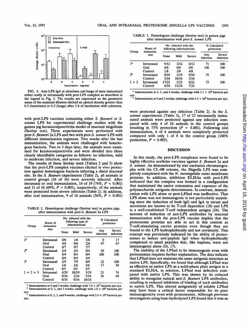
in murine intestine lavage fluids by prot-LPS was similar tothe serum responses in that protocols iii and iv were the mosteffective (P = 0.02) (Fig. 4) and one dose of vaccine failed toelicit detectable antibody levels. Intranasal immunizationwas effective in induction of both intestinal and lung IgA.Induction of higher levels of anti-LPS IgA in lung lavagefluids, however, was far more easily attained by intranasalthan by oral immunization (P < 0.001), while oral immuni-zation was more effective then intranasal immunization ininducing intestinal IgA (P = 0.03) (Fig. 4).
Challenge in guinea pigs. Guinea pigs were immunized
a
0
= 102.
0 10
co4)
32 l,
O Oral imm.
E3 Intranasal imm.
IgA
-'1
a b c
IgG
b ca
Vaccination regimen
FIG. 3. Anti-LPS IgG and IgA in sera of mice immunized eitherorally or intranasally with prot-LPS complex. Four or five animalswere immunized either with two doses at 0 and 1 weeks (a), with twodoses at 0 and 3 weeks (b), or with three doses at 0, 1, and 4 weeks(c). The results are expressed as the geometric mean of the maximaldilution elicited an optical density greater than 0.5 after 1 h ofincubation with substrate.
LPSadProt-LPSad
A Prot-LPS- LPS
INFECT. IMMUN.
-r
on April 16, 2019 by guest
http://iai.asm.org/
Dow
nloaded from
ORAL AND INTRANASAL PROTEOSOME SHIGELLA LPS VACCINES 2393
co
c 102.20
CDX 10'
E Oral imm.O Intranasal imm.
Intestine
a b c a b c
Vaccination regimen
FIG. 4. Anti-LPS IgA in intestines and lungs of mice immunizedeither orally or intranasally with prot-LPS complex as described inthe legend to Fig. 3. The results are expressed as the geometricmean of the maximal dilution elicited an optical density greater than0.5 (intestines) or 0.2 (lungs) after 2 h of incubation with substrate.
with prot-LPS vaccines containing either S. flexneri or S.sonnei LPS for experimental challenge studies with theguinea pig keratoconjunctivitis model of mucosal shigellosis(Sereny test). Three experiments were performed withprot-S. flexneri 2a LPS and two with prot-S. sonnei LPS withdifferent immunization regimens. Two weeks after the lastimmunization, the animals were challenged with homolo-gous bacteria. Two to 3 days later, the animals were exam-ined for keratoconjunctivitis and were divided into threeclearly identifiable categories as follows: no infection, mildto moderate infection, and severe infection.The results of these Sereny tests (Tables 2 and 3) show
that the prot-LPS complex vaccines elicited in vivo protec-tion against homologous bacteria infecting a distal mucosalsite. In the S. flexneri experiments (Table 2), all animals incontrol groups (16 of 16) were severely infected. Afterintranasal or oral immunization, 14 of 19 (74%, P < 0.001)and 11 of 16 (69%, P < 0.001), respectively, of the animalswere protected from severe infection (Table 2). In addition,after oral immunization, 9 of 16 animals (56%, P < 0.001)
TABLE 2. Homologous challenge (Ser6ny test) in guinea pigsafter immunization with prot-S. flexneri 2a LPS
No. infected with the % CalculatedRoute of following type of protection
Group immuniza- infection/total:tion
None Mild Severe Any Severemfection infection
la Intranasal 0/6 1/6 5/6 0 17Oral 4/6 0/6 2/6 67 67Control 0/7 0/7 7/7
2b Intranasal 2/4 2/4 0/4 50 100Oral 4/4 0/4 0/4 100 100Control 0/4 0/4 4/4
3c Intranasal 2/9 7/9 0/9 22 100Oral 1/6 2/6 3/6 17 50Control 0/5 0/5 5/5
1 + 2 + 3 Intranasal 4/19 10/19 5/19 21 74Oral 9/16 2/16 5/16 56 69Control 0/16 0/16 16/16
a Immunization at 0 and 3 weeks; challenge with 7.6 x 10⁷ bacteria per eye.b Immunization at 0, 1, and 3 weeks; challenge with 1.0 x 10i bacteria per
eye.c Immunization at 0, 1, 3, and 8 weeks; challenge with 2.6 x 101 bacteria per
eye.
TABLE 3. Homologous challenge (Sereny test) in guinea pigsafter immunization with prot-S. sonnei LPS
No. infected with the % CalculatedRoute of following infection/total: protection
Group immuniza-tion None Mild Severe Any Severe
infection infection
la Intranasal 9/12 3/12 0/12 71 100Oral 6/6 0/6 0/6 100 100Control 1/8 3/8 4/8
2b Intranasal 8/10 2/10 0/10 75 100Control 3/16 10/16 3/16
1 + 2 Intranasal 17/22 5/22 0/22 73 100Control 4/24 13/24 7/24
a Immunization at 0, 3, and 4 weeks; challenge with 3.1 x 10⁸ bacteria pereye.
b Immunization at 0 and 3 weeks; challenge with 4.3 x 10⁸ bacteria per eye.
were protected against any infection (Table 2). In the S.sonnei experiments (Table 3), 17 of 22 intranasally immu-nized animals were protected against any infection com-pared with only 4 of 24 animals in the control groupsresulting in 73% protection (P < 0.001). Following oralimmunization, 6 of 6 animals were completely protectedcompared with only 1 of 8 in the control group (100%protection, P = 0.005).
DISCUSSION
In this study, the prot-LPS complexes were found to behighly effective acellular vaccines against S. flexneri 2a andS. sonnei. As demonstrated by size exclusion chromatogra-phy with the CL-4B column, the Shigella LPS was com-pletely complexed with the N. meningitidis outer membraneproteins. In addition, inhibition ELISAs with prot-LPSindicated that the complex was formed in a configurationthat maintained the native orientation and exposure of thepolysaccharide antigenic determinants. In contrast, immuni-zation with LPS alone or prot-LPSad was ineffective. ThatLPS alone was not immunogenic is not particularly surpris-ing since the induction of both IgG and IgA in serum andsecretions are known to be T-cell dependent (18) and LPSis a well-established T-cell independent antigen (24). Thesuccess of induction of anti-LPS antibodies by mucosalimmunization with the prot-LPS vaccine implies that theproteosome proteins are able to act in this context asT-cell-stimulating carrier proteins even though they arebound to the LPS hydrophobically and not covalently. Thisconcept was previously indicated by the ability of proteo-somes to induce anti-peptide IgG when hydrophobicallycomplexed to small peptides that, like haptens, were notimmunogenic alone (16, 17).The inability of the LPSad to be immunogenic even with
proteosomes requires further explanation. The data indicatethat LPSad does not maintain the same antigenic structure asnative LPS. Specifically, we found that although LPSad wasas effective as native LPS as a solid-phase plate antigen in astandard ELISA, in solution, LPSad was defective com-pared with native LPS. This was shown by its reducedability to recognize natural anti-S. flexneri LPS antibodies,resulting in reduced inhibition of binding of such antibodiesto native LPS. This altered antigenicity of soluble LPSadmay have been a critical factor responsible for its poorimmunogenicity even with proteosomes. Although previousinvestigators using base-hydrolyzed LPS found that it was as
Lungs
VOL. 61, 1993
on April 16, 2019 by guest
http://iai.asm.org/
Dow
nloaded from

2394 ORR ET AL.
antigenic as native LPS in their assays, S. fle-xneri LPS maybe more sensitive to such treatment than the Escherichia coliLPS they used (21, 23). It is also possible that the adjuvantproperty of lipid A in LPS may act in synergism withproteosomes in the prot-LPS vaccine and that this is abro-gated when ester-linked fatty acids are removed by alkalinetreatment (20, 23, 27). This alteration in lipid A can lead alsoto the inability of LPSad to associate properly with proteo-somes, thus reducing its immunogenicity. Accordingly,since both the gastrointestinal tract and the nasopharynx arereplete with commensal bacteria containing LPS, it is un-likely that alkaline treatment of LPS would be recommendedfor human vaccine development of these mucosal vaccines.The mechanism by which the prot-LPS complex reacts
with the immune system to induce the responses demon-strated here warrants elucidation. Aizpurua and Russel-Jones (1) have noted that proteins with lectin or lectin-likebinding activity are good mucosal immunogens, whereasproteins lacking such activity are ineffective and may besuppressive to serum responses. The B subunit of choleratoxin which binds to GM, ganglioside is a good example ofthis type of antigen. Mucosal adjuvant activity of liposomes(14) and microspheres (5) appears to depend on their phys-ical size and structure that promotes enhanced mucosaluptake and processing. Both of these concepts may berelevant to proteosomes because of their vesicular natureand ability to activate lymphocytes, suggesting lectin-likebinding properties.The data presented here indicate that proteosome-based
vaccines designed to protect against intestinal pathogenssuch as Shigella species would be effective by either the oralor intranasal route. Potential advantages of intranasal immu-nization include lower dosages, perhaps due to more efficientvaccine uptake by avoiding intragastric and intestinal degra-dation. In addition, intranasal immunization induced agreater amount of a specific IgA in murine lungs than did oralimmunization. These differences strongly suggest that asubstantial portion of the intranasal vaccine was absorbed inthe respiratory tract and not swallowed. The data also implythat intranasal proteosome vaccines may be preferable forinduction of immunity to protect against pathogens thatinvade the respiratory tract, especially when antibodies toLPS or polysaccharides confer immunity, such as pneumo-nia caused by gram-negative organisms.The results of the Sereny tests in guinea pigs demonstrate
that both oral and intranasal prot-LPS vaccines can protectin vivo against mucosal infection with homologous bacteria.The inoculation of ca. 108 bacteria in the Sereny test asperformed in this study is comparable to the challengeinoculum given in Sereny tests used to evaluate live vaccinesincluding the T32-Istrati strain which was reported to beprotective in humans (19) and two attenuated vaccine strainswhich were protective in monkeys (8). Further evaluation ofthe relative efficacy of oral and intranasal immunization canbe performed with subhuman primates even though, as in theSereny test, many more Shigella organisms are needed toinfect animals than to infect humans. These results demon-strate that proteosome vaccines are potent mucosal immu-nogens that can stimulate the common mucosal immunesystem to induce antibody production and protection againstpathogenic challenge even at locations distal to the immu-nizing site. The data in this report suggest that such studiesare warranted in preparation for development of such vac-cines as candidates for immunogenicity and efficacy trials inhumans. The results presented here are encouraging for theconcept that safe acellular oral or intranasal vaccines that
protect against shigellosis and other mucosal pathogens maysoon be possible.
REFERENCES1. Aizpurua, H. J. D., and G. L. Russel-Jones. 1988. Identification
of classes of proteins that provide an immune response uponoral feeding. J. Exp. Med. 167:440-451.
2. Black, R. E., M. M. Levine, M. L. Clements, G. Losonsky, D.Herington, S. Berman, and S. B. Formal. 1987. Prevention ofshigellosis by a Salmonella typhi-Shigella sonnei bivalent vac-cine. J. Infect. Dis. 155:1260-1265.
3. Bradford, M. M. 1976. A rapid and sensitive method for thequantitation of microgram quantities of protein utilizing theprinciple of protein-dye binding. Anal. Biochem. 72:248-254.
4. Cohen, D., M. S. Green, C. Block, T. Rouach, and I. Ofek. 1988.Serum antibodies to lipopolysaccharide and natural immunity toshigellosis in an Israeli military population. J. Infect. Dis.157:1068-1071.
5. Eldridge, J. H., J. A. Meulbroek, J. K. Staas, T. R. Tice, andR. M. Gilley. 1989. Vaccine-containing biodegradable micro-spheres specifically enter the gut-associated lymphoid tissuefollowing oral administration and induce a disseminated muco-sal immune response. Adv. Exp. Med. Biol. 251:191-202.
6. Formal, S. B., R. M. Maenza, S. Austin, and E. H. Labrek. 1967.Failure of parenteral vaccines to protect monkeys against ex-perimental shigellosis. Proc. Soc. Exp. Biol. Med. 125:347-349.
7. Hale, T. L., and S. B. Formal. 1990. Live oral vaccinesconsisting of Eschenchia coli or Salmonella typhi expressingShigella antigens, p. 667-676. In G. C. Woodrow and M. M.Levine (ed.), New generation vaccines. Marcel Dekker, Inc.,New York.
8. Hartman, B. A., C. J. Powell, C. L. Schultz, E. V. Oaks, andK. H. Eckels. 1991. Small-animal model to measure efficacy andimmunogenicity of Shigella vaccine strain. Infect. Immun.59:4075-4083.
9. Herrington, D. A., L. V. D. Verg, S. B. Formal, T. L. Hale, B. D.Tall, S. J. Cryz, E. C. Tramont, and M. M. Levine. 1990. Studiesin volunteers to evaluate candidate Shigella vaccines. Furtherexperience with a bivalent Salmonella typhi-Shigella sonneivaccine and protection conferred by previous Shigella sonneidisease. Vaccine 8:353-357.
10. Higgins, A. R., T. M. Floyd, and A. D. Kader. 1955. Studies inshigellosis. 3. A controlled evaluation of a monovalent Shigellavaccine in a highly endemic environment. Am. J. Trop. Med.Hyg. 4:281-288.
11. Keren, D. F., S. E. Kern, D. H. Baver, P. G. Scott, and P. Porter.1982. Direct demonstration in intestinal secretions of an IgAmemory response to orally administered Shigella flexneri anti-gens. J. Immunol. 128:475-479.
12. Keusch, G. T., and M. L. Bennish. 1991. Shigellosis, p. 593-620.In A. S. Evans and P. S. Brachman (ed.), Bacterial infections ofhumans, 2nd ed. Plenum Medical Book Co., New York.
13. Kotloff, K. L., D. A. Herrington, T. L. Hale, J. W. Newland, L.Van De Verg, J. P. Cogan, P. J. Snoy, J. C. Sadoff, S. B. Formal,and M. M. Levine. 1992. Safety, immunogenicity, and efficacy inmonkeys and humans of invasive Escherichia coli K-12 hybridvaccine candidates expressing Shigella flexneri 2a somatic an-tigen. Infect. Immun. 60:2218-2224.
14. Kramp, W. J., H. R. Six, and J. A. Kasel. 1982. Postimmuniza-tion clearance of liposome entrapped adenovirus type 5 hexon.Proc. Soc. Exp. Biol. Med. 169:135-139.
15. Lindberg, A. A. 1990. Aromatic-dependent Shigella strains aslive oral vaccines, p. 677-687. In G. C. Woodrow and M. M.Levine (ed.), New generation vaccines. Marcel Dekker, Inc.,New York.
16. Lowell, G. H. 1990. Proteosomes, hydrophobic anchors, is-coms, and liposomes for improved presentation of peptide andprotein vaccines, p. 141-160. In G. C. Woodrow and M. M.Levine (ed.), New generation vaccines. Marcel Dekker, Inc.,New York.
17. Lowell, G. H., W. R. Ballou, L. F. Smith, R. A. Wirtz, W. D.Zollinger, and W. T. Hockmeyer. 1980. Proteosome-lipopeptidevaccines: enhancement of immunogenicity for malaria CS pep-
INFEC-F. IMMUN.
on April 16, 2019 by guest
http://iai.asm.org/
Dow
nloaded from

ORAL AND INTRANASAL PROTEOSOME SHIGELLA LPS VACCINES 2395
tides. Science 240:800-802.18. McGhee, J. R., J. Mestecky, 0. Elson, and H. Kiyono. 1989.
Regulation of IgA synthesis and immune response by T cells andinterleukins. J. Clin. Immunol. 9:175-199.
19. Meitert, T., E. Pencu, L. Ciudin, and M. Tonciu. 1984. Vaccinestrain Sh. flexneri T32-ISTRATI. Studies in animals and involunteers. Antidysentery immunoprophylaxis and immuno-therapy by live vaccine vadizen (Sh. flexneri T32-istrati). Arch.Roum. Pathol. Exp. Microbiol. 43:251-278.
20. Munford, R. S. 1986. Detoxification of bacterial lipopolysaccha-rides (endotoxins) by human neutrophil enzyme. Science 234:203-205.
21. Neter, E., 0. Westphal, 0. Luderitz, E. A. Gorzynski, and E.Eichenberger. 1956. Studies of enterobacterial lypopolysaccha-rides. J. Immunol. 76:373-385.
22. Sereny, B. 1957. Experimental keratoconjunctivitis shigellosa.Acta Microbiol. Acad. Sci. Hung. 4:367-376.
23. Skidmore, B. J., J. M. Chiller, D. C. Morrison, and W. 0.Weigle. 1975. Immunologic properties of bacterial lipopolysac-charide (LPS): correlation between the mitogenic, adjuvant, andimmunogenic activities. J. Immunol. 114:770-775.
24. Stein, K. E. 1992. Thymus-independent and thymus-dependentresponses to polysaccharide antigens. J. Infect. Dis. 165:S49-S52.
25. Tagliabue, A., L. Nencioni, L. Villa, D. F. Keren, G. H. Lowell,
and D. Boraschi. 1983. Antibody dependent cell-mediated anti-bacterial activity of intestinal lymphocyte with secretory IgA.Nature (London) 306:184-186.
26. Tagliabue, A., L. Villa, M. T. DeMagistris, M. Romano, S.Silvestri, D. Boraschi, and L. Nencioni. 1986. IgA-driven Tcell-mediated antibacterial immunity in human after live oral Ty21a vaccine. J. Immunol. 137:1504-1510.
27. Takayama, K., N. Qureshi, E. Ribi, and J. L. Cantrell. 1984.Separation and characterization of toxic and nontoxic forms oflipid A. Rev. Infect. Dis. 6:439-443.
28. Underdown, B. J., and J. M. Schiff. 1986. Immunoglobulin A:strategic defence initiative at the mucosal surface. Annu. Rev.Immunol. 4:389-417.
29. Westphal, O., and K. Jann. 1965. Bacterial lipopolysaccharides:extraction with phenol-water and further application of proce-dures. Methods Carbohydr. Chem. 5:83-91.
30. Zollinger, W. D. 1990. New and improved vaccines againstmeningococcal disease, p. 325-348. In G. C. Woodrow andM. M. Levine (ed.), New generation vaccines. Marcel Dekker,Inc., New York.
31. Zollinger, W. D., R. E. Mandrell, J. M. Griffiss, P. Altieri, andS. Berman. 1979. Complex of meningococcal group B polysac-charide and type 2 outer membrane protein immunogenic inman. J. Clin. Invest. 63:836-848.
VOL. 61, 1993
on April 16, 2019 by guest
http://iai.asm.org/
Dow
nloaded from