intrinsic tryptophan fluorescence measurements suggest that polylactosaminyl glycosylation affects...
TRANSCRIPT
Eur. J. Biochem. 189, 509-516 (1990) 0 FEBS 1990
Intrinsic tryptophan fluorescence measurements suggest that polylactosaminyl glycosylation affects the protein conformation of the gelatin-binding domain from human placental fibronectin Betty C. R. ZHU', Roger A. LAINE* and Mary D. BARKLEY Departments of ' 9 Biochemistry and *, Chemistry, Louisiana State University, Baton Rouge, Louisiana, USA
Received September 11/November 30,1989) - EJB 891104
Glycosylation can affect the physical and biochemical properties of the polypeptide chain in glycoproteins. Asparagine-N-linked polylactosaminyl glycosylation of the chymotryptic 44-kDa gelatin-binding domain from human placental fibronectin confers protease resistance [Zhu, B. C. R., Fisher, S. F., Panda, H., Calaycay, J., Shively, J. E. & Laine, R. A. (1984) J. Biol. Chem. 259, 3962-39701 and weakens the binding to gelatin [Zhu, B. C. R. & Laine, R. A. (1985) J . Bid . Chem. 260, 4041 -40451. Intrinsic tryptophan fluorescence of the gelatin- binding domain was used to probe glycosylation-dependent protein conformation changes. In gelatin-binding fragments containing incrementally smaller polylactosamine oligosaccharides, the fluorescence intensity progres- sively decreased and the emission spectrum shifted about 7 nm to the blue. Removal of the polylactosamine chains from a highly glycosylated fragment with endo-P-galactosidase from Escherichia,freundii also quenched the protein fluorescence. The fluorescence lifetimes did not appear to be affected by the extent of glycosylation, suggesting static quenching of the tryptophan emission in the low glycosylated fragments. Acrylamide quenching studies showed that the accessibility of the tryptophans to small solutes was not altered by glycosylation. The steady- state emission anisotropy increased with decreasing polylactosamine chain length. The results indicate that the polylactosamine chains alter the tryptophan environments in the gelatin-binding domain, probably by changing the polypeptide conformation. These putative protein conformation changes may be partially responsible for the altered gelatin binding, protease resistance, and cell adhesion functions of fetal tissue fibronectin.
Among other possible sequence-dependent biological functions of carbohydrates, N- and 0-linked glycosylation provide a mechanism for modulating protein function. N- Asparaginyl-linked carbohydrate chains can modify the func- tional properties of the polypeptide to which they are attached [I-41. It has been known for many years that protein N- glycosylation is often heterogeneous with respect to the occu- pancy of glycosylation sites as well as the structural subclasses of carbohydrate occupying a given site. Only recently has it been appreciated that the distinct protein-carbohydrate mo- lecular forms produced by this heterogeneity may serve some purpose. The glycoforms of a glycoprotein comprise a set of identical polypeptides differing in glycosylation [ 5 ] . Glyco- forms of a single polypeptide may have different biochemical properties. This is particularly true when the carbohydrate chain affects the folded conformation of the polypeptide chain or blocks important regions such as proteolytic cleavage, func- tional binding, and catalytic sites.
Numerous studies have been performed showing different biochemical, immunological, and physical characteristics of glycoproteins after laboratory modification of naturally oc-
Correspondence to R. A. Laine, Department of Biochemistry,
Abbreviation. GB44, the chymotryptic 44-kDa gelatin-binding
Enzymes. Chymokrypsin (EC 3.4.21 .I); endo-0-galactosidase (EC
Louisiana State University, Baton Rouge, Louisiana, 70803 USA
domain of human placental fibronectin.
3.2.1.103).
curring carbohydrate chains [6 - 151. However, very little is known about the structural and functional attributes of indi- vidual glycoforms of naturally occurring glycoproteins, in part because of the difficulty of isolating them. Two glycoforms of al-acid glycoprotein have been fractionated on concanavalin- A-Sepharose: one with all five N-linked chains of the bi- antennary complex type, and the other with a mixture of triantennary and tetraantennary types [16]. The solution con- formational properties of these homotypic glycoforms are quite different, though distinct functional roles are as yet unknown [17]. In a few glycoproteins it has been possible to show that individual glycoforms have different biochemical properties. Peterson and Blackburn [3] isolated an antithrom- bin I11 glycosylation idiotype which is missing one of four N- linked biantennary complex oligosaccharides. This antithrom- bin 111 glycoform binds more tightly to heparin and reacts faster with thrombin than the native inhibitor. However, pro- tein conformation changes could not be detected between the two antithrombin 111 glycoforms. In homotropic neural cell adhesion molecules, the early embryonic glycoforms have more extended chains of (2 - 8)-linked poly(sia1ic acid) and slower binding kinetics than the later embryonic and adult glycoforms [I8 - 201. No physical studies of the polysialylated glycoforms have been reported.
Previously, we found that the binding of human placental fibronectin to collagen was weakened in glycoforms with ex- tended polylactosamine substitution on the N-linked carbo- hydrates [4]. This polylactosaminylation is modified steadily
510
throughout gestation, an interesting fact suggesting a pro- grammed glycosylation-dependent functional change in pla- cental fibronectin during pregnancy [21]. The range of poly- merization in the polylactosamine chains produces a larger number of glycoforms than other systems, and consequently greater potential for altering protein function. The polylactosamine chains also protect fibronectin against pro- teolysis [22], in concord with other observations of mitigated protease susceptibility due to the presence of N-linked carbo- hydrates [6,23,24]. Protease protection seems to be one gener- ally accepted purpose of N-linked chains.
In this paper, we use intrinsic tryptophan fluorescence as a probe of protein conformation in the chymotryptic 44-kDa gelatin-binding domain of human placental fibronectin (GB44; Ala260-Trp599). A series of GB44 glycoforms was isolated, differing chiefly in the extent of polymerization of polylactosamine on the three N-linked mannose cores [4, 221. These naturally occurring glycoforms of GB44 were pre- viously shown to have differential protease susecptibility and gelatin-binding affinity. The effects of polylactosamine chain length on the tryptophan emission of GB44 were investigated by fluorescence decay, solute quenching, and emission ani- sotropy measurements.
MATERIALS AND METHODS
Materials
Endo-P-galactosidase from E. freundii was a generous gift from Drs Y. T. Li and S. C. Li (Tulane University Medical Center). Chymotrypsin (treated with tosyllysylchloro- methane), gelatin (type I, swine skin), phenylmethylsulfonyl fluoride, and N-acetyltryptophanamide were obtained from Sigma. Sepharose 4B and 6B and Sephadex G-25 and G-200 were from Pharmacia. Ultra-pure urea and acrylamide were purchased from Schwarz/Mann Biotech. Other reagents were analytical grade.
Preparation qf GB44
Human placental fibronectin was purified from fresh pla- centa by affinity chromatography on gelatin - Sepharose 4B [22]. The gelatin-binding fragment GB44 was isolated by affin- ity chromatography on gelatin - Sepharose 4B after digestion of intact fibronectin (1 mg/ml) with chymotrypsin (20pg/ml) at room temperature for 60 min. Glycoforms of the gelatin- binding fragment were fractionated by gel permeation chromatography on Sephadex (3-200 in 20 mM phosphate pH 7.0 at 4°C [4]. The Sephadex (3-200 column was calibrated with blue dextran (2000 kDa), bovine serum albumin (66 kDa) and ovalbumin (45 kDa). The glycoforms were eluted in the fractions between bovine serum albumin and ovalbumin. For the control experiment polylactosamine chains were removed from a high-polylactosamine glycoform (0.2 mg/ml) by diges- tion with endo-P-galactosidase (0.2 pg/ml) at 37°C for 48 h [22] followed by affinity chromatography on a gelatin- Sepharose 4B column eluted with 4 M urea. The endo-P- galactosidase-treated glycoform was chromatographed on Sephadex G-200 as described above and was eluted in the same fractions as ovalbumin. Protein concentration was deter- mined by amino acid analysis. All glycoprotein samples were dialyzed into 20mM phosphate pH 7.0 and clarified by centrifugation prior to spectroscopic measurements.
Steady-state measurements
Absorbance was measured in a Spectronic 2000 spectro- photometer. Fluorescence was measured at 22°C in a SLM 8000 spectrofluorometer interfaced to an Apple I1 + micro- computer. The excitation wavelength was 295 nm (4-nm bandpass) to avoid exciting tyrosine residues. The protein absorbance at 295 nm was 0.1 with a 4-mm path length. Fluo- rescence emission (1 6-nm bandpass) was measured in the ratio mode and background fluorescence from a solvent blank was subtracted. Emission spectra were corrected for wavelength- dependent instrument response. Spectral and quenching mea- surements were made with the excitation and emission polarizers set at 55 and 0 ", respectively, to eliminate aniso- tropic effects.
Fluorescence quantum yields were determined relative to quinine sulfate (Eastman) by dissolving a crystal in 0.5 M H2S04 (double-distilled, GFS Chemicals) and adjusting the absorbance at 350 nm to < 0.1. Sample yields were calculated assuming a value of 0.550 for quinine sulfate at 22°C [25].
Fluorescence quenching experiments were done by adding small aliquots of 1,2, or 8 M stock solutions of acrylamide in water. Intensities were acquired for five 10-s time intervals and the values were averaged. The quenching data were analyzed according to the modified Stern-Volmer equation,
1, 1 1 -=-+- .L JaKsvLQ1
where A Z = Z,-Z, I, and Z are the intensities in the absence and presence of quencher Q, f a is the fraction of emission accessible to quencher, and K,, is the Stern-Volmer constant. The values of fa and K,, were obtained by linear regression.
Fluorescence polarization data were acquired for five 10-s time intervals and the values were averaged. The emission anisotropy ( r ) was calculated from
where the first and second subscripts refer to the orientation of the excitation and emission polarizers, respectively (v = 0"; h = 90"), and G = zhv/zhh is an instrumental correction factor.
Time-resolved fluorescence measurements
Fluorescence lifetimes were measured at 10°C in a Photochemical Research Associates nanosecond fluorometer with a T optical design. The flash lamp was filled with 170 kPa N2 and operated at 20 - 25 kHz with 5 kV applied across a 1.5-mm electrode gap. Under these conditions the pulse width was about 1.7 ns fwhm. Excitation at 296 nm was selected by a microCoatings (Westford, MA) interference filter (10-nm bandpass). Since the incident light was unpolarized, a single polarizer oriented at 35" was used on the emission side to eliminate anisotropic effects. Emission wavelengths were selected by Instruments SA H-10 monochromators (16-nm bandpass). Fluorescence decays were acquired by alternating between a fluorescent sample and a reference fluorophore for the instrument response. The sample decay curve contained about lo4 counts in the peak channel. Decay curves were stored in 512 channels of 0.108 ns/channel. Data acquisition was controlled by a Digital Equipment Corporation MINC- 11 computer. A solution of terphenyl (Aldrich Chemical Co.) in 75% ethanol, 0.8 M KI (containing a trace of thiosulfate to retard I; formation) was used as the reference fluorophore. A lifetime of 0.25 ns for the quenched terphenyl was deter- mined in separate experiments using N-acetyltryptophan-

51 1
Fig. 1 . SDS/po[yacrylamide gel electrophoresis of GB44 fragments. Coomassie-blue-stained gels o f : (A) fractions 29, 31, 33, 35, and 3 1 from Sephadex G-200 column (lanes 2- 5) and (B) fraction 29 incubated in the absence (lane 2) and presence (lane 3 ) of endo-b-galactosidase. Lane 1 contains molecular mass standards: bovine serum albumin, 66 kDa; ovalbumin, 45 kDa; glyceraldehyde-3-phosphate dehydrogenase, 36 kDa; carbonic anhydrase, 29 kDa; trypsinogen, 24 kDa, cc-lactalbumin, 14 kDa
amide in phosphate-buffered saline as the monoexponential standard.
Fluorescence decay data were fitted by reference deconvolution to a sum of exponentials plus a term for scat- tered light [26],
I(A,t) = Ziai(l) exp( - t/si) + a(l)d( t ) ,
where ai(A) is the amplitude at wavelength A and zi is the fluorescence lifetime for component i, a(1) is the amount of light scattered at wavelength 1, and d( t ) is the delta function. Goodness of fit was evaluated from the value of reduced x: and the shape of the autocorrelation function of the weighted residuals. The reference lifetime was an adjustable parameter in analyses of the monoexponential standard. Its value was fixed at 0.25 ns in analyses of protein samples. Fluorescence decay curves acquired at different emission wavelengths were deconvolved by single-curve analysis and by multiple-curve analysis in a global program [27]. The global analysis assumes that the lifetimes but not the amplitudes and scattered light are independent of emission wavelength.
Decay-associated emission spectra MI..) were calculated by scaling the fractional intensities , f ; ( l ) from the time-resolved spectral data to the corrected steady-state emission spectrum I(A>>
Zi(A) = f i ( A ) Z ( i ) .
The fractional contributionh(A) of component i to the fluores- cence intensity at wavelength 2 is
The above equation neglects the contribution of scattered light to the total intensity. This is justified because stray light rejection is better in the steady-state instrument than in the
nanosecond instrument. The centers of gravity v , ~ , ~ (in nm-') of the decay-associated spectra were calculated from [28]
where the wavelength Iv j increases from 320 nm to 370 nm in 10-nm intervals.
RESULTS
Emission spectra ofGB44 glycoforms
Chromatography of GB44 on Sephadex G-200 resulted in separation of a series of glycoforms of the same polypeptide [4, 221. SDS/polyacrylamide gel electrophoresis of fractions collected across the peak showed that the apparent molecular mass ranged from about 58 kDa for fraction 29 to 46 kDa for fraction 37 (Fig. 1 A). The glycoprotein bands on the gel are wider than the molecular mass markers, probably due to het- erogeneity in carbohydrate chain length. Table 1 gives the approximate molecular mass distribution of individual frac- tions from the Sephadex G-200 column. The glycopeptides from these fractions were previously characterized [4,22]. The glycopeptides are identical and the carbohydrates consist of the three N-linked complex-typr chains with linear lactos- amine oligomers chiefly terminated in sialic acid. The higher- molecular-mass glycoforms contain polylactosamine and the lower-molecular-mass glycoforms contain either single lactos- amine-type or very short oligolactosamine-type complex structures.
The absorption spectrum of the polypeptide was not affec- ted by the extent of glycosylation of GB44 (not shown). How- ever, the intrinsic trptophan fluorescence of GB44 was sensi- tive to glycosylation. Fig. 2 shows that the fluorescence inten-

512
Table 1 . Steady-state fluorescence of GB44 glycoforms Molecular mass was estimated from the data in Fig. 1 A. Anisotropy was determined a t 295 nm excitation wavelength, 350 nm emission wavelength
~
Fraction Molecular A,,, Quantum Anisotropy mass yield
kDa nm 29 54 - 62 349 0.0171 0.068 31 50 - 60 344 - 0.098 33 41 - 56 344 - 0.120 35 46 - 54 342 - 0.138 31 42 - 50 342 0.0087 0.145
01 320 310 360 380 L O O
Wavelength (nrn)
Fig. 2. Fluorescence emission spectra qf GB44 glycoforms. Fractions from the Sephadex G-200 column were dialyzed and diluted to the same absorbance (0.25) at 280nm in 20mM phosphate pH 7.0. Emission spectra of samples measured at 295 nm excitation wave- length, 22'C: (-) fraction 29, (-. - . -) fraction 31, (x--x) fraction 33, (- - - -) fraction 35, and (0-0) fraction 37
sity drops and the emission maximum shifts slightly to the blue with decreasing lactosamine chain length. The protein concentrations of these samples, as determined by amino acid analysis, were the same within experimental error (5.7 + 0.3 pM). Fraction 31, which contains 4-5-kDa saccharide chains, had about 70% of the intensity of fraction 29, which contains 5 - 6.5-kDa chains. The intensities of fractions 33, 35, and 37, which contain 2 - 3-kDa saccharide chains, were only about 50% relative to fraction 29. The emission maximum shifted from about 349 nm in fraction 29 to 342 nm in fraction 37 (Table 1). The fluorescence emission of indole, the chromophore of tryptophan, is highly sensitive to solvent polarity, with a maximum at about 347 nm in water compared to 298 nm in hexane [29]. The emission maxima of the GB44 glycoforms indicate that the tryptophans are in polar environ- ments in the glycoprotein. The fluorescence quantum yields of the glycoforms are low (Table 1). The emission maximum and quantum yield of fraction 37 are similar to the values reported by Isaacs et al. [30] for the thermolytic 42-kDa gelatin-binding domain from human plasma fibronectin. The polypeptides of the gelatin-binding fragments isolated by thermolysin digestion of plasma fibronectin and by
320 340 360 380 L O O Wavelength (nrn)
Fig. 3. Fluorescence emission spectrum of high-molecular-mass GB44 giycaform after removal of polylactosamine chains. Emission spectra measured at 295 nm excitation wavelength, 22°C; (-) fraction 29 and (- - - -) endo-P-galactosidase-treated fraction 29 were dialyzed and diluted to the same absorbance at 280 nm in 20 mM phosphate, pH 7.0
chymotrypsin digestion of placental fibronectin are identical except for a few amino acid differences at the N- and C- termini. Both glycoproteins contain N-linked complex-type carbohydrates : the plasma glycoform has two biantennary chains with a single lactosamine, whereas fraction 37 has three bi- and tetraantennary chains with one or a few lactosamines
The polylactosamine chains were removed from fraction 29 by digestion with endo-P-galactosidase, which cleaves at internal galactose residues linked to glucosamine and releases GlcNAcJl-3Gal disaccharides. The deglycosylated protein eluted from the Sephadex G-200 column at about the same volume as fraction 37. SDS/polyacrylamide gel electro- phoresis shows the decrease in apparent molecular mass of fraction 29 treated with endo-P-galactosidase and conden- sation of the band due to removal of polylactosamine se- quences from the glycopeptide (Fig. 1 B). The fluorescence intensity of fraction 29 dropped to about 40% of its original value and the emission maximum shifted about 7 nm to the blue after digestion with endo-P-galactosidase (Fig. 3). These results confirm that the differences in intrinsic fluorescence of the various glycoforms are induced by the carbohydrate moiety. The effects of carbohydrate chain length on the trypto- phan emission of GB44 were further characterized by fluores- cence lifetime, acrylamide quenching, and anisotropy mea- surements on the high- and low-molecular-mass glycoforms.
[41.
Fluorescence lifetimes Fluorescence decay curves were acquired for fractions 29
and 37 at six emission wavelength between 320-370 nm (10- nm intervals). The decay curves were deconvolved in single- and multiple-curve analyses with and without correction for scattered light. In all cases, three exponential functions were required to fit the data and the lifetimes were about 0.5, 2 , and 7 ns. Including a term for scattered light in the data anaylsis gave slightly better fits with about the same values for the decay parameters, which authenticates the subnanosecond fluorescence decay. Attempts to fit the data to a four-ex-
513
16000r :.
"0° 1 A
0
-0.5 0 J n/2
5
-315
3_L 5
5 15 25 35 Time ( n s )
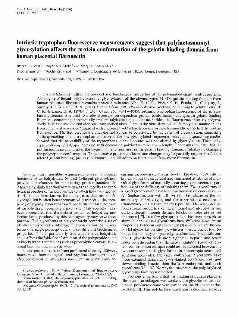
Fig. 4. Fluoresccwce decay of high-molecular-mass GB4 glycoform. Fluorescence decay of fraction 29 from Fig. 2 measured at 295 nm excitation wavelength, 330 nm emission wavelength, 10°C. The data were tit by global analysis of decay curves at six emission wavelength (320 - 370 nm, 10-nm intervals) to three exponentials plus a term for scattered light: c( = 0.793, T~ = 0.49 ns, xz = 0.145, z2 = 2.63 ns, x3 = 0.041, z3 = 7.13 ns, a = 0.021; partial xl = 0.93. C,a, + a = I . Right noisy curve: sample decay; left noisy curve: reference decay; smooth curve: best fit to data. The percentage residuals and autocorrelation function (inset) are also shown
Table 2. Fluorescence decay data of high- and lon~~-mirlecular-mass GB44 glycofoums Results from global analysis of decay curves at six emission wave- lengths with scattered light correction
1 T i I i (at 350 nm) xl vc;,'i
ns nm Fraction 29 1 0.49 0.26 1.07 344 2 2.63 0.36 345 3 7.1 3 0.37 3 SO Fraction 37 1 0.44 0.22 1.14 342 2 2.68 0.12 344 3 7.26 0.12 349
ponential function were unsuccessul. Fig. 4 shows a decay curve for fraction 29 at 330-nm emission wavelength together with the best fit to three exponentials plus scatter obtained by global analysis of the six curves acquired at different emission wavelengths. The global x,? = 1 .I indicates an excellent fit of the data. However, small nonrandom fluctuations persist in the autocorrelation function (inset), suggesting that the scatter term may represent an unresolved fast exponential decay.
Table 2 summarizes the time-resolved fluorescence data for fractions 29 and 37. The similarity of the lifetime values for the two glycoforms implies that the fluorescence lifetimes of GB44 are not affected by carbohydrate chain length. This conclusion was tested by simultaneous analysis of the decay data for fractions 29 and 37, which constrains the lifetimes for all 12 curves to be the same. The combined 12-curve global analysis gave about the same values for x: and the decay parameters as the separate six-curve global analyses.
Table 2 also gives the intensities li at 350 nm and the centers of gravity in nm vL;,\ of the emission spectra associated
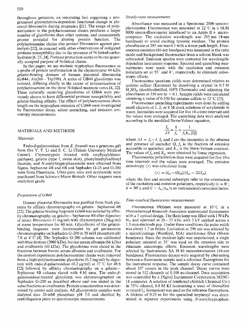
Table 3. Acrylamide quenching of high- and lowmoleculau-mass GB44 glycoforms Determined at 295 nm excitation wavelength, 350 nm emission wave- length
Fraction . f a Ksy (5)
29 37
M- ' ns 0.84 13.9 3.7 0.75 13.0 2.8
with the three exponential decays. The intensity of the 0.5-11s component decreased about 15%, whereas the intensities of the 2-ns and 7-11s components decreased about 65% in fraction 37 compared to fraction 29. Such intensity drops in the ab- sence of lifetime drops suggest static quenching of the trypto- phan fluorescence in the low-molecular-mass glycoform. The 2-ns and 7-11s components each account for almost 40% of the emission intensity at 350 nm in fraction 29, but less than 30% in fraction 37. The preferential quenching of the longer- lived emission in fraction 37 results in an apparent decrease of about 1 ns in the value of the mean lifetime ( T ) = Lih(l")~i at 3.50 nm (Table 3). For both fractions 29 and 37, the decay- associated spectra are closely overlapped, with the spectrum of the 7-11s component shifted about 5 nm to the red. This is apparent in the wavelength dependence of the mean lifetime (z), which increased about 2 ns between 320-370 nm (not shown). Since the centers of gravity of the decay-associated spectra differ little if at all in the two protein fractions, the small blue shift in the steady-state emission spectrum of GB44 with decreasing lactosamine chain length (Fig. 2) is probably due to quenching of the red-shifted emission from the 7-11s component.
Acvylumide quenching
The accessibility of the tryptophan emission in GB44 to small solute molecules was determined by acrylamide quenching experiments. The fluorescence intensity at 350 nm was measured as a function of acrylamide concentration for fractions 29 and 37. The Stern-Volmer plots of the quenching data for both protein fractions showed downward curvature, which suggests that part of the emission is inaccessible to acrylamide (not shown). The modified Stern-Volmer plots were linear (Fig. 5). The values of the quenching parameters are given in Table 3. The fraction of emission accessible to acrylamidef, was roughly the same in the two protein samples, despite the fact that 50% of the fluorescence intensity of fraction 29 is absent in fraction 37. This suggests that the drop in fluorescence intensity of GB44 with decreasing lactosamine chain length involves both exposed and buried tryptophans. The Stern-Volmer constant K,, for the accessible emission is likewise about the same in the two protein fractions. In pro- teins with multiple tryptophans, the apparent Stern-Volmer constant derived from the modified plot is a complicated function of the quenching constants Ksv,i for individual resi- dues. At low concentrations of quencher the inverse slope f ,Ksv of the modified plot is equal to the effective Stern-Volmer constant K,,(eff) = CjfjKsv,j, wherefj is the fractional intensity of tryptophan j [31]. In the absence of static quenching, the individual Stern-Volmer constants are Ksv,j = k,,jzj, where k,,j is the collisional quenching rate constant and z j is the fluorescence lifetime in the absence of quencher. Assuming that K,,(eff) z k,(app)(z), we estimate an apparent quench-

514
7
6
5
Z L
3 \ 0 -
2
1 0 10 20 30 LO 50 60
l/[Ql
Fig. 5. Modified Stern-Volmer plots of' acrylamide quenching data uf higk- and low-molecular-mass GB44 glycoforms. Measured at 295 nm excitation wavelength, 350 nm emission wavelenght, 22°C: (0) frac- tion 29 and ( 0 ) fraction 37
ing rate constant of about 3 x lo9 M- ' s- ' for both fractions 29 and 37. This high value indicates that the accessible trypto- phans in GB44 are on the surface of the glycoprotein [32].
Anisotropy
The fluorescence emission anisotropy of GB44 increased with decreasing lactosamine chain length (Table 1). The rela- tive changes in the anisotropy values were about the same at 350 nm and 370 nm emission wavelength (not shown), in- dicating they are not due to light scattering in the more weakly fluorescent fractions. The steady-state anisotropy ( r ) depends on the ratio of the fluorescence lifetime z and the rotational correlation time 4 . For isotropic rotation of a rigid fluoro- phore with a single lifetime
( r ) = ro/(1 + z / 4 ) , where r, is the limiting anisotropy in a frozen solution. Typical r, values for tryptophan in proteins are 0.17-0.19 at 295 nm excitation wavelength [3]. Substituting these r, values and the mean lifetime (z) into the above equation gives a crude estimate of the apparent rotational correlation time of GB44: 4 = 2.1 -2.5 ns for fraction 29 and 4 = 9- 16 ns for fraction 37. The rotational correlation time of a sphere is 4 = vhy/kT, where v h is its hydrodynamic volume, y is solvent viscosity, k is the Boltzmann constant, and T is absolute temperature. For globular proteins of 40 - 60 kDa, the expected rotational correlation times in aqueous solution at 22°C are about 16- 23 ns. The value of about 2 ns estimated for fraction 29 suggests that the fluorescent tryptophans have greater internal mobility in the high-molecular-mass glycoform than in the low-molecular-mass glycoform.
DISCUSSION
We have compared the intrinsic tryptophan fluorescence of a series of polylactosamine glycoforms of the gelatin-binding domain GB44 of human placental fibronectin. GB44 glyco- forms with high amounts of polylactosamine on the three N- linked carbohydrate chains had greater fluorescence intensity, red-shifted emission spectra, and lower anisotropy values. However, the fluorescence lifetimes and accessibility to solute quencher were essentially the same in high- and low-molecular- mass GB44 glycoforms. The fluorescence intensity and ani- sotropy changes and the emission spectral shifts report differ-
ences in the tryptophan environments of GB44 in the various glycoforms. These could be due to changes in the protein conformation or in the interactions between surface trypto- phans and the carbohydrate chains. The gelatin-binding do- main of fibronectin has nine tryptophans in its 340-amino- acid sequence [34]. The presence of multiple tryptophans in GB44 complicates interpretation of the fluorescence results. The tryptophans are located at positions 291, 354, 414, 453, 475, 522, 545, 566, and 599. Eight of them occur in the four type-I and two type-I1 homology units and one is at the C- terminus. There are two disulfide bridges in each of the six homology units. Five of the tryptophans are one or two resi- dues away from disulfides, which quench tryptophan fluores- cence [35]. Isaacs et al. [30] showed that the unusually low quantum yield of the plasma glycoform is due in part to the disulfides.
The decrease in tryptophan fluorescence of GB44 with decreasing carbohydrate chain length could be caused by static and dynamic quenching processes. Static quenching is due to formation of nonfluorescent ground-state complexes. These could arise in GB44 from interactions between tryptophans and carbohydrate as well as polypeptide functional groups. Dynamic quenching is due to competing nonradiative path- way for deactivation of the excited state, such as collisional quenching, excited-state reactions, and energy transfer. Diffu- sive motion of the carbohydrate chains of GB44 might quench the fluorescence of surface tryptophans. Tryptophan itself is susceptible to dynamic quenching by excited-state proton and electron transfer reactions [36]. In GB44, quenching reactions involving tryptophan and functional groups on the polypep- tide or the carbohydrate may occur. Potential quenching groups on the polylactosamine chains are the carbonyl groups of the internal N-acetylglucosamine and the terminal sialic acids. Alcoholic hydroxyl groups do not quench the fluores- cence of the indole chromophore [37]. Finally, tryptophan - tryptophan energy transfer appears to occur in other multitryptophan proteins [38, 391. In principle, fluorescence decay measurements can distinguish static (amplitude drops) and dynamic (lifetime drops) quenching. We have resolved the fluorescence decay of GB44 into three exponential components and have found no evidence for lifetime decreases in the low-molecular-mass glycoform. The three exponential decays of about 0.5, 2, and 7 ns represent an empirical fit of the data. Even single tryptophans in proteins typically exhibit complex fluorescence decays [40] comprised of discrete life- times or continuous lifetime distributions [41]. With only par- tial resolution of the fluorescence decay of GB44, it is imposs- ible to exclude lifetime drops in which emission has transferred from a longer to a shorter lifetime component. However, the emission of all three lifetime components was quenched in the low-molecular-mass glycoform with no significant shifts in the decay-associated spectra. Thus, static quenching appears to be a reasonable explanation of the decrease in fluorescence intensity of GB44 with decreasing polylactosamine chain length.
The fluorescence lifetimes also help clarify the solute quenching and anisotropy results. In multitryptophan pro- teins the apparent steady-state values are complicated func- tions of the parameters for individual tryptophans. We have used the mean lifetime values to estimate apparent values of the quenching rate constants and rotational correlation times. The time-resolved measurements were performed at lower temperature (10 "C compared to 22 "C) to preserve the protein samples during the long data acquisitions. The fluorescence intensity of GB44 does not vary dramatically in this tempera-

51 5
ture region, so we do not expect the mean lifetimes to be highly temperature-dependent. The apparent values of the quenching rate constants and rotational correlation times are useful for qualitative comparisons of the high- and low-molecular-mass glycoforms of GB44. However, the conclusions are not defini- tive, because the populations of fluorescent tryptophans may not be the same in the two glycoproteins. Nevertheless, the valueofabout 3 x 10' M-'s-'forthequenchingrateconstant indicates that the solute-accessible emission in GB44 comes from surface tryptophans. The polylactosamine chains do not appear to interfere with acrylamide quenching of surface tryp- tophans in either the high- or low-molecular-mass glycoforms. The low apparent values of the correlation times indicate internal motions of the tryptophans in GB44. The tryptophan environments appear to become more rigid with decreasing polylactosamine chain length.
The fluorescence results are consistent with glycosylation- dependent changes in protein conformation. The polar emission maxima and large solute quenching rate constants argue that most of the fluorescence in GB44 is due to surface tryptophans exposed to solvent. Our tentative explanation for the effects of carbohydrate chain length is a mechanical model, in which the highly solvated polylactosamine chains diffusing in solution tug on the protein surface. We image that this would loosen the surface structure and reduce amino acid side-chain contacts, thereby increasing the motional freedom of surface tryptophans and relieving static quenching of their fluorescence. An alternative explanation would be that the polylactosamine chains lie along the protein surface and per- turb the protein structure. NMR studies of the solution con- formation of N-linked oligosaccharides showed that the six- arm in complex structures can fold back over the mannose core [42]. Low-angle neutron and X-ray scattering studies of th al-acid glycoprotein glycoforms revealed two modes of protein - carbohydrate interactions in the single lactosamine- type complex chains [17]. The biantennary chains were loosely bound to the protein surface, while the tri- and tetraantennary chains were tightly anchored through their sialic acid termini. Although we cannot exclude the possibility of tryptophan - carbohydrate interactions in the GB44 glycoforms, we tend to discount this interpretation of the fluorescence results on two grounds. First, the tryptophans are spaced about 20 - 60 residues apart throughout the peptide sequence. We would expect that the longer polylactosamine chains would have more opportunity to interact with surface tryptophans and quench their fluorescence, which is opposite to our findings. And second, the most likely quenching agent on the carbo- hydrate is the carbonyl group of the N-acetylglucosamines and sialic acids. However, the greatest quenching is observed when the carbohydrate is trimmed close to the mannose core by endo-j-galactosidase.
The GB44 glycoforms used in this investigation are mix- tures which have been fractionated on the basis of size. It is not possible with current technology to separate the polylactosamine series into individual molecular idiotypes. The fractions from gel permeation chromatography represent relatively narrow distributions of glycoforms with reproduc- ible centroids. The heterogeneity within a given fraction con- sists mainly in the degree of polymerization of lactosamine subunits on the arms of N-linked tetraantennary chains. The three glycosylated asparagines in the gelatin-binding domain of fibronectin are located at positions 399, 497, and 511 [34]. An average of two of these sites are occupied in the chymotryptic gelatin-binding fragment from plasma, while all three glycosylation sites are occupied in the analogous
fragment from placenta [4]. The plasma glycoform has only biantennary chains, whereas the placental glycoforms have about 30% biantennary, no triantennary, and 70% tetra- antennary carbohydrate [22]. Since none of the glycosylation sites are adjacent to tryptophans, we consider the aggregate effect of the lactosamine subunits to be responsible for the observed differences in the tryptophan fluorescence of the GB44 glycoforms. Although carbohydrate sequence-specific effects may play a role, we favor the notion that the length of the polylactosamine chains is the important variable in this system. In this regard we note that the largest fluorescence changes occur when the apparent molecular mass increases from 52 kDa to 58 kDa (with carbohydrate increasing from about 17 kDa to 23 kDa), suggesting a threshold phenom- enon.
In conclusion, our data show that polylactosamine glycosylation on N-linked chains increases the fluorescence quantum yield of tryptophans in the gelatin-binding domain from fetal tissue fibronectin. The effect can be reversed by removal of the lactosamine oligomers. These long-chain oligosaccharides, which have been shown to inhibit both pro- teolysis and gelatin-binding of this polypeptide, apparently change the conformation of the protein as manifested by changes in the quantum yield and emission anisotropy of the intrinsic tryptophans. The results reported here give the first evidence that polylactosamine elongation of N-linked com- plex saccharides can incrementally alter protein conformation. This suggests that the observed functional differences [4, 12, 221 in various glycoforms of fibronectin and its specific bind- ing domains may be caused not only by steric interference with binding sites, but by subtle changes in the protein struc- ture. Thus, organisms have another mechanism to control functions of a single polypeptide gene product by post-trans- lational modification.
This work was supported by National Institutes of Health Grants GM32594 to R. A. L. and GM35009 to M. D. B.
REFERENCES 1.
2. Kornfeld, S. (1985) Annu. Rev. Biochem. 54,631 -664. 3. Peterson, C. B. & Blackburn, M. N. (1985) J . Bid. Chem. 260,
4. Zhu, B. C. R. & Laine, R. A. (1985) J . Bid. Chem. 60, 4041-
5. Rademacher, T. W., Parekh, R. B. & Dwek, R. A. (1988) Annu.
6. Chu, F. K., Trimble, R. B. & Maley, F. (1978) J . Biol. Chem. 253,
7. Cheng, S.-Y., Morrone, S. & Robbins, J. (1979) J . Bid. Chem.
8. Shifrin, S., Consiglio, E., Laccetti, P., Salvatore, G. & Kohn, L.
9. Shifrin, S., Consiglio, E. & Kohn, L. D. (1983) J . Bid. Chem.
10. Grimaldi, S., Robbins, J. & Edelhoch, H. (1985) Biochemistry24,
11. Keutmann, H. T., Johnson, L. & Ryan, R. J. (1985) FEBS Lett.
12. Jones, G. E., Arumugham, R. G. & Tanzer, M. L. (1986) J . Cell Biol. 103, 1663 - 1670.
13. Basu, S., Mandal, C. &Allen, A. K. (1988) Biochem. J . 254,195- 202.
14. Sakiyama, H., Matsushita, E., Kuwabara, I., Nozue, M., Takahashi, T. & Taniguchi, M. (1988) Cancer Res. 48, 7173- 7178.
Pazur, J. H., Knull, H. R. & Simpson, D. L. (1970) Biochem. Biophqis. Res. Commun. 40, 110- 116.
610-615.
4045.
Rev. Biochem. 57, 785-838.
8691 -8693.
254,8830-8835.
D. (1982) J . Biol. Chem. 257, 9539-9547.
258, 3780- 3786.
3771 - 3776.
185,333-338.
53 6
15. Seidel-Dugan, C., Ponce de Leon, M., Friedman, H. M., Fries, L. F., Frank, M. M., Cohen, G . H. & Eisenberg, R. J . (1988) J . Virol. 62, 4021 - 4036.
16. Bayard, B. & Kerckaert, J.-P. (1980) Biochem. Biophys. Kes. Commun. 95, 771 - 184.
17. Perkins, S. J., Kerckacrt, J.-P. & Loucheux-Lefebvre, M. H. (1 985) Eur. J . Biochem. 147, 525 - 531.
18. Hoffman, S., Sorkin, B. C., Perrin, W. C., Brackenberry, R., Mailhammer, R., Rutishauser, U., Cunningham, B. A. & Edelman, G. M. (1982) J . Biol. Chem. 257,7720-7729.
19. Finne, J . , Finne, U., Deagostini-Bazin, H. & Goridis, C. (1983) Biocheni. Biophys. Res. Commun. 112, 482 -487.
20. Hoffman, S. & Edelman, G. M. (1983) Proc. Nut/ Acud. Sci. USA
21. Zhu, B. C. R. & Laine, R. A. (1987) Arch. Biochem. Biophys. 252, 1-6.
22. Zhu, B. C. R., Fisher, S. F., Pande, H., Calaycay, J., Shively, J . E. & Laine, R. A. (1984) J . Biol. Chem. 259, 3962-3970.
23. Sairam, M. R . & Manjunath, P. (1982) Znt. J. Peptide Protein Res. 19, 315-320.
24. Knudsen, K. A. (1 985) J . Cell. B id . 101, 891 -897. 25. Melhuish, W. H. (1961) J . Phys. Chem. 65, 229-235. 26. Kolber, Z. S. & Barkley, M. D. (1986) Anal. Biochem. 152, 6-
27. Knutson, J. R., Beechem, J . M. & Brand, L. (1983) Chem. Phys.
80, 5762- 5166.
21.
Lett. 102, 501 - 507.
28. Lakowicz, J. R. & Hogcn, D. (1981) Biochemistry 20, 1366-
29. Sun, M. & Song, P.-S. (1977) Photachem. Phatobiol. 25, 3-9. 30. Tsaacs, B. S., Brew, S. A. & Ingham, K. C. (1989) Biochemistry
31. Lehrer, S . S. & Leavis, P. C. (1978) Methuds Enzymol. 49, 222-
32. Eftink, M. R. & Ghiron, C. A. (1981) Anal. Biochem. 114, 199-
33. Lakowicz, J. R., Maliwal, B. P., Cherek, H. & Balter, A. (1983)
34. Skorstengaard, K., Jensen, M. S., Sahl, P., Petersen, T. E. &
35. Cowgill, R. W. (1967) Biochinz. Biophys. Acta 140, 31-44. 36. Creed, D. (1984) Photochem. Photohiol. 3Y, 531-562. 31. Feitelson, J. (1970) Israel J . Chem 8, 241 -252. 38. Desie, G., Boens, N. & De Schryver, F. C. (1986) Biochemistry
39. Eftink, M. R., Wasylewski, Z. & Ghiron, C. A. (1987) Biochemis-
40. Beechem, J. M. & Brand, L. (1985) Annu. Rev. Bioclzenz. 54,43-
41. Alcala, J . R., Gratton, E. & Prendergast, F. G. (2987) Biophys.
42. Brisson, J.-R. &Carver, J. P. (1983) Biochemistry 22,3680-3686.
1373.
28, 842-850.
236.
221.
Biochemistry 22, 1741 - 1752.
Magnusson, S. (1986) Eur. J . Biochem. 61,441 -453.
25, 8301 -8308.
try 26, 8338 - 8346.
71.
J . 51, 597 - 604.