introduction - shodhgangashodhganga.inflibnet.ac.in/bitstream/10603/33784/8/5...2 introduction p....
TRANSCRIPT

1
P. Sathyadevi, 2011
Chapter I
Introduction
Earlier, chemistry, as a science discipline was divided into different subfields
such as analytical, industrial, inorganic, organic, physical and polymer chemistry. Later,
chemistry has become one of the very interesting inter disciplinary subjects owing to its
role in other related science subjects. The field of coordination chemistry that had begun
more than a century ago, has registered a phenomenal growth during last few decades.
There has been a tremendous interest in the pursuit of coordination compounds from the
days of Werner, whose coordination theory opened new avenues to think about the
structure and reactions of coordination compounds.
It is well known fact that precious metals have been used for medicinal purposes
for atleast 3500 years. At that time, precious metals were believed to benefit health
because of their rarity, but research has now well established the link between medicinal
properties of inorganic drugs and specific biological properties. The elucidation of a drug
mechanism is however complex and the activity of many drugs remain unknown. Many
naturally occurring coordination compounds are proved to be vital for living organisms.
The metabolic activity of biological compounds like vitamin B12, chlorophyll and
hemoglobin had been a puzzle to all chemists and induced them to think seriously about
the fundamental studies on the principles of coordination chemistry. Among the
coordination compounds, transition metal complexes constitute a distinct class that are
widely studied due to their interesting structural features and captivating application in
diverse fields such as catalysis, molecular electronics and medicine.
The chemistry of transition elements is being investigated for over several
decades with so much of newer concepts and exciting results. During the past few
decades, these elements and their compounds were proven to be a nearly ideal touchstone
for many of the models that have been developed to understand the principles of structure
and bonding in coordination compounds. Most of the first row transition metals are
biologically essential, with a number of their complexes demonstrating potential

2
Introduction
P. Sathyadevi, 2011
bioactivities such as DNA binding, DNA cleavage, protein binding, cytotoxicity etc.
Among them, nickel, cobalt, copper and ruthenium are of significant and are described in
the following discussions. Recent years have witnessed a phenomenal growth in the
coordination chemistry of transition metal complexes of Schiff base ligands because of
their biochemical significance.
Transition metal complexes act as homogeneous catalysts in many industrially
important reactions such as hydrogenation, hydrosilation, hydroformylation,
polymerisation, isomerisation, acylation and oxidative hydrolysis of olefins. Binuclear
transition metal complexes have also received much attention in recent years and interest
in a particular system is stimulated by a number of factors. Bimetallic coordination
complexes may serve as model for variety of biological reactions such as oxygen
transport, oxygen activation, photosynthesis, electron transfer process, metal-metal
interactions and multi centered catalysis.
Transition metal ions with different oxidation states have a strong role in
bioinorganic chemistry and redox enzyme systems and may provide the basis of models
for active sites of biological systems. To mention a few, nickel is an essential component
in atleast 4 types of enzymes viz. urease, carbon monoxide dehydrogenase (CODH) or
acetyl coenzyme A synthase, hydrogenase and methyl-S-coenzyme M reductase. There is
spectroscopic evidence for nickel-methyl complexes in CODH of anaerobic bacteria. The
interaction of Co(II) complexes in solution with O2 has been the subject of intensive
study. Some of these complexes behave as reversible carriers of O2 and have been used as
models for natural oxygen transport systems as reported by Macke and Williams 1988. In
2003, Cotton et al studied Schiff base complexes such as [Co2+
(acacen)(DMF)O2], which
in solution (pyridine, DMF or similar solvents) pick up O2. Polydentate Co(II) complexes
with ligands capable of intercalation into DNA strands are capable of inducing DNA
cleavage under photochemical conditions were studied by Bhattacharya and Mandal.
Since the recognition of Vitamin B12 and synthesis of cobalmines are responsible for
erythrocytes (RBC) formation in human body, much study of model systems was
undertaken. Copper complexes are known to play important role in the active site of

3
Introduction
P. Sathyadevi, 2011
many copper proteins in vivo as well as in homogeneous and heterogeneous catalysis for
organic chemical reactions. A large body of evidence indicates that copper chelation is an
effective method to inhibit tumor growth and copper chelators have become promising
agents in the treatment of cancer. It has been found that some copper chelators acquire
more effective or novel bioactivity after forming complexes. Recent studies indicated that
Cu2+
was critically needed for PDTC induced apoptosis in HL-60 cells and the copper
complex of salicylaldehyde benzoylhydrazone (SBH) derivatives showed increased
potency of growth inhibition in several cancer cell lines, compared with the metal-free
organics. Copper-SBH complexes were significantly more cytotoxic than complexes of
other transitional metals (Cu > Ni > Zn > Mn > Fe > Cr > Co) in MOLT-4 cells, an
established human T-cell leukaemia cell line. It was noted that SBHs, especially their
copper complexes appeared to be unusually potent inhibitors of DNA synthesis and cell
growth in a variety of human cancer cell lines, including the human lung epithelial cancer
cell line, SKMES-1 and rodent cancer cell lines. A series of, novel salicylaldehyde
pyrazole hydrazone (SPH) derivatives were found capable of inhibiting the growth of
A549 lung carcinoma cells.
Ruthenium complexes are versatile in the field of catalysis of many organic
reactions. Ruthenium complexes are of importance not only because of their use in
catalytic reactions like oxidation and hydrogenation, reduction, dynamic kinetic
resolution, racemisation but also to their medicinal properties. Ruthenium compounds are
well suited for medicinal applications due to the properties such as
Rate of ligand exchange
The range of accessible oxidation states
The ability of ruthenium to mimic iron in binding to certain biological molecules.
Ruthenium anticancer chemistry has already yielded many promising results.
Several compounds display activities comparable to that of cisplatin, and in some cases,
activities are even better. Ruthenium complex ImH[trans-Ru(III)Cl4(DMSO)Im] (Im =
imidazole) was the first to enter clinical trials against metastases and KP 1019 has been
introduced into phase I clinical trials against colon carcinomas and their metastases. More

4
Introduction
P. Sathyadevi, 2011
recent studies demonstrate that ruthenium(II) complexes containing polypyridyl ligand
also show high antitumor activity in vitro and these complexes can effectively inhibit the
cell proliferation. Further, many octahedral ruthenium(II) complexes have also been
proved to bind with DNA through intercalative mode. The complex [Ru(bpy)2(dppz)]2+
(dppz = dipyrido[3,2-a:2',3'-c]phenazine) acted as ‘‘molecular light switch’’ and exhibits
cytotoxic activity at low micromolar IC50 values.
Hydrazones
Intensive research on the physicochemical properties and molecular structure of
complexes with organic derivatives of hydrazine has provided several new results. One of the
reasons for significant attention paid to the formation of complexes of these ligands with
transition metals are their biological activity.1-11 General formula of acid hydrazones is given
in Fig. 1.1.
Fig. 1.1 General formula of acid hydrazones.
Hydrazones are the azomethines, characterized by the tri-atomic grouping
C=N‒ N in their molecule. Hydrazones are of great importance because of their ability
towards complexation with metal ions and their involvement in diversified biological
processes those are attributed to the formation of stable chelates with transition metals
that catalyze the corresponding physiological processes. Aroylhydrazones of carbonyl
compounds coordinate to a metal ion via phenolate- or enolate-O, imine-N and
deprotonated amide-O atoms. With flexible ligands, the competition between bridging
and chelating coordination modes is an important factor in producing mono, di and
polynuclear metal complexes.

5
Introduction
P. Sathyadevi, 2011
R1 N
NR3
R1 N
N R3
R1 N
N R3
R2 H R2
R2
O O O
-H
Fig. 1.2 General formula of acyl-/aroyl-hydrazones and mesomerism of the anion obtained by deprotonation.
In coordination chemistry, hydrazones find application as multidentate ligand
forming chelates with metals, usually from the transition series. Studies have shown that
the azomethine group having a lone pair of electrons in either a p or sp2 hybridized orbital
on trigonally hybridized nitrogen has considerable biological importance. There are
several reports on the synthesis, structural characterization and biological activities of
transition metal complexes containing different types of hydrazone ligands including
Co(II), Ni(II), Cu(II), Cd(II), V(II), Pd(II) and Ru(II).12-29
Diversity in the chelation of hydrazones
The ‒ NH‒ C=O functional group present in hydrazones makes them to behave as
bidentate ligand for metal ions. Acid hydrazones show keto-enol (amido-iminol) tautomerism
as shown in the Fig. 1.3. In solid state, they exist in keto form but in solution, they exist as an
equilibrium mixture of keto and enol forms.
R2
N
R1
HN
R3
O
R2
N
R1
N
R3
OH
Amido form (I) Iminol form (II)
Fig. 1.3 Tautomerisation of hydrazone ligands.
These compounds are expected to exist in a trans form, but in such situation, they
may act as a unidentate ligand, by bonding through enolate oxygen. It is well evident that the
stereochemistry of the ligand is much decided by the steric effects of the various substituents
in the hydrazone moiety. It was found that most of the hydrazones coordinate to the metal ion
through cisform. This phenomenon is assumed to be due to better electron delocalisation
in the chelate ring system that increases the stability upon coordination with metal atoms.
The composition and structure of the complexes are determined by the electronic structure of

6
Introduction
P. Sathyadevi, 2011
metal as well as preparation conditions.30,31 Hydrazones form coordination compounds
through the oxygen atom of either carbonyl32,33 or the enol group and through the imine
nitrogen atom, a five membered chelate ring is being produced as shown in Fig. 1.4.
R2
N
R3
N
R1 OM
Fig. 1.4 General bidentate coordination mode of acid hydrazones.
Hydrazones may behave as a terdentate chelating agent if R1 residue provides a donor
atom in a suitable position, as it occurs in salicyloylhydrazide derivatives, although
hydrazones act commonly as ON donor ligand if R2 and R3 do not show chelating
tendency.34-36 Terdentate ligands may also be obtained if R2 and/or R3 residues are groups
such as o-hydroxyphenyl (ONO) ligands.37-39 Chelating ligand with higher number of
coordination position may be prepared by convenient selection of the condensation products:
Neutral tridentate Uninegative tridentate
N
HN
O
MN
N
N
O
MN
N
HN
OM O
NN
OM O
Uninegative tridentate Binegative tridentate
Uninegative bidentateNeutral bidentate Uninegative bidentate
N
HN
O
M
N
N
O
M
HN
HN
O
M
Fig. 1.5 General coordination modes of acid hydrazones.

7
Introduction
P. Sathyadevi, 2011
aroyl hydrazide (R‒ CONHNH2) and di or tri ketones.40-47 The oxidation state of the metal
ion
plays an important role in predicting the stability of the complexes of acid hydrazones. The
stability of the complexes also increases with an increase in electron delocalisation, size of
the molecule and also the nature and size of the chelate rings formed.48 General coordination
modes of hydrazones are presented in Fig. 1.5.
Applications
The chemistry of transition metals with ligands from the hydrazine family has
been of interest to coordination as well as bioinorganic chemists due to their versatile
bonding modes with both electron-rich and electron-deficient metals.49-52
Heterocyclic
hydrazones constitute an important class of biologically active drugs that have attracted
the attention of medicinal chemists due to their wide range of pharmacological properties
like antifungal, antibacterial, anticonvulsant etc.53-57
The metallochemistry of biomolecules has been largely focused on proteins,
amino acids, nucleic acids and carbohydrates.58-61
During the last two or three decades,
there has been an increasing attention on the binding study of small molecules to DNA,
since it is an important genetic substance in living organisms.62-64
Errors in gene
expression can often cause diseases and play a secondary role in the outcome and severity
of human diseases.65
Hence, a complete understanding of DNA-drug binding is
significant in the rational design of DNA structural probe, DNA footprinting, sequence-
specific cleaving agents and potential anti-cancer drugs.66-69
To design effective
chemotherapeutic agents and better anticancer drugs, it is essential to explore the
interaction of metal complexes with DNA.
Serum albumin is the major soluble protein constituent in the circulatory system
of a wide variety of organisms and it has the ability to reversibly bind to a large variety of
endogenous and exogenous ligands such as fatty acids, drugs and metal ions in the
bloodstream.70,71
The drug-protein complex not only strongly affects the absorption,
distribution, metabolism and excretion properties of drugs, but also influence the drug
stability and toxicity during the chemotherapeutic process. Therefore, knowledge on the

8
Introduction
P. Sathyadevi, 2011
mechanism of interaction between the selected drug and protein has become very vital to
design several new drugs with improved potential.72,73
Many chemicals exert antitumor effects through binding to DNA thereby
changing the replication of it and inhibiting the growth of tumor cells, which is the basis
to design new and more efficient antitumor drugs and their effectiveness depends on the
mode and affinity of the binding. The task of the synthetic inorganic chemists is not only
confined with the synthesis of new complexes having well defined structures, but also to
understand the structure-activity relationship of them.
Hence, we decided to study the chelating properties of hydrazone ligands and the
effect of coordination of them on the composition and geometry of the complexes
containing cobalt(II), nickel(II), copper(I/II) and ruthenium(II) ions were undertaken as
the subject of the present work. Additionally, comparison on the biological properties of
hydrazone complexes with respect to DNA / BSA interactions, free radical scavenging
and cytotoxicity under in vitro conditions have been carried out.
Literature survey
There are numerous reports available in the literature describing the reactions of
transition metal complexes containing hydrazones with various coordination modes and
pharmacological properties. In this connection, some important reports related to our
research work that were published very recently are presented in the following section of
this chapter.
From the reaction between Cu(II) and Ni(II) salts and the Schiff base 6-amino-5-
formyl-1,3-dimethyluracil-benzoylhydrazone (H2BEZDO), in dimethylformamide
(DMF)-water and ammonia media, the complexes [Ni(BEZDO)]·H2O·NH3 (1),
[Cu(BEZDO)]·H2O·NH3 (2) and [Cu(BEZDO)(H2O)]·H2O (3) containing the dianionic
organic ligand have been obtained. These compounds have been studied by IR, electronic
and EPR spectroscopy and magnetic measurements. The structure of
[Cu(BEZDO)(H2O)]H2O has been solved by means of X-ray diffraction methods. The
coordination environment around the Cu(II) may be described as a square planar structure

9
Introduction
P. Sathyadevi, 2011
in which the Schiff base acts as tridentate ligand through the N(6), N(51) and O(52)
atoms, making two five and six membered chelate rings. The coordination sphere is
completed with the oxygen atom of a water molecule.74
NN
ONH
Cu
OHH
O
O
H3C
CH3
3
Cobalt(III) complexes of diacetyl monooxime benzoyl hydrazone (dmoBH2) and
diacetyl monooxime isonicotinoyl hydrazone (dmoInH2) have been synthesized and
characterized by elemental analyses and spectroscopic methods by Naskar et al.75 The X-
ray crystal structure of the hydrazone ligands, as well as that of the cobalt(III) complex
[CoIII
(dmoInH)2]Cl·2H2O (1) were also reported in which the amide and the oxime
hydrogens are deprotonated in both the ligands, while the isonicotine nitrogens are
protonated. In the [CoIII
(dmoBH)2]Cl (2) complex, only the amide nitrogens are
deprotonated. It was shown that the additional hydrogen bonding capability of the
isonicotine nitrogen results in different conformation and supramolecular structure for
dmoInH2, compared to dmoBH2, in the solid state. Comparing the structure of the
[CoIII
(dmoInH)2]Cl·2H2O with that of the Zn(II) complex of the same ligand reported
earlier, it is seen that the metal ion has a profound influence on the supramolecular
structure due to change in geometrical dispositions of the chelate rings.
H3CN
N
NO
N+
CoH3C
O-
N+
NO
NCH3
CH3N
-O
Cl-
1
H H
10
Introduction
P. Sathyadevi, 2011
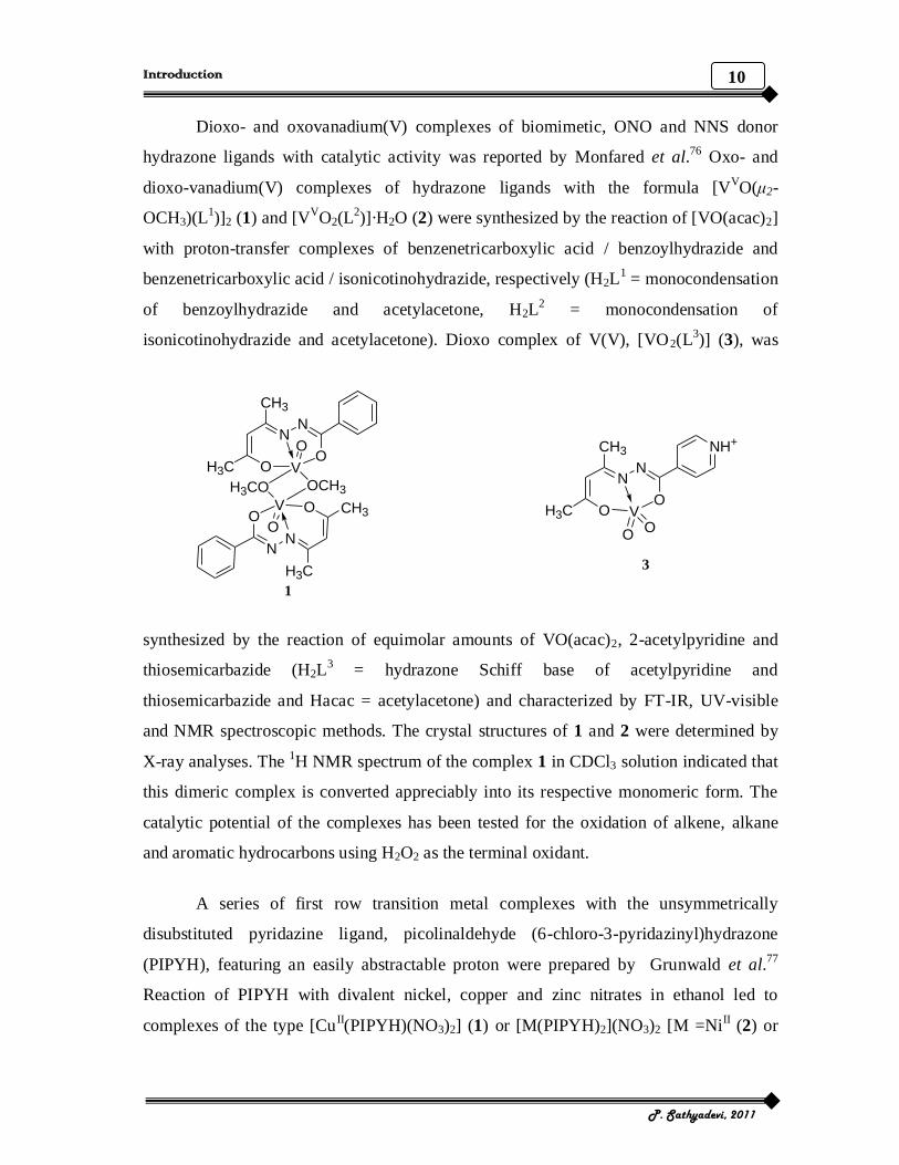
Dioxo- and oxovanadium(V) complexes of biomimetic, ONO and NNS donor
hydrazone ligands with catalytic activity was reported by Monfared et al.76 Oxo- and
dioxo-vanadium(V) complexes of hydrazone ligands with the formula [VVO(μ2-
OCH3)(L1)]2 (1) and [V
VO2(L
2)]·H2O (2) were synthesized by the reaction of [VO(acac)2]
with proton-transfer complexes of benzenetricarboxylic acid / benzoylhydrazide and
benzenetricarboxylic acid / isonicotinohydrazide, respectively (H2L1 = monocondensation
of benzoylhydrazide and acetylacetone, H2L2 = monocondensation of
isonicotinohydrazide and acetylacetone). Dioxo complex of V(V), [VO2(L3)] (3), was
H3C
NN
OO
CH3
V
O
OCH3
CH3
NN
OO
H3C
V
O
H3CO
H3C
NN
OO
NH+CH3
V
OO
1
3
synthesized by the reaction of equimolar amounts of VO(acac)2, 2-acetylpyridine and
thiosemicarbazide (H2L3 = hydrazone Schiff base of acetylpyridine and
thiosemicarbazide and Hacac = acetylacetone) and characterized by FT-IR, UV-visible
and NMR spectroscopic methods. The crystal structures of 1 and 2 were determined by
X-ray analyses. The 1H NMR spectrum of the complex 1 in CDCl3 solution indicated that
this dimeric complex is converted appreciably into its respective monomeric form. The
catalytic potential of the complexes has been tested for the oxidation of alkene, alkane
and aromatic hydrocarbons using H2O2 as the terminal oxidant.
A series of first row transition metal complexes with the unsymmetrically
disubstituted pyridazine ligand, picolinaldehyde (6-chloro-3-pyridazinyl)hydrazone
(PIPYH), featuring an easily abstractable proton were prepared by Grunwald et al.77
Reaction of PIPYH with divalent nickel, copper and zinc nitrates in ethanol led to
complexes of the type [CuII(PIPYH)(NO3)2] (1) or [M(PIPYH)2](NO3)2 [M =Ni
II (2) or

11
Introduction
P. Sathyadevi, 2011
ZnII (3)]. The above synthesis in the presence of triethylamine yielded fully or semi
deprotonated complexes [CuII(PIPY)(NO3)] (4), [Ni
II(PIPYH)(PIPY)](NO3) (5) and
[ZnII(PIPY)2] (6) respectively. Cobalt(II) nitrate is quantitatively oxidized under the
reaction conditions to [CoIII
(PIPY)2](NO3) (7) in both neutral and basic media. X-ray
diffraction analyses reveal a penta (1) or hexa coordinated (2, 3 and 7) metal center
surrounded by one or two tridentate ligands and eventually κ‒ O,O' nitrate ions. The
solid‒ state stoichiometry was confirmed by electron impact (EI) and electrospray
ionization (ESI) mass spectrometry. Diamagnetic complexes 5 and 6 were subjected to
1H NMR spectroscopy suggesting that the ligand to metal ratio remains constant in
solution. Electronic properties were analyzed by means of cyclic voltammetry and in case
of copper complexes 1 and 4, also by electron paramagnetic resonance (EPR) study,
showing increased symmetry upon deprotonation of the latter in accordance with the
proposed stoichiometry [CuII(PIPY)(NO3)].
Six new copper complexes of di-2-pyridyl ketone nicotinoylhydrazone and their
versatile binding properties (HDKN) have been studied by Mangalam et al.78 The
complexes have been characterized by a variety of spectroscopic techniques and the
structure of [Cu(DKN)2]·H2O (1) determined by single crystal X-ray diffraction was
proved to have distorted octahedral geometry. The EPR spectra of compounds
[Cu2(DKN)2(μ-N3)2] (2) and [Cu2(DKN)2(μ-NCS)2] (3) in polycrystalline state suggested
a dimeric structure as they exhibited a half field signal, which indicated the presence of a
weak interaction between two Cu(II) ions in these complexes.
NN
O
N
Cu
NN
O
N
1

12
Introduction
P. Sathyadevi, 2011
The coordination behavior of a new dihydrazone ligand, 2,6-bis[(3-
methoxysalicylidene)hydrazinocarbonyl]pyridine towards manganese(II), cobalt(II),
nickel(II), copper(II), zinc(II) and cadmium(II) has been described by Vadavia et al.79
The metal complexes were characterized by magnetic moments, conductivity
measurements, spectral (IR, NMR, UV-visible, FAB mass and EPR) and thermal studies.
IR spectral studies revealed the nonadentate behavior of the ligand. The X-band EPR
spectra of copper(II) complex at both room temperature and liquid nitrogen temperature
showed unresolved broad signals with giso = 2.106. Cyclic voltammetric studies of
copper(II) complex at different scan rates revealed that all the electrochemical reactions
are irreversible.
O
H3CO
NN
ON
N
O
N
OCH3
OM
Cl ClH2O
MM
OH2 OH2
OH2
H2OH2O
H2O
where, M = Mn(II), Co(II), Ni(II), Cu(II), Zn(II) and Cd(II)
Complexes of Co(II), Ni(II), Cu(II), Mn(II), Cd(II), Zn(II), Hg(II) and U(IV)O2
with N′-(1-(4-hydroxyphenyl)-ethylidene)-2-oxo-2-(phenylamino)acetohydrazide
H3OPAH) have been prepared and characterized by various spectroscopic techniques like
IR, UV-visible, 1H NMR and EPR as well as magnetic and thermal (TG and DTA)
measurements.80
It was found that the ligand behaves as neutral bidentate, monoanionic
tridentate or tetradentate and dianionic tetradentate in these complexes. An octahedral
geometry for [Mn(H3OPAH)2Cl2], [Co2(H2OPAH)2Cl2(H2O)4] and
[(UO2)2(HOPAH)(OAc)2(H2O)2] complexes, a square planar geometry for
[Cu2(H2OPAH)Cl3(H2O)]·H2O complex and a tetrahedral structure for
[Cd(H3OPAH)Cl2], [Zn(H3OPAH)(OAc)2] and [Hg(H3OPAH)Cl2]H2O complexes. The
binuclear [Ni2(HOPAH)Cl2(H2O)2]·H2O complex possessed a mixed tetrahedral and
square planar structures.

13
Introduction
P. Sathyadevi, 2011
A novel hydrazone, N′-(2,4-dimethoxy benzylidene)-2-hydroxybenzohydrazide
(DBH) has been synthesized and characterized, which is an analogue of isonicotinic acid
(3-hydroxy-naphthalen-2-ylmethylene)-hydrazide possessing potent anticancer activity.
The interactions between DBH and bovine serum albumin (BSA) have been investigated
systematically by fluorescence, molecular docking, circular dichroism, UV-visible
absorption and electrochemical impedance spectroscopy methods under physiological
conditions. The fluorescence quenching observed is attributed to the formation of a
complex between BSA and DBH and the reverse temperature effect of the fluorescence
quenching has been found and discussed. The effects of iron on the system of DBH-BSA
have also been investigated and found that iron could compete against BSA to bind DBH.
All of these results are supported by a docking study using a BSA crystal model. It is
shown that DBH can efficiently bind with BSA and be transported to the focuses
needed.81
Anticancer activity, structure and theoretical calculation of N-(1-phenyl-3-methyl-
4-propyl-pyrazolone-5)-salicylidene hydrazone (H2L) and its copper(II) complex
[Cu2L2CH3OH]·2CH3OH has been studied by Zhang et al.82 The crystallographic
structural analysis of the complex revealed that the two Cu centers display different
coordination patterns. The pharmacological result showed that the coordination effect
improves the antitumor activity of the ligand. The calculated Fukui function for H2L and
its deprotonated form L2 predicts that the most probable reactive sites for electrophilic
attack are oxygen atoms and the theoretical data were in good agreement with the
experimental data.
O
N N
O
N N
O
O
CuCuOH3C
H

14
Introduction
P. Sathyadevi, 2011
Binuclear complexes [Zn2(HL1)2(CH3COO)2] (1) and [Zn2(L
2)2] (2) were
synthesized with salicylaldehyde semicarbazones (H2L1) and salicylaldehyde-4-
chlorobenzoyl hydrazone (H2L2) respectively by Parrilha et al.83
Upon recrystallization of
previously prepared [Zn2(HL2)2(Cl)2] (3) in 1:9 DMSO:acetone crystals of
[Zn2(L2)2(H2O)2]·[Zn2(L
2)2(DMSO)4] (3a) were obtained. The crystal structure of 3a was
also determined. All crystal structures revealed the presence of phenoxo-bridged
binuclear zinc(II) complexes.
ONN
H2N O
Zn
OS
CH3
H3C
O NN
NH2O
Zn
OS
H3C
CH3
1
Complexation of iron(III) with the chelating agent 3,5-di-tert-butylsalicylidene
benzoylhydrazine (H2(3,5-tBu2)salbh), in the absence or presence of a base afforded the
complex cation [Fe{H(3,5-tBu2)salbh}2]
+(1) or the neutral compound [Fe{H(3,5-
tBu2)-
salbh}{(3,5-tBu2)salbh}] (2) respectively as revealed by single crystal X-ray analyses.
Such a synthetic and crystallographic demonstration of the coordination versatility of an
aroylhydrazone toward iron is uncommon. The oxidation and spin states of the iron have
been verified with magnetic and spectroscopic measurements.84
Bu2t
N
HN
OO
Fe
tBu2
NN
OOtBu2
tBu2
Bu2t
N
HN
OO
Fe
tBu2
NNH
OOtBu2
tBu2
1 2

15
Introduction
P. Sathyadevi, 2011
Novel, Ln(III) complexes with hesperetin-4-one-(benzoyl) hydrazone (H4L) have
been synthesized and characterized.85
Electronic absorption spectroscopy, fluorescence
spectra, ethidium bromide displacement experiments, iodide quenching experiments, salt
effect and viscosity measurements indicated that the ligand and Ln(III) complexes,
especially the Nd(III) complex strongly bind to calf thymus DNA presumably via an
intercalation mechanism. The intrinsic binding constants of the Nd(III) complex and
ligand with DNA were 2.39×10⁶ and 2.7×10
5 M
-1, respectively. Further, the in vitro
antioxidant activity of the ligand and Ln(III) complexes was determined by superoxide
and hydroxyl radical scavenging method indicated that the ligand and Ln(III) complexes
have the activity to suppress O2-. and HO
. and the Ln(III) complexes were more effective
than the free ligand.
Metal complexes of 2-methyl-1H-benzimidazole-5-carboxylic acid hydrazide and
its Schiff base, 2-methyl-N-(propan-2-ylidene)-1H-benzimidazole-5-carbohydrazide with
copper, silver, nickel, iron and manganese were prepared and their antitumor activity has
been studied. Among the complexes, the silver containing one displayed cytotoxicity
(IC50 = 2 μM) against both human lung cancer cell line A549 and human breast cancer
cell line MCF 7.86
Considering the metal complexation approach with bioactive ligands as one of the
possible strategies for improving the biological efficacy of ATZ, a series of new
ruthenium-ATZ complexes [RuCl2ATZCOD] (where ATZ = aryl-4-
oxothiazolylhydrazones; COD is 1,5-cyclooctadiene) were prepared and characterized.87
R
SN
R1
HN
N
SR2
O
Ru
Cl
Cl
( )n
where, R = ATZ1: R1 = H (or) , R2 = H, n = 1

16
Introduction
P. Sathyadevi, 2011
Two of these complexes presented antitrypanosomal activity at non-cytotoxic
concentrations on mammalian cells and of higher potency than free ligands, while the
metallic precursor [RuCl2COD(MeCN)2] showed only moderate antitrypanosomal
activity. Comparative analysis between the ruthenium complexes and free ligands
demonstrated the usefulness of this approach with the establishment of new SAR data.
Additional pharmacological tests, including a DNA binding assay provided explanation
for the molecular origin of the bioactivity.
A new diferrocene Schiff base 1'-formyl[(2,2-diferrocenyl)propane]isonicotinoyl
hydrazone (HL) was prepared by condensing 1'-formyl[(2,2-diferrocenyl)propane] with
isonictinoyl hydrazine. Its chelates of the type Ln(HL)2Cl3(H2O)n, where Ln = La, Dy,
Yb, Gd, Sm, Nd; n = 1-5, were prepared and characterized by elemental analysis, IR and
NMR spectra by Yao-feng et al.88 In these chelates, the ligand coordinated to lanthanide
ions in the keto form and some chloride ions and water molecules participate in
coordination to the metal ion. The redox properties of the ligand and its complexes were
investigated using cyclic voltammetric method. Both the ligand and its lanthanide
complexes exhibited two distinct pairs of redox peaks displaying electrochemical
characteristics of multi-component system.
Five oxovanadium(IV) complexes of 2-hydroxy-4-methoxybenzaldehyde
nicotinic acid hydrazone (H2L1), 2-hydroxy-4-methoxyacetophenone nicotinic acid
hydrazone (H2L2) and a binuclear oxovanadium(V) complex of H2L
2 have been
synthesized and characterized by different physicochemical techniques like electronic,
infrared and EPR spectral studies by Seena et al.89 The complexes [VOL
1]2·H2O (1) and
[VOL2]2·H2O (4) are binuclear and [VOL
1bipy] (2), [VOL
1phen]·1.5H2O (3) and
[VOL2phen]·2H2O (6) are heterocyclic base adducts and are EPR active. In frozen DMF
at 77 K, all the oxovanadium(IV) complexes show axial anisotropy with two sets of eight
line patterns. The complex [VOL2·OCH3]2 (5) is an unusual product and has distorted
octahedral geometry as revealed by X-ray diffraction studies.

17
Introduction
P. Sathyadevi, 2011
The synthesis and characterization of Co(II), Ni(II), Cu(II), Zn(II), Cd(I1) and
UO22+
complexes of biacetylmonoxime isonicotinoylhydrazone (BMINH) were reported
by Ibrahim et al.90
Elemental analysis, magnetic, thermal and spectral (IR, UV-visible
and NMR) measurements have been used to characterize the complexes. IR spectral data
showed that the ligand behaves in a bidentate and / or tridentate manner. An octahedral
structure was proposed for the Ni(I1) complexes, while a square planar structure is
proposed for both Co(I1) and Cu(II) complexes on the basis of magnetic and spectral
measurements.
The XRD structure and the influence of the conformation in the molecular orbitals
of the pteridine-benzoylhydrazone ligand (1) (BZLMH = benzoylhydrazone of 6-acetyl-
1,3,7-trimethyllumazine, lumazine = (1H,3H)-pteridin-2,4-dione) have been studied.91
Complexes of BZLMH with nickel(II), zinc(II) and mercury(II) have been prepared and
spectroscopically characterized. XRD studies have showed two different coordination
patterns in the complexes [Ni3(BZLMH)3(OH)(H2O)(CH3CN)2](ClO4)5 ·2H2O·CH3CN
(2) and [Zn(NO3)(BZLMH)(H2O)](NO3) (3). Compound (2) is a trinuclear
hydroxo‒ centered complex with a central hydroxo group bridging the three nickel(II)
ions. The [Ni3(μ3-OH)]5+
core is planar with the benzoylhydrazone ligands coordinated in
the bis-bidentate mode. In the case of zinc(II) compound, the BZLMH ligand acted in a
tridentate fashion using N, N and O donor atoms.
N
NN
N
H3C
O
CH3
O
N
HN
O
CH3CH3
Zn
NO3
F
F
F
F
F
H2O
3
NO3-
+
.
A series of new coordination complexes of MnII, Co
II, Ni
II, Cu
II and Hg
II with
the new hydrazones derived from formylferrocene / acetylferrocene and 2-furoyl
hydrazide have been synthesized and characterized by elemental analyses, electrical

18
Introduction
P. Sathyadevi, 2011
conductance, IR spectra, 1H NMR spectra and TGA thermal analyses by Qing Bao et al.92
These organometallic compounds act as uninegative bidentate (NO) ligands in the
complexes. The IR spectra of the complexes revealed that the oxygen in the fury1 ring
does not participate in coordination.
Cyclometallated Ru(II) complexes of the type [Ru(CO)(EPh3)2(L)] (E = P or As;
L = tridentate hydrazone-derived ligand) have been obtained by refluxing an ethanolic
solution of [RuHCl(CO)(PPh3)3] or [RuHCl(CO)(AsPh3)3] with the hydrazone derivatives
H2php = (2-[(2,4-dinitro-phenyl)-hydrazonomethyl]-phenol), H2phm = (2-[(2,4-dinitro-
phenyl)-hydrazonomethyl]-6-methoxy-phenol) and H2phn = (2-[(2,4-dinitro-phenyl)-
hydrazonomethyl]-naphthalen-1-ol). The formation of stable cyclometallated complexes
has been authenticated by single crystal X-ray structure determination of two of the
complexes and the mechanism of C–H activation was discussed in detail.93
The spectral
(IR, UV-visible and 1H NMR) and electrochemical data for all the complexes are
reported. Electrochemistry showed a substantial variation in the metal redox potentials
with regard to the electronic nature of the substituents present in the hydrazone
derivative.
Several new pentacoordinated ruthenium(I1) compounds, [Ru(PPh3)2L], of Schiff
bases were reported by Pardhy et al.94 The Schiff bases used have been derived from
isonicotinoyl hydrazone and β-diketones or salicylaldehyde or azines and mixed azines
obtained from salicylaldehyde, benzoylacetone or 2-hydroxyacetophenone. These new
compounds are highly coloured crystalline solids, stable and monomeric in toluene. They
readily absorb carbon monoxide gas to give a carbonyl derivative of the type
[Ru(PPh3)2CO(L)] in quantitative yields.
NN
OO
Ru
Ph3P PPh3
CO N
N
O
O
Ru
E
PPh3
CO
where, E = PPh3

19
Introduction
P. Sathyadevi, 2011
Barbazan et al.95 have reported the synthesis and studies on a novel ferrocenyl
carbaldehyde benzoylhydrazone ligand H(Fe)L1 and its reaction with [ReBr(CO)3
(CH3CN)2] to afford the complex [ReBr(CO)3{H(Fe)L1}] in good yield. The hydrazide is
O,N-bidentate and afforded a five-membered chelate ring with rhenium in an octahedral
geometry.
Fe
N
HN
O Re
CO
COBr
OC
Ternary copper(II) complexes [Cu(L)(L')](ClO4), where HL is NSO-donor Schiff
base (2-(methylthio)phenyl)salicylaldimine and L' is NN-donor phenanthroline bases like
1,10-phenanthroline (phen), dipyridoquinoxaline (dpq) and 2,9-dimethyl-1,10-
phenanthroline (dmp) were prepared and structurally characterized using X-ray
crystallography by Reddy et al.96 The complexes have a distorted square-pyramidal
CuN3OS coordination geometry, while [CuL(phen)](ClO4) (1) and [CuL(dpq)](ClO4) (2)
showed axial sulfur ligation and [CuL(dmp)](ClO4) (3) has the sulfur bonded at the
equatorial site. The paramagnetic complexes exhibited resonance in dimethylformamide
glass at 77 K. The complexes are redox active and a quasi-reversible electron transfer
process in DMF-Tris buffer involving Cu(II)/Cu(I) couple is observed for the phen and
dpq complexes. The dmp complex exhibits an irreversible reduction process forming
bis(dmp)copper(I) species. A profound effect of the substituents of the phenanthroline
bases was observed on the binding of the complexes to the calf thymus DNA (CT DNA)
and in the cleavage of supercoiled (SC) pUC19 DNA. The phen and dpq complexes
showed DNA cleavage activity in presence of mercaptopropionic acid (MPA). All the
complexes show photocleavage activity when irradiated with a monochromatic UV light
of 312 nm. The dpq complex also cleaves SC DNA on visible light irradiation at 436, 532
and 632 nm but with a longer exposure time and higher complex concentration. The
cleavage reactions in presence of MPA are found to involve hydroxyl radical. The
photocleavage reactions are found to occur under aerobic conditions showing an
enhancement of cleavage in D2O and inhibition with azide addition suggesting formation

20
Introduction
P. Sathyadevi, 2011
of singlet oxygen as a reactive species. The roles of sulfur of the Schiff base as
photosensitizer and the phenanthroline bases as minor groove binder and their influence
on the photocleavage activity are discussed. The quinoxaline ligand exhibits significant
photosensitizing effect assisted by the copper(II) center.
O
N
Cu
S CH3
N
N
N
N
2
N
SCH3Cu
O
N NCH3
H3C
3
Six complexes of the composition [M(HL)2].nH2O (M = Co, Ni and Fe; n = 4)
with two ligands, 2-carboxy-benzaldehydebenzoylhydrazone (H2L1) and 2-
carboxybenzaldehyde-(4'-methoxy)benzoylhydrazone (H2L2) have been synthesized and
characterized on the basis of elemental analyses, molar conductivities, IR spectra and
thermal analyses. In addition, the radical suppression ratio for O2-.
and OH. were
determined with a spectrophotometer. In general, the antioxidative activities increased as
the concentration of these complexes increased up to a selected extent.97
A novel binuclear cobalt(II) complex with N-(2-propionic acid)-salicyloyl
hydrazone was prepared and characterized. The crystal structure of Co2+
ion is six-
coordinated by the carboxyl and acyl O atoms and azomethine N atoms of two tridentate
N-(2-propionicacid)-salicyloyl hydrazone ligands, which form two stable five-numbered
rings sharing one side in the keto form. The coordination environment around the Co2+
ion might be described as a distorted octahedron. Abundant hydrogen bonds of the types
O−H···N and O−H···O between the water molecules and ligands not only form the three-
dimensional network but also provide an extra stability for the crystal. The complex was
studied for the interaction with calf thymus DNA by electronic absorption titration and
emission titration. The results showed that the complex was bound to calf thymus DNA
mainly by intercalation.98

21
Introduction
P. Sathyadevi, 2011
O
HN
N
H3C
O
O
NH
O
N
CH3
O
O
Co
HO OH
Crystal structures, antioxidation and DNA binding properties of Yb(III)
complexes with Schiff-base ligands derived from 8-hydroxyquinoline-2-carbaldehyde
and four aroylhydrazines have been investigated by Liu et al.99 X-ray crystal and other
structural analyses indicated that these newly synthesized ligands yielded binuclear
Yb(III) complexes with a 1:1 metal to ligand stoichiometry with octa-coordination at the
Yb(III) center. Investigations of DNA binding properties show that all the ligands and
Yb(III) complexes can bind to calf thymus DNA through intercalations with binding
constants in the order of magnitude 105-10
7 M
-1. Investigations of antioxidation
properties show that all the ligands and Yb(III) complexes have strong scavenging effects
for hydroxyl radicals and superoxide radicals but Yb(III) complexes show stronger
scavenging effects for hydroxyl radicals than ligands.
OHNN
NO
O
Yb
NO
O
O
D
OH NN
N
O
O
Yb
N O
O
O
D
where, D = DMF
Six organometallic complexes of the general formula [MIICl(η6
-p-cymene)(L)]Cl,
where M = Ru (1a, 2a, 3a) or Os (1b, 2b, 3b) and L = 3-(1H-benzimidazol-2-yl)-1H-
pyrazolo-[3,4-b]pyridines (L1-L3) have been synthesized.100
The latter are known as
potential cyclin-dependent kinase (Cdk) inhibitors. All these have been comprehensively
characterized by elemental analysis, one and two-dimensional NMR ,UV-visible, ESI

22
Introduction
P. Sathyadevi, 2011
mass spectroscopic and X-ray crystallographic (1b and 2b) methods. Structure reactivity
relationships with regard to cytotoxicity and cell cycle effects in human cancer cells as
well as Cdk inhibitory activity are also reported.
NH
N
NHN
N
X
Y
MCl Cl-
+
where, X = H, Y = H (L1): M = Ru (1a), Os (1b); X = Br, Y = H (L2): M = Ru (2a), Os (2b) X = Br, Y = CH2OCH3 (L3): M = Ru (3a), Os (3b)
2-Phenylquinoline-4-carboylhydrazide (HL) and its novel nickel(II) and zinc(II)
complexes, [M(HL)2(L)].2H2O.NO3 (M = Ni (1), M = Zn (2)) have been synthesized and
characterized by elemental analysis, molar conductivity and IR spectra by Xi et al.101 The
crystal structure of [Ni(HL)2(L)].2H2O.NO3 obtained from ethanol solution was
determined by X-ray diffraction analysis. The interactions of the complexes and the
ligand with calf thymus DNA had been investigated using UV-visible spectra, fluorescent
spectra, circular dichroism spectra, cyclic voltammetry and viscosity measurements.
These compounds tested against MFC (mouse forestomach carcinoma) cell lines
demonstrated that 1 showed significant cytotoxic activity against MFC cell lines. The
cleavage reaction on plasmid DNA monitored by agarose gel electrophoresis suggested
that the two complexes 1 and 2 bound to DNA via a groove binding mode.
Inhibition of the growth of LoVo human colon adenocarcinoma and MiaPaCa
pancreatic cancer cell lines by two new organometallic ruthenium(II) complexes of the
general formula [Ru(η5-C5H5)(PP)L][CF3SO3], (where PP is 1,2-bis(diphenylphosphino)
ethane and L is 1,3,5-triazine (Tzn) (1) or PP is 2 molecules of triphenylphosphine and L
is pyridazine (Pyd) (2)) has been investigated.102
Crystal structures of compounds 1 and 2
were determined by X-ray diffraction studies. Atomic force microscopy (AFM) images

23
Introduction
P. Sathyadevi, 2011
suggested different mechanism of interaction with the plasmid pBR322 DNA i.e., the
mode of binding of compound 1 was intercalation between base pairs of DNA while
compound 2 involved in a covalent bond formation with N from the purine base.
RuP
P
L
where, 1: (PP) = dppe; L = Tzn
2 : (PP) = 2 PPh3; L = Pyd
+
(CF3SO3)-
Neutral, mixed ligand mononuclear square-pyramidal copper(II) complexes of the
type [Cu(cpf)(Ln)Cl] (cpf = ciprofloxacin and L
n = phenanthroline derivatives) were
synthesized and characterized.103
The complexes were screened for their antibacterial
activity and bactericidal activity against few Gram(+ve) and Gram(-ve) microorganisms
and the results showed that all complexes studied are more potent than the standard drug
N
O
CH3
N
FO
O
HN
Cu
N N
NHN
R
Cl
where, R = Br (1), Cl (2), F (3), Me (4), OMe (5)

24
Introduction
P. Sathyadevi, 2011
ciprofloxacin. Absorption titration, viscosity and thermal denaturation measurement
studies revealed that each of these square-pyramidal complexes moderately interacts with
calf thymus DNA. Based on the data obtained in the DNA binding studies, an
intercalative mode of binding was suggested for these complexes. The nucleolytic
cleavage activity of adducts and gyrase inhibition assay were studied on double stranded
pUC19 DNA by gel electrophoresis experiments. From the superoxide dismutase mimic
study, it was reported that 0.45 μM to 1.45 μM concentration of complexes are enough to
inhibit the reduction rate of nitroblue tetrazolium (NBT) by 50% (IC50) in NADH/PMS
system.
Three new, diorganotin(IV) derivatives of pyruvic acid picolinoacylhydrazone,
{Me2Sn[(2-C5H4N)CONHNC(CH3)COO]}2·MeOH (1), {nBu2Sn[(2-C5H4N)CO
NHNC(CH3)COO]}2 (2) and {Ph2Sn[(2-C5H4N)CONHNC(CH3)COO]}2 (3) were
synthesized by the reaction of Me2SnCl2, nBu2SnCl2 and Ph2SnCl2 with pyruvic acid
picolinoacylhydrazone, respectively in the presence of sodium ethoxide.104
The prepared
compounds were characterized by elemental analysis, IR and 1H,
13C and
119Sn NMR
spectroscopy. Compounds 1 and 2 were also characterized by X-ray diffraction analysis,
which revealed that the compounds have similar structures containing two-center-two-
ligand skeletons. Further, weak but significant intermolecular hydrogen bonds assemble
these compounds into 1D or 2D supramolecular frameworks.
New copper(II) complexes of the hydrazone ligands H2salhyhb (1), H2salhyhp (2)
and H2salhyhh (3) derived from salicylaldehyde and ω-hydroxy carbonic acid hydrazides
have been synthesized and characterized by Roth et al.105 Two fundamental structures
were found in solid state depending on the pH of the reaction. Acidic conditions lead to
the formation of the di-μ-phenoxo-bridged dicationic complex dimers
[{Cu(Hsalhyhb)}2]2+
(1a), [{Cu(Hsalhyhp)}2]2+
(2a) and [{Cu(Hsalhyhh)}2]2+
(3a),
isolated as perchlorate salts. Higher pH resulted in the aggregation of neutral copper
ligand fragments to the one-dimensional coordination polymers [{Cu(salhyhb)}n] (1b),
[{Cu(salhyhp)}n] (2b) and [{Cu(salhyhh)}n] (3b). 3b has been examined by means of X-

25
Introduction
P. Sathyadevi, 2011
ray crystallography and represents the first example of a structurally characterized
copper(II) N-salicylidenehydrazide complex without additional ligands.
The in situ formed hydrazone Schiff base ligand (E)-N'-(2-hydroxy-3-
methoxybenzylidene)benzohydrazide (H2L1) and nicotinamide (L
2) on reaction with
copper(II) acetate gave the copper(II) complex [Cu(L1)(L
2)]. In the solid state, two
copper atoms are linked by nicotinamide through coordination with its pyridyl nitrogen
atom and amide C=O group to the dicopper(II) complex [CuL1(μ-L
2)Cu(L
1)(L
2)]. The
coordination polyhedra are a CuO2N2 square planar and a CuO3N2 square pyramidal.
Cyclic voltammetric experiments of the solution species [Cu(L1)(L
2)] in DMF revealed
the reduction of ligand L1
along with a reduction at -0.5 V assigned to the decomposition
of the complex.106
The interaction of lanthanum(III) 2-oxo-propionic acid salicyloyl hydrazone
complex [(LaIII
(L)2)] with bovine serum albumin (BSA) was studied under physiological
conditions. Fluorescence spectroscopy in combination with UV-visible absorption and
circular dichroism spectroscopy were used to investigate the binding mechanism, binding
constants and conformational changes of BSA in the presence of [LaIII
(L)2]. It was found
that the fluorescence quenching of BSA by [LaIII
(L)2] resulted mainly from the formation
of a [LaIII
(L)2]-BSA complex. The enthalpy change (ΔH) and entropy change (ΔS) were
found to be -41.03 kJ/mol and -32.61 J/mol/K, respectively which indicated that
Vanderwaals’ interactions and hydrogen bonds were the predominant intermolecular
forces in stabilizing the complex. The distance between the donor (BSA) and acceptor
[LaIII
(L)2] was found to be 4.35 nm, according to Forster theory of non-radioactive
energy transfer. The experimental results confirmed both microenvironmental and
conformational changes of BSA molecules in the presence of [LaIII
(L)2].107
Organometallic Cd(II) compounds have recently attracted attention for their
anticancer activity. The interaction of the dinuclear complex of Cd(II) with the
condensation product of 2-acetylpyridine and malonic acid dihydrazide, N', N'2-bis[(1E)-
1-(2-pyridyl)ethylidene]propanedihydrazide [Cd(II)H2L] with calf thymus DNA (CT
DNA) was monitored by a blue shift in UV-visible spectra of the complex. The binding

26
Introduction
P. Sathyadevi, 2011
constant of [Cd(II)H2L] complex with CT DNA was determined (KB = 1.8×10⁴ M
-1) and
was indicative of minor groove binding. Agarose gel electrophoretic changes in mobility
of supercoiled and circular forms of pBR322 and pUC18 plasmids in the presence of the
complex suggested that conformational changes in the plasmids occur upon binding of
the [Cd(II)H2L] complex. The [Cd(II)H2L] complex induced perturbation of the cell cycle
phase distribution and an increase in the percentage of cells in the sub-G1 phase of
human cervical cancer HeLa cell line and murine melanoma B16 cell line.
Immunoblotting analysis showed the over-expression of Bcl-2 protein with the
[Cd(II)H2L] complex.108
N
N
CH3 HN
O
HN
O
N
CH3
N
N
N
CH3
NH
O
NH
O
N
CH3
N
Cd Cd DD
D = H2O
(ClO4)4. 4H2O
The bimetallic [Ni2(H2L1)2](ClO4)4 (1), [Ni2(HL1)(H2L1)](ClO4)3 (2) and
[Zn2(H2L1)2](BF4)4 (3) complexes (H2L1 = N',N'-2-bis[(1E)-1-(2-pyridyl)ethylidene]
propanedihydrazide) were synthesized and characterized109
and the structure of
complexes (1) and (2) were established by X-ray analysis. The complexes (1) and (2)
were obtained from the same synthetic reaction and two crystal types of these complexes
have been isolated during the fractional crystallization process. In complex (1) each
Ni(II) ion is coordinated with two NNO donor atom sets from two H2L1 ligands forming
an octahedral geometry. Similarly, in complex (2) the octahedral geometry of each Ni(II)
ion is attained by coordination of two NNO donor atom sets, one from the neutral and the
other from the monoanionic form of the ligand. The antimicrobial activity of the ligand
and complexes is also presented.

27
Introduction
P. Sathyadevi, 2011
Control of self-assembly through the influence of terminal hydroxymethyl groups
on the metal coordination of pyrimidine-hydrazone Cu(II) complexes was studied by
Hutchinson et al.110
The synthesis and characterization of 6-hydroxymethylpyridine-2-
carboxaldehyde (2-methyl-pyrimidine-4,6-diyl)bis(1-methylhydrazone) (1) was reported.
Ligand 1 was designed as a ditopic pyrimidine-hydrazone (pym-hyz) molecular strand
with hydroxymethyl groups attached to the terminal pyridine rings. Coordination of the
ligand 1 with Cu(ClO4)2·6H2O or Cu(SO3CF3)2·4H2O in a 1:2 molar ratio resulted in the
dinuclear Cu(II) complexes [Cu21(CH3CN)4](ClO4)4·CH3CN (2) and
[Cu21(SO3CF3)2(CH3CN)2] (SO3CF3)2·CH3CN (3). X-ray crystallography and 1H NMR
NOESY experiments showed that 1 adopted a horseshoe shape with both pyrimidine-
hydrazone (pym-hyz) bonds in a transoid conformation, while 2 and 3 were linear in
shape, with both pym-hyz bonds in a cisoid conformation. Coordination of 1 with
Cu(ClO4)2·6H2O or Cu(SO3CF3)2·4H2O in a 1:1 molar ratio resulted in three different
bent complexes, [Cu(1H)(ClO4)2](ClO4) (4), [Cu(1H)(CH3CN)](ClO4)3·0.5H2O (5) and
[Cu1(SO3CF3)]2-
(SO3CF3)2·CH3CN (6), where the pym-hyz bond of the occupied
coordination site adopted a cisoid conformation, while the pym-hyz bond of the
unoccupied site retained a transoid conformation. Both 4 and 5 showed protonation of the
pyridine nitrogen donor in the empty coordination site; complex 6, however, was not
protonated. A variety of Cu(II) coordination geometries were seen in structures 2 to 6,
including distorted octahedral, trigonal bipyramidal and square pyramidal geometries.
Coordination of the hydroxymethyl arm in the mono nuclear Cu(II) complexes 4, 5 and 6
appeared to inhibit the formation of a [2×2] grid by blocking further access to the Cu(II)
coordination sphere. In addition, the terminal hydroxymethyl groups contributed to the
supramolecular structures of the complexes through coordination to the Cu(II) ions and
hydrogen bonding.
The chemical reactivity, molecular structure and surface characteristics of Cu(I)
camphor hydrazone compounds along with a structural pathway for the conversion of
coordination polymers into dimers and vice versa was presented.111
By X-ray diffraction
analysis, two polymorphic forms of the chain compound [{CuCl}2(Me2NNC10H14O)]n
that essentially differ in the structural arrangement and geometry of the non-linear copper

28
Introduction
P. Sathyadevi, 2011
atom were identified. The characterization of the dimeric complexes [{Cu(Me2NNC10-
H14O)}2 (μ-X)2] (X = Cl or Br) was also achieved by X-ray diffraction analysis showing
the unusual arrangement of the camphor hydrazone ligands that occupy the same side of
the molecule. Bond lengths and torsion angles show that one of the polymorphic forms is
structurally close to the related dimer.
O
NH
N
CuCl
ClCu
NH
O
N
Abdou et al.112 reported the synthesis, characterisation and biological studies of
Mn(II), Fe(III), Co(II), Ni(II), Cu(II), Zn(II), La(III), Ru(III), Hf(IV), Zr(IV) and U(VI)
complexes of 4-methylphenylamino acetoacetylacetone hydrazone. The spectral data
showed that the ligand behaved in various coordination modes as a neutral bidentate,
neutral tridentate, monobasic bidentate, monobasic tridentate and dibasic tridentate type
towards the above metal ions. Antibacterial and antifungal tests of the ligand and some of
its metal complexes were also carried out.
A series of cobalt(II), nickel(II) and copper(II) complexes with two new
aroylhydrazones, 2-hydroxy-1-naphthaldehyde isonicotinoylhydrazone (H2L1) and 2-
hydroxy-1-naphthaldehyde-2-thenoyl-hydrazone (H2L2) have been investigated by Singh
et al.113 IR spectra suggested that the ligands act as a tridentate dibasic donor
coordinating through the deprotonated naphtholic oxygen atom, azomethine nitrogen
atom and enolic oxygen atom. EPR and ligand field spectra suggested an octahedral
geometry for Co(II) and Ni(II) complexes and a square planar geometry for Cu(II)
complexes.
Two lanthanide complexes (Ln = La, Pr) with a Schiff-base, 1-phenyl-3-methyl-5-
hydroxypyrazole-4-carbaldhyde-(benzoyl)hydrazone were synthesized and characterized
by Li et al.114 The crystal structure of the La complex determined by single-crystal X-ray
diffraction revealed that the coordination polyhedron is a tricapped trigonal prism

29
Introduction
P. Sathyadevi, 2011
configuration with the nine coordinate atoms composed of three nitrogens and six
oxygens from three ligands. The electronic absorption titration spectra, fluorescence
titration spectra, ethidium bromide competitive experiment, viscosity measurement and
CD spectra demonstrated that all the complexes were strongly bind with calf thymus
DNA, presumably via groove binding and intercalation mechanism. Further,
investigations of antioxidant properties showed that all the complexes have some
scavenging effects for hydroxyl radicals.
Different coordination modes of the Schiff base ligand [5-methyl-1-H-pyrazole-3-
carboxylic acid(1-pyridin-2-yl-ethylidene)-hydrazide] (H2L) towards metal centers were
reported with the syntheses and characterization of four mononuclear Mn(II), Co(II),
Cd(II) and Zn(II) complexes, [Mn(H2L)(H2O)2](ClO4-)2(MeOH) (1), [Co(H2L)(NCS)2]
(2), [Cd(H2L)(H2O)2](ClO4-) 2 (3) and [Zn(H2L)(H2O)2](ClO4
-)2 (4) and a binuclear Cu(II)
complex, [Cu2(L)2](ClO4-)2 (5) by Konar et al.115
In the complexes 1-4 the neutral ligand
serves as a 3N, 2O donor where the pyridine ring N, two azomethine N and two
carbohydrazine oxygen atoms are coordinatively active, leaving the pyrazole-N atoms
inactive. In the case of complex 5, each ligand molecule behaves as a 4N,O donor
utilizing the pyridine N, one azomethine N, the nitrogen atom proximal to the azomethine
of the remaining pendant arm and one pyrazole-N atom to one metal center and the
carbohydrazide oxygen atom to the second metal center. The complexes 1-4 are
pentagonal bipyramidal in geometry. In each case, the ligand molecule spans the
equatorial plane while the apical positions were occupied by water molecules in 1, 3 and
4 and two N bonded thiocyanate ions in 2. In complex 5, the two Cu(II) centers have
almost square pyramidal geometry. Four N atoms from a ligand molecule form the basal
plane and the carbohydrazide oxygen atom of a second ligand molecule sits in the apex of
the square pyramid. All the complexes have been X-ray crystallographically
characterized. The Zn(II) and Cd(II) complexes show considerable fluorescence emission
while the remaining complexes and the ligand molecule are fluorescent silent.
A novel class of ditopic ligand was synthesized by the reaction of 2,5-pyrazine-
dicarboxaldehyde with 2 equivalents of acyl / aroyl hydrazine and their structures were

30
Introduction
P. Sathyadevi, 2011
confirmed by 1D and 2D NMR and by X-ray crystallography. They formed heteroleptic
Cu(II) dinuclear rack-like complexes of the formula [Cu2(terpy)2](OTf)4. The solid-state
structures of these complexes were determined by X-ray crystallography.116
Three hydrazone ligands, H2L1-H2L3 from salicylaldehyde and ibuprofen or
naproxen derived hydrazides were prepared and transformed into the corresponding
copper(II) complexes [CuIIL1]·H2O, [Cu
IIL2] and [(Cu
II)2(L3)2]·H2O·DMF. The X-ray
crystal structure of the last mentioned complex was solved, showing square-planar
geometry and the single units were found to form a one-dimensional chain structure. The
interactions of these complexes with CT DNA were studied by different techniques
indicated that they all bound to DNA by classical and / or non-classical intercalation
modes.117
A series of mono and binuclear copper(II) complexes of general formulae
[CuL](ClO4) and [Cu2L](ClO4)2, respectively, have been synthesized from lateral
macrocyclic ligands that have different compartments, originated from their
corresponding precursor compounds 3,4:9,10-dibenzo-1,12-[N,N′-bis{(3-formyl-2-
hydroxy-5-methyl)-benzyl}diaza]-5,8-dioxacyclotetradecane (PC-1) and
3,4:9,10-dibenzo-1,12-[N,N′-bis{(3-formyl-2-hydroxy-5-methyl)-benzyl}diaza]-5,8-
O N O
CH3
N
O N O N
CH3
Cu(CH2)m (CH2)nCu
ClO4
+
where, L1a: m = 2, n = 2; L1b: m = 2, n = 3; L1c: m = 2, n = 4;
L2a: m = 3, n = 2; L2b: m = 3, n = 3; L2c: m = 3, n = 4
(ClO4-)2
31
Introduction
P. Sathyadevi, 2011
dioxacyclopentadecane) (PC-2). The structure of the precursor compound PC-1,
mononuclear copper(II) complex [CuL1a
](ClO4) and binuclear copper(II) complex
[Cu2L2c
](ClO4)2 were determined using single crystal XRD. In addition, electrochemical,
magnetic moment and EPR studies evidenced for mono and binuclear species. The
observed initial rate constant values of catechol oxidation, using complexes as catalysts,
range from 4.89×10⁻³
to 5.32×10⁻²
min-1
and the values are found to be higher for
binuclear complexes than for the corresponding mononuclear complexes.118
Copper(II) complexes with the non-steroidal anti-inflammatory drugs (NSAIDs)
naproxen (6-methoxy-α-methyl-2-naphthalene acetic acid) and diclofenac (sodium 2-
(2,6-dichlorophenylamino)phenylacetate) have been synthesized and characterized in the
presence of nitrogen donor heterocyclic ligands (2,2′-bipyridine, 1,10-phenanthroline or
pyridine). Naproxen and diclofenac act as deprotonated ligands coordinated to Cu(II) ion
through carboxylato oxygens. The crystal structures of [(2,2′-bipyridine)bis
(naproxenato)copper(II)] (1), [(1,10-phenanthroline)bis(naproxenato)copper(II)] (2) and
[bis(pyridine)bis(diclofenac)copper(II)] (4) have been determined by X-ray
crystallography. The NSAID ligands and their complexes exhibit good binding
propensity to DNA and human or bovine serum albumin protein having relatively high
binding constant values.119
Cl
ClN H
O
O Cu
Cl
ClNH
O
O
N
N
4
N NOCH3
CH3
O
OH3C
O
O
H3CO
Cu
1
Mononuclear zinc(II) complexes with quinolone antibacterial drug enrofloxacin in
the absence or presence of a nitrogen donor heterocyclic ligand 1,10-phenanthroline or
2,2′-bipyridine have been synthesized and characterized. Enrofloxacin acted as a
bidentate ligand coordinated to zinc ion through the ketone and a carboxylato oxygen

32
Introduction
P. Sathyadevi, 2011
atoms. The crystal structure of [bis(enrofloxacinato)(1,10-phenanthroline)zinc(II)] was
determined by X-ray crystallography. The binding of the complexes to calf thymus DNA
(CT DNA), human and bovine serum albumin proteins has been evaluated with UV and
fluorescence spectroscopies. Both the complexes displayed significant binding ability
with CT DNA, human and bovine serum albumin proteins.120
N
N
N
H3C
F
O
O
O
N
N
N
CH3
F
OO
O
N
NZn

33
Introduction
P. Sathyadevi, 2011
Reference
1. L. F. Lindoy and S. E. Livingstone, Coord. Chem. Rev., 1967, 2, 173-193.
2. M. Carcelli, P. Mazza, C. Pelizzi, G. Pelizzi and F. Zani, J. Inorg. Biochem., 1995,
57, 43-62.
3. D.K. Johnson, T.B. Murphy, N.J. Rose, W.H. Goodwin and L. Pickart, Inorg. Chim. Acta, 1982, 67, 159-165.
4. L. Pickart, W.H Goodwin, W. Burgua, T.B. Murphy and D.K. Johnson, Biochem Pharmacol., 1983, 32, 3868-3871.
5. E.W. Ainscough, A.M. Brodie. A.J. Dobbs, J.D. Ran ford and J.M. Waters, Inorg.
Chim. Acta, 1998, 267, 27-38.
6. P. Domiano, A. Musatti, M. Nardelli and C. Pelizzi, Dalton Trans., 1975, 295-298.
7. H.R. Maghler and E.H. Cordes, Bio. Chem., 1971, 393-397.
8. H.M. Dawes, J.M. Waters and T.N. Waters, Inorg. Chim. Acta, 1982, 66, 29-36.
9. D. Richardson, E. Baker, P. Ponka, P. Wilairat, M.L. Vitolo and J. Webb, S.
Fucharoen, P.T. Rowley, N.W. Paul (eds) , Thalassemia: Pathophysiology and Management, Alan R. Liss, New York, 1988.
10. S.C. Chan, L.L. Koh, P.H. Leung, J.D. Ranford and K.Y. Sim, Inorg. Chim. Acta,
1995, 236, 101-108.
11. O.V. Arapov, O.F. Alferva, E.I. Levocheskaya and I. Krasilnikov, Radiobiologiya, 1987, 27, 843-846.
12. S.S. Massoud, L. Labib and M.F. Iskander, Polyhedron, 1994, 13, 511-524.
13. O.P. Pandey, Polyhedron, 1986, 5, 1587-1591.
14. C. Dongli, J. Handong, Z. Hongyun, C. Deji, Y. Jina and L.B. Jian, Polyhedron, 1994, 13, 51-62.
15. N.R. Sangeetha, S. Pal and S. Pal, Polyhedron, 2000, 19, 2713–2717.
16. V. Mahalingam, N. Chitrapriya, M. Zeller and K. Natarajan, Polyhedron, 2009, 28,
1532-1540.
17. M. Nonoyama and C. Sugiura, Polyhedron, 1982, 1, 179-181.
18. S. Das and S. Pal, J. Organomet. Chem., 2006, 691, 2575-2583.
19. D.K. Rastogi, S.K. Sahni, V.B. Rana, K. Dua and S.K. Dua, J. Inorg. Nucl.Chem., 1979, 41, 21-24.
20. N.M. Samus, A.P.Gulya and V.I. Tsapkov, Russ. J. Gen. Chem., 2006, 76, 1595-1598.
21. W. Xiao, Z.L. Lu, C.Y. Su, K.B. Yu, L.R. Deng, H.Q. Liu and B.S. Kang, J. Mol.
Struct., 2000, 553, 91-99.
34
Introduction
P. Sathyadevi, 2011
22. N.M. Hosny, J. Mol. Struct., 2009, 923, 98-102.
23. S.A. Tysoe, R. Kopelman and D. Schelzig, Inorg. Chem., 1999, 38, 5196-5197.
24. H.G. Aslan, S. Ozcan and N. Karacan, Inorg. Chem. Comm., 2011, 14, 1550- 1553.
25. Z. Xu, X. Zhang, W. Zhang, Y. Gao and Z. Zeng, Inorg. Chem. Comm., 2011, DOI:10.1016/j.inoche.2011.06.005.
26. L.H.S.A. Terra, M. Guekezian, I. Gaubeur, J.R. Matos, M. Encarnacion and V.
Suarez-Iha, Polyhedron, 2002, 21, 2375-2380.
27. J. Chakraborty, S.Thakurta, G. Pilet, D. Luneau and S. Mitra, Polyhedron, 2009, 28, 819-825.
28. S. Biswas, S. Sarkar, I.M. Steele, S. Sarkar, G.Mostafa, B.B. Bhaumik and K. Dey Polyhedron, 2007, 26, 5061-5068.
29. S. Liu and S. Gao, Polyhedron, 1998, 17, 81-84.
30. P.N. Shmanko and S.S. Butsko, Gerbeleu. Zhur. Neorg. Khim., 1976, 21, 10.
(Russian. J. Inorg. Chem.,1976, 9).
31. P.N. Buev, S.S. Butsko, A.V. Nikitin and N.J. Pechurova, J. Shman'ko, Koord. Chim., 1980, 6, 1.
32. T.J. Giordano, G.J. Palenik, R.C. Palenik and D.A. Sullivan, Inorg. Chem., 1979, 18, 2445-2450.
33. J.E. Thomas, R.C. Palenik and G.J. Palenik, Inorg. Chim. Acta, 1979, 37, L459-
L460.
34. H. Ohta, Bull. Chem. Soc. Japan, 1958, 31, 1056-1063.
35. P. Domiano, C. Pelizzi and G. Predieri, Polyhedron, 1984, 3, 281-286.
36. P. Domiano, A. Musatti, M. Nardelli and C. Pelizzi, J. Chem. Soc. Dalton Trans., 1975, 295-298.
37. P. Domiano, C. Pelizzi and G. Predieri, Polyhedron, 1984, 3, 281-286.
38. P. Domiano, A. Musatti, M. Nardelli, C. Pelizzi and G. Predieri, J. Chem. Soc. Dalton Trans., 1975, 2165-2168.
39. G.J. Giordano, R.C. Palenik and D.A. Sullivan, Inorg. Chem., 1979, 18, 2445-2450.
40. J.E. Thomas, R.C. Palenik and G.J. Palenik, Inorg. Chim. Acta, 1979, 37, L459-
L460.
41. C. Pelizzi, G. Pelizzi and G. Predieri, Congr. Naz. Chim. Inorg. (Atti) Ith 1979, 370.
42. S.W. Gaines, G.J. Palenik and R.C. Palenik, Cryst. Struct. Comm.,1981, 10, 673.
43. C. Lorenzini, C. Pelizzi, G. Pelizzi and G. Predieri, J. Chem. Soc. Dalton Trans., 1983, 721-727.
44. C.Pelizzi and G. Pelizzi, J. Chem. Soc. Dalton Trans., 1980, 1970-1973.

35
Introduction
P. Sathyadevi, 2011
45. G. Paolucci, G. Marangoni and G. Bandoli, J. Chem. Soc. Dalton Trans., 1980, 1304-1311.
46. C. Pelizzi, G. Pelizzi, G. Predieri and S. Resola, J. Chem. Soc. Dalton Trans., 1982,
1349-1354.
47. D.B. Pendergrass Jr. and I.C. Paul, J. Am. Chem. Soc., 1972, 94, 8730-8737.
48. R.S. Bottel and D. Quane. J. Inorg. Nucl. Chem., 1964, 26, 1919-1925.
49. M. Amstrong, P.V. Bernhardt, P. Chin and D.R. Richardson, Eur. J. Inorg. Chem., 2003, 1145-1156.
50. A. Basoglu, S. Parlayan, M. Ocak, H. Alp, H. Kantekin, M. Ozdemir and U. Ocak, Polyhedron, 2009, 28, 1115-1120.
51. A.A.R. Despaigne, J.G. Da Silva, A.C.M. Do Carmo, O.E. Piro, E.E. Castellano
and H. Beraldo, J. Mol. Struct., 2009, 920, 97-102.
52. S. Pal, J. Pushparaju, N.J. Sangeetha and S. Pal, Trans. Met. Chem., 2000, 25, 529-533.
53. Y. Kim, Y. Kang and D. Baek, Bull. Korean Chem. Soc., 2001, 22, 141-144.
54. H. Beraldo, W.F. Nacif and D.X. West, Spectrochim. Acta Part A, 2001, 57, 1847-1854.
55. J.R. Dimmock, R.N. Puthucode, J.M. Smith, M. Hetherington, J.W. Quail and U.
Pugazhenthi, J. Med. Chem., 1996, 39, 3984-3997.
56. J.R. Dimmock, S.N. Pandeya, J.W. Quail, U. Pugazhenthi, T.M. Allen and G.I. Kao, Eur. J. Med. Chem., 1995, 30, 287-301.
57. J.R. Dimmock, S.N. Pandeya, J.W. Quail, U. Pugazhenthi, T.M. Allen and G.I. Kao, Eur. J. Med. Chem., 1995, 30, 303-314.
58. W. Kaim, B. Schewederski, O. Heilmann and F.M. Hornung, Coord. Chem. Rev.,
1999, 182, 323-342.
59. W. Kaim and B. Schwederski, Bioinorganic Chemistry, Wiley, Chichester, 1994.
60. B. Lippert, BioMetals, 1992, 5, 195-208.
61. D.M. Whitfield, S. Stoijkowski and B. Sarkar, Coord. Chem. Rev., 1993, 122, 171-225.
62. K.E. Erkkila, D.T. Odom and J.K. Barton, Chem. Rev., 1999, 99, 2777-2795.
63. J.K. Barton and A.L. Raphael, J. Am. Chem. Soc., 1984, 106, 2466-2468.
64. A. Chouai, S.E. Wicke, C. Turro, J. Bacsa, K.R. Dunbar, D. Wang and R.P. Thummel, Inorg. Chem., 2005, 44, 5996-6003.
65. J. Hooda, D. Bednarski, L. Irish and S.M. Firestine, Bioorg. Med. Chem., 2006, 14,
1902-1909.
66. F. Liang, P. Wang, X. Zhou, T. Li, Z.Y. Li, H.K. Lin, D.Z. Gao, C.Y. Zheng and C.T. Wu, Bioorg. Med. Chem. Lett., 2004, 14, 1901-1904.
36
Introduction
P. Sathyadevi, 2011
67. J.M. Kelly, A.B. Tossi, D.J. McConnel and C. O. Huigin, Nucleic Acids Res., 1985, 13, 6017-6034.
68. V. Uma, V.G. Vaidyanathan and B.U. Nair, Bull. Chem. Soc. Jpn., 2005, 78, 845-
850.
69. W. Szczepanik, J. Ciesiolka, J. Wrzesinski, J. Skala and M. Jezowska-Bojczuk, Dalton Trans., 2003, 1488-1494.
70. Y.P. Wang, Y.L. Wei and C. Dong, J. Photochem. Photobiol. A Chem., 2006, 177, 6-11.
71. J.N. Tian, J.Q. Liu, Z.D. Hu and X.G. Chen, Bioorg. Med. Chem., 2005, 13, 4124-
4129.
72. L.B. Qu, X.L. Chen, R. Yang, L. Wang and H.J. Zeng, Chin. J. Chem., 2007, 25, 1151-1155.
73. Y.J. Hu, H.G. Yu, J.X. Dong, X. Yang and Y. Liu, Spectrochim. Acta Part A, 2006, 65, 988-992.
74. F. Hueso-Urena, N.A. Illan-Cabeza, M.N. Moreno-Carretero, Antonio L. Penas-
Chamorro and R. Faure, Polyhedron, 2000, 19, 689-693.
75. S. Naskar, S. Naskar, S. Mondal, P. K. Majhi, M.G.B. Drew and S.K. Chattopadhyay, Inorg. Chim. Acta, 2011, 371, 100-106.
76. H.H. Monfared, S. Kheirabadi, N.A. Lalami and P.Mayer, Polyhedron, 2011, 30, 1375-1384.
77. K.R. Grunwald, M. Volpe, P. Cias, G. Gescheidt and N.C. Mosch-Zanetti, Inorg.
Chem., 2011, 50, 7478-7488.
78. N.A. Mangalam and M.R.P. Kurup, Spectrochim. Acta Part A, 2011, 78, 926-934.
79. R.S. Vadavia, R.V. Shenoya, D.S. Badigera, K.B. Gudasia, L.G. Devib and M. Nethaji, Spectrochim. Acta Part A, 2011, 79, 348-355.
80. S.F. Ahmed, O.A. El-Gammal and G.A. El-Reash, Spectrochim. Acta Part A, 2011, 83, 17-27.
81. F.F. Tian, F.L. Jiang, X.L. Han, C. Xiang, Y.S. Ge, J.H. Li, Y. Zhang, R. Li, X.L.
Ding and Y. Liu, J. Phys. Chem. B, 2010, 114, 14842-14853.
82. Y. Zhang, L. Zhang, L. Liu, J. Guo, D. Wu, G. Xu, X. Wang and D. Jia, Inorg. Chim. Acta, 2010, 363, 289-293.
83. G.L. Parrilha, R.P. Vieira, A.P. Rebolledo, I.C. Mendes, L.M. Lima, E.J. Barreiro, O.E. Piro, E.E. Castellano and H. Beraldo, Polyhedron, 2011, 30, 1891-1898.
84. P.G. Avaji, C.H.V. Kumar, S.A. Patil, K.N. Shivananda and C. Nagaraju, Eur. J.
Med. Chem., 2009, 44, 3552-3559.
85. Z.C. Liu, B.D. Wang, Z.Y. Yang, Y.Li, D.D. Qin and T.R. Li, Eur. J. Med. Chem., 2009, 44, 4477-4484.

37
Introduction
P. Sathyadevi, 2011
86. S.A. Galal, K.H. Hegab, A.S. Kassab, M.L. Rodriguez, S.M. Kerwin, A.M.A. El-Khamry and H.I. El Diwani, Eur. J. Med. Chem., 2009, 44, 1500-1508.
87. C.L. Donnici, M.H. Araujo, H.S. Oliveira, D.R.M. Moreira, V.R.A. Pereira, M. de
Assis Souza, M.C.A. Brelaz de Castro and A.C.L. Leite, Bioorg. Med. Chem., 2009, 17, 5038-5043.
88. Y. Yao-feng, Z. Ling-yun, H. Ai-guo, W. Ji-tao, L. Wan-yi and D. Ting-zhen, Polyhedron, 1999, 18, 1247-1251.
89. E.B. Seena, N. Mathew, M. Kuriakose and M.R.P. Kurup, Polyhedron, 2008, 27,
1455-1462.
90. K.M. Ibrahim, M.Iv. Bekheit, G.M.A. El-Reash and M.M. Mostafa, Polyhedron, 1986, 5, 1633-1638.
91. S.B. Jimenez-Pulido, F.M. Linares-Ordonez and M.N. Moreno-Carretero, Polyhedron, 2009, 28, 2641-2648.
92. S. Qingbao, W. Xiaoli, L. Yongmin and M. Yongxiang, Polyhedron, 1994, 13,
2395-2399.
93. N. Chitrapriya, V. Mahalingam, M. Zeller and K. Natarajan, Polyhedron, 2008, 27, 1573-1580.
94. S.A. Pardhy, K. Joseph, S. Gopinathan and C. Gopinathan, Polyhedron, 1985, 4, 307-310.
95. P. Barbazan, R. Carballo, U. Abram, G. Pereiras-Gabian and E.M. Vazquez-Lopez,
Polyhedron, 2006, 25, 3343-3348.
96. P.A.N. Reddy, B.K. Santra, M. Nethaji and A.R. Chakravarty, J. Inorg. Biochem., 2004, 98, 377-386.
97. Y. Wang, Z. Yang and B. Wang, Trans. Met. Chem., 2005, 30, 879-883.
98. F. Liu, Wie-Ping Zhang, Shui-Yang He and Liu-Jie Wang, Russ. J. Coord. Chem., 2009, 35, 454-459.
99. Yong-chun Liu and Zheng-yin Yang, Biometals, 2009, 22, 733-751.
100. I.N. Stepanenko, M.S. Novak, G. Meuhlgassner, A. Roller, M. Hejl, V.B. Arion,
M.A. Jakupec and B.K. Keppler, Inorg. Chem., DOI: doi.org/10.1021/ic201704.
101. Pin-xian Xi, Zhi-hong Xu, Feng-juan Chen Zheng-zhi Zeng and Xiao-wen Zhang, J. Inorg. Biochem., 2009, 103, 210-218.
102. M. Helena Garcia, T.S. Morais, P. Florindo, M.F.M. Piedade, V. Moreno, C. Ciudad and V. Noe, J. Inorg. Biochem., 2009, 103, 354-361.
103. M.N. Patel, P.A. Dosi and B.S. Bhatt, Z. Anorg. Allg. Chem., 2011, 637, 1602-
1611.
104. J. Cui, Y. Qiao, H. Yin and M. Liu, Z. Anorg. Allg. Chem., 2010, 636, 2508-2512.
105. A. Roth, A. Buchholz and W. Plass, Z. Anorg. Allg. Chem., 2007, 633, 383-392.

38
Introduction
P. Sathyadevi, 2011
106. H.H. Monfareda, Z. Kalantaria, Mohammad-Ali Kamyabia and C. Janiakb, Z. Anorg. Allg. Chem., 2007, 633, 1945-1948.
107. Ye-Zhong Zhang, Xiao-Xia Chen, J. Dai, Xiao-Ping Zhang, Yan-Xia Liu and Y.
Liu, Luminescence, 2008, 23, 150-156.
108. M. Vujcic, M. Lazic, M. Milenkovic, D. Sladic, S. Radulovic, N. Filipovic and K. And-elkovic, J. Biochem. Mol. Toxicol., 2011, 25, 175-182.
109. T.R. Todorovic, U. Rychlewska, B. War zajtis, D.D. Radanovic, N.R. Filipovic, I.A. Pajic, D.M. Sladic and K.K. Andelkovic, Polyhedron, 2009, 28, 2397-2402.
110. D.J. Hutchinson, L.R. Hanton and S.C. Moratti, Inorg. Chem., 2010, 49, 5923-5934.
111. M.F.N.N. Carvalho, M.T. Duarte, T.A. Fernandes, A.M. Galvao and A.M. Botelho
do Rego, Inorg. Chem., 2010, 49, 10330-10337.
112. A.S. EL-Tabl, F.A. EL-Saied and A.N. AL-Hakimi, Trans. Met. Chem., 2007, 32, 689-701.
113. P.K. Singh and D.N. Kumar, Spectrochim. Acta Part A, 2006, 64, 853-858.
114. H.G. Li, Z.Y. Yang, B.D. Wang and J.C. Wu, J. Organomet. Chem., 2010, 695, 415-422.
115. S. Konar, A. Jana, K. Das, S. Ray, S. Chatterjee, J.A. Golen, A.L. Rheingold and
S.K. Kar, Polyhedron, 2011, 30, 2801-2808.
116. J. Ramirez, A. Stadler, G. Rogez, M. Drillon and J. Lehn, Inorg. Chem., 2009, 48, 2456-2463.
117. L. Wu, H. Teng, X. Ke, W. Xu, J. Su, S. Liang and X. Hu, Chem. Biodiversity, 2007, 4, 2198- 2209.
118. M. Thirumavalavan, P. Akilan, M. Kandaswamy, K. Chinnakali, G. Senthil Kumar
and H. K. Fun, Inorg. Chem., 2003, 42, 3308-3317.
119. F. Dimiza, F. Perdih, V. Tangoulis, I. Turel, D.P. Kessissoglou and G. Psomas, J. Inorg. Biochem., 2011, 105, 476-489.
120. A. Tarushi, C.P. Raptopoulou, V. Psycharis, A. Terzis, G. Psomas and D.P. Kessissoglou, Bioorg. Med. Chem., 2010, 18, 2678-2685.