invitro and invivo aggregationofafragmentofhuntingtin ... · tebufenozide inducible cell line was a...
TRANSCRIPT

In Vitro and in Vivo Aggregation of a Fragment of HuntingtinProtein Directly Causes Free Radical Production*□S
Received for publication, September 29, 2011 Published, JBC Papers in Press, October 7, 2011, DOI 10.1074/jbc.M111.307587
Sarah Hands, Mohammad U. Sajjad1, Michael J. Newton, and Andreas Wyttenbach2
From the Southampton Neuroscience Group, School of Biological Sciences, University of Southampton,Southampton SO17 1BJ, United Kingdom
Background: Neurodegenerative diseases are associated with intracellular protein aggregation and free radical damage.Results: Protein aggregation of polyglutamine-containing proteins directly causes free radical production in vitro and withincells.Conclusion: Protein aggregation during polyglutamine diseases could be targeted to prevent oxidative stress.Significance: Intracellular protein aggregation during chronic neurodegeneration is closely linked to abnormal production offree radicals.
Neurodegenerative diseases are characterized by intra-and/or extracellular protein aggregation and oxidative stress.Intense attention has been paid to whether protein aggregationitself contributes to abnormal production of free radicals andensuing cellular oxidative damage. Although this question hasbeen investigated in the context of extracellular protein aggre-gation, it remains unclear whether protein aggregation insidecells alters the redox homeostasis. To address this, we have usedin vitro and in vivo (cellular) models of Huntington disease, oneof nine polyglutamine (poly(Q)) disorders, and examined thecausal relationship among intracellular protein aggregation,reactive oxygen species (ROS) production, and toxicity. Liveimaging of cells expressing a fragment of huntingtin (httExon1)with a poly(Q) expansion shows increased ROS production pre-ceding cell death. ROS production is poly(Q) length-dependentand not due to the httExon 1 flanking sequence. Aggregationinhibition by the MW7 intrabody and Pgl-135 treatment abol-ishes ROS production, showing that increased ROS is caused bypoly(Q) aggregation itself. To examine this hypothesis further,we determined whether aggregation of poly(Q) peptides in vitrogenerated free radicals.Monitoring poly(Q) protein aggregationusing atomic force microscopy and hydrogen peroxide (H2O2)productionover time inparallelwe show that oligomerizationofhttEx1Q53 results in early generation of H2O2. Inhibition ofpoly(Q) oligomerization by the single chain antibody MW7abrogates H2O2 formation. These results demonstrate thatintracellular protein aggregation directly causes free radicalproduction, and targeting potentially toxic poly(Q) oligomersmay constitute a therapeutic target to counteract oxidativestress in poly(Q) diseases.
Neurodegenerative disorders, such as Huntington disease(HD),3 Alzheimer disease, Parkinson disease, and other pro-teinopathies, which involve misfolding of cellular proteinsresulting in the formation of intra- or extracellular proteinaggregates, have all been associated with an abnormal redoxhomeostasis and oxidative damage due to increased productionof reactive oxygen species (ROS) (1, 2). Although the mainte-nance of ROS at low levels is critical to normal cell functions (3),a prolonged increase in ROS due to an impairment of the oxi-dative metabolism can be highly damaging to macromoleculessuch as DNA, proteins, and lipids (4). Although various cellularmechanisms of abnormal ROS generation have been hypothe-sized to occur during disease progression of proteinopathies,protein misfolding and ensuing protein aggregation reactionsin the presence of metal ions themselves were suggested to par-ticipate in the production of free radicals (5–7). This hypothesisis supported by in vitro experiments using several amyloid-forming and redox-active proteins and peptides (A�, �-sy-nuclein, prion-, amylin-, and British dementia (ABri) peptides)(for review, see Ref. 8) and cell studies of extracellular proteinaggregation such as A� (5, 9). However, it is unknown whetherintracellular aggregation causes abnormal ROS production.We have used existing and novel models of polyglutamine
(poly(Q)) misfolding to investigate the causal relationshipsbetween intracellular protein aggregation, ROS production andcellular toxicity. By altering the length of the poly(Q) stretchwithin a protein the magnitude and kinetics of protein aggre-gation in vitro and in vivo can be achieved. As a model we usedN-terminal fragments of the huntingtin (htt) protein includingthe first exon (httEx1)with expanded poly(Q) stretches becausethese are aggregation-prone cleavage products found to aggre-gate within cells in the HD brain (10) and N-terminal or full-length HD mouse models (11, 12). Expression of poly(Q)-ex-
* This work was supported by the Medical Research Council (to S. H. andA. W.).
□S The on-line version of this article (available at http://www.jbc.org) containssupplemental Figs. S1–S12.
1 Supported by the Gerald Kerkut Trust, Bestway Foundation, and the Univer-sity of Southampton.
2 To whom correspondence should be addressed. Tel.: 0044-2380595998;Fax: 0044-2380594459; E-mail: [email protected].
3 The abbreviations used are: HD, Huntington disease; AFM, atomic forcemicroscopy; CM-H2DCF, 5-(-6)-chloromethyl-2�,7�-dichlorodihydrofluo-rescein diacetate acetyl ester; DAF-FM diacetate, 4-amino-5-methylamino-2�,7�-difluorofluorescein diacetate; DHE, dihydroethidium; EGFP,enhanced GFP; htt, huntingtin; httEx1, htt exon 1; IB, inclusion body;L-NAME, N(G)-nitro-L-arginine methyl ester; mRFP, monomeric red fluores-cent protein; ROS, reactive oxygen species; poly(Q), polyglutamine.
THE JOURNAL OF BIOLOGICAL CHEMISTRY VOL. 286, NO. 52, pp. 44512–44520, December 30, 2011© 2011 by The American Society for Biochemistry and Molecular Biology, Inc. Printed in the U.S.A.
44512 JOURNAL OF BIOLOGICAL CHEMISTRY VOLUME 286 • NUMBER 52 • DECEMBER 30, 2011
by guest on March 17, 2020
http://ww
w.jbc.org/
Dow
nloaded from

panded htt has also been associated with oxidative stress inseveral cell and animals models (13–19) and the HD brain (20–23), but themechanisms bywhich the cellular redox homeosta-sis is altered in HD remain unclear.Given that httEx1 oligomerization and amyloid-like fibril
formation can be modeled in vitro (in the test tube), we showhere that both in vitro and in vivo (using cellular HD models)httEx1 aggregation is sufficient to cause an increased, detri-mental poly(Q) length-dependent production of free radicals.Because increased ROS strongly coincides with the formationof oligomeric poly(Q) protein species that when suppressedalso decreases ROS, our data suggest that targeting poly(Q)oligomerization could be an important avenue to prevent theabnormal redox homeostasis occurring inHD and indeed otherdisorders associated with intracellular protein aggregation.
EXPERIMENTAL PROCEDURES
Plasmids, Cell Culture, and Antibodies—All chemicals werepurchased fromSigmaunless otherwise stated. pcDNA3.1 plas-mids containing httEx1 with 25, 47, 72, or 97 glutamines fusedto enhanced green fluorescent protein (EGFP) at the C termi-nus were described previously (13). Identical httEx1 plasmids,but fused to monomeric red fluorescent protein (mRFP), wereproduced by excising EGFP using BamHI and XbaI restrictionenzymes (Promega) and ligatingmRFP that was PCR-amplifiedfrommRFP of pRSETB (a gift from R. Tsien, University of Cal-ifornia San Diego) using primers flanked by BamHI and XbaIsites. pCDNA3.1 plasmids encoding stretches of 15 or 81 glu-tamines fused to GFP were obtained from W. Strittmatter(Duke University Medical Center, Durham, NC). The MW7intrabody was a gift from A. Khoshnan (Caltech, Pasadena,CA). Plasmid DNA preparations were sequenced after eachpreparation using an endonuclease-free Maxi kit (Qiagen).HeLa cells were grown in DMEM with 2 mM L-glutamine, 10%fetal bovine serum (FBS), and 100 units/ml penicillin with 100�g/ml streptomycin at 37 °C, 10% CO2. PC12 cells were grownin RPMI 1640 medium with 2 mM L-glutamine, 10% horseserum, 5% FBS, 4.5 g/liter glucose, 10 mM Hepes, 1 mM sodiumpyruvate at 37 °C, 5% CO2. The PC12 httEx1Q25/103-EGFPtebufenozide inducible cell line was a gift from E. Schweitzer(24), and the ponasterone A-inducible 14.1A PC12 cell line,originally described in Ref. 25, was cultured in DMEM with 5mM Hepes, 5% FBS, 5% horse serum, 2 mM L-glucose, 100units/ml penicillin with 100 �g/ml streptomycin and G418 (0.5mg/ml) at 37 °C, 5% CO2. 1 �M tebufenozide or 5 �M ponaster-one A was added to induce expression of httEx1. For all PC12cell experiments surfaces were precoated with poly-L-lysine.24 h after plating, cells were exposed to the appropriate DNAconstruct and Lipofectamine (Invitrogen) for 5 h in serum-freemedium (Opti-MEM; Invitrogen) as described previously (13,26, 27), after which 2� FBS-containing medium was added.Where appropriate, prior to transfection cells were treatedwithPgl-135, Trolox,N-acetyl-L-cysteine (L-NAC), orN(G)-nitro-L-arginine methyl ester (L-NAME). For co-expression of intra-body MW7, cells were first transfected for 24 h followed bytransfection of httEx1 constructs. Antibodies used were: S830(polyclonal sheep anti-httEx1 antibody, gift from G. Bates,London), EGFP (Abcam), MW7 (Developmental Studies
HybridomaBank,University of Iowa, IowaCity, IA), anti-FLAG(Sigma), cytochrome c (BD Biosciences), active caspase-3 (Pro-mega), H2A.X-Ser(P)-139 (Upstate Biotech).Analysis of Cellular Poly(Q) Aggregation, Toxicity, and
Immunocytochemistry—Cells on coverslips were washed with1� PBS, fixedwith 4% paraformaldehyde in 1� PBS for 20min,and then processed for immunocytochemistry or mounted inFluoromount G medium (SouthernBiotech) supplementedwith 1 �g/ml 4�,6-diamidino-2-phenylindole (DAPI) to allowvisualization of nuclear morphology. Immunocytochemistrywas performed as described (26). For quantification of histone-2A.X phosphorylation at serine 139 (H2A.X-Ser-139) 10–30cells transfected with the respective mRFP constructs wereimaged by confocal microscopy followed by analysis of the flu-orescence intensity using Metamorph software (MolecularDevices). For this purpose, the pixel intensity within a circleoverlaying the nucleus compared with the cytoplasmic staining(background) was determined using three randomly chosenareas within each compartment and themean ratio for each cellwas calculated. Microscopic analysis was done using aninvertedZeissAxioplan2 epifluorescentmicroscope. Cellswerecounted as aggregate-positive if one/several inclusion bodies(IBs) were visible. We counted 200–300 mRFP/EGFP-positivecells in multiple random visual fields/coverslip in duplicate forindependent experiments. Poly(Q) aggregation was also moni-tored by the filter trap assay (27, 28). For native gels, cell lysateswere prepared in a nonreducing lysis buffer, and samples wererun on 7.5% nonreducing Tris/glycine gels at 30 mA for 2–4 hand blotted overnight at 4 °C at 30 V.ForWestern blotting, epitopes were detected by ECL (Amer-
sham Biosciences) using Kodak Scientific Imaging film. Celldeath was monitored by scoring the proportion of httEx1-ex-pressing cells with fragmented and/or pyknotic nuclei asdescribed (26), release of cytochrome c or activation ofcaspase-3 as described in Ref. 29 or using the MTS assay (Pro-mega). Cell death end points were analyzed by scoring the pro-portion of httEx1mRFP/EGFP-expressing cells with frag-mented or pyknotic nuclei, as described previously (26),showing that both apoptotic and nonapoptotic types of deathcan be detected. To monitor the release of cytochrome c oractivation of caspase-3, cells were processed via immunocyto-chemistry (see above) with the respective antibodies and thehttEx1-expressing cells with a clear signal for active caspase-3antibody or homogeneous or no cytochrome c staining noted(as opposed to mitochondrially localized staining when cyto-chrome c is not released).Cellular and in Vitro Free RadicalMeasurements—Dihydro-
ethidium (DHE, 5�M) and 5-(-6)-chloromethyl-2�,7�-dichloro-dihydrofluorescein diacetate acetyl ester (CM-H2DCF, 8 �M)were used to measure ROS. Reactive nitrogen species weremeasured by 4-amino-5-methylamino-2�,7�-difluorofluores-cein diacetate (DAF-FM diacetate, 5 �M). All dyes (Invitrogen)were freshly suspended for each experiment. At each time pointafter transfection, adherent cells were washed in the 35-mmplastic dishes (used for transfection) once with DMEMwithoutserum, and medium was replaced with 1 ml of nonsupple-mented DMEM containing dyes and incubated for 20 min at37 °C, washed 3–5 times with nonsupplemented DMEM, and
Poly(Q) Aggregation Causes Free Radical Production
DECEMBER 30, 2011 • VOLUME 286 • NUMBER 52 JOURNAL OF BIOLOGICAL CHEMISTRY 44513
by guest on March 17, 2020
http://ww
w.jbc.org/
Dow
nloaded from

multiple images of cells were taken using an excitation wave-length of 488 nm (DCF/EGFP) and 543 nm (DHE/mRFP)sequentially by confocal microscopy. The integrated morphol-ogy function inMetamorph software was employed tomeasureaverage pixel intensity of each transfected cell normalized byarea (40–60 cells per experiment and condition). A -foldchange comparing mean pixel intensity of cells expressinghttEx1Q97 compared with mean pixel intensity of cellsexpressing httEx1Q25 for each experiment was calculated. Forthe Amplex Red assays, used to monitor in vitroH2O2 produc-tion, a working solution consisting of 10-acetyl-3;7-dihydroxy-phenoxazine (Cambridge Bioscience) in dimethyl sulfoxide andhorseradish peroxidase (HRP) in PBS was produced asdescribed in Ref. 6. Standard curves were obtained using H2O2solutions. A 15-�l sample of standard H2O2 solution or testsample of httEx1 was added to each well of a black, flat-bot-tomed, 384-well microtiter plate (Nunc), in triplicate, with sub-sequent addition of Amplex Red working solution. After a30-min incubation at room temperature, fluorescence of reso-rufin was read on a plate reader (BMG Labtech) with �Ex � 544nm and �Em � 590 nm. Solutions containing no H2O2 wereused as a measure of background fluorescence, which was sub-tracted from all values. As a control, 0.1 unit of catalase wasadded prior to performance of the Amplex Red assay withhttEx1Q53.Protein Purification, in Vitro Aggregation, Atomic Force
Microscopy (AFM) Analysis, andWestern Blotting—Expressionof GST-httEx1-Q20/Q53 plasmids was performed as described(28). The protein was purified using FPLC with a 5-ml GSTaffinity column (GE Healthcare), and the integrity of the pro-tein was evaluated using SDS-PAGE. The protein was dialyzedin 20 mM Tris-HCl, pH 8, 150 mM NaCl, 0.1 mM EDTA, 5%glycerol. Freshly prepared protein samples were used for eachexperiment. Before each experiment the GSThttEx1Q20/53was centrifuged at 20,000 � g for 30 min to remove any pre-formed aggregates. GST cleavage and aggregation were initi-ated by addition of PreScission protease (2 units/100 �g of pro-tein) (GE Healthcare) to 50 �M GST-httEx1Q20/53. Atindicated times a volume corresponding to 12 �g of protein ofthe reaction was spotted onto a freshly cleaved mica disc (AgarScientific), incubated for 2 min, and then rinsed with 200 �l ofultrapure water and dried with compressed air. Samples wereimaged by AFM in air with an uncoated silicon cantilevers(FM-W, Nanoworld Innovative Technologies, Switzerland,nominal spring constant 2.8 N/m) operating in tapping mode.Western blotting was performed as described (27). Fluorescentsecondary antibody-labeled membranes were scanned with aLi-COR scanner at 700 or 800 nm and quantified by usingOdyssey version 1.2 software. The total number of fibrils perarea was calculated by counting the number of fibrils in each10-�m2 image with at least five random images obtained fromseveral experiments. A fibril was defined as an elongated struc-ture of greater than 200 nm in length. Oligomer sizes weredetermined by cross-sectional height analysis of individualglobular aggregates from at least five AFM 2-�m2 images.Fibrils were excluded from this analysis. Lyophilized A�1–40or A�1–42 peptide (rPeptide) was dissolved in 1 ml of 0.001%ammoniumhydroxide (pH10) and sonicated four times for 30 s
in an ice beaker, with vortexing in between the sonication steps.After distribution into aliquots, ammonium hydroxide wasremoved using a speed vacuum, and each aliquot was stored at�20 °C until use. For aggregation experiments the peptide wasresuspended in 1 mg/ml hexafluoroisopropyl alcohol, soni-cated four times for 30 s in an ice beaker, and vortexed over aperiod of 30 min. Hexafluoroisopropyl alcohol was removedusing a speed vacuum. The peptide was then resolubilized in 10mM PBS to a final concentration of 50 �M and sonicated fourtimes for 30 s in an ice beaker, with vortexing between sonica-tion. Following resuspension, A� peptides were incubated at37 °C for 24–72 h before imaging with AFM or performing theAmplex Red assay as described above.
RESULTS
Poly(Q) Expanded httEx1 Causes Increased ROS Coincidingwith Aggregation but Preceding Cell Death—Previous studiesshowed that expression of poly(Q) expanded proteins are asso-ciated with toxicity and simultaneous ROS production thatinvariably occurs during cell death processes (13, 14, 30). Wesought to separate ROS potentially due to protein aggregationand ROS caused by cell death. Therefore, using transient trans-fection of httEx1 constructs with different poly(Q) expansions(Gln-25, Gln-47, Gln-72, and Gln-97) fused to either mRFP orEGFP, we monitored the kinetics of ROS, IB formation, SDS-insoluble poly(Q) protein, and cell death in several httEx1 cellsystems. The use of two fluorescent httEx1 proteins with differ-ent absorption/emission spectra allowed employment of twooxidation-sensitive molecules to determine intracellular ROS.AlthoughhttEx1Q25did not showany IBs or formation of SDS-insoluble material (filter trap assay), aggregation of httEx1 withlonger poly(Q) stretches followed a poly(Q) length- and time-dependent aggregation process as described previously (26, 30),mainly in the cytoplasm (Fig. 1, A and B, and supplemental Fig.S1,A and B). No significant differences in transgene expressionlevels, IB formation, and the production of SDS-insoluble pro-tein of mRFP versus EGFP control and mutant constructs wasobserved (supplemental Fig. S1, C–F). Although httEx1Q97aggregation was detectable at 24 h after transfection (Fig. 1Band supplemental Fig. S1F), a small increase in toxicity com-pared with httEx1Q25 expression was only detectable at 48 hthat became statistically significant after 72 h (Fig. 1A). Hence,poly(Q) aggregation in this cell system occurred significantlyearlier compared with nuclear abnormalities that correlatedwith release of cytochrome c and caspase-3 activation (supple-mental Fig. S1, G and H).We nextmeasured ROSwithin single, living cells by confocal
microscopy using the fluorescein derivative CM-H2DCF inconjunction with mRFP constructs or DHE in combinationwith EGFP constructs (see supplemental Fig. S2, A and B). Themean -fold fluorescence intensity for both CM-H2DCF andDHE (as calculated by pixel intensities of cells expressinghttEx1Q97 compared with httEx1Q25) showed a 2-foldincrease and hence a significant ROS elevation due tohttEx1Q97 expression at 24 h that decreased afterward (Fig.1C). Comparison with untransfected (nonfluorescent) cells ofthe same culture dish or parallel culture dishes of untransfectedcells, both httEx1Q25- and Q97mRFP-expressing cells showed
Poly(Q) Aggregation Causes Free Radical Production
44514 JOURNAL OF BIOLOGICAL CHEMISTRY VOLUME 286 • NUMBER 52 • DECEMBER 30, 2011
by guest on March 17, 2020
http://ww
w.jbc.org/
Dow
nloaded from
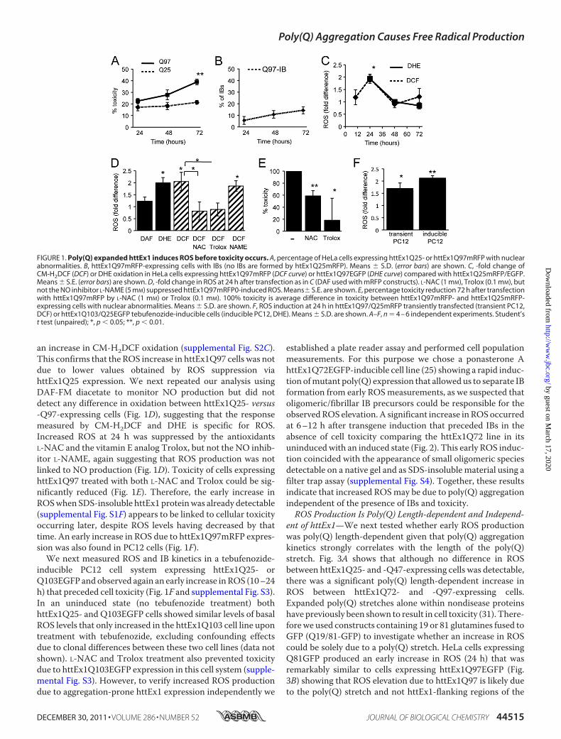
an increase in CM-H2DCF oxidation (supplemental Fig. S2C).This confirms that the ROS increase in httEx1Q97 cells was notdue to lower values obtained by ROS suppression viahttEx1Q25 expression. We next repeated our analysis usingDAF-FM diacetate to monitor NO production but did notdetect any difference in oxidation between httEx1Q25- versus-Q97-expressing cells (Fig. 1D), suggesting that the responsemeasured by CM-H2DCF and DHE is specific for ROS.Increased ROS at 24 h was suppressed by the antioxidantsL-NAC and the vitamin E analog Trolox, but not the NO inhib-itor L-NAME, again suggesting that ROS production was notlinked to NO production (Fig. 1D). Toxicity of cells expressinghttEx1Q97 treated with both L-NAC and Trolox could be sig-nificantly reduced (Fig. 1E). Therefore, the early increase inROSwhen SDS-insoluble httEx1 proteinwas already detectable(supplemental Fig. S1F) appears to be linked to cellular toxicityoccurring later, despite ROS levels having decreased by thattime. An early increase in ROS due to httEx1Q97mRFP expres-sion was also found in PC12 cells (Fig. 1F).We next measured ROS and IB kinetics in a tebufenozide-
inducible PC12 cell system expressing httEx1Q25- orQ103EGFP andobserved again an early increase inROS (10–24h) that preceded cell toxicity (Fig. 1F and supplemental Fig. S3).In an uninduced state (no tebufenozide treatment) bothhttEx1Q25- and Q103EGFP cells showed similar levels of basalROS levels that only increased in the httEx1Q103 cell line upontreatment with tebufenozide, excluding confounding effectsdue to clonal differences between these two cell lines (data notshown). L-NAC and Trolox treatment also prevented toxicitydue to httEx1Q103EGFP expression in this cell system (supple-mental Fig. S3). However, to verify increased ROS productiondue to aggregation-prone httEx1 expression independently we
established a plate reader assay and performed cell populationmeasurements. For this purpose we chose a ponasterone AhttEx1Q72EGFP-inducible cell line (25) showing a rapid induc-tion ofmutant poly(Q) expression that allowedus to separate IBformation from early ROSmeasurements, as we suspected thatoligomeric/fibrillar IB precursors could be responsible for theobservedROS elevation.A significant increase inROSoccurredat 6–12 h after transgene induction that preceded IBs in theabsence of cell toxicity comparing the httEx1Q72 line in itsuninducedwith an induced state (Fig. 2). This early ROS induc-tion coincided with the appearance of small oligomeric speciesdetectable on a native gel and as SDS-insoluble material using afilter trap assay (supplemental Fig. S4). Together, these resultsindicate that increased ROSmay be due to poly(Q) aggregationindependent of the presence of IBs and toxicity.ROS Production Is Poly(Q) Length-dependent and Independ-
ent of httEx1—We next tested whether early ROS productionwas poly(Q) length-dependent given that poly(Q) aggregationkinetics strongly correlates with the length of the poly(Q)stretch. Fig. 3A shows that although no difference in ROSbetween httEx1Q25- and -Q47-expressing cells was detectable,there was a significant poly(Q) length-dependent increase inROS between httEx1Q72- and -Q97-expressing cells.Expanded poly(Q) stretches alone within nondisease proteinshave previously been shown to result in cell toxicity (31). There-fore we used constructs containing 19 or 81 glutamines fused toGFP (Q19/81-GFP) to investigate whether an increase in ROScould be solely due to a poly(Q) stretch. HeLa cells expressingQ81GFP produced an early increase in ROS (24 h) that wasremarkably similar to cells expressing httEx1Q97EGFP (Fig.3B) showing that ROS elevation due to httEx1Q97 is likely dueto the poly(Q) stretch and not httEx1-flanking regions of the
FIGURE 1. Poly(Q) expanded httEx1 induces ROS before toxicity occurs. A, percentage of HeLa cells expressing httEx1Q25- or httEx1Q97mRFP with nuclearabnormalities. B, httEx1Q97mRFP-expressing cells with IBs (no IBs are formed by htEx1Q25mRFP). Means � S.D. (error bars) are shown. C, -fold change ofCM-H2DCF (DCF) or DHE oxidation in HeLa cells expressing httEx1Q97mRFP (DCF curve) or httEx1Q97EGFP (DHE curve) compared with httEx1Q25mRFP/EGFP.Means � S.E. (error bars) are shown. D, -fold change in ROS at 24 h after transfection as in C (DAF used with mRFP constructs). L-NAC (1 mM), Trolox (0.1 mM), butnot the NO inhibitor L-NAME (5 mM) suppressed httEx1Q97mRFP0-induced ROS. Means� S.E. are shown. E, percentage toxicity reduction 72 h after transfectionwith httEx1Q97mRFP by L-NAC (1 mM) or Trolox (0.1 mM). 100% toxicity is average difference in toxicity between httEx1Q97mRFP- and httEx1Q25mRFP-expressing cells with nuclear abnormalities. Means � S.D. are shown. F, ROS induction at 24 h in httEx1Q97/Q25mRFP transiently transfected (transient PC12,DCF) or httEx1Q103/Q25EGFP tebufenozide-inducible cells (inducible PC12, DHE). Means � S.D. are shown. A–F, n � 4 – 6 independent experiments. Student’st test (unpaired); *, p � 0.05; **, p � 0.01.
Poly(Q) Aggregation Causes Free Radical Production
DECEMBER 30, 2011 • VOLUME 286 • NUMBER 52 JOURNAL OF BIOLOGICAL CHEMISTRY 44515
by guest on March 17, 2020
http://ww
w.jbc.org/
Dow
nloaded from
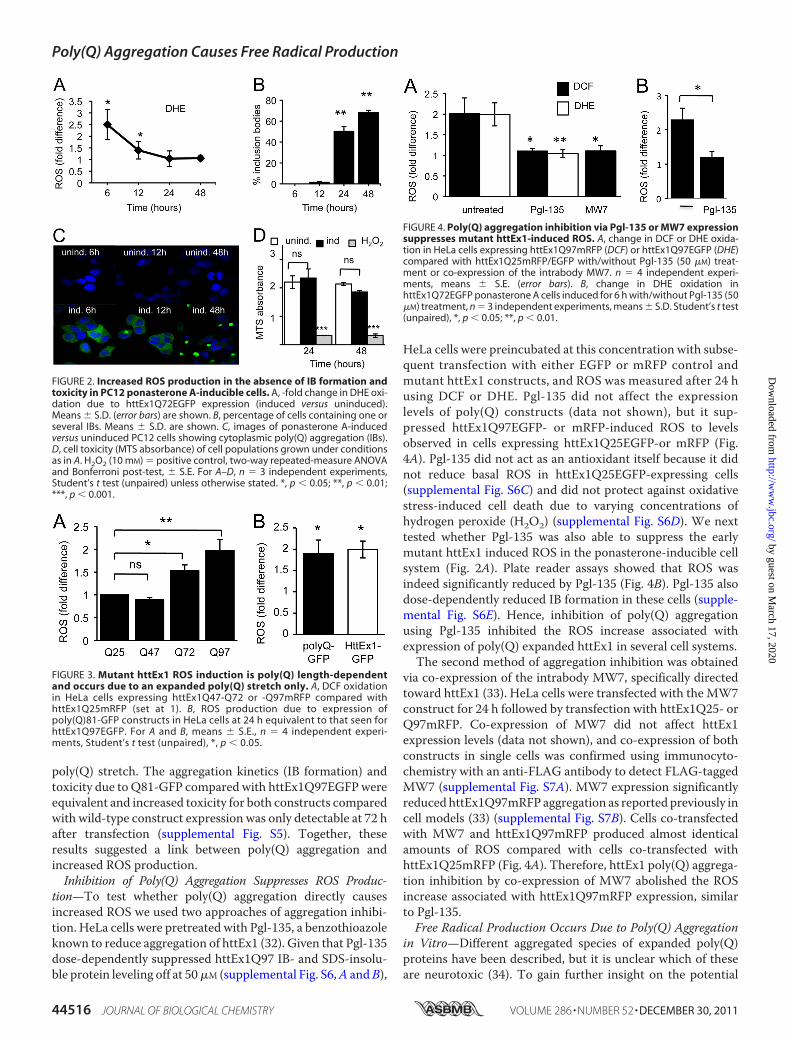
poly(Q) stretch. The aggregation kinetics (IB formation) andtoxicity due to Q81-GFP compared with httEx1Q97EGFPwereequivalent and increased toxicity for both constructs comparedwith wild-type construct expression was only detectable at 72 hafter transfection (supplemental Fig. S5). Together, theseresults suggested a link between poly(Q) aggregation andincreased ROS production.Inhibition of Poly(Q) Aggregation Suppresses ROS Produc-
tion—To test whether poly(Q) aggregation directly causesincreased ROS we used two approaches of aggregation inhibi-tion. HeLa cells were pretreated with Pgl-135, a benzothioazoleknown to reduce aggregation of httEx1 (32). Given that Pgl-135dose-dependently suppressed httEx1Q97 IB- and SDS-insolu-ble protein leveling off at 50�M (supplemental Fig. S6,A andB),
HeLa cells were preincubated at this concentration with subse-quent transfection with either EGFP or mRFP control andmutant httEx1 constructs, and ROS was measured after 24 husing DCF or DHE. Pgl-135 did not affect the expressionlevels of poly(Q) constructs (data not shown), but it sup-pressed httEx1Q97EGFP- or mRFP-induced ROS to levelsobserved in cells expressing httEx1Q25EGFP-or mRFP (Fig.4A). Pgl-135 did not act as an antioxidant itself because it didnot reduce basal ROS in httEx1Q25EGFP-expressing cells(supplemental Fig. S6C) and did not protect against oxidativestress-induced cell death due to varying concentrations ofhydrogen peroxide (H2O2) (supplemental Fig. S6D). We nexttested whether Pgl-135 was also able to suppress the earlymutant httEx1 induced ROS in the ponasterone-inducible cellsystem (Fig. 2A). Plate reader assays showed that ROS wasindeed significantly reduced by Pgl-135 (Fig. 4B). Pgl-135 alsodose-dependently reduced IB formation in these cells (supple-mental Fig. S6E). Hence, inhibition of poly(Q) aggregationusing Pgl-135 inhibited the ROS increase associated withexpression of poly(Q) expanded httEx1 in several cell systems.The second method of aggregation inhibition was obtained
via co-expression of the intrabody MW7, specifically directedtoward httEx1 (33). HeLa cells were transfected with the MW7construct for 24 h followed by transfection with httEx1Q25- orQ97mRFP. Co-expression of MW7 did not affect httEx1expression levels (data not shown), and co-expression of bothconstructs in single cells was confirmed using immunocyto-chemistry with an anti-FLAG antibody to detect FLAG-taggedMW7 (supplemental Fig. S7A). MW7 expression significantlyreducedhttEx1Q97mRFP aggregation as reported previously incell models (33) (supplemental Fig. S7B). Cells co-transfectedwith MW7 and httEx1Q97mRFP produced almost identicalamounts of ROS compared with cells co-transfected withhttEx1Q25mRFP (Fig. 4A). Therefore, httEx1 poly(Q) aggrega-tion inhibition by co-expression of MW7 abolished the ROSincrease associated with httEx1Q97mRFP expression, similarto Pgl-135.Free Radical Production Occurs Due to Poly(Q) Aggregation
in Vitro—Different aggregated species of expanded poly(Q)proteins have been described, but it is unclear which of theseare neurotoxic (34). To gain further insight on the potential
FIGURE 2. Increased ROS production in the absence of IB formation andtoxicity in PC12 ponasterone A-inducible cells. A, -fold change in DHE oxi-dation due to httEx1Q72EGFP expression (induced versus uninduced).Means � S.D. (error bars) are shown. B, percentage of cells containing one orseveral IBs. Means � S.D. are shown. C, images of ponasterone A-inducedversus uninduced PC12 cells showing cytoplasmic poly(Q) aggregation (IBs).D, cell toxicity (MTS absorbance) of cell populations grown under conditionsas in A. H2O2 (10 mM) � positive control, two-way repeated-measure ANOVAand Bonferroni post-test, � S.E. For A–D, n � 3 independent experiments,Student’s t test (unpaired) unless otherwise stated. *, p � 0.05; **, p � 0.01;***, p � 0.001.
FIGURE 3. Mutant httEx1 ROS induction is poly(Q) length-dependentand occurs due to an expanded poly(Q) stretch only. A, DCF oxidationin HeLa cells expressing httEx1Q47-Q72 or -Q97mRFP compared withhttEx1Q25mRFP (set at 1). B, ROS production due to expression ofpoly(Q)81-GFP constructs in HeLa cells at 24 h equivalent to that seen forhttEx1Q97EGFP. For A and B, means � S.E., n � 4 independent experi-ments, Student’s t test (unpaired), *, p � 0.05.
FIGURE 4. Poly(Q) aggregation inhibition via Pgl-135 or MW7 expressionsuppresses mutant httEx1-induced ROS. A, change in DCF or DHE oxida-tion in HeLa cells expressing httEx1Q97mRFP (DCF) or httEx1Q97EGFP (DHE)compared with httEx1Q25mRFP/EGFP with/without Pgl-135 (50 �M) treat-ment or co-expression of the intrabody MW7. n � 4 independent experi-ments, means � S.E. (error bars). B, change in DHE oxidation inhttEx1Q72EGFP ponasterone A cells induced for 6 h with/without Pgl-135 (50�M) treatment, n � 3 independent experiments, means � S.D. Student’s t test(unpaired), *, p � 0.05; **, p � 0.01.
Poly(Q) Aggregation Causes Free Radical Production
44516 JOURNAL OF BIOLOGICAL CHEMISTRY VOLUME 286 • NUMBER 52 • DECEMBER 30, 2011
by guest on March 17, 2020
http://ww
w.jbc.org/
Dow
nloaded from

species involved in ROS productionwithin cells (see above) andto test whether poly(Q) aggregation itself, independent of a cel-lular context, could produce free radicals, we purified recombi-nant httEx1Q20 and -Q53 fused to GST. Cleavage of the GSTtag from httEx1Q20 or -Q53 and subsequent aggregation weremonitored using AFM and filter trap assays. Consistent withprevious studies we found a time-dependent increase in aggre-gation for httEx1Q53 protein that was absent for httEx1Q20(Fig. 5A), with oligomeric globular or annular structures andfibrils (34) (supplemental Fig. S8). We noted that some smallaggregates were also present in the httEx1Q20 sample at latertime points, likely due to a relatively high starting concentra-tion enhanced by surface deposition, but at no stage were fibrilsidentified for httExQ20 and this aggregatedmaterial was alwaysSDS-soluble. Given that Pgl-135 suppressed ROS in cell models(see above), it was of interest to examine whether it reducedoligomer size and fibril formation. Pgl-135 indeed reduced theappearance of fibrils, mean oligomer size, and relative numbersof small oligomers (Fig. 5, B–D, and supplemental Fig. S9) andthe production of SDS-insoluble httEx1Q53 protein (Fig. 5E).Using this in vitromodel of httEx1 aggregation, we next used anAmplex Red assay to measure the production of H2O2 asdescribed previously for aggregating proteins (6). Incubation ofhttEx1Q53, but not -Q20, caused a rapid increase in H2O2 pro-duction after 5 h (Fig. 6A). The H2O2 formed by httEx1Q53could be removed by addition of catalase (decomposing H2O2)before cleavage of the GST tag from httEx1Q53 (Fig. 6A).Because H2O2 production reached its maximum 5 h afterhttEx1Q53 cleavage when no major fibrils were present (Fig.
FIGURE 6. In vitro poly(Q) aggregation is associated with hydrogen per-oxide (H2O2) formation that is inhibited by the single chain antibodyMW7. A, H2O2 production due to incubation of httEx1Q20/Q53. httEx1Q53was also incubated in the presence of catalase, n � 6 independent experi-ments. B, addition of MW7 (1:1 molar ratio) prior to cleavage of GST fromhttEx1Q53 and reduction of oligomer size (5 h, AFM height measurements),n � 3 independent experiments. C, H2O2 production after 5 h of httEx1Q20/Q53 incubation with/without MW7 as in B, n � 4 independent experiments,means � S.E. (error bars). Student’s t test (unpaired), *, p � 0.05; **, p � 0.01;***, p � 0.001.
FIGURE 5. Pgl-135 reduces httEx1Q53 oligomer size and fibril formation. A, aggregation of httEx1Q53 versus httEx1Q20 analyzed by AFM. Poly(Q) fibrils arevisible at 5 h. B, representative AFM images of httEx1Q53 aggregation (24 h) in the presence/absence (untreated) of Pgl-135 (50 �M). C, number of fibrillarstructures/10-�m AFM scan of httEx1Q53 aggregation as in B, n � 3 independent experiments, means � S.D. (error bars). D, httEx1Q53 oligomer size (AFMheight measurements), n � 3 independent experiments, means � S.D. E, dot blot showing that Pgl-135 (50 �M) and MW7 antibody reduce httEx1Q53aggregation (for details, see “Experimental Procedures”). Note that MW7 24 h time point was not done.
Poly(Q) Aggregation Causes Free Radical Production
DECEMBER 30, 2011 • VOLUME 286 • NUMBER 52 JOURNAL OF BIOLOGICAL CHEMISTRY 44517
by guest on March 17, 2020
http://ww
w.jbc.org/
Dow
nloaded from

5A), early stages of httEx1Q53 aggregation (e.g. oligomers orprotofibrils) could be responsible for H2O2 production.
We next asked whether inhibition of poly(Q) oligomeriza-tion was able to suppress H2O2 production. Given that theMW7 intrabody was shown to specifically inhibit httEx1 aggre-gation in cell models (see above; 33), MW7 antibody was addedto httEx1Q20/53 reactions in a 1:1 stoichiometry before theGST tag was cleaved, and AFM samples were prepared afterH2O2 levels reached its maximum (5 h). Because it was likelythat early stages of aggregation caused ROS production, andonly occasional fibrils were detected at this early time point (5h), wemeasuredmean oligomer size for httEx1Q53 reactions inparallel in the presence/absence of MW7. Fig. 6B shows thatoligomer size was significantly reduced when MW7 was pres-ent. The relative number of small oligomers was similarlydecreased in the presence ofMW7 (supplemental Fig. S9). Thisfinding of poly(Q) aggregation inhibition is consistent with adrastic reduction of SDS-insoluble material produced byhttEx1Q53 when co-incubated withMW7measured by the fil-ter trap assay (Fig. 5D). httEx1Q20- and -Q53 samples werethen co-incubated with MW7 and used in the Amplex Redassay to monitor H2O2 production. Although MW7 did notchange basal H2O2 production measured for httEx1Q20, itreduced H2O2 in httEx1Q53 reactions to levels measured forhttEx1Q20 (Fig. 6C). Therefore, reducing httEx1Q53 aggrega-tion also suppressed httEx1Q53-mediated ROS production invitro. Because Pgl-135 interfered with the Amplex Red assay wecould not use this compound.
DISCUSSION
We have determined the relationship between intracellularprotein aggregation and the production of ROS using cellularmodels of HD. Expansions of poly(Q) repeats led to earlyincreased ROS coinciding with poly(Q) aggregation. Inhibitionof poly(Q) aggregation suppressed ROS. We then employedrecombinant httEx1 protein andmonitored its aggregation andROS production in vitro. H2O2 production peaked at 1–5 hbefore substantial amounts of httEx1 fibrils were formed. Wepropose that poly(Q) aggregation intermediates are responsiblefor increased H2O2 production.This finding is consistent with in vitro experiments per-
formed with other amyloid-forming proteins (5–8). For com-parison with poly(Q) experiments we have also used A�1–40and A�1–42 peptides and found a time-dependent productionof H2O2 using Amplex Red assays correlating with the aggrega-tion kinetics of each peptide, similar to previous reports (sup-plemental Fig. S10). Interestingly, Tabner and colleagues (6)showed that there is a tight correlation between the time ofoligomer formation and a ROS burst during such reactions,suggesting that it is the early aggregation steps and not theproduction of fibrillar structures that may be associated withfree radical production.We tested this idea in a poly(Q) contextusing a single chain antibody specifically directed towardhttEx1 (MW7) that altered early in vitro poly(Q) oligomeriza-tion by decreasing oligomer size (and subsequent poly(Q)aggregation steps). MW7 suppressed H2O2 and httEx1Q53oligomerization in parallel and at an early stage (5 h), when nosignificant numbers of fibrils were formed (Fig. 5A). This find-
ing strongly indicates that it is prefibrillar poly(Q) protein thatmay be responsible for H2O2 production. This hypothesis isalso supported by the absence of a continued rise in H2O2 pro-duction in parallel with an increase in fibril formation byhttEx1Q53. The mechanisms by which aggregation-pronehttEx1Q53 and other aggregation-prone peptides generateH2O2, in concert with metal ions (e.g. Cu2�), is unclear and amatter of debate (35) (see below).Our proposition of prefibrillar poly(Q) aggregates causing
ROS production is supported by our httEx1 cell assays: usingseveral cell types and ROS assays we consistently observedincreased ROS production early after intracellular expressionof httEx1mRFP/EGFP with poly(Q) expansions, before the for-mation of IBs. In the ponasterone A-inducible cell system wedetected small SDS-soluble oligomeric httEx1Q72EGFP spe-cies at 6 h after transgene induction coinciding with the ROSelevation. This suggests that soluble oligomeric species ofmutant huntingtin fragments may cause increased free radicalproduction within cells (supplemental Fig. S4). Both Pgl-135and cellular co-expression of MW7 intrabody reduced earlypoly(Q) aggregation and ROS in parallel, and because bothMW7 and Pgl-135 acted on early poly(Q) oligomerization stepsin vitro (Figs. 5,B andD, and 6B) it is likely that both treatmentsalso impacted on cellular poly(Q) oligomerization. Therefore,our findings in cell models and the in vitro data show a directlink between poly(Q) aggregation and ROS production.Our approach of determining ROS levels in a defined set of
single, living cells when no increased mutant httEx1 toxicitywas present allowed us to dissociate ROS due to poly(Q)expression from secondary redox alterations that usually occurduring later stages of apoptosis (e.g.when cytochrome c releaseoccurs) and other forms of cell death (36, 37). Basal toxicity dueto mutant and wild-type httEx1 at 24 h was similar, varyingfrom 10 to 20% in transient transfection systems, and hence thedoubling of ROS in cells expressing httEx1Q97 compared withhttEx1Q25 at 24 h cannot be ascribed to differences in toxicity.This finding in HeLa cells was supported by experiments per-formed with PC12 cells in which httEx1 transgenes were tran-siently transfected or stably induced with tebufenozide. Anincrease in ROS after only a fewhours of httExQ72EGFP induc-tion also occurred in the ponasterone A-inducible cell model inthe absence of toxicity (Fig. 2). Such early induction of ROS isconsistent with a recent study in Tet-Off PC12 cells (16) andshows that ROS can be induced long before the onset of celldeath. The study by Bertoni et al. (16) showed a link betweenROS and downstream histone protein phosphorylation, aknown marker for DNA damage (16), suggesting ROS inducedDNAdamage. In theHeLa cell system used in the present studywe also observed that mutant httEx1 expression increasedH2A.X-Ser-139 phosphorylation (supplemental Fig. S11).Interestingly, the increase in phosphorylation only occurred at24 h (supplemental Fig. S11C), mimicking the ROS kineticsmeasured with oxidation sensitive molecules (Fig. 1C).The likely cellular sources of ROS, or redox imbalances due
to poly(Q)-expanded proteins, are manifold. As shown in thisstudy, poly(Q) aggregation itself causes increased ROS. Here, itis possible that aggregating structures other than oligomers (asproposed in the present report), also contribute to abnormal
Poly(Q) Aggregation Causes Free Radical Production
44518 JOURNAL OF BIOLOGICAL CHEMISTRY VOLUME 286 • NUMBER 52 • DECEMBER 30, 2011
by guest on March 17, 2020
http://ww
w.jbc.org/
Dow
nloaded from
ROS. IBs have been suggested to be centers of oxidative reac-tions (38). It has been shown that oligomeric poly(Q) speciesare present at IBs in vivo (39), and hence it is possible thatoxidative events at IBs are also driven by oligomerization reac-tions, likely in concert with certainmetal ions. Both copper andiron ions have been proposed to be involved in amyloid-associ-ated in vitroH2O2productionwith subsequent hydroxyl radicalformation via Fenton chemistry and/or the Haber-Weiss reac-tion (for review, see Ref. 8). Interestingly, N-terminal httincluding httEx1 has been shown to be redox-active, bindingcopper (40). Additionally, we have demonstrated that copperincreased aggregation of httEx1 with long glutamine stretchesboth in vitro and in vivo (cells) (41). Therefore, it is possible thatcopper is involved in the httEx1-driven elevation of ROS. Wedo not believe that poly(Q)-induced ROS itself modulatespoly(Q) aggregation in a major way in the cell systems used inthe present study because both L-NAC and Trolox treatmentdid not alter IB formation in all cell systems (supplemental Fig.S12). However, poly(Q)-induced ROS could still affect early,oligomerization reactions together with transition metals.Poly(Q) aggregation-independentmechanisms are also likely
to play significant roles in contributing to the redox imbalanceinHD including transcriptional,mitochondrial, and endosomaltrafficking abnormalities. Expression of PGC-1�, a key regula-tor of antioxidant gene expression and mitochondrial biogene-sis, has been shown to be transcriptionally dysregulated in HD(42), and both full-length and httEx1 directly interact with theoutermitochondrialmembrane (43, 44).Hence, transcriptionaland mitochondrial abnormalities (for example alterations ofthe mitochondrial fusion-fission machinery) (45) are furthercandidate mechanisms for involvement of redox alterations inHD. Long poly(Q) stretches have also been shown to directlyalter respiration of isolated mitochondria and increase ROS(46).Recently, Li et al. proposed that aberrant trafficking of the
neuronal glutamate transporter EAAC1, which regulatesuptake of cysteine required for glutathione (GSH) synthesis,leads to an increase in ROS in neurons expressing mutant full-length htt (15). This study showed that ROS accumulated inneurons expressing mutant Gln-140 full-length htt in knock-inmice due to an early GSH depletion. This proposedmechanismfor redox alteration is likely different from the mechanism ofincreased oxidative stress proposed in our study.As pointed outby Li et al. (15), it is possible that the full-length model used intheir study is representative of redox abnormalities at earlystages of HD whereas httEx1 model systems represent a laterstage ofHD.However, it should be noted that the production ofhtt cleavage products, including fragment sizes consistent withhttEx1, is an early event inHD (12), and hence httEx1 oligomer-ization could also contribute to alter the redox homeostasis inearly HD. Another study on proteins with poly(Q) expansionsargued that the lipid-localized NADPH oxidase may be anenzyme involved in abnormal redox homeostasis (16). Thisstudy showed that the relatively specificNADPHoxidase inhib-itor apocynin was able to reduce ROS due to expression ofhttEx1 with 43 glutamines, and httEx1 interacted with gp91 (aNADPH oxidase subunit).
We propose that there are several independent sources ofabnormal free radical production in HD. It is likely that on theone hand, alterations in transcription and trafficking events dueto poly(Q) expansions result in increased susceptibility to oxi-dative stress via disruption of the glutathione and the antioxi-dant defense system. On the other hand poly(Q) aggregationitself and intracellularmechanisms disrupted by poly(Q) aggre-gation (e.g.mitochondrial functions) may contribute in paralleland/or sequentially to increased oxidative stress during poly(Q)pathology. Therefore, a beneficial approach for antioxidanttherapy during chronic neurodegeneration associated withprotein aggregation should involve the targeting of severalmechanisms. In this context it will be important to explore theroles of non-cell autonomous mechanisms that could contrib-ute to oxidative stress in HD. As both microglial and astrocyticcellular dysfunction due to expanded poly(Q) proteins havebeen reported (47, 48), glial cells may contribute to oxidativealterations in the HD brain. Although microglial cells areknown for their role in the production of ROS, astrocytes reg-ulate antioxidant control in the central nervous system.Whether increased ROS is induced due to intracellular aggre-gation of non-poly(Q)-containing proteins is an exciting ave-nue for future investigation.
Acknowledgments—We thank G. Bates for the S830 antibody, theMuchowski laboratory for plasmids, A. Koshnan for the MW7 con-struct, M. Cuttle and H. Schuppe for assistance with microscopy(Imaging and Microscopy Centre, Southampton), S. Moore and D.Allsop for advice with the Amplex Red assay, and V. O‘Connor forfruitful discussions.
REFERENCES1. Barnham, K. J.,Masters, C. L., and Bush, A. I. (2004)Nat. Rev. DrugDiscov.
3, 205–2142. Martínez, A., Portero-Otin, M., Pamplona, R., and Ferrer, I. (2010) Brain
Pathol. 20, 281–2973. D’Autréaux, B., and Toledano, M. B. (2007) Nat. Rev. Mol. Cell Biol. 8,
813–8244. Halliwell, B., and Gutteridge, J. (2007) Free Radicals in Biology and Med-
icine, 4th Ed., Oxford University Press, New York5. Opazo, C., Huang, X., Cherny, R. A.,Moir, R. D., Roher, A. E.,White, A. R.,
Cappai, R., Masters, C. L., Tanzi, R. E., Inestrosa, N. C., and Bush, A. I.(2002) J. Biol. Chem. 277, 40302–40308
6. Tabner, B. J., El-Agnaf, O. M., Turnbull, S., German, M. J., Paleologou,K. E., Hayashi, Y., Cooper, L. J., Fullwood,N. J., andAllsop,D. (2005) J. Biol.Chem. 280, 35789–35792
7. Turnbull, S., Tabner, B. J., Brown,D. R., andAllsop,D. (2003)Biochemistry42, 7675–7681
8. Allsop, D., Mayes, J., Moore, S., Masad, A., and Tabner, B. J. (2008)Biochem. Soc. Trans. 36, 1293–1298
9. Huang, X., Cuajungco, M. P., Atwood, C. S., Hartshorn, M. A., Tyndall,J. D., Hanson, G. R., Stokes, K. C., Leopold,M.,Multhaup, G., Goldstein, L.E, Scarpa, R. C., Saunders, A. J., Lim, J., Moir, R. D., Glabe, C., Bowden,E. F., Masters, C. L., Fairlie, D. P., Tanzi, R. E., and Bush, A. I. (1999) J. Biol.Chem. 274, 37111–37116
10. DiFiglia, M., Sapp, E., Chase, K. O., Davies, S. W., Bates, G. P., Vonsattel,J. P., and Aronin, N. (1997) Science 277, 1990–1993
11. Davies, S.W., Turmaine,M., Cozens, B. A., DiFiglia,M., Sharp, A.H., Ross,C. A., Scherzinger, E.,Wanker, E. E.,Mangiarini, L., and Bates, G. P. (1997)Cell 90, 537–548
12. Landles, C., Sathasivam, K., Weiss, A., Woodman, B., Moffitt, H., Fink-beiner, S., Sun, B., Gafni, J., Ellerby, L. M., Trottier, Y., Richards, W. G.,
Poly(Q) Aggregation Causes Free Radical Production
DECEMBER 30, 2011 • VOLUME 286 • NUMBER 52 JOURNAL OF BIOLOGICAL CHEMISTRY 44519
by guest on March 17, 2020
http://ww
w.jbc.org/
Dow
nloaded from

Osmand, A., Paganetti, P., and Bates, G. P. (2010) J. Biol. Chem. 285,8808–8823
13. Wyttenbach, A., Sauvageot, O., Carmichael, J., Diaz-Latoud, C., Arrigo,A. P., and Rubinsztein, D. C. (2002) Hum. Mol. Genet. 11, 1137–1151
14. van Roon-Mom, W. M., Pepers, B. A., ’t Hoen, P. A., Verwijmeren, C. A.,denDunnen, J. T., Dorsman, J. C., and vanOmmen,G. B. (2008)BMCMol.Biol. 9, 84
15. Li, X., Valencia, A., Sapp, E., Masso, N., Alexander, J., Reeves, P., Kegel,K. B., Aronin, N., and Difiglia, M. (2010) J. Neurosci. 30, 4552–4561
16. Bertoni, A., Giuliano, P., Galgani, M., Rotoli, D., Ulianich, L., Adornetto,A., Santillo, M. R., Porcellini, A., and Avvedimento, V. E. (2011) J. Biol.Chem. 286, 4727–4741
17. Pérez-Severiano, F., Santamaría, A., Pedraza-Chaverri, J., Medina-Cam-pos, O. N., Ríos, C., and Segovia, J. (2004) Neurochem. Res. 29, 729–733
18. Bogdanov,M. B., Andreassen,O. A., Dedeoglu, A., Ferrante, R. J., and Beal,M. F. (2001) J. Neurochem. 79, 1246–1249
19. Tabrizi, S. J., Workman, J., Hart, P. E., Mangiarini, L., Mahal, A., Bates, G.,Cooper, J. M., and Schapira, A. H. (2000) Ann. Neurol. 47, 80–86
20. Browne, S. E., Bowling, A. C., MacGarvey, U., Baik, M. J., Berger, S. C.,Muqit,M.M., Bird, E. D., and Beal,M. F. (1997)Ann. Neurol. 41, 646–653
21. Browne, S. E., Ferrante, R. J., and Beal, M. F. (1999) Brain Pathol. 9,147–163
22. Polidori,M. C.,Mecocci, P., Browne, S. E., Senin, U., and Beal,M. F. (1999)Neurosci. Lett. 272, 53–56
23. Tabrizi, S. J., Cleeter,M.W., Xuereb, J., Taanman, J.W., Cooper, J.M., andSchapira, A. H. (1999) Ann. Neurol. 45, 25–32
24. Aiken, C. T., Tobin, A. J., and Schweitzer, E. S. (2004) Neurobiol. Dis. 16,546–555
25. Apostol, B. L., Kazantsev, A., Raffioni, S., Illes, K., Pallos, J., Bodai, L.,Slepko, N., Bear, J. E., Gertler, F. B., Hersch, S., Housman, D. E., Marsh,J. L., and Thompson, L. M. (2003) Proc. Natl. Acad. Sci. U.S.A. 100,5950–5955
26. Wyttenbach, A., Carmichael, J., Swartz, J., Furlong, R. A., Narain, Y.,Rankin, J., and Rubinsztein, D. C. (2000) Proc. Natl. Acad. Sci. U.S.A. 97,2898–2903
27. King, M. A., Hands, S., Hafiz, F., Mizushima, N., Tolkovsky, A. M., andWyttenbach, A. (2008)Mol. Pharmacol. 73, 1052–1063
28. Wacker, J. L., Zareie, M. H., Fong, H., Sarikaya, M., and Muchowski, P. J.(2004) Nat. Struct. Mol. Biol. 11, 1215–1222
29. Wyttenbach, A., and Tolkovsky, A. M. (2006) J. Neurochem. 96,1213–1226
30. Firdaus,W. J.,Wyttenbach, A., Diaz-Latoud, C., Currie, R.W., andArrigo,
A. P. (2006) FEBS J. 273, 3076–309331. Marsh, J. L., Walker, H., Theisen, H., Zhu, Y. Z., Fielder, T., Purcell, J., and
Thompson, L. M. (2000) Hum. Mol. Genet. 9, 13–2532. Hockly, E., Tse, J., Barker, A. L., Moolman, D. L., Beunard, J. L., Revington,
A. P., Holt, K., Sunshine, S., Moffitt, H., Sathasivam, K., Woodman, B.,Wanker, E. E., Lowden, P. A., and Bates, G. P. (2006) Neurobiol. Dis. 21,228–236
33. Khoshnan, A., Ko, J., and Patterson, P. H. (2002) Proc. Natl. Acad. Sci.U.S.A. 99, 1002–1007
34. Hands, S. L., andWyttenbach, A. (2010)Acta Neuropathol. 120, 419–43735. Nadal, R. C., Rigby, S. E., and Viles, J. H. (2008) Biochemistry 47,
11653–1166436. Kirkland, R. A., and Franklin, J. L. (2001) J. Neurosci. 21, 1949–196337. Orrenius, S. (2007) Drug Metab. Rev. 39, 443–45538. Firdaus,W. J.,Wyttenbach, A., Giuliano, P., Kretz-Remy, C., Currie, R.W.,
and Arrigo, A. P. (2006) FEBS J. 273, 5428–544139. Legleiter, J., Mitchell, E., Lotz, G. P., Sapp, E., Ng, C., DiFiglia, M., Thomp-
son, L. M., and Muchowski, P. J. (2010) J. Biol. Chem. 285, 14777–1479040. Fox, J. H., Kama, J. A., Lieberman, G., Chopra, R., Dorsey, K., Chopra, V.,
Volitakis, I., Cherny, R. A., Bush, A. I., and Hersch, S. (2007) PLoS One 2,e334
41. Hands, S. L., Mason, R., Sajjad, M. U., Giorgini, F., and Wyttenbach, A.(2010) Biochem. Soc. Trans. 38, 552–558
42. Cui, L., Jeong, H., Borovecki, F., Parkhurst, C. N., Tanese, N., and Krainc,D. (2006) Cell 127, 59–69
43. Choo, Y. S., Johnson, G. V., MacDonald, M., Detloff, P. J., and Lesort, M.(2004) Hum. Mol. Genet. 13, 1407–1420
44. Panov, A. V., Gutekunst, C. A., Leavitt, B. R., Hayden, M. R., Burke, J. R.,Strittmatter,W. J., andGreenamyre, J. T. (2002)Nat. Neurosci. 5, 731–736
45. Song,W., Chen, J., Petrilli, A., Liot, G., Klinglmayr, E., Zhou, Y., Poquiz, P.,Tjong, J., Pouladi,M. A., Hayden,M. R.,Masliah, E., Ellisman,M., Rouiller,I., Schwarzenbacher, R., Bossy, B., Perkins, G., and Bossy-Wetzel, E. (2011)Nat. Med. 17, 377–382
46. Puranam, K. L., Wu, G., Strittmatter, W. J., and Burke, J. R. (2006)Biochem. Biophys. Res. Commun. 341, 607–613
47. Bradford, J., Shin, J. Y., Roberts, M., Wang, C. E., Li, X. J., and Li, S. (2009)Proc. Natl. Acad. Sci. U.S.A. 106, 22480–22485
48. Björkqvist, M., Wild, E. J., Thiele, J., Silvestroni, A., Andre, R., Lahiri, N.,Raibon, E., Lee, R. V., Benn, C. L., Soulet, D., Magnusson, A., Woodman,B., Landles, C., Pouladi, M. A., Hayden, M. R., Khalili-Shirazi, A., Lowdell,M.W., Brundin, P., Bates, G. P., Leavitt, B. R., Möller, T., and Tabrizi, S. J.(2008) J. Exp. Med. 205, 1869–1877
Poly(Q) Aggregation Causes Free Radical Production
44520 JOURNAL OF BIOLOGICAL CHEMISTRY VOLUME 286 • NUMBER 52 • DECEMBER 30, 2011
by guest on March 17, 2020
http://ww
w.jbc.org/
Dow
nloaded from

Sarah Hands, Mohammad U. Sajjad, Michael J. Newton and Andreas WyttenbachCauses Free Radical Production
Aggregation of a Fragment of Huntingtin Protein Directlyin Vivo and In Vitro
doi: 10.1074/jbc.M111.307587 originally published online October 7, 20112011, 286:44512-44520.J. Biol. Chem.
10.1074/jbc.M111.307587Access the most updated version of this article at doi:
Alerts:
When a correction for this article is posted•
When this article is cited•
to choose from all of JBC's e-mail alertsClick here
Supplemental material:
http://www.jbc.org/content/suppl/2011/10/07/M111.307587.DC1
http://www.jbc.org/content/286/52/44512.full.html#ref-list-1
This article cites 47 references, 16 of which can be accessed free at
by guest on March 17, 2020
http://ww
w.jbc.org/
Dow
nloaded from