b-mediated modulation of inducible nitric oxide synthase...
TRANSCRIPT
JOURNAL OF VIROLOGY, July 2011, p. 6502–6512 Vol. 85, No. 130022-538X/11/$12.00 doi:10.1128/JVI.02560-10Copyright © 2011, American Society for Microbiology. All Rights Reserved.
NF-�B-Mediated Modulation of Inducible Nitric Oxide SynthaseActivity Controls Induction of the Epstein-Barr Virus Productive
Cycle by Transforming Growth Factor Beta 1�†Lassad Oussaief,1,4 Vanessa Ramírez,2,5 Aurelie Hippocrate,1 Hratch Arbach,2 Chantal Cochet,2
Alexis Proust,3 Martine Raphael,3 Ridha Khelifa,4 and Irene Joab1*UMR 1014 Inserm-Universite Paris 11, Hopital Paul Brousse, 14 Avenue Paul Vaillant Couturier, 94807 Villejuif Cedex, France1;
U716 Inserm, 27 rue Juliette Dodu, 75010 Paris, France2; Universite Paris-Sud, INSERM U802, AP-HP, Hopital Bicetre, andHopital Paul Brousse Service d’Hematologie et Immunologie Biologiques, Cytogenetique, Le Kremlin-Bicetre F-94270,
France3; Viral and Molecular Tumor Diagnostics Unit, Habib Thameur Hospital, 1008 Tunis, Tunisia4; andInvestigations Health Institute, Costa Rica University, San Jose, Costa Rica5
Received 2 December 2010/Accepted 7 April 2011
Transforming growth factor beta 1 (TGF-�1) signal transduction has been implicated in many second-messenger pathways, including the NF-�B pathway. We provide evidence of a novel TGF-�1-mediated pathwaythat leads to extracellular signal-regulated kinase (ERK) 1/2 phosphorylation, which in turn induces expres-sion of an Epstein-Barr virus (EBV) protein, ZEBRA, that is responsible for the induction of the viral lyticcycle. This pathway includes two unexpected steps, both of which are required to control ERK 1/2 phosphor-ylation: first, a quick and transient activation of NF-�B, and second, downregulation of inducible nitric oxidesynthase (iNOS) activity that requires the participation of NF-�B activity. Although necessary, NF-�B aloneis not sufficient to produce downregulation of iNOS, suggesting that another uncharacterized event(s) isinvolved in this pathway. Dissection of the steps involved in the switch from the EBV latent cycle to the lyticcycle will be important to understand how virus-host relationships modulate the innate immune system.
Epstein-Barr virus (EBV) is a ubiquitous human herpesvirusassociated with numerous malignancies of both B-cell and ep-ithelial cell origins (32, 43). In vitro, infection of B lymphocytesusually results in viral latency, with expression of six nuclearproteins (EBNA 1, 2, 3A, 3B, 3C, and LP), three latent mem-brane proteins (LMP1, LMP2A, and LMP2B), two small EBV-encoded RNAs (EBERs) (latency III pattern of expression)(24), and microRNAs (37). Entry into the viral lytic cycle isinitiated by expression of two immediate-early (IE) EBV pro-teins, i.e., Z Epstein-Barr virus replication activator (ZEBRA),encoded by BZLF1(44), and Rta, encoded by BRLF1 (50).The two IE proteins activate the viral early genes, resulting ina cascade of events that leads to progeny virions. EBV reacti-vation can be modeled by treating EBV-positive B-cell lineswith pleiotropic agents, such as phorbol 12-myrisate 13-acetate(PMA) (an activator of protein kinase C), calcium ionophore(inducing calcium entry and upregulation of the calcium-dependent factors calcineurin and calcium/calmodulin-de-pendent kinase type IV [CAMK-IV]), n-butyrate (a histonedeacetylase inhibitor), cross-linking of cell surface immuno-globulin G (which activates the B cell receptor) (for a reviewsee reference 23), transforming growth factor beta 1 (TGF-�1) (11, 19, 31, 48), and nitric oxide (NO) inhibitor (26). Type2 nitric oxide synthase (iNOS or NOS2) generates nitric oxide
(NO) from the amino acid L-arginine and thereby contrib-utes to the control of replication or killing of intracellularmicrobial pathogens.
The transforming growth factor beta family of peptides isinvolved in the control of several biological processes, includ-ing the immune response (inflammation) (13, 39). Binding ofTGF-� to the TGF-� type II receptor (T�RII) triggers het-erodimerization and phosphorylation of the TGF-� type I re-ceptor (T�RI). The signal is then propagated through phos-phorylation of receptor-regulated Smads (R-Smads), Smad2and Smad3, which oligomerize with the common mediator(Co-Smad) Smad4 and then translocate to the nucleus, wherethey induce the expression of a large number of target genes(27, 41, 51). However, depending on the cell type, TGF-�1signal transduction has been implicated in many second-mes-senger pathways, including the NF-�B pathway (3, 14). NF-�Bis a dimeric transcription factor composed of homo- or het-erodimers of p50 (NF-�B1), p52 (NF-�B2), p65 (RelA or NF-�B3), RelB, and c-Rel. These are endogenously complexed toinhibitor proteins called I�B that sequester NF-�B in the cy-toplasm. In response to various stimuli, inhibitory proteins arephosphorylated at specific serine residues and are rapidly pro-cessed by the proteasome after polyubiquitination. This ex-poses the nuclear localization signal of NF-�B, leading to itsnuclear translocation. The phosphorylation step is usually con-trolled by the I�B kinase (IKK) complex (38). The most fre-quent NF-�B dimer is p50/p65, which hence is considered to bethe prototype (29).
Activation of NF-�B is a feature of many viral infections(30). Viruses may modulate the NF-�B pathway to enhanceviral replication or prevent virus-induced apoptosis (18, 40).
Because NF-�B is involved in the maintenance of EBV
* Corresponding author. Mailing address: UMR 1014 Inserm-Uni-versite Paris 11, Hopital Paul Brousse, Batiment Andre Lwoff, 14Avenue Paul Vaillant Couturier, 94807 Villejuif Cedex, France. Phone: 331 45 59 60 32. Fax: 33 1 45 59 53 43. E-mail: [email protected].
† Supplemental material for this article may be found at http://jvi.asm.org/.
� Published ahead of print on 20 April 2011.
6502
on May 10, 2018 by guest
http://jvi.asm.org/
Dow
nloaded from
latency and has an effect on lytic cycle progression, we inves-tigated NF-�B activation in TGF-�1-treated positive Burkitt’slymphoma (BL) cell lines which exhibit a restricted, latency I(Lat I) pattern, in which EBNA1 is the only expressed viralprotein. We identified a novel pathway that includes twounexpected steps, (i) a quick transient activation of NF-�Band (ii) NF-�B modulation of iNOS activity, both of whichare required to control extracellular signal-regulated kinase(ERK) 1/2 phosphorylation; this last event is necessary forexpression of the EBV protein ZEBRA, which is responsiblefor the induction of the viral lytic cycle.
MATERIALS AND METHODS
Cell culture and reagents. The EBV-positive BL cell lines Mutu-I, Kem-I,Sav-I, and B95-8 and the EBV-negative BL cell lines BJAB and DG75 weregrown in RPMI 1640 supplemented with 2 mM glutamine (GIBCO BRL LifeTechnologies, Grand Island, NY), 100 �g/ml primocin (Cayla, Toulouse,France), and 10% heat-inactivated fetal calf serum (FCS) (GIBCO BRL LifeTechnologies, Grand Island, NY). For induction of the EBV lytic cycle, Mutu-I,Kem-I, and Sav-I cells were stimulated with 2 ng/ml of TGF-�1 (R&D SystemsMinneapolis, MN) in reduced 0.5% FCS medium. U0126, phorbol 12-myristate13-acetate (PMA), BAY1170-82, IKK2 inhibitor V, and MG262 were obtainedfrom Calbiochem (VWR International, France), and SB-431542, NG-mono-methyl-L-arginine acetate salt (L-NMMA) and S-nitroso-N-acetylpenicil-lamine (SNAP) were purchased from Sigma (St. Louis, MO).
Antibodies. The anti-ZEBRA monoclonal antibody Z125 was obtainedfrom E. Drouet (Faculte de Pharmacie, Grenoble, France). Antibodiesagainst p44/p42 mitogen-activated protein kinase (MAPK), phospho-p44/p42MAPK (Thr202/Tyr204), HP1�, tubulin, p65, and IKB� were purchased fromOzyme (St. Quentin-en-Yvelines, France). Human antiserum to EA (D, R) waskindly provided by Jean H. Joncas (Sainte Justine Hospital, Montreal, Canada).Human anti-VCA antibody was obtained from Ortho Diagnostic Systems, Inc.(Raritan, NJ), and human anti-IgG was obtained from Dako (Denmark). Rabbitperoxidase-conjugated Ig, human peroxidase-conjugated IgG, and mouse perox-idase-conjugated IgG from Ozyme (St. Quentin-en-Yvelines, France) were usedas secondary antibodies.
Cell fractionation. Cells were harvested at 4°C and lysed, and cytoplasmic andnuclear extracts were prepared using an NE-PER nuclear and cytoplasmic ex-traction reagent kit (Pierce Biotechnology) as described by the manufacturer.Protease inhibitors were added (complete mini-protease inhibitor cocktail tab-lets, obtained from Roche Diagnostic, Meylan, France). Briefly, 10 mg of cellswas resuspended in 50 �l cytoplasmic extraction reagent I (CERI), mixed, andincubated on ice for 10 min; 2.75 �l of cytoplasmic extraction reagent II (CERII)was then added, followed by mixing and incubation on ice for 1 min. The intactnuclei were pelleted, and the supernatant cytoplasmic extract was collected. Thenuclei were resuspended in 25 �l of ice-cold nuclear extraction reagent (NER),incubated on ice repeatedly, and centrifuged to obtain the supernatant contain-ing nuclear proteins. Protein concentrations were determined using micro-bicin-choninic acid (BCA) protein assay kit from Pierce Biotechnology.
NF-�B activity. The activity of NF-�B was assessed by the levels of p65 in thenuclear fractions using an NF-�B/p65-active enzyme-linked immunosorbent as-say (ELISA) kit (Imgenex, San Diego, CA). Nuclear and cytoplasmic extractswere prepared and subjected to an enzyme-linked immunosorbent assay accord-ing to the manufacturer’s instructions. All experiments were repeated at leasttwice at different times.
Immunoblot analysis. Cells were harvested, washed briefly with phosphate-buffered saline, resuspended in a buffer composed of 100 mM Tris-Cl (pH 7.6),50 mM NaCl, 2 mM EDTA, 0.5% NP-40, phenylmethylsulfonyl fluoride (100�g/ml), and 1 �g each of leupeptin, pepstatin, and aprotinin per ml, and soni-cated. The protein concentration was determined using a micro-BCA proteinassay kit from Pierce Biotechnology. Equal amounts of protein in loading buffer,heated for 5 min at 100°C and separated by sodium dodecyl sulfate-polyacryl-amide gel electrophoresis (SDS-PAGE) on 10 to 12% gels, were transferred byelectroblotting to a nitrocellulose membrane (Schleicher & Schuell, Ecquevilly,France). Membranes were reacted with the indicated primary antibodies usingthe appropriate dilution as indicated by the manufacturer, followed by treatmentwith a horseradish peroxidase-conjugated IgG (anti-rabbit, anti-mouse, or anti-human IgG antibody [1:2,000 to 1:4,000]) from Sigma (St. Louis, MO). Mem-branes were visualized with enhanced chemiluminescence (West-Pico or -Femto;
Pierce Biotechnology, Rockford, IL). Images were captured using a charge-coupled device (CCD) camera (LAS-1000; Fuji system).
Reverse transcription-PCR (RT-PCR). Total RNA from each uninduced andinduced EBV-positive cell lines was isolated using TRIzol reagent and RNaseinhibitors (Invitrogen, Carlsbad, CA) according to the manufacturer’s protocol.The RNA concentration was determined spectrophotometrically. The templatecDNA was synthesized by reverse transcription of total RNA (3 �g) with Molo-ney murine leukemia virus reverse transcriptase (Promega, Madison, WI) usinga dT15 oligonucleotide primer (Promega). The primers used for PCR amplifica-tions included 5�-TTACACCTGACCCATACCAG-3� and 5�-ACATCTGCTTCAACAGGAGG-3� for ZEBRA. Hypoxanthine phosphoribosyltransferase (HPRT)cDNA was used as internal control as previously described (2, 36).
Determination of cell viability. Cells were treated with different combinationsof TGF-�1, BAY11-7082, IKK2 inhibitor V, U0126, SNAP, L-NMMA, for theindicated periods of time and then tested for viability using the LIVE/DEADreduced-biohazard viability/cytotoxicity test (Molecular Probes, Invitrogen,Cergy Pontoise, France). Staining and analysis were performed as recommendedby the manufacturer. The ability of this kit to detect cell death was controlledusing a treatment with 0.4 mM MnCl2 (10). Cell number was determined with aKova counting chamber (Hycor, Edinburgh, United Kingdom). All experimentswere repeated at least twice at different times.
Quantification of iNOS protein. The amount of iNOS in cell lysates wasquantified with the Quantikine human iNOS immunoassay (R&D) according tothe recommendations of the manufacturer. This assay employs a quantitativesandwich enzyme immunoassay technique. Briefly, a monoclonal antibody spe-cific for iNOS is preapplied to a microplate. Standards and samples are pipettedinto the wells, and any iNOS present is bound by the immobilized antibody. Afterany unbound substances are washed away, an enzyme-linked monoclonal anti-body that is specific for iNOS is added to the wells. Following a wash to removeany unbound antibody-enzyme reagent, a substrate solution is added to the wells,and color develops (450 nm) in proportion to the amount of iNOS bound in theinitial step.
Nitrate/nitrite assays. The amount of NO produced by cells was indirectlymeasured by the formation of nitrate (NO3�) and nitrite (NO2�) (two stable endproducts of NO) in culture supernatants. The total amounts of nitrate and nitritewere determined with a nitrate/nitrite assay kit (Cayman Chemical Co., AnnArbor, MI), using the manufacturer’s protocols. Briefly, supernatant (100 �l) wasincubated with nitrate reductase (20 �l) at room temperature for 1 h, followedby incubation with 100 �l Griess reagent (1% sulfanilic acid, 0.1% naphthylethylenediamine dihydrochloride, 2.5% phosphoric acid) for 10 min. The opticaldensity of each sample was analyzed at 540 nm in a microplate reader withsodium nitrite as a standard.
Expression and reporter plasmids. Plasmids pEGFP-p65 and active mutantNRAS have been described (33, 46). pEGFP, pRL-TK, and pNF-�B-Luc werepurchased from Promega (Madison, WI). The plasmid coding for NF-�B-induc-ing kinase (NIK) was obtained from Open Biosystems (Thermo Scientific OpenBiosystems, Huntsville, AL). The plasmid �234Zp-CAT(16) was generouslyprovided by Henri Gruffat and contained bp �234 to �12 (relative to thetranscription initiation site) of the BZLF1 promoter cloned upstream of thebacterial chloramphenicol acetyltransferase (CAT) reporter gene.
Transient transfection and CAT assay. Plasmid DNA (5 �g) and pRL-TKvector (0.1 �g) were mixed with DG75 cells in 500 �l of RPMI 1640. The cellswere exposed to a single pulse at 230 V and 960 mF (Bio-Rad, Richmond, CA).The transfected cells were resuspended in 5 ml of complete culture medium.Cells were harvested 24 h later and washed with phosphate-buffered saline, andthe cell extract was subjected to the CAT ELISA as recommended by themanufacturer (Roche Diagnostics Mannheim, Germany). Each transfection andreporter assay result shown was compiled from two independent experiments. ARenilla luciferase assay was performed as recommended by the manufacturer(Promega, Madison, WI).
Luciferase assay. A total of 107 DG75 cells were transfected with 5 �g ofpNF-�B-luc (Promega, Madison, WI) and 0.1 �g of pRL-TK internal controlplasmid (Promega, Madison, WI), and combined with 5 �g pEGFP-p65 or 5 �gNIK (Open Biosystems, Thermo Scientific Open Biosystems, Huntsville, AL)-expressing plasmids in 6-well plates. After 6 h of incubation in completeculture medium, transfected cells were lysed, and the luciferase activity wasassayed using a dual-luciferase reporter assay system (Promega, Madison,WI). Both firefly and Renilla luciferase activities were monitored with aFLUOstar Optima luminometer (BMG, Labtech, France). Normalized re-porter activity was determined by dividing the firefly luciferase value by theRenilla luciferase value.
Silencing of p65. DG75 cells were transfected with a 20 nM concentration ofeither a small interfering oligonucleotide RNA (siRNA) specific for p65 or a
VOL. 85, 2011 TGF-�1-MEDIATED PATHWAY AND EBV LYTIC CYCLE INDUCTION 6503
on May 10, 2018 by guest
http://jvi.asm.org/
Dow
nloaded from
scrambled nonsilencing control oligonucleotide (control siRNA) purchased fromAmbion (Applied Biosystems, Courtaboeuf, France). Assessment of knockdownefficiency was performed 2 days later by immunoblotting.
RESULTS
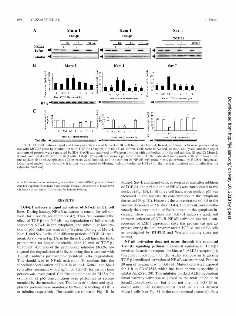
TGF-�1 induces a rapid activation of NF-�B in BL celllines. During latency, NF-�B activation is crucial for cell sur-vival (for a review, see reference 42). Thus, we examined theeffect of TGF-�1 on NF-�B, i.e., degradation of I�B�, whichsequesters NF-�B in the cytoplasm, and subcellular localiza-tion of p65. I�B� was assayed by Western blotting of Mutu-I,Kem-I, and Sav-I cells after different periods of TGF-�1 treat-ment. As shown in Fig. 1A, in the three BL cell lines, the I�B�protein was no longer detectable after 10 min of TGF-�1treatment. Addition of the proteasome inhibitor MG262 ab-rogated the degradation of I�B�, showing that treatment withTGF-�1 induces proteasome-dependent I�B� degradation.This should lead to NF-�B activation. To confirm this, thesubcellular localization of RelA in Mutu-I, Kem-I, and Sav-Icells after treatment with 2 ng/ml of TGF-�1 for various timeperiods was investigated. Cell fractionation and an ELISA forestimation of p65 concentration were performed as recom-mended by the manufacturer. The loads of nuclear and cyto-plasmic proteins were monitored by Western blotting of HP1�or tubulin, respectively. The results are shown in Fig. 1B. In
Mutu-I, Sav-I, and Kem-I cells, as soon as 30 min after additionof TGF-�1, the p65 subunit of NF-�B was translocated to thenucleus (Fig. 1B). In all three cell lines, when nuclear p65 wasincreased in the nucleus, its concentration in the cytoplasmdecreased (Fig. 1C). However, the concentration of p65 in thenucleus decreased at 2 h after TGF-�1 treatment, and simulta-neously the concentration of RelA protein in the cytoplasm in-creased. These results show that TGF-�1 induces a quick andtransient activation of NF-�B. NF-�B activation was not a con-sequence of LMP1 expression, since this protein was not ex-pressed during the Lat I program and in TGF-�1-treated BL cellsas investigated by RT-PCR and Western blotting (data notshown).
NF-�B activation does not occur through the canonicalTGF-�1 signaling pathway. Canonical signaling of TGF-�1involves the activin receptor-like kinase 5 (ALK5) receptor (4);therefore, involvement of the ALK5 receptor in triggeringTGF-�1-mediated activation of NF-�B was examined. Prior to30 min of treatment with TGF-�1, Mutu-I cells were exposedfor 1 h to SB-431542, which has been shown to specificallyinhibit ALK5 (6, 20). This inhibitor blocked ALK5-dependentSmad pathway activation as judged by the total inhibition ofSmad2 phosphorylation, but it did not alter the TGF-�1-in-duced subcellular localization of RelA in TGF-�1-treatedMutu-I cells (see Fig. S1 in the supplemental material). As a
FIG. 1. TGF-�1 induces rapid and transient activation of NF-�B in BL cell lines. (A) Mutu-I, Kem-I, and Sav-I cells were pretreated ornot with MG262 prior to stimulation with TGF-�1 (2 ng/ml) for 10, 15, or 30 min. Cells were harvested, washed, and lysed, and then equalamounts of protein were separated by SDS-PAGE and analyzed by Western blotting with antibodies to I�B� and tubulin. (B and C) Mutu-I,Kem-I, and Sav-I cells were treated with TGF-�1 (2 ng/ml) for various periods of time. At the indicated time points, cells were harvested;the nuclear (B) and cytoplasmic (C) extracts were isolated, and the content of NF-�B p65 protein was determined by ELISA (Imgenex).Loading of nuclear and cytosolic fractions was assayed by blotting with antibodies to HP1� (for the nuclear fraction) and tubulin (for thecytosolic fraction).
6504 OUSSAIEF ET AL. J. VIROL.
on May 10, 2018 by guest
http://jvi.asm.org/
Dow
nloaded from
control, pretreatment with BAY11-7082 (a specific inhibitor ofNF-�B) completely abolished p65 translocation. This resultindicates that a noncanonical signaling pathway, independentof ALK5, was used to activate NF-�B.
Inhibition of the NF-�B pathway prevents TGF-�1-inducedprogression of the EBV lytic cycle. The contribution of theNF-�B pathway to the balance between the latent and lyticphases of the EBV life cycle was addressed using two different
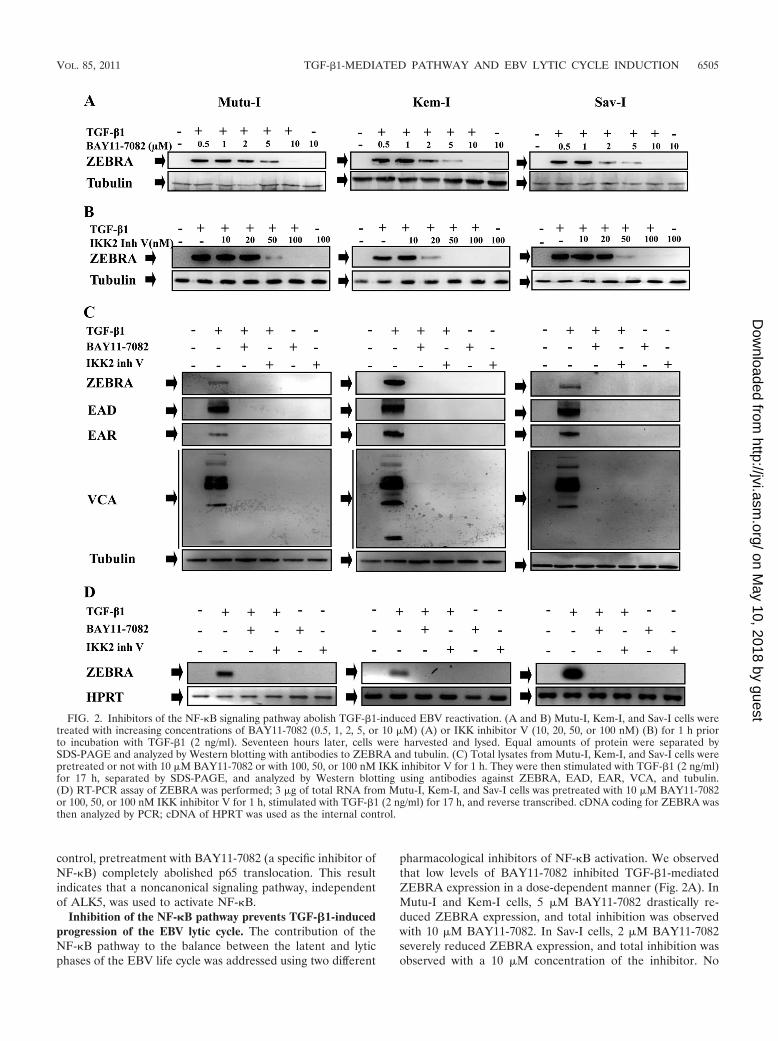
pharmacological inhibitors of NF-�B activation. We observedthat low levels of BAY11-7082 inhibited TGF-�1-mediatedZEBRA expression in a dose-dependent manner (Fig. 2A). InMutu-I and Kem-I cells, 5 �M BAY11-7082 drastically re-duced ZEBRA expression, and total inhibition was observedwith 10 �M BAY11-7082. In Sav-I cells, 2 �M BAY11-7082severely reduced ZEBRA expression, and total inhibition wasobserved with a 10 �M concentration of the inhibitor. No
FIG. 2. Inhibitors of the NF-�B signaling pathway abolish TGF-�1-induced EBV reactivation. (A and B) Mutu-I, Kem-I, and Sav-I cells weretreated with increasing concentrations of BAY11-7082 (0.5, 1, 2, 5, or 10 �M) (A) or IKK inhibitor V (10, 20, 50, or 100 nM) (B) for 1 h priorto incubation with TGF-�1 (2 ng/ml). Seventeen hours later, cells were harvested and lysed. Equal amounts of protein were separated bySDS-PAGE and analyzed by Western blotting with antibodies to ZEBRA and tubulin. (C) Total lysates from Mutu-I, Kem-I, and Sav-I cells werepretreated or not with 10 �M BAY11-7082 or with 100, 50, or 100 nM IKK inhibitor V for 1 h. They were then stimulated with TGF-�1 (2 ng/ml)for 17 h, separated by SDS-PAGE, and analyzed by Western blotting using antibodies against ZEBRA, EAD, EAR, VCA, and tubulin.(D) RT-PCR assay of ZEBRA was performed; 3 �g of total RNA from Mutu-I, Kem-I, and Sav-I cells was pretreated with 10 �M BAY11-7082or 100, 50, or 100 nM IKK inhibitor V for 1 h, stimulated with TGF-�1 (2 ng/ml) for 17 h, and reverse transcribed. cDNA coding for ZEBRA wasthen analyzed by PCR; cDNA of HPRT was used as the internal control.
VOL. 85, 2011 TGF-�1-MEDIATED PATHWAY AND EBV LYTIC CYCLE INDUCTION 6505
on May 10, 2018 by guest
http://jvi.asm.org/
Dow
nloaded from
significant effects of the inhibitor on cell viability were ob-served (see Fig. S2A in the supplemental material).
IKK2 inhibitor V is known to selectively block I�B� phos-phorylation and prevent NF-�B p65 nuclear translocation(22, 34). TGF-�1-induced production of ZEBRA in Mutu-I,
Kem-I, and Sav-I cells was reduced by treatment with thisinhibitor in a dose-dependent manner (Fig. 2B). In Mutu-I andSav-I cells, 50 nM IKK2 inhibitor V dramatically diminishedZEBRA expression, and complete inhibition was observedwith 100 nM. For Kem-I cells, 20 nM IKK2 inhibitor V dra-
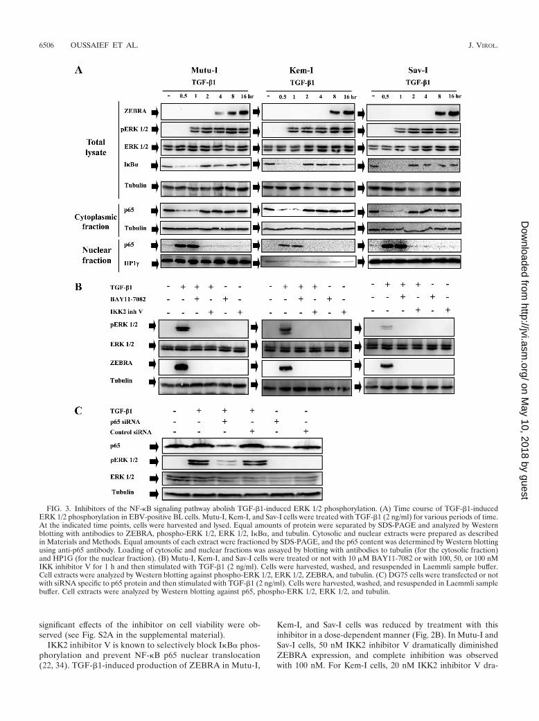
FIG. 3. Inhibitors of the NF-�B signaling pathway abolish TGF-�1-induced ERK 1/2 phosphorylation. (A) Time course of TGF-�1-inducedERK 1/2 phosphorylation in EBV-positive BL cells. Mutu-I, Kem-I, and Sav-I cells were treated with TGF-�1 (2 ng/ml) for various periods of time.At the indicated time points, cells were harvested and lysed. Equal amounts of protein were separated by SDS-PAGE and analyzed by Westernblotting with antibodies to ZEBRA, phospho-ERK 1/2, ERK 1/2, I�B�, and tubulin. Cytosolic and nuclear extracts were prepared as describedin Materials and Methods. Equal amounts of each extract were fractioned by SDS-PAGE, and the p65 content was determined by Western blottingusing anti-p65 antibody. Loading of cytosolic and nuclear fractions was assayed by blotting with antibodies to tubulin (for the cytosolic fraction)and HP1G (for the nuclear fraction). (B) Mutu-I, Kem-I, and Sav-I cells were treated or not with 10 �M BAY11-7082 or with 100, 50, or 100 nMIKK inhibitor V for 1 h and then stimulated with TGF-�1 (2 ng/ml). Cells were harvested, washed, and resuspended in Laemmli sample buffer.Cell extracts were analyzed by Western blotting against phospho-ERK 1/2, ERK 1/2, ZEBRA, and tubulin. (C) DG75 cells were transfected or notwith siRNA specific to p65 protein and then stimulated with TGF-�1 (2 ng/ml). Cells were harvested, washed, and resuspended in Laemmli samplebuffer. Cell extracts were analyzed by Western blotting against p65, phospho-ERK 1/2, ERK 1/2, and tubulin.
6506 OUSSAIEF ET AL. J. VIROL.
on May 10, 2018 by guest
http://jvi.asm.org/
Dow
nloaded from
matically diminished ZEBRA expression, and complete inhi-bition was observed with 50 nM (Fig. 2B). No significanteffects of the inhibitor on cell viability were observed (seeFig. S2B in the supplemental material). In Mutu-I, Kem-I,and Sav-I TGF-�1-stimulated cells, the whole EBV lyticprotein pattern, i.e., ZEBRA, EAD, EAR, and VCA, wasexpressed, showing that the virion proteins were produced.NF-�B inhibitors (BAY11-7082 and IKK2 inhibitor V) ab-rogated this expression (Fig. 2C).
Expression of the transcripts encoding ZEBRA was investi-gated by RT-PCR (Fig. 2D). Pretreatment with NF-�B inhib-itors (BAY11-7082 or IKK2 inhibitor V) prevents amplifica-tion of BZLF1 cDNA. Consequently, inhibition of ZEBRAexpression might be the result of inhibited production oftranscripts encoding the protein. These results suggest thattransient activation of NF-�B is required for TGF-�1-in-duced ZEBRA expression.
TGF-�1 induces NF-�B-dependent ERK 1/2 phosphoryla-tion. We already established a key role of ERK 1/2 MAPK inthe TGF-�1 signaling pathway (11). Given that transient
NF-�B activation was also shown to be required for triggeringZEBRA expression upon TGF-�1 simulation, we focused onthe possible interaction between the ERK 1/2 and NF-�B path-ways. A detailed time course study of the kinetics of ERK 1/2pathway activation in response to TGF-�1 stimulation indi-cated that activation of ERK 1/2 (1 h after addition of TGF-�1) occurred after IKB� degradation and nuclear p65 trans-location (30 min) (Fig. 3A). The possible role of NF-�B inTGF-�1-induced ERK 1/2 signaling activation was then inves-tigated in Mutu-I, Kem-I, and Sav-I cells. Those cells weretreated or not with 10 �M BAY11-7082 or with 100, 50, or 100nM IKK inhibitor V for 1 h and were then stimulated withTGF-�1 (2 ng/ml). Total proteins were analyzed for phosphor-ylated forms of ERK 1/2 by Western blotting with phospho-specific antibodies. The membranes were reprobed to evaluatetotal ERK 1/2 expression. Figure 3B shows that treatment witheach NF-�B inhibitor completely inhibited ERK 1/2 phosphor-ylation and consequently ZEBRA expression. This result wasconfirmed by examining the effect of transfection a small in-terfering oligonucleotide RNA (siRNA) specific for p65 in
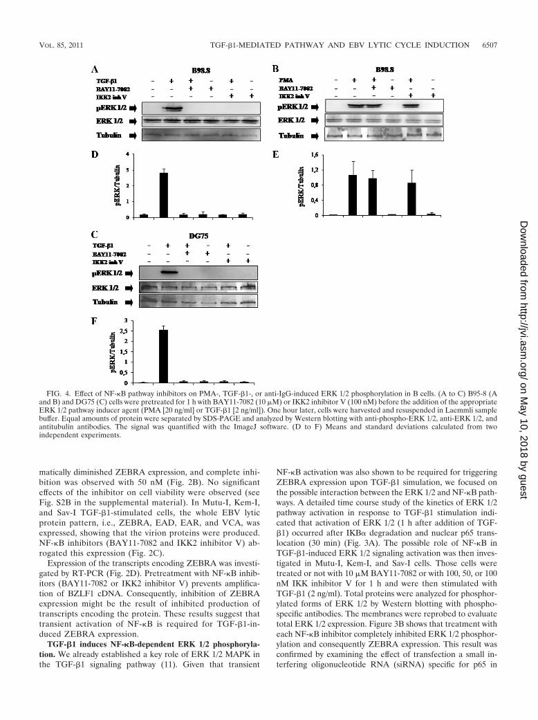
FIG. 4. Effect of NF-�B pathway inhibitors on PMA-, TGF-�1-, or anti-IgG-induced ERK 1/2 phosphorylation in B cells. (A to C) B95-8 (Aand B) and DG75 (C) cells were pretreated for 1 h with BAY11-7082 (10 �M) or IKK2 inhibitor V (100 nM) before the addition of the appropriateERK 1/2 pathway inducer agent (PMA [20 ng/ml] or TGF-�1 [2 ng/ml]). One hour later, cells were harvested and resuspended in Laemmli samplebuffer. Equal amounts of protein were separated by SDS-PAGE and analyzed by Western blotting with anti-phospho-ERK 1/2, anti-ERK 1/2, andantitubulin antibodies. The signal was quantified with the ImageJ software. (D to F) Means and standard deviations calculated from twoindependent experiments.
VOL. 85, 2011 TGF-�1-MEDIATED PATHWAY AND EBV LYTIC CYCLE INDUCTION 6507
on May 10, 2018 by guest
http://jvi.asm.org/
Dow
nloaded from
DG75 cells. This transfection dramatically reduced p65 expres-sion as well as ERK 1/2 phosphorylation (Fig. 3C). As ex-pected, inhibition of ERK 1/2 phosphorylation by the specificinhibitor U0126 did not affect TGF-�1-induced NF-�B activa-tion (see Fig. S3 in the supplemental material). These data arein accordance with the hypothesis of NF-�B-dependent ERK1/2 activation on TGF-�1 stimulation of BL cells.
NF-�B-dependent ERK 1/2 phosphorylation triggered byTGF-�1 also occurs in an LCL with a latency III pattern ofEBV expression. TGF-�1-treated EBV-positive lymphoblas-toid cell line (LCL) B95-8 cells also exhibited NF-�B-depen-dent ERK 1/2 phosphorylation, as pretreatment with eitherBAY11-7082 or IKK2 inhibitor V completely abrogatedTGF-�1-induced ERK 1/2 phosphorylation as shown byWestern blotting with antibodies to phospho-ERK 1/2 (Fig.4A). Thus, in either latency I (Fig. 3B) or latency III (Fig.4A), EBV-positive cells exhibited NF-�B-dependent ERK1/2 activation.
However, when B95-8 cells were treated with PMA, al-though ERK 1/2 phosphorylation was observed, this activationwas NF-�B independent since inhibition of this pathway didnot affect PMA-induced ERK 1/2 phosphorylation (Fig. 4B).Thus, NF-�B-mediated ERK 1/2 phosphorylation was TGF-�1dependent.
NF-�B-dependent ERK 1/2 phosphorylation triggered byTGF-�1 also occurs in EBV-negative BL cells. DG75 cells weretreated with TGF-�1 for 1 h; ERK 1/2 was phosphorylated,and pretreatments with NF-�B inhibitors (BAY11-7082 orIKK2 inhibitor V) abolished TGF-�1-induced ERK 1/2 phos-phorylation (Fig. 4C). This suggests that the effect of TGF-�1-mediated ERK 1/2 activation that occurs through NF-�B isindependent of EBV.
NO inhibits TGF-�1-induced ERK 1/2 phosphorylation. Ithas been shown that inducible nitric oxide synthase (iNOS)
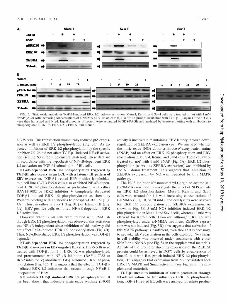
activity is involved in maintaining EBV latency through down-regulation of ZEBRA expression (26). We analyzed whetherthe nitric oxide (NO) donor S-nitroso-N-acetylpenicillamine(SNAP) had an effect on ERK 1/2 phosphorylation and EBVreactivation in Mutu-I, Kem-I, and Sav-I cells. These cells weretreated (or not) with 1 mM SNAP (Fig. 5A). ERK 1/2 phos-phorylation (as well as ZEBRA expression) was inhibited bythe NO donor treatment. This suggests that inhibition ofZEBRA expression by NO was mediated by this MAPKpathway.
The NOS inhibitor NG-monomethyl-L-arginine acetate salt(L-NMMA) was used to investigate the effect of NOS activityon ERK 1/2 phosphorylation. Mutu-I, Kem-I, and Sav-Icells were treated for 1 h with increasing concentrations ofL-NMMA (2, 5, 10, or 20 mM), and cell lysates were assayedfor ERK 1/2 phosphorylation and ZEBRA expression. Asshown in Fig. 5B, 5 mM NOS inhibitor induced ERK 1/2phosphorylation in Mutu-I and Sav-I cells, whereas 10 mM wasefficient for Kem-I cells. However, although ERK 1/2 wasphosphorylated under L-NMMA treatment, ZEBRA expres-sion was not induced (Fig. 5B); this suggests that activation ofthis MAPK pathway is insufficient, even though it is necessary,to provoke EBV reactivation in the cells explored. No changein cell viability was observed under treatments with eitherSNAP or L-NMNA (see Fig. S4 in the supplemental material).Activity of the promoter directing expression of the ZEBRAprotein could be achieved in DG75 cells by coexpression ofSmad2 to -4 with Ras (which induced ERK 1/2 phosphoryla-tion). This suggests that expression from Zp necessitated bothERK 1/2 MAPK and Smad activation (see Fig. S5 in the sup-plemental material).
TGF-�1 mediates inhibition of nitrite production throughNF-�B activation. As NO influences ERK 1/2 phosphoryla-tion, TGF-�1-treated BL cells were assayed for nitrite produc-
FIG. 5. Nitric oxide modulates TGF-�1-induced ERK 1/2 pathway activation. Mutu-I, Kem-I, and Sav-I cells were treated or not with 1 mMSNAP (A) or with increasing concentrations of L-NMMA (2, 5, 10, or 20 mM) (B) for 1 h prior to incubation with TGF-�1 (2 ng/ml) for 8 h. Cellswere then harvested and lysed. Equal amounts of protein were separated by SDS-PAGE and analyzed by Western blotting with antibodies tophosphorylated-ERK 1/2, ERK 1/2, ZEBRA, and tubulin.
6508 OUSSAIEF ET AL. J. VIROL.
on May 10, 2018 by guest
http://jvi.asm.org/
Dow
nloaded from
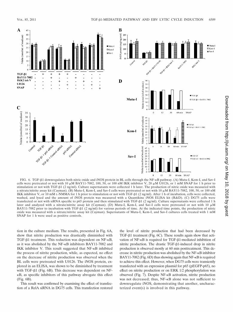
tion in the culture medium. The results, presented in Fig. 6A,show that nitrite production was drastically diminished withTGF-�1 treatment. This reduction was dependent on NF-�B,as it was abolished by the NF-�B inhibitors BAY11-7082 andIKK inhibitor V. This result suggested that NF-�B inhibitedthe process of nitrite production, while, as expected, no effecton the decrease of nitrite production was observed when theBL cells were pretreated with U0126. The iNOS protein, ex-plored in an ELISA, was shown to be diminished by treatmentwith TGF-�1 (Fig. 6B). This decrease was dependent on NF-�B, as specific inhibitors of this pathway abrogate this effect(Fig. 6B).
This result was confirmed by examining the effect of transfec-tion of a RelA siRNA in DG75 cells. This transfection restored
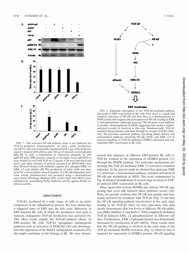
the level of nitrite production that had been decreased byTGF-�1 treatment (Fig. 6C). These results again show that acti-vation of NF-�B is required for TGF-�1-mediated inhibition ofnitrite production. The drastic TGF-�1-induced drop in nitriteproduction is observed mostly at 60 min posttreatment. This de-crease in nitrite production was abolished by the NF-�B inhibitorBAY11-7082 (Fig. 6D) thus showing again that NF-�B is requiredto achieve this effect. However, when DG75 cells were transientlytransfected with an expression plasmid for p65 (pEGFP-p65), noeffect on nitrite production or on ERK 1/2 phosphorylation wasobserved (Fig. 7). Despite NF-�B activation, nitrite productionwas not decreased; thus, NF-�B alone was not sufficient todownregulate iNOS, demonstrating that another, uncharac-terized event(s) is involved in this pathway.
FIG. 6. TGF-�1 downregulates both nitric oxide and iNOS protein in BL cells through the NF-�B pathway. (A) Mutu-I, Kem-I, and Sav-Icells were pretreated or not with 10 �M BAY11-7082, 100, 50, or 100 nM IKK inhibitor V, 20 �M U0126, or 1 mM SNAP for 1 h prior tostimulation or not with TGF-�1 (2 ng/ml). Culture supernatants were collected 1 h later. The production of nitric oxide was measured witha nitrate/nitrite assay kit (Cayman). (B) Mutu-I, Kem-I, and Sav-I cells were pretreated or not with 10 �M BAY11-7082, 100, 50, or 100 nMIKK inhibitor V, or 10 mM L-NMMA for 1 h prior to stimulation or not with TGF-�1 (2 ng/ml). After 1 h of incubation, cells were collected,washed, and lysed and the amount of iNOS protein was measured with a Quantikine iNOS ELISA kit (R&D). (C) DG75 cells weretransfected or not with siRNA specific to p65 protein and then stimulated with TGF-�1 (2 ng/ml). Culture supernatants were collected 1 hlater and analyzed with a nitrate/nitrite assay kit (Cayman). (D) Mutu-I, Kem-I, and Sav-I cells were pretreated or not with 10 �MBAY11-7082 prior to incubation with TGF-�1 (2 ng/ml) for various periods of time. At the indicated time points, the production of nitricoxide was measured with a nitrate/nitrite assay kit (Cayman). Supernatants of Mutu-I, Kem-I, and Sav-I cultures cells treated with 1 mMSNAP for 1 h were used as positive controls.
VOL. 85, 2011 TGF-�1-MEDIATED PATHWAY AND EBV LYTIC CYCLE INDUCTION 6509
on May 10, 2018 by guest
http://jvi.asm.org/
Dow
nloaded from
DISCUSSION
TGF-�1, produced by a wide range of cells, is an activecomponent of the inflammatory process. We have shown thatit triggered entry of EBV into the lytic cycle. Moreover, inEBV-infected BL cells in which the productive viral cycle isinduced, endogenous TGF-�1 production was activated (9).This effect would amplify the TGF-�1-induced effects. InEBV-positive BL cells TGF-�1 upregulates antiapoptoticpathways such as activation of PI3K/Akt (36) and downregu-lates the expression of the BimEL antiapoptotic molecule (35);this might contribute to the biology of BL. We have demon-
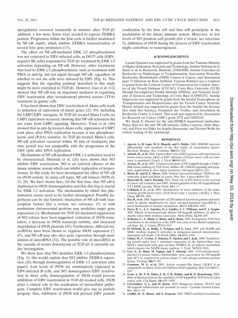
strated that exposure of different EBV-positive BL cells toTGF-�1 resulted in the expression of ZEBRA protein (11)through the MAPK pathway. The molecular mechanisms un-derlying this TGF-�1-mediated ERK 1/2 activation remainedunknown. In the present study we showed that upstream ERK1/2 activation, a noncanonical pathway, included activation ofNF-�B and modulation of iNOS. This work (summarized inFig. 8) allowed identification of several steps involved in TGF-�1-induced EBV reactivation in BL cells.
Many agents that activate MAPKs also activate NF-�B, sug-gesting that cross talk between these pathways occurs (20).Here, we provide evidence that NF-�B was quickly and tran-siently activated by treatment with TGF-�1 (Fig. 1) and thatthe NF-�B signaling pathway was involved in the early stepsleading to the TGF-�1 effect on viral expression. Our dataclearly demonstrate that low levels of different NF-�B inhibi-tors (IKK2 inhibitor V and BAY11-7082) completely abolishedTGF-�1-induced ERK 1/2 phosphorylation in different celllines. Furthermore, ERK 1/2 phosphorylation was dramaticallydecreased by transfection of p65 siRNA. These results showthat the NF-�B pathway is involved in the early steps of theTGF-�1-mediated MAPK activation (Fig. 3), which in turn isrequired for expression of ZEBRA protein. NF-�B signaling
FIG. 7. The activated NF-�B pathway alone is not sufficient forTGF-�1-mediated downregulation of nitric oxide production.(A) DG75 cells were transiently transfected with 5 �g of the luciferasereporter plasmid pNF-�B-Luc and 100 ng of internal control plasmidpRL-TK or with a combination of expression plasmids for p65(pEGFP-p65), NIK (positive control), or an empty vector (pEGFP) orwere treated (or not) with TGF-�1 (2 ng/ml). Cells were harvested andlysed, and equal amounts of protein separated by SDS-PAGE werethen Western blotted with antibodies against p65, phospho-ERK 1/2,ERK 1/2, and tubulin. (B) The production of nitric oxide was mea-sured by a nitrate/nitrite assay (Cayman). (C) NF-�B-dependent lucif-erase activity (luminescence) was measured using a dual-luciferaseassay system (Promega, Madison, WI); relative light units (RLU) werecalculated by normalizing firefly luciferase activity against Renilla lu-ciferase activity.
FIG. 8. Schematic description of the TGF-�1-mediated pathwaythat leads to EBV reactivation in BL cells. First there is a quick andtransient activation of NF-�B, and then there is a downregulation ofiNOS activity that requires the participation of NF-�B, leading to ERK1/2 phosphorylation. Although necessary, NF-�B alone is not sufficientto produce downregulation of iNOS, suggesting that another, unchar-acterized event(s) is involved at this step. Simultaneously, TGF-�1mediates Smad pathway activation through its receptor I/ALK5. How-ever, the activated canonical pathway (involving Smad factors) andnoncanonical pathways (involving NF-�B, iNOS, and ERK 1/2) inconcert contribute to TGF-�1-mediated ZEBRA expression and con-sequently EBV reactivation in BL cells.
6510 OUSSAIEF ET AL. J. VIROL.
on May 10, 2018 by guest
http://jvi.asm.org/
Dow
nloaded from
upregulation occurred transiently in minutes after TGF-�1addition; a few more hours were needed to express ZEBRAprotein. Progression within the lytic cycle is further modulatedby NF-�B Asp65, which inhibits ZEBRA transactivation ofseveral lytic gene promoters (17).
The effect on NF-�B-mediated ERK 1/2 phosphorylationwas not restricted to EBV-infected cells, as DG75 cells (EBV-negative BL cells) responded to TGF-�1 treatment by ERK 1/2activation depending on NF-�B. However, other treatmentsthat lead to ERK 1/2 phosphorylation, such as treatment withPMA or anti-Ig, did not signal through NF-�B, regardless ofwhether or not the cells were infected by EBV (Fig. 4). Thissuggests that the signaling pathway described in this studymight be more restricted to TGF-�1. However, Gao et al. (12)showed that NF-�B was an important mediator in regulatingEBV reactivation after tetradecanoyl phorbol acetate (TPA)treatment in gastric cells.
It has been shown that EBV reactivation of Akata cells leadsto induction of expression of latent genes (25, 49), includingthe LMP1 EBV oncogene. In TGF-�1-treated Mutu-I cells, noLMP1 expression occurred, showing that NF-�B activation didnot come from LMP1 signaling. Moreover, Yuan et al. (49)showed that in anti-Ig-treated-Akata cells, expression of LMP1took place after DNA replication because it was phosphono-acetic acid (PAA) sensitive. In TGF-�1-treated Mutu-I cells,NF-�B activation occurred within 30 min of treatment; thistime period was not compatible with the progression of theEBV cycle after DNA replication.
Mechanisms of NF-�B-mediated ERK 1/2 activation need tobe characterized. Mannick et al. (26) have shown that NOinhibits EBV reactivation. NO is an antiviral effector of theinnate immune system which can inhibit replication of herpes-viruses. In this study, we have investigated the effect of NF-�Bon iNOS activity. In many cell types, NF-�B induces iNOS (8,21, 28). We have shown here, for the first time, that NF-�B isimplicated in iNOS downregulation and that this step is crucialfor ERK 1/2 activation. The mechanisms by which this phe-nomenon occurs need to be further investigated. Possible hy-potheses can be put forward: interaction of NF-�B with tran-scription factors (for a review, see reference 15) or withnonhistone chromosomal proteins may modulate target geneexpression (1). Mechanisms for TGF-�1-mediated suppressionof NO release have been suggested: reduction of iNOS trans-lation, a decrease in iNOS transcript stability, and increaseddegradation of iNOS proteins (45). Furthermore, different mi-croRNAs have been shown to regulate iNOS expression (7,47), and NF-�B may also alter gene expression through mod-ulation of microRNA (52). The possible role of microRNA inthe cascade of events downstream of TGF-�1 is currently un-der investigation.
We show here that NO abolishes ERK 1/2 phosphorylation(Fig. 5); this would explain that NO inhibits ZEBRA expres-sion (26) through downregulation of ERK 1/2 activation (thispaper). Low levels of iNOS are constitutively expressed inEBV-infected B cells, and NO downregulates EBV reactiva-tion in those cells; downregulation of iNOS would preventinhibition of EBV reactivation in TGF-�1-treated cells. iNOSplays a critical role in the eradication of intracellular patho-gens. Complete EBV reactivation would give rise to particleprogeny; thus, inhibition of iNOS will prevent EBV particle
eradication by the host cell and thus will participate in themodulation of the innate immune system. Moreover, as lowlevels of NO promote cell growth (for a review, see reference5), inhibition of iNOS during the process of EBV reactivationmight contribute to tumorigenesis.
ACKNOWLEDGMENTS
Lassad Oussaief was supported by grants from the Tunisian Ministryof Higher Education, Research and Technology, Institut National de laSante et de la Recherche Medicale (INSERM), Institut Francilien deRecherche en Nephrologie et Transplantation, Association NouvellesRecherches Biomedicales (NRB) Vaincre le Cancer, and Associationpour l’Utilization du Rein Artificiel. Vanessa Ramírez was a recipientof grants from the Cultural Center of Cooperation for Central Amer-ica of the French Embassy (CCCAC), Costa Rica University (UCR)through Investigations Health Institute (INISA), and National Acad-emy of Sciences and Technology of Costa Rica (CONICIT). AurelieHippocrate was supported by grants from the Societe Francophone deTransplantation and Herpesviruses and the French Cancer Network.Hratch Arbach was supported by grants from the Societe De SecoursDes Amis Des Sciences, Fondation des Treilles, and Association deRecherche Contre le Cancer. This work was supported by Associationfor Research on Cancer (ARC) grant 3572 and GEFLUC.
We thank E. Drouet for the anti-ZEBRA monoclonal antibodies,Nancy Rice for the anti NF-�B subunits, Gilles Courtois, Alain Mau-viel, and Peter ten Dijke for helpful discussions, and Dermot Walls forcritical reading of the manuscript.
REFERENCES
1. Agresti, A., R. Lupo, M. E. Bianchi, and S. Muller. 2003. HMGB1 interactsdifferentially with members of the Rel family of transcription factors.Biochem. Biophys. Res. Commun. 302:421–426.
2. Arbach, H., et al. 2006. Epstein-Barr virus (EBV) genome and expression inbreast cancer tissue: effect of EBV infection of breast cancer cells on resis-tance to paclitaxel (Taxol). J. Virol. 80:845–853.
3. Arsura, M., et al. 2003. Transient activation of NF-kappaB through a TAK1/IKK kinase pathway by TGF-beta1 inhibits AP-1/SMAD signaling and apop-tosis: implications in liver tumor formation. Oncogene 22:412–425.
4. Bierie, B., and H. L. Moses. 2006. Tumour microenvironment: TGFbeta: themolecular Jekyll and Hyde of cancer. Nat. Rev. Cancer 6:506–520.
5. Bonavida, B., and S. Baritaki. 2011. Dual role of NO donors in the reversalof tumor cell resistance and EMT: Downregulation of the NF-kappaB/Snail/YY1/RKIP circuitry. Nitric Oxide 24:1–11.
6. Callahan, J. F., et al. 2002. Identification of novel inhibitors of the trans-forming growth factor beta1 (TGF-beta1) type 1 receptor (ALK5). J. Med.Chem. 45:999–1001.
7. Dai, R., et al. 2008. Suppression of LPS-induced interferon-gamma and nitricoxide in splenic lymphocytes by select estrogen-regulated microRNAs: anovel mechanism of immune modulation. Blood 112:4591–4597.
8. Davis, R. L., A. C. Sanchez, D. J. Lindley, S. C. Williams, and P. J. Syapin.2005. Effects of mechanistically distinct NF-kappaB inhibitors on glial in-ducible nitric-oxide synthase expression. Nitric Oxide 12:200–209.
9. di Renzo, L., A. Altiok, G. Klein, and E. Klein. 1994. Endogenous TGF-betacontributes to the induction of the EBV lytic cycle in two Burkitt lymphomacell lines. Int. J. Cancer 57:914–919.
10. El Mchichi, B., A. Hadji, A. Vazquez, and G. Leca. 2007. p38 MAPK andMSK1 mediate caspase-8 activation in manganese-induced mitochondria-dependent cell death. Cell Death Differ. 14:1826–1836.
11. Fahmi, H., C. Cochet, Z. Hmama, P. Opolon, and I. Joab. 2000. Transform-ing growth factor beta 1 stimulates expression of the Epstein-Barr virusBZLF1 immediate-early gene product ZEBRA by an indirect mechanismwhich requires the MAPK kinase pathway. J. Virol. 74:5810–5818.
12. Gao, X., K. Ikuta, M. Tajima, and T. Sairenji. 2001. 12-O-tetradecanoyl-phorbol-13-acetate induces Epstein-Barr virus reactivation via NF-kappaBand AP-1 as regulated by protein kinase C and mitogen-activated proteinkinase. Virology 286:91–99.
13. Goumans, M. J., et al. 2003. Activin receptor-like kinase (ALK)1 is anantagonistic mediator of lateral TGFbeta/ALK5 signaling. Mol. Cell 12:817–828.
14. Grau, A. M., P. K. Datta, J. Zi, S. K. Halder, and R. D. Beauchamp. 2006.Role of Smad proteins in the regulation of NF-kappaB by TGF-beta in coloncancer cells. Cell Signal. 18:1041–1050.
15. Grivennikov, S. I., and M. Karin. 2010. Dangerous liaisons: STAT3 andNF-kappaB collaboration and crosstalk in cancer. Cytokine Growth FactorRev. 21:11–19.
16. Gruffat, H., E. Manet, and A. Sergeant. 2002. MEF2-mediated recruitment
VOL. 85, 2011 TGF-�1-MEDIATED PATHWAY AND EBV LYTIC CYCLE INDUCTION 6511
on May 10, 2018 by guest
http://jvi.asm.org/
Dow
nloaded from
of class II HDAC at the EBV immediate early gene BZLF1 links latency andchromatin remodeling. EMBO Rep. 3:141–146.
17. Gutsch, D. E., et al. 1994. The bZIP transactivator of Epstein-Barr virus,BZLF1, functionally and physically interacts with the p65 subunit of NF-kappa B. Mol. Cell. Biol. 14:1939–1948.
18. Hiscott, J., H. Kwon, and P. Genin. 2001. Hostile takeovers: viral appropri-ation of the NF-kappaB pathway. J. Clin. Invest. 107:143–151.
19. Inman, G. J., U. K. Binne, G. A. Parker, P. J. Farrell, and M. J. Allday. 2001.Activators of the Epstein-Barr virus lytic program concomitantly induceapoptosis, but lytic gene expression protects from cell death. J. Virol. 75:2400–2410.
20. Inman, G. J., et al. 2002. SB-431542 is a potent and specific inhibitor oftransforming growth factor-beta superfamily type I activin receptor-like ki-nase (ALK) receptors ALK4, ALK5, and ALK7. Mol. Pharmacol. 62:65–74.
21. Kadowaki, S., et al. 2004. Down-regulation of inducible nitric oxide synthaseby lysophosphatidic acid in human respiratory epithelial cells. Mol. Cell.Biochem. 262:51–59.
22. Kamon, J., et al. 2004. A novel IKKbeta inhibitor stimulates adiponectinlevels and ameliorates obesity-linked insulin resistance. Biochem. Biophys.Res. Commun. 323:242–248.
23. Kenney, S. 2007. Reactivation and lytic replication of EBV. In C.-F. G.Arvin, E. Mocarski, P. S. Moore, B. Roizman, R. Whitley, and K. Yamanishi(ed.), Human herpesviruses: biology, therapy, and immunoprophylaxis.Cambridge University Press, Cambridge, United Kingdom.
24. Kieff, E. 2007. Epstein-Barr virus and its replication, vol. 2. Lippincott Wil-liam & Wilkins, Philadelphia, PA.
25. Lu, C. C., et al. 2006. Genome-wide transcription program and expression ofthe Rta responsive gene of Epstein-Barr virus. Virology 345:358–372.
26. Mannick, J. B., K. Asano, K. Izumi, E. Kieff, and J. S. Stamler. 1994. Nitricoxide produced by human B lymphocytes inhibits apoptosis and Epstein-Barrvirus reactivation. Cell 79:1137–1146.
27. Massague, J., and D. Wotton. 2000. Transcriptional control by the TGF-beta/Smad signaling system. EMBO J. 19:1745–1754.
28. Mizel, S. B., A. N. Honko, M. A. Moors, P. S. Smith, and A. P. West. 2003.Induction of macrophage nitric oxide production by Gram-negative flagellininvolves signaling via heteromeric Toll-like receptor 5/Toll-like receptor 4complexes. J. Immunol. 170:6217–6223.
29. Mogensen, T. H., J. Melchjorsen, P. Hollsberg, and S. R. Paludan. 2003.Activation of NF-kappa B in virus-infected macrophages is dependent onmitochondrial oxidative stress and intracellular calcium: downstream in-volvement of the kinases TGF-beta-activated kinase 1, mitogen-activatedkinase/extracellular signal-regulated kinase kinase 1, and I kappa B kinase.J. Immunol. 170:6224–6233.
30. Mogensen, T. H., and S. R. Paludan. 2001. Molecular pathways in virus-induced cytokine production. Microbiol. Mol. Biol. Rev. 65:131–150.
31. Nagata, Y., et al. 2004. Activation of Epstein-Barr virus by saliva fromSjogren’s syndrome patients. Immunology 111:223–229.
32. Niller, H. H., et al. 2004. EBV-associated neoplasms: alternative pathoge-netic pathways. Med. Hypotheses 62:387–391.
33. Oliveira, J. B., et al. 2007. NRAS mutation causes a human autoimmunelymphoproliferative syndrome. Proc. Natl. Acad. Sci. U. S. A. 104:8953–8958.
34. Onai, Y., et al. 2004. Inhibition of IkappaB phosphorylation in cardiomyo-
cytes attenuates myocardial ischemia/reperfusion injury. Cardiovasc. Res.63:51–59.
35. Oussaief, L., et al. 2009. Activation of the lytic program of the Epstein-Barrvirus in Burkitt’s lymphoma cells leads to a two steps downregulation ofexpression of the proapoptotic protein BimEL, one of which is EBV-late-gene expression dependent. Virology 387:41–49.
36. Oussaief, L., et al. 2009. Phosphatidylinositol 3-kinase/Akt pathway targetsacetylation of Smad3 through Smad3/CREB-binding protein interaction:contribution to transforming growth factor beta1-induced Epstein-Barr virusreactivation. J. Biol. Chem. 284:23912–23924.
37. Pfeffer, S., et al. 2005. Identification of microRNAs of the herpesvirus family.Nat. Methods 2:269–276.
38. Saito, N., et al. 2003. Two carboxyl-terminal activation regions of Epstein-Barr virus latent membrane protein 1 activate NF-kappaB through distinctsignaling pathways in fibroblast cell lines. J. Biol. Chem. 278:46565–46575.
39. Sanchez-Capelo, A. 2005. Dual role for TGF-beta1 in apoptosis. CytokineGrowth Factor Rev. 16:15–34.
40. Santoro, M. G., A. Rossi, and C. Amici. 2003. NF-kappaB and virus infection:who controls whom. EMBO J. 22:2552–2560.
41. Shi, Y., and J. Massague. 2003. Mechanisms of TGF-beta signaling from cellmembrane to the nucleus. Cell 113:685–700.
42. Soni, V., E. Cahir-McFarland, and E. Kieff. 2007. LMP1 TRAFficking acti-vates growth and survival pathways. Adv. Exp. Med. Biol. 597:173–187.
43. Thompson, M. P., and R. Kurzrock. 2004. Epstein-Barr virus and cancer.Clin. Cancer Res. 10:803–821.
44. Urier, G., M. Buisson, P. Chambard, and A. Sergeant. 1989. The Epstein-Barr virus early protein EB1 activates transcription from different responsiveelements including AP-1 binding sites. EMBO J. 8:1447–1453.
45. Vodovotz, Y., C. Bogdan, J. Paik, Q. W. Xie, and C. Nathan. 1993. Mecha-nisms of suppression of macrophage nitric oxide release by transforminggrowth factor beta. J. Exp. Med. 178:605–613.
46. Wan, F., et al. 2007. Ribosomal protein S3: a KH domain subunit in NF-kappaB complexes that mediates selective gene regulation. Cell 131:927–939.
47. Wang, X., et al. 2009. Inducible nitric-oxide synthase expression is regulatedby mitogen-activated protein kinase phosphatase-1. J. Biol. Chem. 284:27123–27134.
48. Yin, Q., K. Jupiter, and E. K. Flemington. 2004. The Epstein-Barr virustransactivator Zta binds to its own promoter and is required for full pro-moter activity during anti-Ig and TGF-beta1 mediated reactivation. Virology327:134–143.
49. Yuan, J., E. Cahir-McFarland, B. Zhao, and E. Kieff. 2006. Virus and cellRNAs expressed during Epstein-Barr virus replication. J. Virol. 80:2548–2565.
50. Zalani, S., E. Holley-Guthrie, and S. Kenney. 1996. Epstein-Barr viral la-tency is disrupted by the immediate-early BRLF1 protein through a cell-specific mechanism. Proc. Natl. Acad. Sci. U. S. A. 93:9194–9199.
51. Zhang, Y., X. Feng, R. We, and R. Derynck. 1996. Receptor-associated Madhomologues synergize as effectors of the TGF-beta response. Nature 383:168–172.
52. Zhou, R., G. Hu, A. Y. Gong, and X. M. Chen. 2010. Binding of NF-kappaBp65 subunit to the promoter elements is involved in LPS-induced transacti-vation of miRNA genes in human biliary epithelial cells. Nucleic Acids Res.38:3222–3232.
6512 OUSSAIEF ET AL. J. VIROL.
on May 10, 2018 by guest
http://jvi.asm.org/
Dow
nloaded from