ir thermal imaging
DESCRIPTION
IR thermal imagingTRANSCRIPT

This content has been downloaded from IOPscience. Please scroll down to see the full text.
Download details:
IP Address: 188.25.193.123
This content was downloaded on 23/03/2016 at 11:28
Please note that terms and conditions apply.
Infrared thermal imaging in medicine
View the table of contents for this issue, or go to the journal homepage for more
2012 Physiol. Meas. 33 R33
(http://iopscience.iop.org/0967-3334/33/3/R33)
Home Search Collections Journals About Contact us My IOPscience

IOP PUBLISHING PHYSIOLOGICAL MEASUREMENT
Physiol. Meas. 33 (2012) R33–R46 doi:10.1088/0967-3334/33/3/R33
TOPICAL REVIEW
Infrared thermal imaging in medicine
E F J Ring1,3 and K Ammer1,2
1 Medical Imaging Research Unit, Faculty of Advanced Technology, University of Glamorgan,Pontypridd, CF37 1DL, UK2 Institute for Physical Medicine and Rehabilitation, Hanuschkrankenhaus, Vienna, Austria
E-mail: [email protected]
Received 13 October 2011, accepted for publication 16 December 2011Published 28 February 2012Online at stacks.iop.org/PM/33/R33
AbstractThis review describes the features of modern infrared imaging technology andthe standardization protocols for thermal imaging in medicine. The techniqueessentially uses naturally emitted infrared radiation from the skin surface.Recent studies have investigated the influence of equipment and the methodsof image recording. The credibility and acceptance of thermal imaging inmedicine is subject to critical use of the technology and proper understandingof thermal physiology. Finally, we review established and evolving medicalapplications for thermal imaging, including inflammatory diseases, complexregional pain syndrome and Raynaud’s phenomenon. Recent interest in thepotential applications for fever screening is described, and some other areasof medicine where some research papers have included thermal imaging asan assessment modality. In certain applications thermal imaging is shownto provide objective measurement of temperature changes that are clinicallysignificant.
Keywords: infrared, skin temperature, inflammation, fever
Introduction
Quantitative thermal imaging was reviewed 21 years ago by Ring in this journal (Ring 1990).Considerable progress has been made over the last 20 years in the performance of infraredimaging equipment, standardization of technique and clinical protocols for thermal imaging.The physiological mechanisms of temperature distribution on the body surface are now betterunderstood. This has resulted in more evidence for the diagnostic accuracy of thermal imagingin defined disorders. The value of temperature mapping as an outcome measure has also beenestablished.
3 Author to whom any correspondence should be addressed.
0967-3334/12/030033+14$33.00 © 2012 Institute of Physics and Engineering in Medicine Printed in the UK & the USA R33

R34 Topical Review
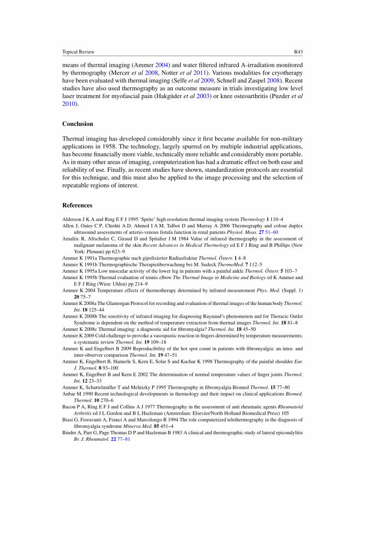
Figure 1. (Left) Thermogram of lateral face recorded in 1995 with 320 × 240 pixels; (right)thermogram recorded in 2011 with a new 640 × 480 pixel infrared camera.
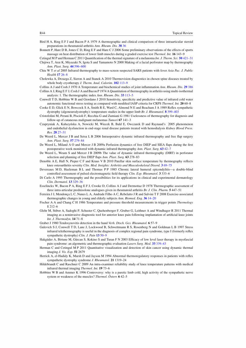
Figure 2. Thermograms of dorsal hand (left) recorded in 1990 with 240 × 320 pixels and rightrecorded in 2011 with modern focal plane array camera with 640 × 480 pixels.
The thermal image
Infrared imagers
An infrared thermogram is an image of temperature distribution of the target. Although thesecond generation of infrared detectors was in use for military applications in the latter half ofthe 20th century (Rogalski 2011), thermal imagers used in medicine were almost exclusivelyscanning detector units with one to ten elements. Average speed rates were 1 to 16 framesper second, temperature resolution was 0.5 ◦C and spatial resolution was about 5 mm ata target size of 50 cm2 (Ring 1984). However, high resolution in temperature (better than0.1 ◦C) and spatial resolution (less than 0.1 mm) at frame rates of 25 per second were availablein special designed equipment (Alderson and Ring 1995). In addition, all detectors needed acooling mechanism such as nitrogen, argon gas or a sterling cooler. Focal plane array infraredcameras became common in the 1990s, and this new equipment provided improved spatialresolution necessary to resolve thermal patterns caused by superficial skin vessels (Ring andDicks 1999) (figures 1, 2). Smaller camera units and the use of microbolometers lead to highermobility and imaging of objects in the perpendicular view i.e. with the camera mounted inthe vertical position, which can now be used with modern uncooled equipment. However for

Topical Review R35
(A) (B)
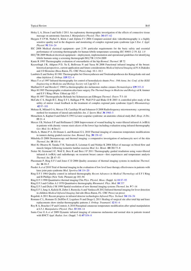
Figure 3. (A) Chronic inflammation of the forefoot following a sports injury; (B) rheumatoidarthritis of one knee (left of the image).
(A) (B) (C) (D)
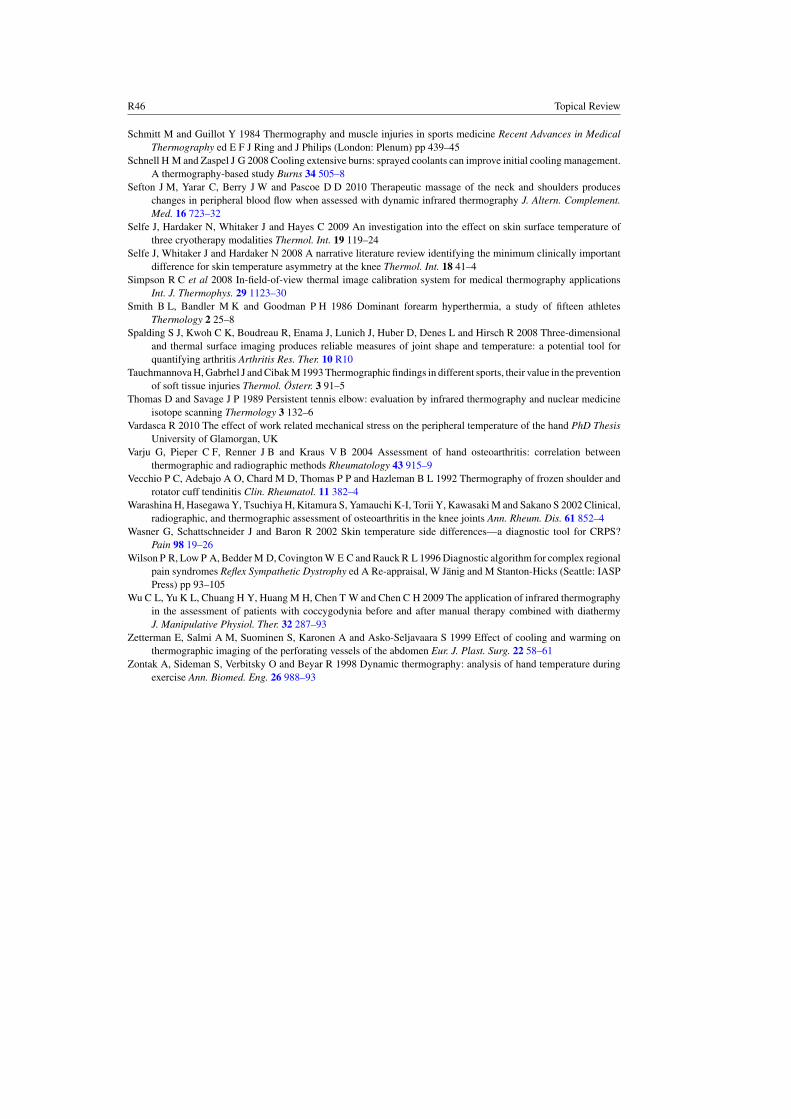
Figure 4. The effects of stress on hand thermograms, (A) 10 min full normal recovery from 1 minimmersion in water at 20 ◦C, (B) in a patient with Raynaud’s phenomenon after 10 min. (C), (D)Examples of hand arm vibration injury to certain fingers, showing delayed recovery after vibrationand thermal stress have been applied. The affected fingers are cooler.
very high sensitivity detectors such as the quantum well infrared photodetector, cooling is stillnecessary.
The non-uniformity value is a measure for the variation in temperature recorded byeach measuring point within the focal plane array. The defined threshold of 0.04% can only beachieved by correcting the raw data with software, meaning that inbuilt computing is necessaryto achieve accurate temperature measurements.
Standardization
Within recent years some progress has been made in improved standardization protocols.The International Standards Organization has published two new documents defining theuse of a thermal imaging camera for fever screening. The first in September 2008 (IEC 2008)describes the essential design and performance characteristics of a radiometric infrared camerafor screening, where differences on the face can be little more than 1 ◦C, and the second inMarch 2009 defines the recommended mode of deployment including the testing of the systemand the training of its users (ISO 2009).
There is now increased awareness of the potential measurement errors caused by theimaging system itself. A series of simple tests have now been proposed for regular qualityassurance for thermal imaging in medicine (Plassmann et al 2006). The use of externalreference temperature sources is an important recommendation for regular calibration checks,especially with the modern uncooled detector-based cameras (Simpson et al 2008).
Today, digital images and image processing have become the standard, with many differentcolour palettes applied by different users. For example in engineering applications a colour

R36 Topical Review
palette called ‘iron’ is used where yellow is hotter than red, and white is hotter than yellow,as found when iron is heated in a furnace. In medicine, where the temperature range is morelimited (typically 10 ◦C) a ‘rainbow’ palette is preferred, with red as hot and blue/black as cold.These colour scales may also be linear or logarithmic in distribution, and in some softwarepackages, the user can generate his own colour scale for a specific application if required. Ingeneral, the temperature colour scale used at the time of image capture should be displayedalongside the final image. Without this, the image is poorly defined, since the range and levelof temperatures are essential to the full information provided in the thermogram. These issuesare frequently defined in any standardization protocol, since they are essential elements for thecomparison of thermograms to indicate change. They are also essential in evidential materialfor forensic and legal issues (ISO 2009).
Over the last 20 years, there has been a growing interest in standardization of proceduresfor clinical protocols including patient preparation, body positions for image recording andevaluation of thermal imaging. Previous guidelines for thermal imaging in medicine haveincluded specific patient positioning to obtain the views required for medical thermography.Reference temperature values of healthy subjects have yet to be systematically obtained. Thereare some data that the mean temperature varies little between regions of the right and left sideof the body, but absolute temperature values have seldom been reported. Some of these datawill become available from the ‘reference atlas for clinical thermography’ project which beganat the University of Glamorgan in 2001. A total of 24 body positions and 90 regions of interesthave been defined in order to construct a clinical database of reference thermograms (Ammer2008a). The improved reproducibility of body positions and location of regions of interest hasbeen shown to have a marked influence on both the accuracy and precision of temperaturemeasurements obtained from thermal images.
The influence of regions of interest of different shape and size of diagnostic accuracyof thermal imaging for thoracic outlet syndrome has been reported. Also the thermographicdiagnosis of Raynaud’s phenomenon has been shown to be dependent on the definition andpositioning of regions of interest (Ammer 2008b). A systematic review on the cold challengetest for provocation of vasospastic reactions of finger blood vessels found a wide variationin water temperature of the immersion bath and also of duration of immersion. More than20 different methods for evaluation of hand temperatures have been reported (Ammer 2009).However, immersion in water of 20 ◦C for 1 min and use of a temperature gradient combiningthe temperature gradient from the fingertip to the dorsum of the hand prior and 20 min afterthe cold challenge has evolved as a proposed standard from the systematic review (Ammer2009).
Applications of thermal imaging in medicine
The study of temperature has widespread applications across science and industry.Thermal imaging offers the great advantage of real time two-dimensional temperature
measurement. With modern technology, a single image may contain several thousands oftemperature points, recorded in a fraction of a second.
The human body is homeothermic, i.e. self-generating and regulating the essential levelsof temperature for survival. As humans we increase our ‘comfort’ by added clothing forinsulation in winter, or decreasing clothing levels in the summer. The body core is relativelystable in temperature, but the shell of the body (the surface tissues mainly the skin) forms partof the regulatory process. Human skin behaves as an almost blackbody with an emissivity of0.96–0.98. An American physiologist J D Hardy showed in 1934, that the emission of human

Topical Review R37
skin peaks at 9–12 μm. However detectors operating at 2–5 μm and bolometer systemsoperating up to 15 μm have all proved to be equally successful in medical applications.
The association between human body temperature and disease is almost as old as medicineitself. For generations physicians had to rely on the clinical thermometer, a simple maximumthermometer for a narrow range of body temperature close to 37 ◦C. The level of temperaturewas measured in a cavity such as the mouth, and principally used for the detection of fever.
Thermal imaging has been used mainly for research over the last 50 years. It has beenused to study a number of diseases where skin temperature can reflect the presence ofinflammation in underlying tissues, or where blood flow is increased or decreased due toa clinical abnormality. In principle, thermal imaging can be applied in medicine either as adiagnostic test or as outcome measure for clinical trials.
Inflammatory arthritis
From early times physicians have used the cardinal signs of inflammation, i.e. pain, swelling,heat, redness and loss of function. When a joint is acutely inflamed, the increase in heat canbe readily detected by touch. However, subtle changes in joint surface temperature occur, andincrease and decrease in temperature can have a direct expression of reduction or exacerbationof inflammation. This means that changes due to treatment, whether pharmaceutical, physicalor surgical, can be objectively measured (See figure 3).
Studies were conducted throughout the 1960s to establish the best analogues of cortico-steroids and their effective dose. Work by Collins and Cosh in 1970 and Ring and Collins in1970 showed that the surface temperature of an arthritic joint was related to the intra-articularjoint, and to other biochemical markers of inflammation obtained from the exudate. In a seriesof experiments with different analogues of prednisolone (all cortico-steroids), the temperaturewas measured by thermal imaging in groups of patients, and used to objectively determine theduration and degree of reduction in inflammation (Esselinckx et al 1978, Bird et al 1979).
A number of new non-steroid anti-inflammatory agents were introduced intorheumatology in the 1970s and 1980s. Infrared imaging was also shown to be a powerful toolfor the clinical testing of these drugs, using temperature changes in the affected joints as anobjective marker. The technique had been successfully used on animal models of inflammation,and effectively showed that optimal dose response curves could be obtained from temperaturechanges at the experimental animal joints. The process with human patients suffering fromacute rheumatoid arthritis was adapted, to include a washout period from previous medication.The compound used by all the pharmaceutical companies was paracetamol. A study byBacon et al (1977) found that small joints such as fingers and metacarpal joints increasedin temperature quite rapidly while paracetamol (analgesic) treatment was given, even if painwas still suppressed. Larger joints, such as knees and ankles required more than 1 week offactive anti-inflammatory treatment to register the same effect. Nevertheless, the commonlyaccepted protocol was to switch from analgesic to the new test anti-inflammatory treatmentafter 1 week of washout therapy. In every case if the dose was ineffective the joint temperaturewas not reduced. At an effective dose, a fall in temperature was observed, first in the smalljoints, then later in the larger joints. Statistical studies were able to show an objective decreasein joint temperature by infrared imaging as a result of a new and successful treatment. However,temperature measurements over joints have not generally been considered for inclusion in coresets of outcome measurements for the assessment of response to newer biological agents forinflammatory arthritis. Recently, a pilot study from the United States found a high coincidencebetween high temperature and swelling of finger joints detected by three-dimensional images.The authors created a heat distribution index which had a diagnostic sensitivity of 67% and aspecificity of 100% for arthritic swelling (Spalding et al 2008).
R38 Topical Review
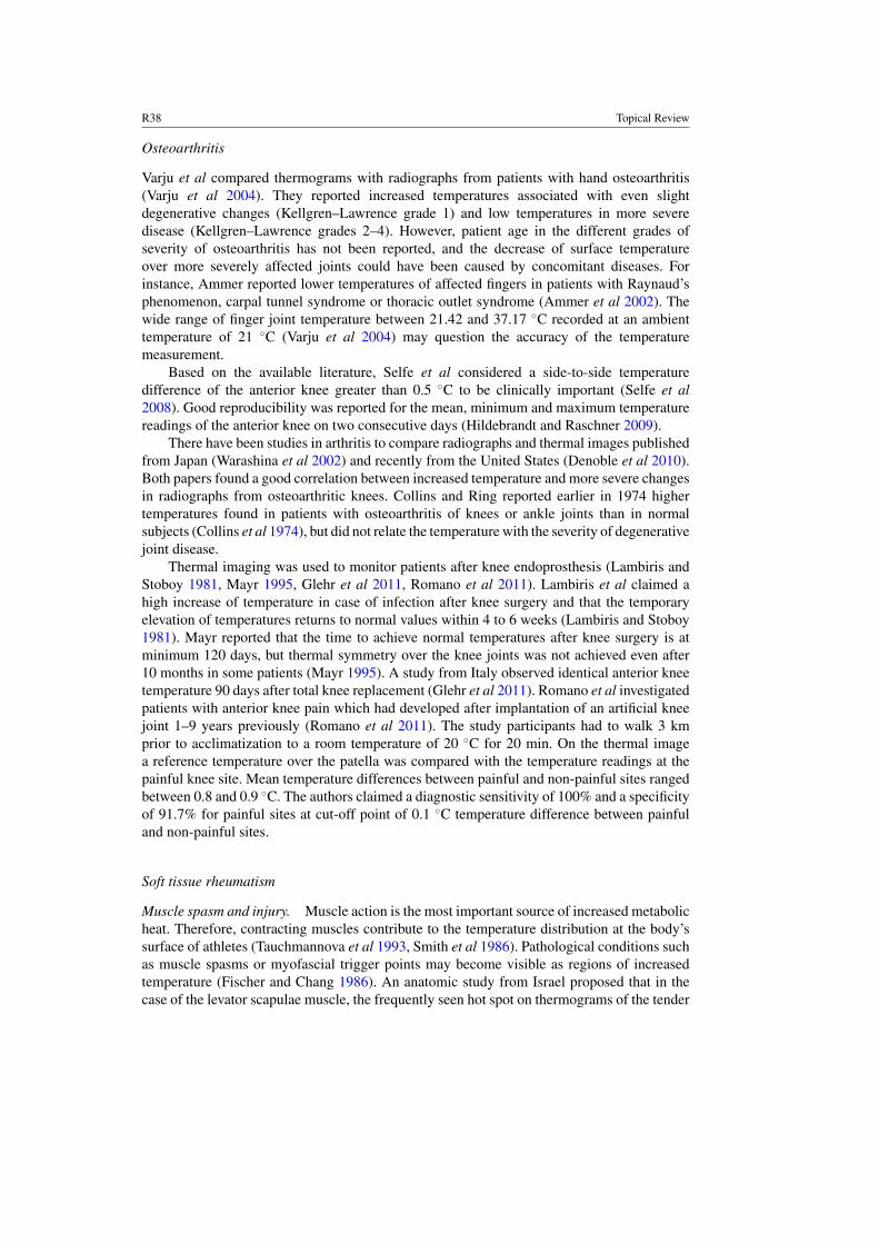
Osteoarthritis
Varju et al compared thermograms with radiographs from patients with hand osteoarthritis(Varju et al 2004). They reported increased temperatures associated with even slightdegenerative changes (Kellgren–Lawrence grade 1) and low temperatures in more severedisease (Kellgren–Lawrence grades 2–4). However, patient age in the different grades ofseverity of osteoarthritis has not been reported, and the decrease of surface temperatureover more severely affected joints could have been caused by concomitant diseases. Forinstance, Ammer reported lower temperatures of affected fingers in patients with Raynaud’sphenomenon, carpal tunnel syndrome or thoracic outlet syndrome (Ammer et al 2002). Thewide range of finger joint temperature between 21.42 and 37.17 ◦C recorded at an ambienttemperature of 21 ◦C (Varju et al 2004) may question the accuracy of the temperaturemeasurement.
Based on the available literature, Selfe et al considered a side-to-side temperaturedifference of the anterior knee greater than 0.5 ◦C to be clinically important (Selfe et al2008). Good reproducibility was reported for the mean, minimum and maximum temperaturereadings of the anterior knee on two consecutive days (Hildebrandt and Raschner 2009).
There have been studies in arthritis to compare radiographs and thermal images publishedfrom Japan (Warashina et al 2002) and recently from the United States (Denoble et al 2010).Both papers found a good correlation between increased temperature and more severe changesin radiographs from osteoarthritic knees. Collins and Ring reported earlier in 1974 highertemperatures found in patients with osteoarthritis of knees or ankle joints than in normalsubjects (Collins et al 1974), but did not relate the temperature with the severity of degenerativejoint disease.
Thermal imaging was used to monitor patients after knee endoprosthesis (Lambiris andStoboy 1981, Mayr 1995, Glehr et al 2011, Romano et al 2011). Lambiris et al claimed ahigh increase of temperature in case of infection after knee surgery and that the temporaryelevation of temperatures returns to normal values within 4 to 6 weeks (Lambiris and Stoboy1981). Mayr reported that the time to achieve normal temperatures after knee surgery is atminimum 120 days, but thermal symmetry over the knee joints was not achieved even after10 months in some patients (Mayr 1995). A study from Italy observed identical anterior kneetemperature 90 days after total knee replacement (Glehr et al 2011). Romano et al investigatedpatients with anterior knee pain which had developed after implantation of an artificial kneejoint 1–9 years previously (Romano et al 2011). The study participants had to walk 3 kmprior to acclimatization to a room temperature of 20 ◦C for 20 min. On the thermal imagea reference temperature over the patella was compared with the temperature readings at thepainful knee site. Mean temperature differences between painful and non-painful sites rangedbetween 0.8 and 0.9 ◦C. The authors claimed a diagnostic sensitivity of 100% and a specificityof 91.7% for painful sites at cut-off point of 0.1 ◦C temperature difference between painfuland non-painful sites.
Soft tissue rheumatism
Muscle spasm and injury. Muscle action is the most important source of increased metabolicheat. Therefore, contracting muscles contribute to the temperature distribution at the body’ssurface of athletes (Tauchmannova et al 1993, Smith et al 1986). Pathological conditions suchas muscle spasms or myofascial trigger points may become visible as regions of increasedtemperature (Fischer and Chang 1986). An anatomic study from Israel proposed that in thecase of the levator scapulae muscle, the frequently seen hot spot on thermograms of the tender
Topical Review R39
tendon insertion on the medial angle of the scapula could be caused by an inflamed bursae andnot by a taut band of muscle fibres (Menachem et al 1993).
Acute muscle injuries may also be recognized by areas of increased temperature (Schmittand Guillot 1984) due to inflammation in the early state of trauma. However, long lastinginjuries and also scars appear as hypothermic areas caused by reduced muscle contraction, andtherefore reduced heat production. Similar areas of decreased temperature have been foundadjacent to peripheral joints with reduced range of motion due to inflammation or pain (Ammer1995a). Reduced skin temperatures have also been related to osteoarthritis of the hip (Kanie1995) or to the frozen shoulder (Vecchio et al 1992, Ammer et al 1998). The impact of muscleweakness on hypothermia in patients suffering from paresis has been discussed elsewhere(Hobbins and Ammer 1996).
Enthesiopathies. Muscle overuse or repetitive strain may lead to painful tendon insertions,or where the tendons are shielded by the tendon sheath or adjacent to bursae, with painfulswelling. It has been shown that tendovaginitis in the hand was sucessfully diagnosed by skintemperature measurement (Graber 1980). The acute bursitis at the tip of the elbow was alsodetected through an intense hot spot adjacent to the olecranon (Mayr 1997).
Tennis elbow. Painful muscle insertion of the extensor muscles at the elbow is associatedwith hot areas on a thermogram (Binder et al 1983). Thermal imaging can detect persistenttendon insertion problems of the elbow region in a similar way as isotope bone scanning(Thomas and Savage 1989). Hot spots at the elbow have also been described as having a highassociation with a low threshold for pain on pressure (Ammer 1995b). Such hot areas havebeen sucessfully used as outcome measure for monitoring treatment (Devereaux et al 1985,Meknas et al 2008). In patients suffering from fibromyalgia, bilateral hot spots at the elbowsis a common finding (Ammer et al 1995).
Fibromyalgia. There are two terms used by physicians in the examination of muscular pain:tender points (important for the diagnosis of fibromyalgia) and trigger points (main featureof the myofascial pain syndrome). Tender points and trigger points may give a similar imageon the thermogram. If this is true, patients suffering from fibromyalgia may present witha high number of hot spots in typical regions of the body. A study from Italy did not finddifferent patterns of heat distribution in patients suffering from fibromyalgia and patients withosteoarthritis of the spine (Biasi et al 1994). However, they reported a correspondence ofnon-specific hyperthermic pattterns with painful muscle areas in both groups of patients. In anAustrian study, thermographic investigations in fibromyalgia revealed a diagnostic accuracyof 60% of hot spots for tender points (Ammer et al 1995). The number of hot spots found wasgreatest in fibromyalgia patients and least in healthy subjects. It was therefore concluded thatmore than seven hot spots could be predictive for tenderness in 11 or more of 18 specific sites(Ammer 2008c). Based on the count of hot spots, 74.2% of 252 subjects (161 fibromyalgia,71 with widespread pain, but less than 11 tender sites out of 18, and 20 healthy controls) werecorrectly diagnosed. However, the intra- and inter-observer reproducibility of hot spot countis poor (Ammer and Engelbert 2009). Software assisted identification of hot or cold spotsbased on the angular distribution around a thermal irregularity (Anbar 1990) might improvereproducibility.
Complex regional pain syndrome
A temperature difference between the affected and the non-affected limb equal or greater than1 ◦C is one of the diagnostic criteria of the complex regional pain syndrome (CRPS) (Wilson

R40 Topical Review
et al 1996). Ammer conducted a study in patients after radius fracture treated conservativelywith a plaster cast (Ammer 1991a). Thermal images were recorded within 2 h after plasterremoval and 1 week later. After the second thermogram an x-ray image of both handswas taken. The mean temperature difference between the affected and unaffected hand was0.6 ◦C after plaster removal and 0.63 ◦C 1 week later. In some 50% of 41 radiographs slightbone changes suspected of algodystropy were found. It was also shown that the temperaturedifference decrease during successful therapeutic intervention and the temperature changewas paralleled by reduction of pain and swelling and the resolution of radiological changes(Ammer 1991b).
Disturbances in the vascular adaptation mechanism and delayed response to temperaturestimuli have been observed in patients suffering from CRPS (Cooke et al 1989, Herrick et al1994). These alterations were interpreted as being caused by abnormalities of the autonomicnerve system. It was suggested that a cold challenge on the contralateral side of the injuredlimb for prediction and early diagnosis of CRPS could be used. Gulevich et al (1997) andConwell et al (2010) confirmed the high diagnostic sensitivity and specificity of cold challengefor the CRPS. Wasner et al achieved similar results by whole body cooling or whole bodywarming (Wasner et al 2002). Recently a Dutch study found that the asymmetry factor whichwas based on histograms of temperatures from the affected and non-affected hand had thehighest diagnostic power for CRPS; however the difference in mean temperatures did notdiscriminate between disease and health (Huygen et al 2004). McCabe et al investigatingthe use of mirror visual feedback as a means of treating type1 CRPS found that patientswho were successfully treated in this way during the early stages of the disease did show areturn to thermal symmetry between the affected and non-affected limb, which was objectivelymeasured by thermography. The thermal changes were associated with the relief of symptoms(McCabe et al 2003).
Peripheral circulation
In some circulatory disturbances, such as Raynaud’s phenomenon, or hand arm vibrationsyndrome, damage to small blood vessels from exposure to vibrating machinery and the effectof local blood circulation on skin temperature can be assessed by thermal imaging. Thisis found after exposure of the hands to a temperature stimulus or contact with a vibratingsurface at a known frequency. Most commonly, a number of studies have reported the valueof the thermal challenge test, particularly for quantifying the ‘vasospastic’ reaction found inRaynaud’s phenomenon (Ammer 2009). After a baseline thermogram of both hands (usuallyof the dorsal surfaces) the hands are protected by plastic gloves and immersed in a water bath(typically 18–20 ◦C) for 1 min. The thermal recovery is then monitored. In normal healthysubjects this can lead to a reactive hyperaemia of the fingers, while in Raynaud’s sufferers thereis a slow protracted recovery of more than 15 min to baseline. This test has been applied inmany different studies and trials of vasodilator treatments. In most cases, some improvementscan be measured, but ultimately normal recovery is rarely achieved, even when there is animprovement in clinical symptoms.
A recent study by Vardasca has shown that hand arm vibration syndrome can be objectivelymeasured by a combination of a vibration stress and a thermal stress to the hands (Vardasca2010). Since this remains an important issue in occupational health, it is promising that thistechnique may improve the diagnostic discrimination in those industrial workers who areaffected (See figure 4).
In all medical applications, the technique can only provide an image of skin temperaturedistribution; it does not provide data at a specific depth inside the body, as is common in other
Topical Review R41
imaging methods. However, thermal imaging is non-invasive and objective, and therefore safeand harmless.
The small size and weight of the modern cameras are similar to a domestic camcorder. Thismeans that they can now be employed in the operating theatre, and have been used successfullyto monitor the surgical procedures in open-heart surgery. Another evolving application ofthermal imaging is the identification and localization of nutrient vessels in free perforatorflap surgery. Based on work from Finland (Zetterman et al 1999) and Japan (Chijiwa et al2000), a group from Norway had clearly shown that the design of deep inferior epigastricartery perforator flaps which are used for breast reconstruction can be based on dynamicinfrared thermal imaging for pre-surgical selection of the vessel (De Weerd et al 2009b). Inthe abdominal region, blood vessels in the subcutaneous tissue are connected with the vascularsystem of abdominal muscles by perforating vessels which terminate in the subdermal layer.These vessels transport warm blood from the deep tissue to the surface of the body and areeasily detected on a thermal image after the skin has been cooled by forced convection. In therewarming phase the perforators appear as a rapidly growing hot spot on the skin. Infraredimaging is also a valuable tool for intraoperative (De Weerd et al 2006) and postoperative (DeWeerd et al 2009a) monitoring in flap surgery.
Fever screening
Currently, there is interest in the use of thermal imaging for fever screening. Following theSARS (severe acute respiratory syndrome) outbreak in South East Asia, increasing use ofthermal imaging had been made to screen travelling passengers at the time of pandemic fever.For this reason The International Standards Organization has published two new documentsdefining the use of a thermal imaging camera for fever screening. An essential part of thesecond standards paper is that only a close up image of the upper face, where a minimum of9 pixels can be located in each corner of the eye (inner canthus), will provide a true indicationof the presence or absence of fever. The widespread idea that a camera can be used to survey agroup of moving passengers at a distance is entirely wrong, since it is possible to have a provenfever, yet not have a generalized increase in facial temperature, as may have been found in theSARS outbreak (Chiu et al 2005). Ring and Jung et al have measured the facial temperaturesin children using the new ISO recommendations in a recent study with a hospital population.In 354 afebrile children, mean temperatures at the inner canthi of the eyes were 36.48 ◦C(SD of 0.49 ◦C) while in 52 children with clinically proven fever just prior to medication, themean temperature was 38.9 ◦C (SD of 0.84 ◦C). These were compared with axilla temperaturemeasured by thermometry, and forehead and tympanic membrane temperatures measured byradiometry. A good correlation was found between the eye measurements obtained from thethermogram and the clinical thermometry data, which supports the methodology described inthe new standard (Ring et al at press).
However, the main concerns of fever screening with a thermal infrared camera is theuncertainty of the accuracy of some of the equipment currently used in airports, and the wayin which it is employed, where the ISO recommendations are often ignored.
Malignant diseases
Many of the early investigations with thermal imaging some 50 years ago were entirely focusedon the potential of this technique to become a useful tool in breast cancer diagnostics. Formany of the reasons cited above, large expensive and unstable camera systems, before evencomputing and image processing was available, made this a difficult and unreliable tool. There
R42 Topical Review
were undoubtedly some interesting ‘positive’ findings, but the arrival of both mammographyand ultrasound effectively made thermal imaging of less interest. In addition, many proposalswere made for identification of breast cancer, often based on models of heat transfer from themalignant lesion to the surface or on pattern recognition, but none of these proposals havebeen applied in large samples of patients or in screening programmes for breast cancer. Oneproblem with breast imaging of surface temperature is the curved surfaces. This does meanthat a single view in not sufficient and many investigators have adopted a standard series ofimages with full anterior, then left and right oblique positions. A single breast close up view isalso used for each of these positions in order to maximize the thermal data. In some cases thepatient may be imaged in the seated position with arms raised. Further anterior views with thepatient lying flat on an examination couch may also be used to provide data from the inferiorquadrants of the breast.
While much activity has been devoted to the complimentary role of thermography indiagnostic medicine, the monitoring of known cancerous lesions has been investigated.Monitoring chemotherapy was investigated by Keyserlingk et al (2008) in Canada (Med.Inf. Imaging Diakides 1. Ch 10 2008). They found that infrared imaging can be used to addfunctional information on the course of tumour development, and that these changes canprecede and linger after structural tumour-induced changes have occurred. This in itself wasfound to be variable in a small group of patients, which might be due to the variable volumeof angiogenesis, the inability of chemotherapy to affect it, or could be due to the deficienciesin scoring and grading the thermal changes. There is scope for continued research, but theauthors believe that standardization of technique and better methods for quantification needsto be related to early tumour genesis, and may have a future role in studying clinical treatmentregimen.
The success of locating malignant melanomas by their increased temperature on the skinsurface is more interesting. This has been shown to be more easily detected in rewarmingafter cooling the skin by convection (Herman and Cetingul 2011) or by contact cooling(Di Carlo 1995). Melanoma identification is one of the oldest applications of infrared thermalimaging (Maillard and Hessler 1969). The value of infrared imaging for diagnosis andmonitoring malignant melanomas has been a subject of contradiction in the past (Di Carlo1995, Cristofolini et al 1981, Amalric et al 1984), but recent studies from Poland (Mikulska2008), Argentina (Santa Cruz et al 2009) and the United States (Cetingul and Herman 2011)have shown different patterns of temperature changes in malignant and benign melanoma skinlesions. These promising results have yet to be confirmed in larger samples of patients.
Other applications
The evaluation of burns and areas of skin grafts explored initially in the 1960s in the UK hasbeen shown to be a useful non-invasive tool of particular value where early assessment of fullthickness burns improves the outcome of skin grafts.
In renal dialysis patients several papers from Austria (Maca et al 1997), UK (Allen et al2006) and Poland (Czupryniak et al 2005) have shown that monitoring stent insertion sitesand peripheral circulation with thermal imaging is an efficient means of assessing the need forrevision of the arterio-venous fistula.
Thermal imaging is now increasingly used for imaging different physiological reactionsinduced by non-drug treatments such as massage (Bonnett et al 2006, Sefton et al 2010, Holeyet al 2011, Wu et al 2009) or manual therapy (Mori et al 2004, Roy et al 2010). Temperaturedistribution of the skin during and after physical exercise has been reported (Zontak et al1998, Ferreira et al 2008, Merla et al 2010). The effects of thermotherapy were recorded with
Topical Review R43
means of thermal imaging (Ammer 2004) and water filtered infrared A-irradiation monitoredby thermography (Mercer et al 2008, Notter et al 2011). Various modalities for cryotherapyhave been evaluated with thermal imaging (Selfe et al 2009, Schnell and Zaspel 2008). Recentstudies have also used thermography as an outcome measure in trials investigating low levellaser treatment for myofascial pain (Hakguder et al 2003) or knee osteoarthritis (Puzder et al2010).
Conclusion
Thermal imaging has developed considerably since it first became available for non-militaryapplications in 1958. The technology, largely spurred on by multiple industrial applications,has become financially more viable, technically more reliable and considerably more portable.As in many other areas of imaging, computerization has had a dramatic effect on both ease andreliability of use. Finally, as recent studies have shown, standardization protocols are essentialfor this technique, and this must also be applied to the image processing and the selection ofrepeatable regions of interest.
References
Alderson J K A and Ring E F J 1995 ‘Sprite’ high resolution thermal imaging system Thermology 1 110–4Allen J, Oates C P, Chishti A D, Ahmed I A M, Talbot D and Murray A 2006 Thermography and colour duplex
ultrasound assessments of arterio-venous fistula function in renal patients Physiol. Meas. 27 51–60Amalric R, Altschuler C, Giraud D and Spitalier J M 1984 Value of infrared thermography in the assessment of
malignant melanoma of the skin Recent Advances in Medical Thermology ed E F J Ring and B Phillips (NewYork: Plenum) pp 623–9
Ammer K 1991a Thermographie nach gipsfixierter Radiusfraktur Thermol. Osterr. 1 4–8Ammer K 1991b Thermographische Therapieuberwachung bei M. Sudeck ThermoMed. 7 112–5Ammer K 1995a Low muscular activity of the lower leg in patients with a painful ankle Thermol. Osterr. 5 103–7Ammer K 1995b Thermal evaluation of tennis elbow The Thermal Image in Medicine and Biology ed K Ammer and
E F J Ring (Wien: Uhlen) pp 214–9Ammer K 2004 Temperature effects of thermotherapy determined by infrared measurement Phys. Med. (Suppl. 1)
20 75–7Ammer K 2008a The Glamorgan Protocol for recording and evaluation of thermal images of the human body Thermol.
Int. 18 125–44Ammer K 2008b The sensitivity of infrared imaging for diagnosing Raynaud’s phenomenon and for Thoracic Outlet
Syndrome is dependent on the method of temperature extraction from thermal images Thermol. Int. 18 81–8Ammer K 2008c Thermal imaging: a diagnostic aid for fibromyalgia? Thermol. Int. 18 45–50Ammer K 2009 Cold challenge to provoke a vasospastic reaction in fingers determined by temperature measurements;
a systematic review Thermol. Int. 19 109–18Ammer K and Engelbert B 2009 Reproducibility of the hot spot count in patients with fibromyalgia: an intra- and
inter-observer comparison Thermol. Int. 19 47–51Ammer K, Engelbert B, Hamerle S, Kern E, Solar S and Kuchar K 1998 Thermography of the painful shoulder Eur.
J. Thermol. 8 93–100Ammer K, Engelbert B and Kern E 2002 The determination of normal temperature values of finger joints Thermol.
Int. 12 23–33Ammer K, Schartelmuller T and Melnizky P 1995 Thermography in fibromyalgia Biomed Thermol. 15 77–80Anbar M 1990 Recent technological developments in thermology and their impact on clinical applications Biomed.
Thermol. 10 270–6Bacon P A, Ring E F J and Collins A J 1977 Thermography in the assessment of anti rheumatic agents Rheumatoid
Arthritis ed J L Gordon and B L Hazleman (Amsterdam: Elsevier/North Holland Biomedical Press) 105Biasi G, Fioravanti A, Franci A and Marcolongo R 1994 The role computerized telethermography in the diagnosis of
fibromyalgia syndrome Minerva Med. 85 451–4Binder A, Parr G, Page Thomas D P and Hazleman B 1983 A clinical and thermographic study of lateral epicondylitis
Br. J. Rheumatol. 22 77–81
R44 Topical Review
Bird H A, Ring E F J and Bacon P A 1979 A thermographic and clinical comparison of three intraarticular steroidpreparations in rheumatoid arthritis Ann. Rheum. Dis. 38 36
Bonnett P, Hare D B, Jones C D, Ring E F and Hare C J 2006 Some preliminary observations of the effects of sportsmassage on heat distribution of lower limb muscles during a graded exercise test Thermol. Int. 16 143–9
Cetingul M P and Herman C 2011 Quantification of the thermal signature of a melanoma Int. J. Therm. Sci. 50 421–31Chijiwa T, Arai K, Miyazaki N, Igota S and Yamamoto N 2000 Making of a facial perforator map by thermography
Ann. Plast. Surg. 44 596–600Chiu W T et al 2005 Infrared thermography to mass-screen suspected SARS patients with fever Asia Pac. J. Public
Health 17 26–8Cholewka A, Drzazga Z, Sieron A and Stanek A 2010 Thermovision diagnostics in chosen spine diseases treated by
whole body cryotherapy J. Therm. Anal. Calorim. 102 113–9Collins A J and Cosh J 1970 A Temperature and biochemical studies of joint inflammation Ann. Rheum. Dis. 29 386Collins A J, Ring E F J, Cosh J A and Bacon P 1974 A Quantitation of thermography in arthritis using multi-isothermal
analysis: 1. The thermographic index Ann. Rheum. Dis. 33 113–5Conwell T D, Hobbins W B and Giordano J 2010 Sensitivity, specificity and predictive value of infrared cold water
autonomic functional stress testing as compared with modified IASP criteria for CRPS Thermol. Int. 20 60–8Cooke E D, Glick E N, Bowcock S A, Smith R E, Ward C, Almond N E and Beacham J A 1989 Reflex sympathetic
dystrophy (algoneurodystrophy): temperature studies in the upper limb Br. J. Rheumatol. 8 399–403Cristofolini M, Perani B, Piscioli F, Recchia G and Zumiani G 1981 Uselessness of thermography for diagnosis and
follow-up of cutaneous malignant melanomas Tumori 67 141–3Czupryniak A, Kałuzynska A, Nowicki M, Wiecek B, Bald E, Owczarek D and Raynaud’s 2005 phenomenon
and endothelial dysfunction in end-stage renal disease patients treated with hemodialysis Kidney Blood Press.Res. 28 27–31
De Weerd L, Mercer J B and Setsa L B 2006 Intraoperative dynamic infrared thermography and free flap surgeryAnn. Plast. Surg. 57 279–84
De Weerd L, Miland A O and Mercer J B 2009a Perfusion dynamics of free DIEP and SIEA flaps during the firstpostoperative week monitored with dynamic infrared thermography Ann. Plast. Surg. 63 42–7
De Weerd L, Weum S and Mercer J B 2009b The value of dynamic infrared thermography (DIRT) in perforatorselection and planning of free DIEP flaps Ann. Plast. Surg. 63 278–83
Denoble A E, Hall N, Pieper C F and Kraus V B 2010 Patellar skin surface temperature by thermography reflectsknee osteoarthritis severity Clin. Med. Insights: Arthritis and Musculoskeletal Disord. 3 69–75
Devereaux M D, Hazleman B L and Thomas P P 1985 Chronic lateral humeral epicondylitis—a double-blindcontrolled assessment of pulsed electromagnetic field therapy Clin. Exp. Rheumatol. 3 333–6
Di Carlo A 1995 Thermography and the possibilities for its applications in clinical and experimental dermatologyClin. Dermatol. 13 329–36
Esselinckx W, Bacon P A, Ring E F J, Crooke D, Collins A J and Dermottaz D 1978 Thermographic assessment ofthree intra-articular prednisolone analogues given in rheumatoid arthritis Br. J. Clin. Pharm. 5 447–51
Ferreira J J, Mendonca L C, Nunes L A, Andrade Filho A C, Rebelatto J R and Salvini T F 2008 Exercise-associatedthermographic changes in young and elderly subjects Ann. Biomed. Eng. 36 14–20
Fischer A A and Chang C H 1986 Temperature and pressure threshold measurements in trigger points Thermology1 212–6
Glehr M, Stibor A, Sadoghi P, Schuster C, Quehenberger F, Gruber G, Leithner A and Windhager R 2011 Thermalimaging as a noninvasive diagnostic tool for anterior knee pain following implantation of artificial knee jointsInt. J. Thermodyn. 14 71–8
Graber J 1980 Tendosynovitis detection in the hand Verh. Dtsch. Ges. Rheumatol. 6 57–9Gulevich S J, Conwell T D, Lane J, Lockwood B, Schwettmann R S, Rosenberg N and Goldman L B 1997 Stress
infrared telethermography is useful in the diagnosis of complex regional pain syndrome, type I (formerly reflexsympathetic dystrophy) Clin. J. Pain 13 50–9
Hakguder A, Birtane M, Gurcan S, Kokino S and Turan F N 2003 Efficacy of low level laser therapy in myofascialpain syndrome: an algometric and thermographic evaluation Lasers Surg. Med. 33 339–43
Herman C and Cetingul M P 2011 Quantitative visualization and detection of skin cancer using dynamic thermalimaging J. Vis. Exp. 51 2679
Herrick A, el-Hadidy K, Marsh D and Jayson M 1994 Abnormal thermoregulatory responses in patients with reflexsympathetic dystrophy syndrome J. Rheumatol. 21 1319–24
Hildebrandt C and Raschner C 2009 An intra-examiner reliability study of knee temperature patterns with medicalinfrared thermal imaging Thermol. Int. 19 73–6
Hobbins W B and Ammer K 1996 Controversy: why is a paretic limb cold, high activity of the sympathetic nervesystem or weakness of the muscles? Thermol. Osterr. 6 42–5
Topical Review R45
Holey L A, Dixon J and Selfe J 2011 An exploratory thermographic investigation of the effects of connective tissuemassage on autonomic function J. Manipulative Physiol. Ther. 34 457–62
Huygen F J P M, Niehof S, Klein J and Zijlstra F J 2004 Computer-assisted skin videothermography is a highlysensitive quality tool in the diagnosis and monitoring of complex regional pain syndrome type I Eur. J. Appl.Physiol. 91 516–24
IEC 2008 Medical electrical equipment—part 2-59: particular requirements for the basic safety and essentialperformance of screening thermographs for human febrile temperature screening IEC 80601-2-59, Ed. 1.0
ISO/TR 2009 Medical electrical equipment—deployment, implementation and operational guidelines for identifyingfebrile humans using a screening thermograph ISO/TR 13154:2009
Kanie R 1995 Thermographic evaluation of osteoarthritis of the hip Biomed. Thermol. 15 72Keyserlingk J R, Ahlgren P D, Yu E, Belliveau N and Yassa M 2008 Functional infrared imaging of the breast:
historical perspectives, current application and future considerations Medical Infrared Imaging ed N A Diakidesand J D Bronzino (Boca Raton, FL: CRC Press) chap. 10.1–10.3
Lambiris E and Stoboy H 1981 Thermographie bei Osteosynthesen und Totalendoprothesen des Kniegelenks mit undohne Infektion Z. Orthop. 119 521–4
Maca T et al 1997 Infrared thermography for control of hemodialysis shunts Proc. 19th Annu. Int. Conf. of the IEEEEngineering in Medicine and Biology Society vol 2 pp 621–4
Maillard G F and Hessler C 1969 La thermographie des melanomes malins cutanes Dermatologica 139 353–8Mayr H 1995 Thermographic evaluation after knee surgery The Thermal Image in Medicine and Biology ed K Ammer
and E F J Ring (Wien: Uhlen) pp 182–7Mayr H 1997 Thermografische Befunde bei Schmerzen am Ellbogen Thermol. Osterr. 7 5–10McCabe C S, Haigh R C, Ring E F J, Halligan P W, Wall P D and Blake D R 2003 A controlled pilot study of the
utility of mirror visual feedback in the treatment of complex regional pain syndrome (type1) Rheumatology42 97–101
Meknas K, Miland O A, Mercer J B, Castillejo M and Johansen O 2008 Radiofrequency microtenotomy: a promisingmethod for treatment of lateral epicondylitis Am. J. Sports Med. 36 1960–5
Menachem A, Kaplan O and Dekel S 1993 Levator scapulae syndrome: an anatomic-clinical study Bull. Hosp. Jt Dis.53 21–4
Mercer J B, Nielsen S P and Hoffmann G 2008 Improvement of wound healing by water-filtered infrared-A (wIRA)in patients with chronic venous stasis ulcers of the lower legs including evaluation using infrared thermographyGer. Med. Sci. 6 Doc11
Merla A, Mattei P A, Di Donato L and Romani G L 2010 Thermal imaging of cutaneous temperature modificationsin runners during graded exercise Ann. Biomed. Eng. 38 158–63
Mikulska D 2008 Dermatoscopy and thermal imaging: a comparative investigation of melanocytic nevi of the skinThermol. Int. 18 101–6
Mori H, Ohsawa H, Tanaka T H, Taniwaki E, Leisman G and Nishijo K 2004 Effect of massage on blood flow andmuscle fatigue following isometric lumbar exercise Med. Sci. Monit. 10 CR173–8
Notter M, Germond J-F, Wolf E, Berz R and Berz J P 2011 Thermography guided irradiation using water-filteredinfrared-A (wIRA) and radiotherapy on recurrent breast cancer—first experiences and temperature analysisThermol. Int. 21 47–53
Plassmann P, Ring E F J and Jones C D 2006 Quality assurance of thermal imaging systems in medicine Thermol.Int. 16 10–5
Puzder A et al 2010 Trial of thermal imaging in the evaluation of low level laser therapy effectiveness in patients withknee joint pain syndrome Med. Sportiva 14 114–20
Ring E F J 1984 Quality control in infrared thermography Recent Advances in Medical Thermology ed E F J Ringand B Phillips (New York: Plenum) pp 185–94
Ring E F J 1990 Quantitative thermal imaging Clin Phys. Physiol. Meas. (Suppl. A) 11 87–95Ring E F J and Collins A J 1970 Quantitative thermography Rheumatol. Phys. Med. 10 337Ring E F J and Dicks J M 1999 Spatial resolution of new thermal imaging systems Thermol. Int. 9 7–14Ring E F J, Jung A, Kalicki B, Zuber J, Rustecka A and Vardasca R 2012 Infrared thermal imaging for fever detection
in children Medical Infrared Imaging 2nd edn (Boca Raton, FL: CRC Press) (at press)Rogalski A 2011 Recent progress in infrared detector technologies Infrared Phys. Technol. 54 136–54Romano C L, Romano D, DellOro F, Loguluso N and Drago L 2011 Healing of surgical site after total hip and knee
replacements show similar thermographic patterns J. Orthop. Traumatol. 12 81–6Roy R A, Boucher J P and Comtois A 2010 Paraspinal cutaneous temperature modification after spinal manipulation
at L5 J. Manipulative Physiol. Ther. 33 308–14Santa Cruz G A et al 2009 Dynamic infrared imaging of cutaneous melanoma and normal skin in patients treated
with BNCT Appl. Radiat. Isot. (Suppl. 7–8) 67 S54–8
R46 Topical Review
Schmitt M and Guillot Y 1984 Thermography and muscle injuries in sports medicine Recent Advances in MedicalThermography ed E F J Ring and J Philips (London: Plenum) pp 439–45
Schnell H M and Zaspel J G 2008 Cooling extensive burns: sprayed coolants can improve initial cooling management.A thermography-based study Burns 34 505–8
Sefton J M, Yarar C, Berry J W and Pascoe D D 2010 Therapeutic massage of the neck and shoulders produceschanges in peripheral blood flow when assessed with dynamic infrared thermography J. Altern. Complement.Med. 16 723–32
Selfe J, Hardaker N, Whitaker J and Hayes C 2009 An investigation into the effect on skin surface temperature ofthree cryotherapy modalities Thermol. Int. 19 119–24
Selfe J, Whitaker J and Hardaker N 2008 A narrative literature review identifying the minimum clinically importantdifference for skin temperature asymmetry at the knee Thermol. Int. 18 41–4
Simpson R C et al 2008 In-field-of-view thermal image calibration system for medical thermography applicationsInt. J. Thermophys. 29 1123–30
Smith B L, Bandler M K and Goodman P H 1986 Dominant forearm hyperthermia, a study of fifteen athletesThermology 2 25–8
Spalding S J, Kwoh C K, Boudreau R, Enama J, Lunich J, Huber D, Denes L and Hirsch R 2008 Three-dimensionaland thermal surface imaging produces reliable measures of joint shape and temperature: a potential tool forquantifying arthritis Arthritis Res. Ther. 10 R10
Tauchmannova H, Gabrhel J and Cibak M 1993 Thermographic findings in different sports, their value in the preventionof soft tissue injuries Thermol. Osterr. 3 91–5
Thomas D and Savage J P 1989 Persistent tennis elbow: evaluation by infrared thermography and nuclear medicineisotope scanning Thermology 3 132–6
Vardasca R 2010 The effect of work related mechanical stress on the peripheral temperature of the hand PhD ThesisUniversity of Glamorgan, UK
Varju G, Pieper C F, Renner J B and Kraus V B 2004 Assessment of hand osteoarthritis: correlation betweenthermographic and radiographic methods Rheumatology 43 915–9
Vecchio P C, Adebajo A O, Chard M D, Thomas P P and Hazleman B L 1992 Thermography of frozen shoulder androtator cuff tendinitis Clin. Rheumatol. 11 382–4
Warashina H, Hasegawa Y, Tsuchiya H, Kitamura S, Yamauchi K-I, Torii Y, Kawasaki M and Sakano S 2002 Clinical,radiographic, and thermographic assessment of osteoarthritis in the knee joints Ann. Rheum. Dis. 61 852–4
Wasner G, Schattschneider J and Baron R 2002 Skin temperature side differences—a diagnostic tool for CRPS?Pain 98 19–26
Wilson P R, Low P A, Bedder M D, Covington W E C and Rauck R L 1996 Diagnostic algorithm for complex regionalpain syndromes Reflex Sympathetic Dystrophy ed A Re-appraisal, W Janig and M Stanton-Hicks (Seattle: IASPPress) pp 93–105
Wu C L, Yu K L, Chuang H Y, Huang M H, Chen T W and Chen C H 2009 The application of infrared thermographyin the assessment of patients with coccygodynia before and after manual therapy combined with diathermyJ. Manipulative Physiol. Ther. 32 287–93
Zetterman E, Salmi A M, Suominen S, Karonen A and Asko-Seljavaara S 1999 Effect of cooling and warming onthermographic imaging of the perforating vessels of the abdomen Eur. J. Plast. Surg. 22 58–61
Zontak A, Sideman S, Verbitsky O and Beyar R 1998 Dynamic thermography: analysis of hand temperature duringexercise Ann. Biomed. Eng. 26 988–93