is called specific immunity the body ’ s ability to recognize and defend itself against distinct...
TRANSCRIPT
Is called specific immunityThe body’s ability to recognize and defend itself against distinct invaders and their products
Is a “smart” system whose “memory’ allows it to respond rapidly to a second encounter with a pathogen
Third Line of Defense

Is acquired over time
Antigens trigger specific immune responses
Various cells, tissues, and organs are part of specific immunity
Includes B and T lymphocytes
Elements of Specific Immunity
Molecules that trigger a specific immune response
Include components of bacterial cell walls, capsules, pili, and flagella, as well as proteins of viruses, fungi, and protozoa
Food and dust can also contain antigenic particles
Enter the body by various methodsThrough breaks in the skin and mucous membranes
Direct injection, as with a bite or needle
Through organ transplants and skin grafts
Antigens

Types of Antigens

Properties of antigens
Antigens are recognized by the 3-dimensional shapes of their antigenic determinants (epitopes).
Large molecular masses (5,000-100,000).
Chemical nature
Complexity
Incomplete antigens (haptens)

Screens the tissues of the body for foreign antigens
Composed of lymphatic vessels and lymphatic cells
Lymphatic System
Form a one-way system that conducts lymph from local tissues and returns it to the circulatory system
Lymph is a liquid with similar composition to blood plasma that arises from fluid leaked from blood vessels into surrounding tissues
Lymphatic Vessels


Develop from stem cells in the red bone marrow
Includes lymphocytes, the smallest of the leukocytes
Lymphoid Cells


Houses leukocytes that recognize and attack foreign antigens present in the lymph
Concentrated in the cervical (neck), inguinal (groin), axillary (armpit), and abdominal regions
Receives lymph from afferent lymphatic vessels and drains lymph into efferent lymphatic vessels
Lymph Nodes


SpleenSimilar in structure and function to the lymph nodes
Filters bacteria, viruses, toxins, and other foreign matter from the blood
Tonsils and mucosa-associated lymphoid tissue (MALT)
Physically trap foreign particles and microbes
MALT includes the appendix, lymphoid tissue of the respiratory tract, and Peyer’s patches in the wall of the small intestine
Other Lymphoid Tissues and Organs
Arise and mature in the red bone marrow
Found primarily in the spleen, lymph nodes, red bone marrow, and Peyer’s patches
Small percentage of B cells circulate in the blood
Major function is the secretion of antibodies
B Lymphocytes

Also called immunoglobulins (Ig) & make up 20% of plasma proteins.Soluble, proteinaceous molecules that react specifically with the antigen that stimulated their production.Secreted by plasma cells, which are B cells actively fighting exogenous antigenConsidered part of the humoral immune response since bodily fluids such as lymph and blood were once called humorsAntibody mediated immunity is important mainly
in toxin-induced disorders against capsulated bacteria in the prevention of some viral infections.
Antibodies
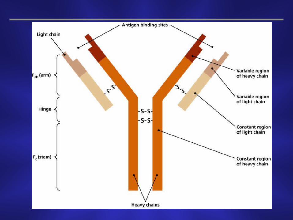


Antigen-binding sites are complementary to antigenic determinants (epitopes), they are hypervariable regions within the variable domains of heavy and light chains
Due to the close match can form strong, noncovalent interactions
Hydrogen bonds and other attractions (van der Waals, electrostatic) may also be involved
Antibody Function

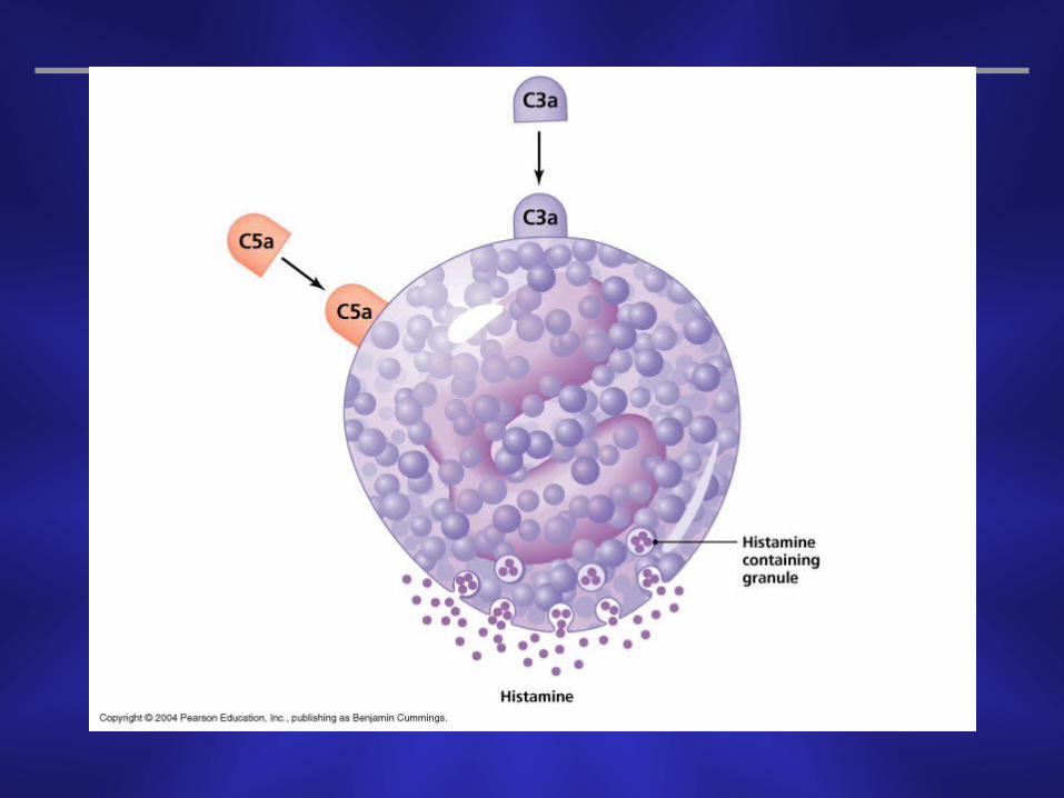
Function in several waysActivation of complement
Stimulation of inflammation
Agglutination
Neutralization
Opsonization
Antibody Function


A single type of antibody is not sufficient for the multiple types of invaders to the body
The class involved in the immune response depends on the type of foreign antigen, the portal of entry, and the antibody function needed
5 different classes of antibodies
Classes of Antibodies
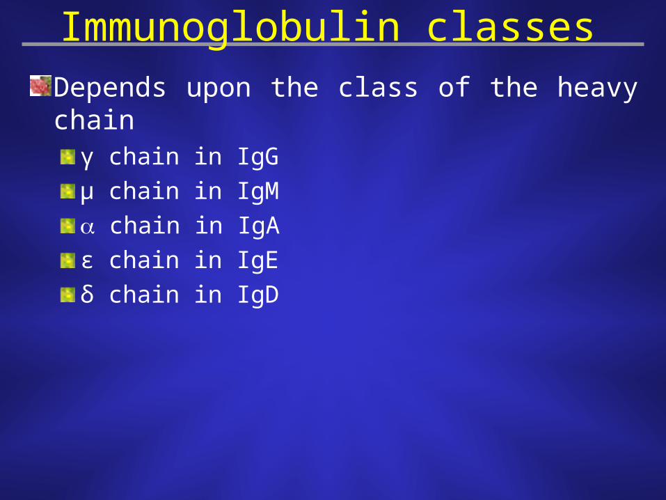
Immunoglobulin classes
Depends upon the class of the heavy chainγ chain in IgG
µ chain in IgM
chain in IgA
ε chain in IgE
δ chain in IgD

Immunoglobulin classesIgG is divalent. It crosses the placenta. 80% in serum. 2ry immune response.
It is agglutinating, fixes complement, neutralizing.
IgM is a pentamer. 6% in serum. 1ry immune response.
It is agglutinating, fixes complement, neutralizing.
IgA is dimer in secretions (milk, saliva, tears, respiratory & intestinal secretions) , monomer in serum (13%)
IgE bound to the surface of basophils and mast cells, <1%
Increases in anaphylactic reactions and helminth infections.
IgD bound to the surface of some cells, <1%.


Characteristics of the Five Classes of IGs

Characteristics of the Five Classes of Immunoglobulins
Structure ( Molecular Weight In Daltons)
Name function(s) Location(s) percentage in serum
IgG Complement activation, agglutination Monomer (180,000) serum, intercellular fluid 85 opsonization, and neutralization;
crosses placenta to protect fetus IgM Complement activation, agglutination, Pentamer (900,000) serum 5-10 and neutralizationIgA Agglutination and neutralization Monomer (150,000) External secretions, 5 Dimer (300,000) including milk IgE Triggers release of histamines Monomer (200,000) Serum, <1 from basophil mast cells mast cell surfaces IgD Unknown Monomer (180,000) B cell surface (as receptors) <1

Is an antibody that remains associated with the cytoplasmic membrane
Each B lymphocyte has multiple copies of a single type of BCR (105)
Antigen binding site is identical to that of the secreted antibody for that particular cell
The randomly generated antibody variable region determines the BCR (it is not formed in response to antigens)
B Cell Receptor (BCR)
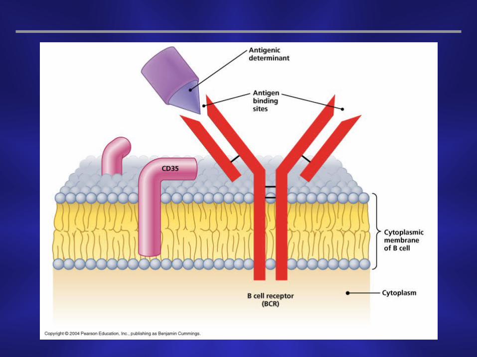

Each BCR is complementary to only one antigenic determinant
The BCRs on all of an individual’s B cells are capable of recognizing millions (107-108) of different antigenic determinants
B Cell Receptor (BCR)

Produced in the red bone marrow and mature in the thymus
Circulate in the lymph and blood and migrate to the lymph nodes, spleen, and Peyer’s patches
Part of the cell-mediated immune response because they act directly against various antigens
Endogenous invaders
Many of the body’s cells that harbor intracellular pathogens
Abnormal body cells such as cancer cells that produce abnormal cell surface proteins
T Lymphocytes

3 typesCytotoxic T cells
2 types of helper T cells
T Lymphocytes


Distinguished by the CD8 cell-surface glycoprotein
Directly kill certain cellsCells infected with viruses and other intracellular pathogens
Abnormal cells, such as cancer cells
Cytotoxic T cells (TC Cells)
Distinguished by the CD4 cell-surface glycoprotein
Function to “help” regulate the activities of B cells and cytotoxic T cells during an immune response
Secrete various soluble protein messengers, called cytokines, that determine which immune response will be activated
Helper T Cells (TH Cells)

2 typesType 1 helper T cell (TH
1)
Assist cytotoxic T cells
Express CD26 and a cytokine receptor named CCR5
Type 2 helper T cell (TH2)
Assist B cells
Have cytokine receptors CCR3 and CCR4
Helper T Cells (TH Cells)

Soluble regulatory proteins that act as intercellular signals when released from certain body cells
Immune system cytokines signal among various leukocytes
The complex web of signals among all the cell types of the immune system is referred to as the cytokine network
Cytokines
Interleukins (ILs)- signal among leukocytes(IL-1 produced by macrophages and activates various cells eg T & B cells)
(IL-2 produced by T helpers and stimulate growth of T cells)
Interferons (IFNs)- antiviral proteins that may act as cytokines (especially gamma)
Growth factors- proteins that stimulate stem cells to divide, maintaining an adequate supply of leukocytes
Tumor necrosis factors (TNFs)- Secreted by macrophages and T cells to kill tumor cells and regulate immune responses and inflammation (cachectin)
Chemokines- signal leukocytes to go to a site of inflammation or infection and stimulate other leukocytes
Cytokines of the Immune System

Vital that immune responses not be directed against autoantigens
Body “edits” lymphocytes to eliminate any self-reactive cells
Lymphocyte Editing by Clonal Deletion


Group of antigens first identified in graft patients
Important in determining the compatibility of tissues in successful grafting
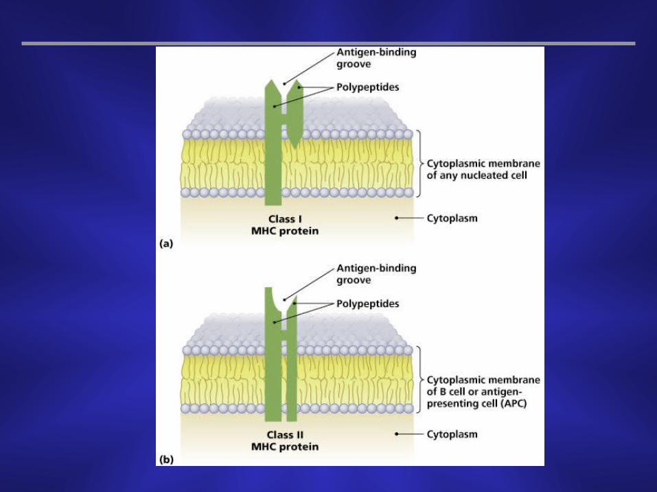
Major histocompatibility antigens are glycoproteins found in the membranes of most cells of vertebrate animals
Function to hold and position antigenic determinants for presentation to T cells
Major Histocompatibility Complex (MHC)
Antigens bind in the antigen-binding groove of MHC molecules
2 classes of MHC proteinsMHC class I (A, B, C)
MHC class II (Dp, Dq, Dr)
Major Histocompatibility Complex (MHC)

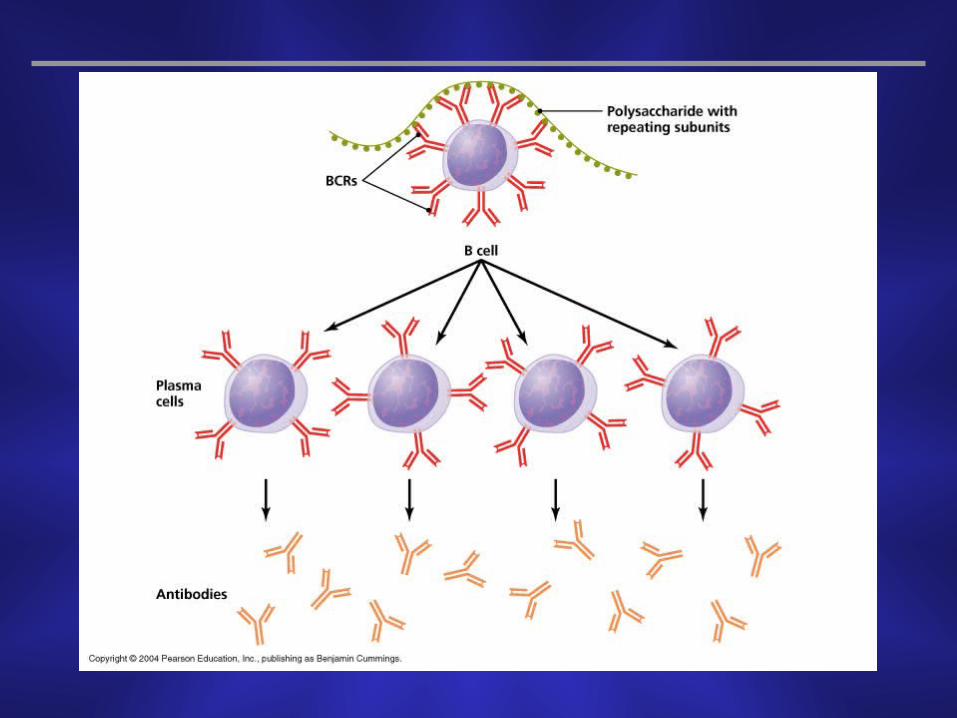
T-independent antigenLarge antigen molecules with readily accessible, repeating antigenic determinants
B cells can bind these directly without being processedStimulates B cells to differentiate into a plasma cell and produce antibodies
Antigen Processing

T-dependent antigensSmaller antigens with less accessible antigenic determinants
B cells require involvement from helper T cells to target these antigens
Helper T cells are assisted by leukocytes that process the antigen to make the antigenic determinants more accessible
Processing is different based on whether the antigen is exogenous or endogenous
Antigen Processing

APC internalizes the invading pathogen and enzymatically digests it into smaller antigenic fragments which are contained within a phagolysosome
Phagolysosome fuses with a vesicle containing MHC II molecules
Each fragment binds to the antigen-binding groove of a complementary MHC II molecule
The fused vesicle then inserts the MHCII-antigen complex into the cytoplasmic membrane so the antigen is presented on the outside of the cell
Processing of Exogenous Antigens
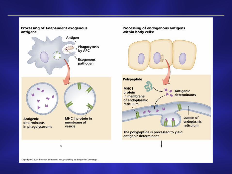


The intracellular pathogens are also digested into smaller antigenic determinants
Each fragment binds to a MHC I molecule located in the endoplasmic reticulum membrane
The membrane is packaged into a vesicle by a Golgi body which is inserted into the cytoplasmic membrane so the antigen is displayed on the cell’s surface
Processing of Endogenous Antigens

Body mounts humoral immune responses against exogenous pathogens
Components of a humoral immune responseB cell activation and clonal selection
Memory B cells and the establishment of immunological memory
Humoral Immune Response

Make up the majority of cells produced during B cell proliferation
Each plasma cell secretes only antibody molecules complementary to the specific antigenic determinant
Are short-lived cells that die within a few days of activation, though their antibodies and progeny can persist
Plasma Cells


Cells produced by B cell proliferation that do not secrete antibodies
Cells that have BCRs complementary to the specific antigenic determinant that triggered their production
Long-lived cells that divide only a few times and then persist in the lymphoid tissue
Are available to initiate antibody production if the same antigen is encountered again
Memory B Cells


Responds to intracellular pathogens and abnormal body cells
The most common intracellular pathogens are viruses but the response is also effective against intracellular bacteria
Triggered when antigenic determinants of the pathogen are displayed on the host cell’s surface
Cell-Mediated Immune Response


The perforin-granzyme cytotoxic pathway
Tc cells have vesicles containing cytotoxins (perforins & granzymes)
Upon attachment of Tc cell to a target, the vesicles release the cytotoxins
Perforins aggregate into a tubular structure in the infected cell membrane forming a channel
Granzymes move through this channel into target cells and activate apoptotic enzymes
Tc disengages and moves on to another infected cell.
The CD95 cytotoxic pathway
CD95 is present on cell membrane of most body cells
Its receptor CD95L is present on activated Tc
CD95L from activated Tc binds to CD95 on target cell, this activates enzymes that trigger apoptosis


Careful regulation of cell-mediated immune response to prevent T cells from responding to autoantigens
T cells require additional signals from an antigen presenting cell
Interaction of the T cell and antigen presenting cell at an immunological synapse stimulates the T cell to respond to the antigen
T Cell Regulation

Specific immunity acquired during an individuals life
2 typesNaturally acquired- immune response against antigens encountered in daily life
Artificially acquired- response to antigens introduced via a vaccine
Further distinguished as either active or passiveActive- active response to antigens via humoral or cell-mediated responses
Passive- passively receive antibodies from another individual
Acquired Immunity

A Comparison of the Types of Acquired Immunity