isolation of spherosomes (oleosomes) from onion, cabbage, and
TRANSCRIPT

Plant Physiol. (1971) 48, 675-682
Isolation of Spherosomes (Oleosomes) from Onion, Cabbage, andCottonseed Tissues
Received for publication April 5, 1971
L. Y. YATSU, T. J. JACKS, AND T. P. HENSARLINGSouthern Regionial Research Laboratory,' New Orleans, Louisiana 70119
ABSTRACT
Subeellular particles, identical in appearance to spherosomesobserved in situ, were isolated from onions (Allium cepa L.)and cabbages (Brassica capitata L.). They were minutespherules about 1 micron in diameter, filled with an evenlystained osmiophilic matrix and delimited by a single, fine.line membrane 20 to 30 A thick."Spherosomes" isolated from onions and cabbages were com-
pared with "oil-droplets" isolated from cottonseeds. Morpho-logically, they were similar, even at ultrastructural levels. Theirchemical compositions were also similar: both types of parti-cles containing primarily lipid with very little phospholipid orprotein. Neither type of particle possessed acid phosphataseactivity.
These results indicate that oil-droplets of oleaginous tis-sues correspond to spherosomes of nonoily tissues. Therefore,both types of particles should be referred to by the same name.Since these particles are rich in lipids, it is suggested that thename "spherosome" be abandoned in favor of "oleosome,"which is also entitled to priority.
Spherosomes are intracellular particles which have beenfamiliar to botanical cytologists for more than half a cen-tury; yet, surprisingly, they are still the object of much con-troversy (4, 5, 7, 8, 10, 13, 16, 19, 23). The term "spherosome"was introduced into the literature in 1922 when Dangeard(2) distinguished between spherical (spherosomes) and rod-shaped (mitosomes) "microsomes of the spherome." Earlier,Dangeard (1) had described the spherome as consisting of"microsomes" which he reported as highly refringent spherulesof an oily appearance that blacken, more or less, with osmicacid. From the very outset, there was controversy concerningspherosomes: one faction (1) which looked on spherosomesas organelles, and the other (6) which considered spherosomesmerely as products of cellular metabolism (lipids).One of the most recent eruptions of this controversy was
the objection voiced over the identification of "oil-droplets"from oleaginous tissues with "spherosomes" of nonoily tissues(19). Oil-droplets were purported to be droplets of oil thatwere free in the cytoplasm without delimiting unit-membranes,whereas spherosomes were alleged to be composed of phos-pholipids and proteins which were bounded by unit-mem-
I One of the laboratories of the Southern Marketing and Nutri-tion Research Division, Agricultural Research Service, UnitedStates Department of Agriculture.
branes. Many investigators (17, 22) agreed with this distinctionbetween oil-droplets and spherosomes-both at the light andelectron microscope levels. Yet, some continued to identifyoil-droplets with spherosomes (10, 23).
Clearly, this problem requires investigation. Since sphero-somes do not possess distinct morphological features as seenin other organelles such as mitochondria, chloroplasts, ordictyosomes, it was felt that a morphological and chemicalcharacterization was necessary. In this communication wedescribe, for the first time, the isolation of spherosomes fromnonoily tissues and compare their morphology and chemicalcomposition with oil-droplets isolated from cottonseeds.
MATERIAIS AND METHODSLight-microscopic Procedures. Microscopic examinations of
fresh tissues were conducted with a Zeiss Ultraphot II lightmicroscope using brightfield, darkfield, and phase contrast.The effects of osmium tetroxide on tissues were observeddirectly as the fixative contacted the tissues under the coverslips.
Electron-microscopic Procedures. Electron microscopic ex-aminations were conducted on tissues: (a) fixed overnight atroom temperature in 2% osmium tetroxide dissolved in 0.1 Mphosphate buffer, pH 7.2, (b) fixed for 1 hr in 2% aqueouspotassium permanganate at room temperature, and (c) doublyfixed with aldehyde and osmic acid fixatives. The tissue wasfirst fixed overnight in an aldehyde fixative which consistedof 2.8% glutaraldehyde and half-saturated picric acid adjustedto pH 7.3 with 50 mm sodium cacodylate. The tissue wasthoroughly rinsed in 50 mm sodium cacodylate, pH 7.3,and postfixed overnight in 1% OsO dissolved in 50 mm sodiumcacodylate, pH 7.3.The tissues were serially dehydrated in a graded series of
aqueous acetone and embedded in Maraglas. Sections werecut on a Servall MT-1 Porter-Blum ultramicrotome with adiamond knife, stained with uranyl acetate and lead citrateand examined with a Phillips EM 200 or EM 300 electronmicroscope.
Isolation of Spherosomes. Basically, spherosomes were iso-lated from tissue homogenates by differential centrifugation.Fifty pounds of onion bulbs (Allium cepa L.) were sliced intosmall pieces and blended in a Waring Blendor with approxi-mately an equal volume of 0.5 M NaCl containing 50 mMtris-HCl buffer, pH 7.2. The mixture was blended for 1 min inan ice bath. The homogenate was squeezed through eightlayers of cheesecloth, and the effluent was passed through aSharples Type T-41-248RY-34 continuous-flow centrifuge at20,000g to remove dense particles. The supernatant was thenpassed through a De Laval Gyro test unit separator to collectparticles less dense than the grinding medium. No "cream" wascollected from the separator; however, a light layer of creamysubstance adhered to the cones inside the separator. This mate-
675
Dow
nloaded from https://academ
ic.oup.com/plphys/article/48/6/675/6091512 by guest on 27 D
ecember 2021

YATSU, JACKS, AND HENSARLING Plant Physiol. Vol. 48, 1971
FIG. 1. Dark field light micrograph of an onion epidermal cell showing the highly refractile spherosomes in the thin layer of cytoplasm nextto the cell wall. Because the highly refringent spherosomes are so bright, many of the organelles which are not in focus are visible as white dots.The apparent size is deceptive for various reasons, e.g., diffraction, refraction, etc. Bar indicates 10 ,u. a: Brightfeld photom crograph of a por-tion of an onion epidermal cell showing highly osmiophilic spherosomes (see arrows). The darkening of the spherosomes by osmium tetroxidewas observed while the object was under the microscope (see text). Bar indicates 5 ,u. Fixed in I cc osmium tetroxide in 0.1 M phosphate buffer,pH 7.2.
rial was rinsed off the cones with buffer and centrifuged at25,QOOg for 20 min in the swinging bucket rotor of a ServwCllRC2B centrifuge. A light, creamy layer came to the top of thecentrifuge tube but was present in such sparse quantity thatno analytical procedures were prvcticable. A sample was takenfrom the top of the centrifuge tube with a platinum loop andexamined with a light microscope.
Fifty pounds of cabbages (Brassica capiatt L.) were slicedand treated aus above, but the procedures were carried out indistilled water instead of buffered saline. Again. cream wasnot obtained from the separator. but the rinses from theseparator cones yielded about 1 ml of fatty material. Sampleswere taken for light and electron microscopic studies and forbiochemical analyses.
676
Dow
nloaded from https://academ
ic.oup.com/plphys/article/48/6/675/6091512 by guest on 27 D
ecember 2021
ISOLATION OF SPHEROSOMES
Studies of Isolated Spherosomes. Unfixed, isolated sphero-somes exhibited Brownian motion under the light microscope.To eliminate this for photographic purposes, a droplet ofspherosomal preparation was allowed to dry on a cover slipbriefly and then observed in contact with a fresh drop of water.Many spherosomes then adhered to the cover slip enablingphotomicrographs to be taken.
Electron microscopic examination of isolated spherosomeswas conducted as before (11). Electron microscopic histo-chemical studies were carried out as described in a previouscommunication (24).
Materials for chemical analyses were dried to constantweights over P20 in vacuo at room temperature. The driedsamples (from 100-200 mg) were extracted of total lipidsaccording to the method of Martin and Morton (14), exceptthat the residues were collected in small, fritted glass funnels.Solvents were removed by evaporation, and the residues andtotal lipids were determined gravimetrically after drying toconstant weight in vacuo.
Lipid-phosphorus was determined by procedures describedpreviously (12); nitrogen was determined by the method ofMinari and Zilversmit (15); acid phosphatase (acid phos-phomonoesterase, E.C. 3.1 .3.2) activity was assayed accord-ing to Salomon et al. (18).
RESULTS
Light Microscopy. Spherosomes, in the classical sense, werereadily observable in fresh, nonoily plant tissue, e.g., epidermalcells of onion bulb scales. They were minute spherules about1 ,. in diameter, highly refractile, and prone to Brownianmotion. We confirmed, under direct observation in the lightmicroscope, the fact that spherosomes stain brown to blackwith osmic acid. The highly refractile spherules (spherosomes)were the most intensely stained (osmiophilic) subcellular par-ticles in the cell; the hyaloplasm and other organelles were veryfaintly stained (Fig. 1).
Electron Microscopy. The quality of ultrastructural preser-vation achieved varied with the fixative employed. Osmium.tetroxide, even when buffered, produced the poorest images
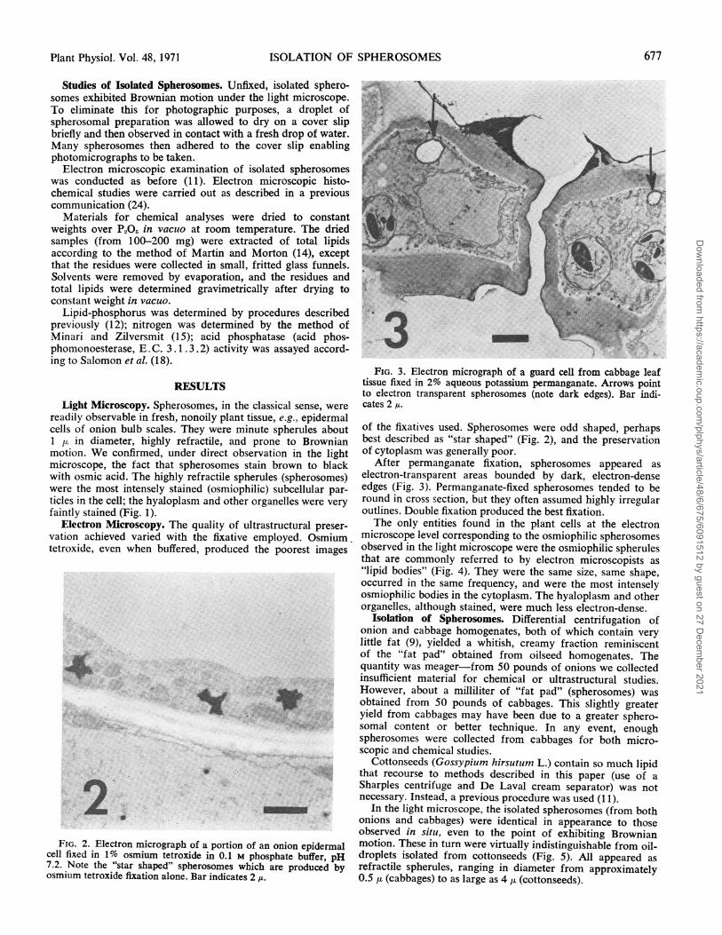
FIG. 2. Electron micrograph of a portion of an onion epidermalcell fixed in 1% osmium tetroxide in 0.1 M phosphate buffer, pH7.2. Note the "star shaped" spherosomes which are produced byosmium tetroxide fixation alone. Bar indicates 2 IA.
FIG. 3. Electron micrograph of a guard cell from cabbage leaftissue fixed in 2% aqueous potassium permanganate. Arrows pointto electron transparent spherosomes (note dark edges). Bar indi-cates 2 u.
of the fixatives used. Spherosomes were odd shaped, perhapsbest described as "star shaped" (Fig. 2), and the preservationof cytoplasm was generally poor.
After permanganate fixation, spherosomes appeared aselectron-transparent areas bounded by dark, electron-denseedges (Fig. 3). Permanganate-fixed spherosomes tended to beround in cross section, but they often assumed highly irregularoutlines. Double fixation produced the best fixation.The only entities found in the plant cells at the electron
microscope level corresponding to the osmiophilic spherosomesobserved in the light microscope were the osmiophilic spherulesthat are commonly referred to by electron microscopists as"lipid bodies" (Fig. 4). They were the same size, same shape,occurred in the same frequency, and were the most intenselyosmiophilic bodies in the cytoplasm. The hyaloplasm and otherorganelles, although stained, were much less electron-dense.
Isolation of Spherosomes. Differential centrifugation ofonion and cabbage homogenates, both of which contain verylittle fat (9), yielded a whitish, creamy fraction reminiscentof the "fat pad" obtained from oilseed homogenates. Thequantity was meager-from 50 pounds of onions we collectedinsufficient material for chemical or ultrastructural studies.However, about a milliliter of "fat pad" (spherosomes) wasobtained from 50 pounds of cabbages. This slightly greateryield from cabbages may have been due to a greater sphero-somal content or better technique. In any event, enoughspherosomes were collected from cabbages for both micro-scopic and chemical studies.
Cottonseeds (Gossypium hirsutum L.) contain so much lipidthat recourse to methods described in this paper (use of aSharples centrifuge and De Laval cream separator) was notnecessary. Instead, a previous procedure was used (1 1).
In the light microscope, the isolated spherosomes (from bothonions and cabbages) were identical in appearance to thoseobserved in situ, even to the point of exhibiting Brownianmotion. These in turn were virtually indistinguishable from oil-droplets isolated from cottonseeds (Fig. 5). All appeared asrefractile spherules, ranging in diameter from approximately0.5 u (cabbages) to as large as 4 p. (cottonseeds).
677Plant Physiol. Vol. 48.,1971
".DI; .."t..
. *t'.,., . x.
z.i, - A.F;e" 6 ,o
'u-
*1 ..
_S~~
Dow
nloaded from https://academ
ic.oup.com/plphys/article/48/6/675/6091512 by guest on 27 D
ecember 2021

YATSU, JACKS, AND HENSARLING Plant Physiol. Vol. 48, 1971
4
7
4a#
FIG. 4. A low power electron micrograph of an onion epidermal cell similar to the light micrograph shown in Figure 1. Note the intenseosmiophilia of the spherosomes compared with the other cellular constituents (see arrows). Even though the electron microscore has a muchgreater depth of focus than the light microscope, the section is only about one-tenth of a micron in thickness, therefore fewer spherosomes arevisible than in the light micrograph. Fixed in glutaraldehyde-picric acid and postfixed in osmium tetroxide. Bar indicates 10 ,u. a: Higher mag-nification of a portion of an onion epidermal cell fixed with aldehyde/osmium with no poststaining. The spherosomes are the most intenselyosmiophilic'organelle in the cell. Bar indicates 2 ,u.
In the electron microscope, isolated cabbage spherosomesfixed with osmium tetroxide were identical in appearance tothose observed in situ (Fig. 6). They were bounded by asingle fine-line on the order of 20 to 30 A thick. The matricesof the spherosomes, after osmium tetroxide fixation, wereuniformly electron dense with no apparent internal structure.
They ranged in size from about 0.4 ,u to slightly over 2 ,uin diameter, averaging under 1 tu in diameter.
Isolated cottonseed oil-droplets were essentially the same ascabbage spherosomes in appearance; however, they were abouttwo to three times as large in diameter (Fig. 7).
Biochemistry. When cottonseed oil-droplets were dried over
678
Dow
nloaded from https://academ
ic.oup.com/plphys/article/48/6/675/6091512 by guest on 27 D
ecember 2021

ISOLATION OF SPHEROSOMES
FIG. 5. Darkfield light micrographs of isolated spherosomes from; (a) onion tissue, (b) cabbage tissue, and (c) cottonseed. Bar indicates 10 At.
P205 in a vacuum desiccator, the white, mayonnaise-like mate-rial assumed a clear, translucent, jelly-like appearance. Themass did not become a liquid but maintained a lumpy ap-pearance, indicating a maintenance of structure. Cabbagespherosomes, on the other hand, never lost their white ap-pearance; dried cabbage spherosomes appeared as a white,powdery mass. The bulk of cottonseed oil-droplets was lipid(98.8%). Cabbage spherosomes were also highly lipoidal,yielding 81.0% lipid (Table I).The phosphorus content of lipid from cottonseed oil-
droplets was 0.015% and cabbage spherosomal lipid 0.007%,corresponding to 0.38% and 0.16% phospholipid, respectively(Table I). Protein content of cottonseed oil-droplets and cab-bage spherosomes were 0.63% and 4.0% respectively.
Since spherosomes have been shown to possess acid phos-phatase activity (20), it was of interest to determine acid phos-phatase activity in our spherosomal preparations. The oil-droplet fraction from cottonseeds contained virtually no acidphosphatase activity. Cabbage spherosomes. on the other hand,contained a slight amount of acid phosphatase activity; how-ever, when distribution of the activity was analyzed (Table II),only 0.001% of the activity in the homogenate was associatedwith the spherosomal fraction, leading us to suspect con-tamination. This suspicion was corroborated by electron-microscopic histochemistry (Fig. 6c). Spherosomes allowedto react with Gomori's reagent for 4 hr showed a slight amountof acid phosphatase activity in the "fat pad" fraction. How-ever, the activity was not associated with spherosomes; instead,it was associated with fine, membranous material found withthe spherosomes as "contaminant." The presence of "contami-nation" was not surprising. Due to apprehension over the lossof our meager spherosomal preparation, this particular samplewas not washed. All preparations of oilseed oil-droplets, on theother hand, were routinely washed and rewashed at least fivetimes.
DISCUSSIONThe object of this study was to eliminate the confusion con-
cerning the cytological status of spherosomes, particularly atthe electron microscope level, and to compare spherosomes of
nonoily plant tissues with oil-droplets of oily plant tissues. Inorder to achieve these goals, spherosomes were isolated fromnonoily plant tissues and studied in the cell-free state.
Identification of Spherosomes. Spherosomes are easily recog-nizable in the light microscope. They are highly refractile,rapidly displaced spherules about 1 ju in diameter which be-come black after contact with osmium tetroxide. Spherosomesstain so intensely that they stand out in stark contrast fromthe rest of the cytoplasm. There is no question concerning theidentity between the bright, refractile particles and the blackspherules obtained after osmium tetroxide staining; this can beshown under direct observation. Difficulty seems to arise atthe electron microscope level.At low magnification in the electron microscope, the only
cytoplasmic inclusions that were intensely osmiophilic andspherical (corresponding to spherosomes observed in the lightmicroscope) were the bodies commonly referred to as "lipiddroplets" (compare Fig. 1 and Fig. 4). At these low magnifica-tions, mitochondria and plastids, although stained, were not asconspicuously osmiophilic nor did not stand out as markedlyfrom the groundplasm as did the spherosomes. The spherulesnot only exhibited intense osmiophilia, they were the same sizeand shape as spherosomes observed under light microscopy.We concluded, therefore, that the highly osmiophilic roundbodies observed in the electron microscope (often referred toas "lipid bodies") and the highly refractile, osmiophilic spheri-cal bodies (spherosomes) seen in the light microscope are oneand the same.One might wonder why confusion arose in the first place.
Undoubtedly, the profiles observed in the electron microscopefollowing osmium tetroxide or permanganate fixation werelargely responsible for this problem. Spherosomes in doublyfixed (aldehyde and osmium) tissues are spherical bodies simi-lar to the spherosomes seen in the light microscope; however,spherosomes in tissues fixed with permanganate or osmiumtetroxide (fixatives employed prior to the introduction ofdouble fixation) are not. Spherosomes in tissues fixed withpermanganate are irregular-shaped with an electron-trans-parent matrix. The edges are generally very electron dense andmembranes are not discernible. Spherosomes in tissues fixed
679Plant Physiol. Vol. 48, 1971
Dow
nloaded from https://academ
ic.oup.com/plphys/article/48/6/675/6091512 by guest on 27 D
ecember 2021

YA7SU, JACKS, AND HENSARLING Plant Physiol. Vol. 48, 1971
4%
Mv4t<.@ .-E.w; @.t>'.,rg5w* ..... ::
| F. F
i
.... r
4;..:
i...I
.: 1 t
..IX
A11 A
FIG. 6. Electron micrograph of a portion of a leaf cell showing the highly osmiophilic spherosomes in cabbage tissue. Fixed in glutaraldehyde,postfixed in osmium tetroxide. Bar indicates 0.5 ,u. a: High magnification of a spherosome from cabbage leaf tissue fixed with glutaraldehyde, post-fixed in osmium tetroxide followed by uranyl acetate and lead citrate staining. Note the "unit membrane" indicated by the double arrows andthe "single membrane" bounding the spherosome indicated by the single arrow. Bar indicates 0.1 M. b: Electron micrograph of isolated sphero-somes from cabbage tissue. Note the debris between the spherosomes. Fixed with glutaraldehyde-osmium tetroxide. Bar indicates 1 ,s. c: Elec-tron microscopic histochemical study of isolated spherosomes from cabbage tissue. Note deposition of lead phosphate indicating enzymic ac-tivity associated with "debris" rather than the spherosomes. Bar indicates 1 ,z.
with osmium tetroxide have very irregular and aberrantforms. Drawert and Mix (4) correctly identified spherosomeswith "sternformige" bodies observed in the tissues afterosmium tetroxide fixation, but apparently other workers (priorto double fixation techniques) could not, or would not,identify a star-shaped or irregular-shaped body with the neat,spherical body seen in the light microscope. In their quest fortrim, round bodies (the counterpart of the neat, refractilespheres observed in the light microscope), many microscopistsapparently ignored the star-shaped bodies and sought a roundbody. Consequently, many bodies labeled "spherosome" inthe literature are not spherosomes but simply sphericalorganelles such as microbodies or round vesicles. Perhaps, ifdoubly fixed tissue had been observed first, this issue may nothave arisen.
Isolation of Spherosomes. Our working hypothesis in thisstudy was that spherosomes are lipid bodies. The knownproperties of spherosomes support our thesis. Firstly, lipiddroplets would form spherical shapes in an aqueous milieu.Since vegetable oils have a higher refractive index than water,lipid droplets should be refractile in water. Lipid dropletswould also tend to exhibit Brownian motion more readily thanproteinaceous bodies because of their lower specific densities(lower inertia), and, since they are composed of highly un-saturated fatty acids, they should exhibit very high osmiophilia.
Thus, if our thesis is correct, a fraction of plant homogenateshould be obtained that moves centripetally in a centrifugalfield. Since low fat tissues would be expected to contain verysparse quantities of fatty spherosomes, a continuous methodof centrifugation was sought to handle the large quantities of
680
M...
A, qd.
Ak
...
. . d. , Dow
nloaded from https://academ
ic.oup.com/plphys/article/48/6/675/6091512 by guest on 27 D
ecember 2021

ISOLATION OF SPHEROSOMES
plant material required. By utilizing a Sharples continuouscentrifuge (which sediments dense particles and yields effluentcontinuously) together with a De Laval separator (whichseparates the light particles from the denser medium con-tinuously), large volumes of material could be handled.
Although modest in quantity, a "fat pad" which was com-posed of fatty particles was obtained from both onion andcabbage tissues using the techniques described above. Theisolated particles were identical in appearance to the sphero-somes in situ, both at the light and electron microscope levels.They were the same size and shape, exhibited the same refrac-tile quality, the same osmiophilia, and even the same Brownianmotion.The very low yield of spherosomes obtained from cabbages
was disappointing. A rough calculation, however, shows thatthe yield of spherosomes is reasonably satisfactory. Assumingthat 50 pounds of cabbages occupies about 1 bushel (approxi-mately 0.35 cubic meters), half of which is air, and also assum-ing that the average cell is about 50 tt in diameter, one cancalculate that 50 pounds of cabbages should contain about3 X 10 cells. If we assign a value of 0.5 y diameter for theaverage spherosome, 1 ml of spherosomes would contain about8 X 1012 spherosomes. This corresponds to about 25 sphero-somes per cell, which agrees with the values Sorokin andSorokin (20) reported for epidermal cells of Campanula.Therefore, although this is a crude calculation (e.g., sphero-somal populations vary from cell to cell, they vary in size, cellsizes vary), the yield appears to be of the correct order ofmagnitude.The chemical data which were obtained show that isolated
spherosomes are mainly lipid with very small quantities ofphospholipid and protein. These data argue well for thecorrectness of our hypothesis.
Comparison between "Spherosomes" and "Oil-Droplets."Aside from differences in size, isolated cabbage spherosomeswere virtually identical in appearance to oil-droplets isolatedfrom cottonseeds. In the light microscope, cottonseed oil-droplets were larger than spherosomes from onions or cab-
FIG. 7. Electron micrograph of isolated cottonseed "oil-droplets"(spherosomes). Fixed with glutaraldehyde-osmium tetroxide. Barindicates 2 I.
Table I. Composition of Spherosomal FractionzsSpherosomes were prepared and analyzed as described in
"Materials and Methods." Values represent averages of duplicatedeterminations on two samples and are given as percent of the dryweight of recovered material.
Materiall Total |Nolipid Recovery2 Pbospho- Protein4
Peanut5 99.55 0.45 100.3 0.09 0.22Cotton6 98.8 1.2 100.0 0.37 0.63Cabbage7 81.0 19.0 100.9 0.16 4.0
1 Total lipid + nonlipid residue = 100%.2 Dry weight of sample = 100%7o.3Phospholipid = lipid phosphorus X 24.8.4Protein = nonlipid nitrogen X 6.25.5 Washed lOX.6 Washed 5X.7Washed 2X.
Table II. Distribution ofAcid Phosphatase Activity in Spherosomaland Nonspherosomal Fractions
Fractions were prepared and analyzed as described in "Ma-terials and Methods." Values are given as percent of recoveredactivity.
Fraction' Peanut Cotton Cabbage
Spherosomal 0 0 trace4Nonspherosomal2 100 100 100Recovery3 99 98 100
1 Activities of spherosomal + nonspherosomal fractions =100%. The original homogenate contained, per ml, 3.24 units ofenzyme from peanut, 3.23 units from cottonseed and 4.48 unitsfrom cabbage. An enzyme unit is defined as the amount of enzymewhich produces an increase in absorbance of 1 per 15 min.
2 Nonspherosomal fraction is mitochondrial and supernatantliquid fractions combined.
3Activity in supernatant liquid after centrifuging homogenateat 1000 X g for 5 min = 100%.
I Trace = 0.001%c.
bages, but both types of bodies were spherical, refractile, andhighly osmiophilic. In the electron microscope, both entitieswere round in cross section, filled with an evenly stainedosmiophilic ground substance (oil), and bounded by the sametype of single-line membrane that is distinctly different fromtripartite, unit-membranes. These particles are similar to the"lipid bodies" of corn embryo cells described by Trelease (21),namely lipid droplets bounded by a single-line membrane 25to 40 A in thickness.
Chemically, the composition of spherosomes from cabbageswas similar to that of oil-droplets isolated from cottonseeds.Since the cabbage spherosomal preparation was not washed,a certain amount of contamination occurred; however, bothtypes of bodies were mainly lipid with very small quantitiesof phospholipid and protein.
It has been reported that spherosomes possess acid phos-phatase activity (20). However, we were unable to detect acidphosphatase activity in our spherosomal preparations. Sincethe enzymic activity (20) appeared to be a function of physio-logical status, this negative finding is inconclusive-the lack ofacid phosphatase activity in spherosomes from the quiescentcabbage and onion tissues could be analogous to the situation
681Plant Physiol. Vol. 48, 1971
Dow
nloaded from https://academ
ic.oup.com/plphys/article/48/6/675/6091512 by guest on 27 D
ecember 2021
682 YATSU, JACKS, A
in quiescent cottonseeds. It may be of interest, however, thatacid phosphatase activity was also not detected in oil-dropletpreparations from 3-day germinated castor seed endospermand 7-day germinated peanut seed cotyledons.Our results, therefore, lead us to the conclusion that sphero-
somes are lipid droplets bounded by a single fine-line mem-brane and that they are the same as the so-called "oil-droplets"found in oleaginous plant tissues. Therefore, it is deemed in-correct to distinguish between "oil-droplets" and "sphero-somes." Finally, since the name spherosome was chosenthrough default (17), we would like to suggest the more mean-ingful name "oleosome" (which has equal priority) (2) as thepreferred terminology for "microsome" of the spherome.
Acknowledgment-We thank Miss Mona L. Brown for her assistance in trans-lating articles and Mr. Jacques J. Hebert for his technical assistance on the lightnicroscope.
LITERATURE CITED
1. DANGEARD, P. A. 1919. Sur la distinction du chondriome des auteurs en vac-uome, plastidome et spherome. C. R. Acad. Sci. 169: 1005-1010.
2. DANGEARD, P. A. 1922. Sur la structure de la cellule chez les Iris. C. R. Acad.Sci. 175: 7-12.
3. DANGEARD, P. A. 1925. La structure des Vaucheries dans ses rapports avec laterminologie nouvelle des elements celluaires. La Cellule 35: 239-250.
4. DRAWERT, H. AND M. MIX. 1963. Elektronenmikroskopische Studien an denOberepidermiszellen der Schuppenblatter von Allium cepa L. Protoplasma57: 270-289.
5. FREY-WYSSLING, A., E. GRIESHABER, AND K. MUHLETHALER. 1963. Origin ofspherosomes in plant cells. J. Ultrastruct. Res. 8: 506-516.
6. GUILLIERMOND, A. 1921. Sur les microsomes et les formations lipoides de lacellule vegetale. C. R. Acad. Sci. 172: 1676-1678.
7. GUILLIERMOND, A. 1941. The Cytoplasm of the Plant Cell. The ChronicaBotanica Company, Waltham, Mass.
8. HORNER, H. T. AND J. J. ARNOTT. 1966. A histochemical and ultrastructuralstudy of pre- and post-germinated Yucca seeds. Bot. Gaz. 127: 48-64.
Al?ND HENSARLING Plant Physiol. Vol. 48, 1971
9. HOWARD, F. D., J. H. MACGILLIVRAY, AND M. YAMAGUCHI. 1962. Nutrientcomposition of fresh California-grown vegetables. Calif. Agr. Exp. Sta.Bull. 788.
10. HRSEL, I. 1966. The morphology and function of vesicles and of vesicle re-lated formations in cell ultrastructure. Biol. Plant. (Praha) 8: 263-272.
11. JACKS, T. J., L. Y. YATSU, AND A. M. ALTSCHUL. 1967. Isolation and charac-terization of peanut spherosomes. Plant Physiol. 42: 585-597.
12. JACKS, T. J., L. Y. YATSU, AND T. P. HENSARLING. 1970. Extraction of lipidfrom cottonseed tissue. I. Comparison of hexane-acetone-water, its non-aqueous components, and chloroform-m-nethanol. J. Amer. Oil Chem. Soc.47: 222-223.
13. JENSEN, W. A. 1965. The ultrastructure and histochemistry of the synergidsof cotton. Amer. J. Bot. 52: 238-256.
14. MARTIN, E. M. AND R. K. MORTON. 1956. The chemical composition of micro-somes and mitochondria from silver beet. Biochem. J. 64: 221-235.
15. MINARI, 0. AND D. B. ZILVERSMIT. 1963. Use of KCN for stabilization of colorin direct nesslerization of Kjeldahl digests. Anal. Biochem. 6: 320-327.
16. NIEUIWDROP, P. J. 1963. Electron microscopic structure of the epithelial cellsof the scutellum of barley. The structure of the epithelial cells before ger-mination. Acta Bot. Neerl. 12: 295-301.
17. PERNER, E. S. 1953. Die Spharosomen (Mikrosomen) pflanzlicherzellen.Protoplasma 42: 457-481.
18. SALOMON, L. L., J. JAMIES, AND P. R. WEAVER. 1964. Assay of phosphataseactivity by direct spectrophotometric determination of phenolate ion. Anal.Chem. 36: 1162-1164.
19. SOROKIN, H. P. 1967. The spherosomes and the reserve fat in plant cells. Amer.J. Bot. 54: 1008-1016.
20. SOROKIN, H. P. AND S. SOROKIN. 1968. Fluctuations in the acid phosphataseactivity of spherosomes in guard cells of Campanula persicifolia. J. Histo-chem. Cytochem. 16: 791-802.
21. TRELEASE, R. N. 1969. Ultrastructural characterization, composition, andutilization of lipid bodies in the maize shoot apex during postgerminativedevelopment. J. Cell Biol. 43: 147a.
22. WALEK-CZERNECKA, A. 1965. Histochemical demonstration of some hydrolyticenzymes in the spherosomes of plant cells. Acta Soc. Bot. Pol. 34: 573-588.
23. YATSu, L. 1965. The ultrastructure of cotyledonary tissue from Gossypiumhirsutum L. seeds. J. Cell Biol. 25: 193-199.
24. YATSIJ, L. Y. AND T. J. JACKS. 1968. Association of lysosomal activity witkaleurone grains in plant seeds. Arch. Biochem. Biophys. 124: 460471.
Dow
nloaded from https://academ
ic.oup.com/plphys/article/48/6/675/6091512 by guest on 27 D
ecember 2021