itral tricusid transcateter teraies echocardiography for
TRANSCRIPT
62 CARDIAC INTERVENTIONS TODAY JULY/AUGUST 2018 VOL. 12, NO. 4
M I T R A L & T R I C U S P I D T R A N S C AT H E T E R T H E R A P I E S
Essentials for understanding echocardiographic imaging in percutaneous tricuspid
interventions.
BY MENHEL KINNO, MD, MPH; SASAN R. RAISSI, MD; JYOTHY J. PUTHUMANA, MD;
JAMES D. THOMAS, MD; AND CHARLES J. DAVIDSON, MD
Echocardiography for Tricuspid Valve Intervention
Recognition of the significant morbidity and mor-tality associated with tricuspid valve disease has shifted attention to the often-referred to “forgot-ten” valve.1,2 Tricuspid regurgitation (TR) is the
most common pathology involving the tricuspid valve and secondary or functional TR is by far the most com-mon etiology encountered in clinical practice. Although primary disorders of the tricuspid valve can also lead to TR, including infective endocarditis, prolapse, carcinoid, right ventricle (RV) biopsy-related trauma, RV pace-maker/defibrillator lead, Ebstein anomaly, and rheumatic heart disease,3 functional TR is usually secondary to left ventricular dysfunction, left-sided valvular diseases, pul-monary hypertension, atrial fibrillation, isolated/idiopathic annular dilation, and regional/global RV dysfunction.4,5
Despite the well-recognized adverse outcomes associ-ated with functional TR, the only class I surgical indica-tion to repair/replace a tricuspid valve with severe TR is during cardiac surgery for left-sided valve disease.3,6 Recent studies have shown improvement in clinical out-comes and RV reverse remodeling when the timing of tricuspid repair for functional TR (in the setting of left-sided heart disease) is based on the tricuspid annulus size irrespective of TR severity.7,8 This was reflected in the cur-rent valve guidelines by giving a class IIa indication to repair for progressive functional TR on the basis of tricuspid valve annulus dilation (> 40 mm [> 21 mm/m2] on transthoracic echocardiogram [TTE] or > 70 mm on direct intraoperative measurement) at the time of left-sided valve surgery.3,6
Despite this recommendation, many patients do not undergo tricuspid valve surgery at the time of left-sided valve surgery. The reason treatment options for TR have lagged is due in part to the complexity of the valve
anatomy and high recurrence rates. There is significantly higher perioperative mortality for isolated tricuspid valve surgery in comparison to any other valve surgery.9-12 In addition, in the subset of patients in whom mild TR pro-gressed in severity after a cardiac surgery, repeated open
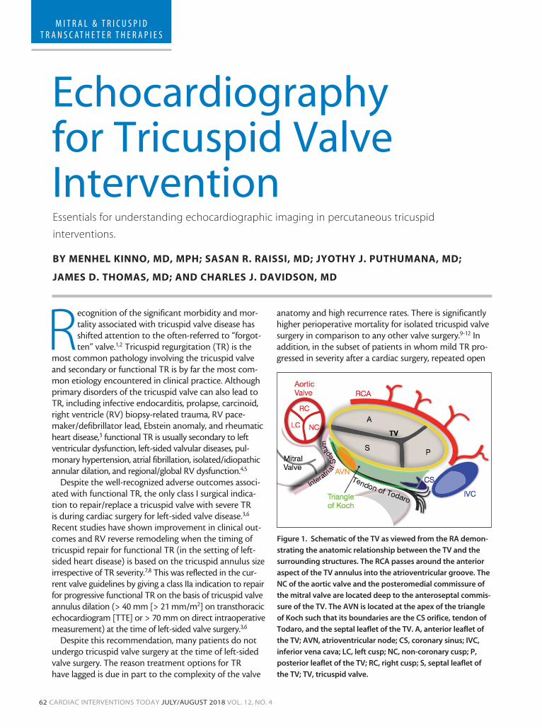
Figure 1. Schematic of the TV as viewed from the RA demon-
strating the anatomic relationship between the TV and the
surrounding structures. The RCA passes around the anterior
aspect of the TV annulus into the atrioventricular groove. The
NC of the aortic valve and the posteromedial commissure of
the mitral valve are located deep to the anteroseptal commis-
sure of the TV. The AVN is located at the apex of the triangle
of Koch such that its boundaries are the CS orifice, tendon of
Todaro, and the septal leaflet of the TV. A, anterior leaflet of
the TV; AVN, atrioventricular node; CS, coronary sinus; IVC,
inferior vena cava; LC, left cusp; NC, non-coronary cusp; P,
posterior leaflet of the TV; RC, right cusp; S, septal leaflet of
the TV; TV, tricuspid valve.
VOL. 12, NO. 4 JULY/AUGUST 2018 CARDIAC INTERVENTIONS TODAY 63
M I T R A L & T R I C U S P I D T R A N S C AT H E T E R T H E R A P I E S
heart surgery carries a high mortality rate that can reach up to 20%.13 Moreover, substantial progress in percutaneous interventional therapies in the management of left-sided valve diseases (aortic and mitral valves) has further empha-sized the negative consequences of functional TR on the survival of those patients.14,15
In light of the accumulating evidence that TR is not a passive bystander, and in the current era in which percu-taneous options have successfully progressed in the treat-ment of left-sided valve disease, there is an unmet need for transcatheter therapies for tricuspid valve pathologies. As
for any successful transcatheter image-guided intervention, it is imperative to have a solid understanding of the valve anatomy and to acquire comprehensive knowledge of the TTE and transesophageal echocardiographic (TEE) image acquisition to provide an early and accurate assessment of the valve structure and function.
In this article, we discuss tricuspid valve anatomy, echo-cardiographic image acquisition for assessing tricuspid valve morphology and function, and the role of echocardiog-raphy in currently available minimally invasive treatment options for TR (mainly functional TR).
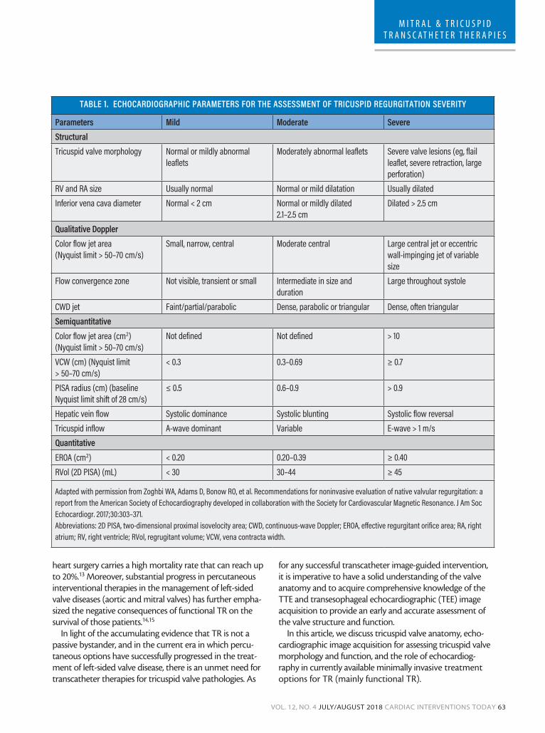
TABLE 1. ECHOCARDIOGRAPHIC PARAMETERS FOR THE ASSESSMENT OF TRICUSPID REGURGITATION SEVERITY
Parameters Mild Moderate SevereStructuralTricuspid valve morphology Normal or mildly abnormal
leafletsModerately abnormal leaflets Severe valve lesions (eg, flail
leaflet, severe retraction, large perforation)
RV and RA size Usually normal Normal or mild dilatation Usually dilatedInferior vena cava diameter Normal < 2 cm Normal or mildly dilated
2.1–2.5 cmDilated > 2.5 cm
Qualitative DopplerColor flow jet area(Nyquist limit > 50–70 cm/s)
Small, narrow, central Moderate central Large central jet or eccentric wall-impinging jet of variable size
Flow convergence zone Not visible, transient or small Intermediate in size and duration
Large throughout systole
CWD jet Faint/partial/parabolic Dense, parabolic or triangular Dense, often triangularSemiquantitative Color flow jet area (cm2) (Nyquist limit > 50–70 cm/s)
Not defined Not defined > 10
VCW (cm) (Nyquist limit > 50–70 cm/s)
< 0.3 0.3–0.69 ≥ 0.7
PISA radius (cm) (baseline Nyquist limit shift of 28 cm/s)
≤ 0.5 0.6–0.9 > 0.9
Hepatic vein flow Systolic dominance Systolic blunting Systolic flow reversalTricuspid inflow A-wave dominant Variable E-wave > 1 m/sQuantitative EROA (cm2) < 0.20 0.20–0.39 ≥ 0.40RVol (2D PISA) (mL) < 30 30–44 ≥ 45
Adapted with permission from Zoghbi WA, Adams D, Bonow RO, et al. Recommendations for noninvasive evaluation of native valvular regurgitation: a report from the American Society of Echocardiography developed in collaboration with the Society for Cardiovascular Magnetic Resonance. J Am Soc Echocardiogr. 2017;30:303–371.Abbreviations: 2D PISA, two-dimensional proximal isovelocity area; CWD, continuous-wave Doppler; EROA, effective regurgitant orifice area; RA, right atrium; RV, right ventricle; RVol, regrugitant volume; VCW, vena contracta width.

64 CARDIAC INTERVENTIONS TODAY JULY/AUGUST 2018 VOL. 12, NO. 4
M I T R A L & T R I C U S P I D T R A N S C AT H E T E R T H E R A P I E S
TRICUSPID VALVE ANATOMYThe tricuspid apparatus is a complex structure that
consists of the tricuspid valve fibrous annulus, three leaflets (septal, anterior, and posterior), and the papillary muscles and their associated chordae tendineae. What contributes more to the complexity of the tricuspid valve is its proximity to important structures: the atrio-ventricular node, right coronary artery, inferior vena cava, and coronary sinus (Figure 1).5,16-19
The tricuspid valve annulus is nonplanar, dynamic, and oval or triangular in shape. The most basal/superior point of the annulus is at the anteroseptal commissure near the RV outflow tract and aortic valve, while the most apical/inferior point is at the posteroseptal com-missure where the coronary sinus drains into the right atrium (RA). The tricuspid valve annulus is a dynamic structure and its size and perimeter change during the cardiac cycle (with the largest dimensions during atrial contraction). It is important to note that the tricuspid annulus becomes more planar and round with function-al TR as the annulus dilates anteriorly and posteriorly.
The tricuspid valve is the largest and most apically located valve; its three leaflets vary in their circumferen-tial and area sizes. The anterior and septal leaflets have a comparable circumferential extent that is larger than the circumference of the posterior leaflet. The anterior leaflet has the largest area with the highest mobility. The septal leaflet usually has the smallest area with the least mobil-ity. The restricted mobility of the septal leaflet is due to its immediate proximity to the membranous interven-tricular septum with many chordae directly attached to the septum. The posterior leaflet is highly variable as it may have one or multiple deep scallops. Not infrequent-
ly, the posterior leaflet might be absent or incorporated into the anterior or septal leaflet.
The tricuspid apparatus consists of two papillary mus-cles, anterior and posterior, with a variable septal papil-lary muscle (absent in approximately 20%). The anterior papillary muscle is the largest, with its associated chordae mainly attached to the anterior and posterior leaflets. The posterior papillary muscle is smaller in size and its chordae are attached to the posterior and septal leaflets.
ECHOCARDIOGRAPHIC IMAGING OF THE TRICUSPID VALVE
Echocardiography is the most common imaging modality used for assessing the tricuspid valve structure and func-tion and for determining the suitability of catheter-based interventional therapies. Comprehensive echocardiography with a systematic approach is essential for identifying the mechanism of TR and the severity.20-22 The three tricuspid valve leaflets cannot be visualized simultaneously by means of two-dimensional (2D) echocardiography, and therefore, multiple acoustic windows are essential to completely evaluate the tricuspid valve.21 Because of the frequently encountered anatomic variability in the tricuspid valve apparatus and the inconsistency in the 2D imaging planes related to changes in the degrees of transducer angulation, there is controversy regarding which leaflets are visualized in each imaging plane.17,23,24
Three-dimensional (3D) echocardiography has emerged as an essential tool in evaluating the tricuspid valve because it allows for simultaneous visualization of all three leaflets and the entirety of their associated anatomic components. In addition to 2D and 3D imaging of the tricuspid valve, color Doppler and spectral Doppler interrogations are essential for the determination of TR severity, as suggested by the current American Society of Echocardiography (ASE) guidelines for the evaluation of native valvular regurgitation (Table 1).21 Many of the investigational transcatheter thera-pies are evaluating advanced TR that is considerably more severe than the guidelines describe. Therefore, alternative grading schemes with expansion of the “severe” grade have been described to better quantify baseline TR severity and the response to treatment (Table 2).25 Moreover, evaluation of the dimension and function of the right heart chamber is important to determine the hemodynamic consequence of TR.20,22
Transthoracic EchocardiographyTTE is the first-line diagnostic tool for evaluating the
tricuspid valve. Due to the anterior location of the tricus-pid valve, visualization and assessment of TR is often better performed from the planes obtained by TTE in comparison to those of TEE. Because of the aforementioned variability
TABLE 2. PROPOSED EXPANSION OF THE SEVERE GRADE OF TRICUSPID REGURGITATION
Quantitative Parameter
Massive Torrential
VCW (biplane) (cm) 1.4–2 ≥ 2.1EROA (2D PISA) (cm2)
0.6–0.79 ≥ 0.8
3D VCA or quantita-tive/volumetric Doppler EROA (cm2)
0.95–1.14 ≥ 1.15
Adapted from Hahn RT, Zamorano JL. The need for a new tricuspid regurgitation grading scheme. Eur Heart J Cardiovasc Imaging. 2017;18:1342–1343.Abbreviations: 2D PISA, two-dimensional proximal isovelocity area; 3D VCA, three-dimensional vena contracta area; EROA, effective regurgitant orifice area; VCW, vena contracta width.

VOL. 12, NO. 4 JULY/AUGUST 2018 CARDIAC INTERVENTIONS TODAY 65
M I T R A L & T R I C U S P I D T R A N S C AT H E T E R T H E R A P I E S
in the visualized leaflets in standard 2D imaging planes, it is imperative to have an in-depth understanding of tricuspid valve anatomy and the surrounding structures to accurately identify the tricuspid valve leaflets.
The parasternal RV inflow view allows for consistent visualization of the anterior leaflet in the near field, whereas the far-field leaflet can either be the septal leaflet (if the coronary sinus and/or the interventricular septum is visual-ized) or the posterior leaflet (if the coronary sinus and the interventricular septum are not visualized as the transducer is rotated rightward with inferior angulation) (Figure 2A and 2B).4,22 The parasternal short-axis view captures the ante-rior leaflet that might either be visualized alone, with the posterior leaflet (especially if the tricuspid valve annulus is
dilated), or with the septal leaflet (if the transducer is angled toward the left ventricular outflow tract) (Figure 3).4,22
The other important imaging plane is the apical view, which consistently shows the septal leaflet (adjacent to the interventricular septum) with the opposing leaflet being the anterior leaflet (if the transducer is angulated anteriorly, showing the aorta) or the posterior leaflet (if the transducer is angulated posteriorly, showing the coronary sinus) (Figure 2C and 2D).4,22
Transesophageal EchocardiographyTEE examination is imperative for transcatheter
interventions on the tricuspid valve because it pro-vides real-time, continuous monitoring during the entire procedure. The ASE guidelines for performing comprehensive TEE evaluation provide detailed infor-mation about how to maneuver the TEE probe to obtain a complete assessment of the tricuspid valve.26 In general, multiple acoustic windows acquired from several depths and plane angles, along with the use of simultaneous biplane imaging, are needed to com-pletely visualize the tricuspid valve and accurately identify its leaflets.
Figure 2. Schematic of the TV as viewed from the RA and the
correlating TTE parasternal RV inflow view and apical four-
chamber view. In the parasternal inflow view, if the CS and/or
the IVS are visualized then the leaflets demonstrated are the
A and S leaflets (panel A). In the same view, if the transducer
is rotated more rightward and inferiorly, the CS and IVS will
disappear and the leaflets imaged are the A and P leaflets
(panel B). In the apical view, when the transducer is angled
anteriorly, the Ao, the S and A leaflets are imaged (panel C).
In this view, if the transducer is angled inferiorly and the CS is
visualized, the leaflets that are imaged are the S and P leaflets
(panel D). A, anterior leaflet; Ao, aorta; AV, aortic valve; CS,
coronary sinus; IVS, interventricular septum; LMA, left main
artery; LV, left ventricle; MV, mitral valve; P, posterior leaflet;
RA, right atrium; RCA, right coronary artery; RV, right ven-
tricle; S, septal leaflet; TTE, transthoracic echocardiography;
TV, tricuspid valve.
Figure 3. Schematic of the TV as viewed from the RA
and the correlating TTE parasternal short-axis window.
Depending on the transducer rotation, different leaflets can
be visualized, ranging from only the A leaflet (panel A), to
the A and P leaflets (panel B), or to even the S and A leaflets
if the transducer is angled toward the left ventricular inflow
tract. A, anterior leaflet; AV, aortic valve; CS, coronary sinus;
IVS, interventricular septum; LMA, left main artery; MV,
mitral valve; P, posterior leaflet; RA, right atrium; RCA, right
coronary artery; RV, right ventricle; S, septal leaflet; TTE,
transthoracic echocardiography; TV, tricuspid valve.
A
A
C
B
B
D
66 CARDIAC INTERVENTIONS TODAY JULY/AUGUST 2018 VOL. 12, NO. 4
M I T R A L & T R I C U S P I D T R A N S C AT H E T E R T H E R A P I E S
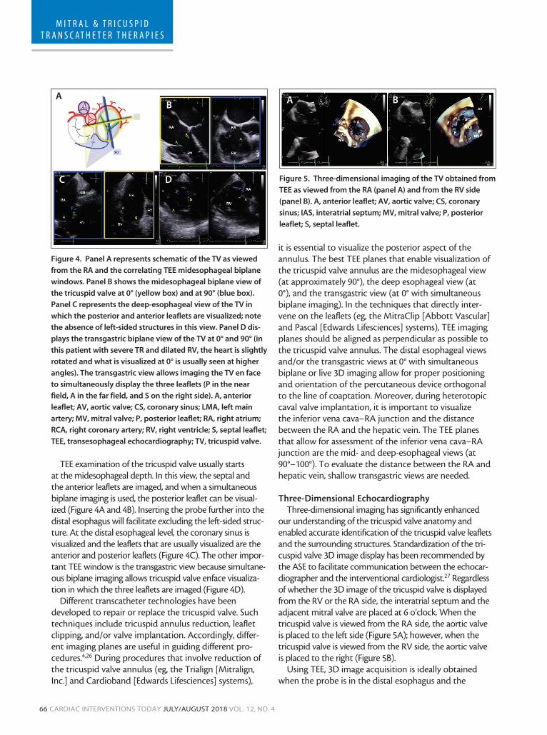
TEE examination of the tricuspid valve usually starts at the midesophageal depth. In this view, the septal and the anterior leaflets are imaged, and when a simultaneous biplane imaging is used, the posterior leaflet can be visual-ized (Figure 4A and 4B). Inserting the probe further into the distal esophagus will facilitate excluding the left-sided struc-ture. At the distal esophageal level, the coronary sinus is visualized and the leaflets that are usually visualized are the anterior and posterior leaflets (Figure 4C). The other impor-tant TEE window is the transgastric view because simultane-ous biplane imaging allows tricuspid valve enface visualiza-tion in which the three leaflets are imaged (Figure 4D).
Different transcatheter technologies have been developed to repair or replace the tricuspid valve. Such techniques include tricuspid annulus reduction, leaflet clipping, and/or valve implantation. Accordingly, differ-ent imaging planes are useful in guiding different pro-cedures.4,26 During procedures that involve reduction of the tricuspid valve annulus (eg, the Trialign [Mitralign, Inc.] and Cardioband [Edwards Lifesciences] systems),
it is essential to visualize the posterior aspect of the annulus. The best TEE planes that enable visualization of the tricuspid valve annulus are the midesophageal view (at approximately 90°), the deep esophageal view (at 0°), and the transgastric view (at 0° with simultaneous biplane imaging). In the techniques that directly inter-vene on the leaflets (eg, the MitraClip [Abbott Vascular] and Pascal [Edwards Lifesciences] systems), TEE imaging planes should be aligned as perpendicular as possible to the tricuspid valve annulus. The distal esophageal views and/or the transgastric views at 0° with simultaneous biplane or live 3D imaging allow for proper positioning and orientation of the percutaneous device orthogonal to the line of coaptation. Moreover, during heterotopic caval valve implantation, it is important to visualize the inferior vena cava–RA junction and the distance between the RA and the hepatic vein. The TEE planes that allow for assessment of the inferior vena cava–RA junction are the mid- and deep-esophageal views (at 90°–100°). To evaluate the distance between the RA and hepatic vein, shallow transgastric views are needed.
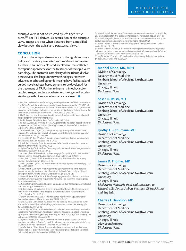
Three-Dimensional EchocardiographyThree-dimensional imaging has significantly enhanced
our understanding of the tricuspid valve anatomy and enabled accurate identification of the tricuspid valve leaflets and the surrounding structures. Standardization of the tri-cuspid valve 3D image display has been recommended by the ASE to facilitate communication between the echocar-diographer and the interventional cardiologist.27 Regardless of whether the 3D image of the tricuspid valve is displayed from the RV or the RA side, the interatrial septum and the adjacent mitral valve are placed at 6 o’clock. When the tricuspid valve is viewed from the RA side, the aortic valve is placed to the left side (Figure 5A); however, when the tricuspid valve is viewed from the RV side, the aortic valve is placed to the right (Figure 5B).
Using TEE, 3D image acquisition is ideally obtained when the probe is in the distal esophagus and the
Figure 4. Panel A represents schematic of the TV as viewed
from the RA and the correlating TEE midesophageal biplane
windows. Panel B shows the midesophageal biplane view of
the tricuspid valve at 0° (yellow box) and at 90° (blue box).
Panel C represents the deep-esophageal view of the TV in
which the posterior and anterior leaflets are visualized; note
the absence of left-sided structures in this view. Panel D dis-
plays the transgastric biplane view of the TV at 0° and 90° (in
this patient with severe TR and dilated RV, the heart is slightly
rotated and what is visualized at 0° is usually seen at higher
angles). The transgastric view allows imaging the TV en face
to simultaneously display the three leaflets (P in the near
field, A in the far field, and S on the right side). A, anterior
leaflet; AV, aortic valve; CS, coronary sinus; LMA, left main
artery; MV, mitral valve; P, posterior leaflet; RA, right atrium;
RCA, right coronary artery; RV, right ventricle; S, septal leaflet;
TEE, transesophageal echocardiography; TV, tricuspid valve.
Figure 5. Three-dimensional imaging of the TV obtained from
TEE as viewed from the RA (panel A) and from the RV side
(panel B). A, anterior leaflet; AV, aortic valve; CS, coronary
sinus; IAS, interatrial septum; MV, mitral valve; P, posterior
leaflet; S, septal leaflet.
AB
C D
A B
VOL. 12, NO. 4 JULY/AUGUST 2018 CARDIAC INTERVENTIONS TODAY 67
M I T R A L & T R I C U S P I D T R A N S C AT H E T E R T H E R A P I E S
tricuspid valve is not obstructed by left-sided struc-tures.4,26 For TTE-derived 3D acquisition of the tricuspid valve, images are best when obtained from a modified view between the apical and parasternal views.4
CONCLUSIONDue to the indisputable evidence of the significant mor-
bidity and mortality associated with moderate and severe TR, there is an undeniable need for effective transcatheter therapeutic approaches for the treatment of tricuspid valve pathology. The anatomic complexity of the tricuspid valve poses several challenges for new technologies. However, advances in echocardiographic imaging have facilitated and guided novel catheter-based systems to be developed for the treatment of TR. Further refinements in echocardio-graphic imaging and transcatheter technologies will acceler-ate the growth of an area of unmet clinical need. n
1. Nath J, Foster E, Heidenreich PA. Impact of tricuspid regurgitation on long-term survival. J Am Coll Cardiol. 2004;43:405-459.2. Lee JW, Song JM, Park JP, et al. Long-term prognosis of isolated significant tricuspid regurgitation. Circ J. 2010;74:375-380.3. Nishimura RA, Otto CM, Bonow RO, et al. 2017 AHA/ACC focused update of the 2014 AHA/ACC guideline for the management of patients with valvular heart disease: a report of the American College of Cardiology/American Heart Association Task Force on Clinical Practice Guidelines. J Am Coll Cardiol. 2017;70:252-289.4. Hahn RT. State-of-the-art review of echocardiographic imaging in the evaluation and treatment of functional tricuspid regurgitation. Circ Cardiovasc Imaging. 2016;9.5. Shah PM, Raney AA. Tricuspid valve disease. Curr Probl Cardiol. 2008;33:47-84.6. Nishimura RA, Otto CM, Bonow RO, et al. 2014 AHA/ACC guideline for the management of patients with valvular heart disease: a report of the American College of Cardiology/American Heart Association Task Force on Practice Guidelines. J Am Coll Cardiol. 2014;63:e57-e185.7. Van de Veire NR, Braun J, Delgado V, et al. Tricuspid annuloplasty prevents right ventricular dilatation and progression of tricuspid regurgitation in patients with tricuspid annular dilatation undergoing mitral valve repair. J Thorac Cardiovasc Surg. 2011;141:1431-1439.8. Dreyfus GD, Corbi PJ, Chan KM, Bahrami T. Secondary tricuspid regurgitation or dilatation: which should be the criteria for surgical repair? Ann Thorac Surg. 2005;79:127-132.9. Ejiofor JI, Neely RC, Yammine M, et al. Surgical outcomes of isolated tricuspid valve procedures: repair versus replacement. Ann Cardiothorac Surg. 2017;6:214-222.10. Alqahtani F, Berzingi CO, Aljohani S, et al. Contemporary trends in the use and outcomes of surgical treatment of tricuspid regurgitation. J Am Heart Assoc. 2017;6.11. Beckmann A, Funkat AK, Lewandowski J, et al. Cardiac surgery in Germany during 2012: a report on behalf of the German Society for Thoracic and Cardiovascular Surgery. Thorac Cardiovasc Surg. 2014;62:5-17.12. Kilic A, Shah AS, Conte JV, Yuh DD. Nationwide outcomes of surgical embolectomy for acute pulmonary embolism. J Thorac Cardiovasc Surg. 2013;145:373-377.13. Hornick P, Harris PA, Taylor KM. Tricuspid valve replacement subsequent to previous open heart surgery. J Heart Valve Dis. 1996;5:20-25.14. Ohno Y, Attizzani GF, Capodanno D, et al. Association of tricuspid regurgitation with clinical and echocar-diographic outcomes after percutaneous mitral valve repair with the MitraClip System: 30-day and 12-month follow-up from the GRASP Registry. Eur Heart J Cardiovasc Imaging. 2014;15:1246-1255.15. Lindman BR, Maniar HS, Jaber WA, et al. Effect of tricuspid regurgitation and the right heart on survival after transcatheter aortic valve replacement: insights from the Placement of Aortic Transcatheter Valves II inoperable cohort. Circ Cardiovasc Interv. 2015;8.16. Martinez RM, O’Leary PW, Anderson RH. Anatomy and echocardiography of the normal and abnormal tricuspid valve. Cardiol Young. 2006;16(suppl 3):4-11.17. Stankovic I, Daraban AM, Jasaityte R, et al. Incremental value of the en face view of the tricuspid valve by two-dimensional and three-dimensional echocardiography for accurate identification of tricuspid valve leaflets. J Am Soc Echocardiogr. 2014;27:376-384.18. Fawzy H, Fukamachi K, Mazer CD, et al. Complete mapping of the tricuspid valve apparatus using three-dimensional sonomicrometry. J Thorac Cardiovasc Surg. 2011;141:1037-1043.19. Fukuda S, Saracino G, Matsumura Y, et al. Three-dimensional geometry of the tricuspid annulus in healthy subjects and in patients with functional tricuspid regurgitation: a real-time, 3-dimensional echocardiographic study. Circulation. 2006;114:I492-498.20. Rudski LG, Lai WW, Afilalo J, et al. Guidelines for the echocardiographic assessment of the right heart in adults: a report from the American Society of Echocardiography endorsed by the European Association of Echocardiogra-phy, a registered branch of the European Society of Cardiology, and the Canadian Society of Echocardiography. J Am Soc Echocardiogr. 2010;23:685-713; quiz 786-788.21. Zoghbi WA, Adams D, Bonow RO, et al. Recommendations for noninvasive evaluation of native valvular regurgitation: a report from the American Society of Echocardiography developed in collaboration with the Society for Cardiovascular Magnetic Resonance. J Am Soc Echocardiogr. 2017;30:303-371.22. Lang RM, Badano LP, Mor-Avi V, et al. Recommendations for cardiac chamber quantification by echocar-diography in adults: an update from the American Society of Echocardiography and the European Association of Cardiovascular Imaging. J Am Soc Echocardiogr. 2015;28:1-39.e14.
23. Addetia K, Yamat M, Mediratta A, et al. Comprehensive two-dimensional interrogation of the tricuspid valve using knowledge derived from three-dimensional echocardiography. J Am Soc Echocardiogr. 2016;29:74-82.24. Anwar AM, Geleijnse ML, Soliman OI, et al. Assessment of normal tricuspid valve anatomy in adults by real-time three-dimensional echocardiography. Int J Cardiovasc Imaging. 2007;23:717-724.25. Hahn RT, Zamorano JL. The need for a new tricuspid regurgitation grading scheme. Eur Heart J Cardiovasc Imaging. 2017;18:1342-1343.26. Hahn RT, Abraham T, Adams MS, et al. Guidelines for performing a comprehensive transesophageal echo-cardiographic examination: recommendations from the American Society of Echocardiography and the Society of Cardiovascular Anesthesiologists. J Am Soc Echocardiogr. 2013;26:921-964.27. Lang RM, Mor-Avi V, Sugeng L, et al. Three-dimensional echocardiography: the benefits of the additional dimension. J Am Coll Cardiol. 2006;48:2053-2069.
Menhel Kinno, MD, MPHDivision of CardiologyDepartment of MedicineFeinberg School of Medicine Northwestern UniversityChicago, IllinoisDisclosures: None.
Sasan R. Raissi, MDDivision of CardiologyDepartment of MedicineFeinberg School of Medicine Northwestern UniversityChicago, IllinoisDisclosures: None.
Jyothy J. Puthumana, MDDivision of CardiologyDepartment of MedicineFeinberg School of Medicine Northwestern UniversityChicago, IllinoisDisclosures: None.
James D. Thomas, MDDivision of CardiologyDepartment of MedicineFeinberg School of Medicine Northwestern UniversityChicago, IllinoisDisclosures: Honoraria from and consultant to Edwards Lifesciences, Abbott Vascular, GE Healthcare, and Bay Labs.
Charles J. Davidson, MDDivision of CardiologyDepartment of MedicineFeinberg School of Medicine Northwestern UniversityChicago, [email protected]: None.