journal of ginseng research
TRANSCRIPT

Q8
Q2
lable at ScienceDirect
J Ginseng Res xxx (2018) 1e12
123456789101112131415161718192021222324252627282930313233343536373839404142434445464748495051525354
55
JGR353_proof ■ 25 April 2018 ■ 1/12
Contents lists avai
Journal of Ginseng Research
journal homepage: ht tp: / /www.ginsengres.org
565758596061626364
Research Article 656667686970717273Evaluation of the gastroprotective effects of 20 (S)-ginsenoside Rg3 ongastric ulcer models in mice
Kai Zhang 1,2, Ying Liu 2, Cuizhu Wang 1, Jiannan Li 2, Lingxin Xiong 1, Zhenzhou Wang 1,Jinping Liu 1,**, Pingya Li 1,*1 School of Pharmaceutical Sciences, Jilin University, Changchun 130021, China2Department of General Surgery, The Second Hospital of Jilin University, Changchun, Jilin 130041, China
7475
76777879808182838485868788a r t i c l e i n f o
Article history:Received 7 October 2017Received in Revised form15 January 2018Accepted 3 April 2018Available online xxx
Keywords:AntiinflammationAntioxidant20 (S)-ginsenoside Rg3Gastric ulcerUlcer index
* Corresponding author. School of Pharmaceutical S** Corresponding author. School of Pharmaceutical S
E-mail addresses: [email protected] (J. Liu), lipy@jlu
https://doi.org/10.1016/j.jgr.2018.04.001p1226-8453 e2093-4947/$ e see front matter � 2018license (http://creativecommons.org/licenses/by-nc-n
Please cite this article in press as: Zhang K, emice, Journal of Ginseng Research (2018), h
899091929394959697
a b s t r a c t
Background: Gastric ulcer (GU) is a common gastrointestinal disease that can be induced by many fac-tors. Finding an effective treatment method that contains fewer side effects is important. 20 (S)-ginse-noside Rg3 is a kind of protopanaxadiol and has shown superior antiinflammatory and antioxidanteffects in many studies, especially cancer studies. In this study, we examined the treatment efficacy of 20(S)-ginsenoside Rg3 on GU.Methods: Three kinds of GU models, including an alcohol GU model, a pylorus-ligated GU model, and anacetic acid GU model, were used. Mouse endothelin-1 (ET-1) and nitric oxide (NO) levels in blood andepidermal growth factor (EGF), superoxide dismutase, and NO levels in gastric mucosa were evaluated.Hematoxylin and eosin staining of gastric mucosa and immunohistochemical staining of ET-1, induciblenitric oxide synthase (NOS2), and epidermal growth factor receptors were studied. Ulcer index (UI)scores and UI ratios were also analyzed to demonstrate the GU conditions in different groups. Further-more, Glide XP from Schrödinger was used for molecular docking to clarify the interactions between 20(S)-ginsenoside Rg3 and EGF and NOS2.Results: 20 (S)-ginsenoside Rg3 significantly decreased the UI scores and UI ratios in all the three GUmodels, and it demonstrated antiulcer effects by decreasing the ET-1 and NOS2 levels and increasing theNO, superoxide dismutase, EGF, and epidermal growth factor receptor levels. In addition, high-dose 20(S)-ginsenoside Rg3 showed satisfactory gastric mucosa protection effects.Conclusion: 20 (S)-ginsenoside Rg3 can inhibit the formation of GU and may be a potential therapeuticagent for GU.� 2018 The Korean Society of Ginseng, Published by Elsevier Korea LLC. This is an open access article
under the CC BY-NC-ND license (http://creativecommons.org/licenses/by-nc-nd/4.0/).
98
99100101102103104105106107108109110111112
1. Introduction
Gastric ulcer (GU) is one of the major gastrointestinal diseasesthat occur on the surface of the gastric mucosa. It is a kind of localtissue necrosis because of the formation of ulceration lesions. Inrecent years, the incidence of gastric ulcer increases because ofmany external damaging factors, such as smoking, excessivedrinking, stress, poor diet, and long-term ingestion of nonste-roidal antiinflammation drugs (NSAIDs) [1]. Generally, theimbalance of the damage to the gastric mucosa caused by exog-enous factors and the mucosal self-repair effect lead to the inci-dence of GU.
ciences, Jilin University, Fujin Roaciences, Jilin University, Fujin Roa.edu.cn (P. Li).
The Korean Society of Ginseng, Pud/4.0/).
t al., Evaluation of the gastropttps://doi.org/10.1016/j.jgr.201
Gastric mucosa can be damaged by excessive intake of alcohol.In detail, alcohol can lead to direct injury of mucosa vascularendothelial cells, disrupt the cells continuity, induce the formationof reactive oxygen radicals and inflammatory cytokines, and causelocal ischemia of the gastric mucosa [2]. In addition, most of theNSAIDs, such as aspirin and diclofenac sodium, are very commonfactors in GU formation. The NSAIDs are mainly maintained in anonionic state in the gastric fluid and can easily pass through thegastric mucosa cell membrane and accumulate within cells. Highconcentrations of NSAIDs can change the permeability of the cellmembrane, which may lead to edema, degeneration, necrosis, andshedding of gastric mucosal epithelial cells. As a result, the barrier
d 1266, Changchun 130021, China.d 1266, Changchun 130021, China.
blished by Elsevier Korea LLC. This is an open access article under the CC BY-NC-ND
rotective effects of 20 (S)-ginsenoside Rg3 on gastric ulcer models in8.04.001
113114115116117118119

J Ginseng Res 2018;-:1e122
1234567891011121314151617181920212223242526272829303132333435363738394041424344454647484950515253545556575859606162636465
66676869707172737475767778798081828384858687888990919293949596979899
100101102103104105106107108109110111112113114115116117118119120121122123124125126127128129130
JGR353_proof ■ 25 April 2018 ■ 2/12
effect of the gastric mucosa is damaged, and GU forms [3].Furthermore, poor dieting and many kinds of stress can also lead togastric hyperacidity and local blood supply disorders of the gastrictissue, which can easily cause GU formation. Therefore, drugs thatpossess the ability to reduce the amounts of inflammatory andoxidative stress and protect the gastric mucosa from local ischemiainjury can be used in the treatment of GU.
Currently, the most common drugs used in GU treatment areproton pump inhibitors and H2 receptor antagonists. However,the associated undesirable side effects and the recurrence of GUafter treatment have attracted increasing attention [4]. Thus, it isstill necessary and important to search for an ideal antiulcer drugthat possesses fewer side effects and can protect the gastric mu-cosa well. Panax notoginseng, also known as Tianqi or Sanqi inChinese, is a traditional Chinese medicine that has been used for along time because of its multiple pharmacological effects [5]. Ithas been reported that Yunnan Baiyao, in which the main ingre-dient is P. notoginseng, was efficient in treating uterine hemor-rhaging, ulcerative colitis, and skin ulcers [6]. The bioactivecomponents of P. notoginseng, including many kinds ofprotopanaxadiol-type saponins, are believed to be ginseno-sides [7]. Previous studies have shown that 20 (S)-ginsenosideRg3, a deglycosylated derivative of the ginsenoside Rb3, exertsobvious antiinflammation, antiischemia, and antioxidative stresseffects [8e11]. Based on these findings, we aimed to explore theanti-GU effects of ginsenoside Rg3 in mice. In this study, three GUmodels, alcohol, pylorus-ligated, and acetic acid, were used tosystematically study the GU treatment efficacy of 20 (S)-ginse-noside Rg3.
2. Materials and methods
2.1. Materials
20 (S)-ginsenoside Rg3 (C42H72O13, HPLC > 98%) was pur-chased from Jilin Yatai Pharmaceutical Co., Ltd. (Jilin, China).Cimetidinewas bought from Sigma (St. Louis, MO, USA) and used asthe model control in this study. Mouse endothelin-1 (ET-1), nitricoxide (NO), epidermal growth factor (EGF), and superoxide dis-mutase (SOD) enzymeelinked immunosorbent assay (ELISA) kitswere purchased from Longton Co. Ltd. (Shanghai, P. R. China). Theantibodies of ET-1, inducible nitric oxide synthase (NOS2), andepidermal growth factor receptor (EGFR) for immunohistochemicalstaining were bought from Abcam (Cambridge, USA).
2.2. Animals
The animal experiments were conducted based on the guide forthe administration of laboratory animals (Directive 86/609/EEC inthe Protection of Animals Used for Experimental and Other Scien-tific Purposes, 1986) and were approved by the Institutional AnimalCare and Use Committee of Jilin University (No. SCXK-2013-0001).Male Wistar rats, weighing 180e220 g, were obtained from theLaboratory Animal Center of Jilin University. For each GUmodel, theanimals were divided into six groups: the blank control group,model control group, cimetidine group, low-dose Rg3 group (L-Rg3), moderate-dose Rg3 group (M-Rg3), and high-dose Rg3 group(H-Rg3). Ten animals were included in each group. For rats in theblank control andmodel control groups, intragastric administrationof 10 mL/kg/day 0.9% saline solution was performed. For rats in thecimetidine group, intragastric administration of 2 mg/kg/daycimetidine was performed. For rats in the L-Rg3, M-Rg3, and H-Rg3groups, intragastric administrations of 5 mg/kg/day, 10 mg/kg/day,and 20 mg/kg/day 20 (S)-ginsenoside Rg3 were performed,respectively.
Please cite this article in press as: Zhang K, et al., Evaluation of the gastropmice, Journal of Ginseng Research (2018), https://doi.org/10.1016/j.jgr.20
2.3. Establishment of GU
For the alcohol GU model, intragastric administration was per-formed for 7 days for each group. Half an hour after the lastintragastric administration, the rats were orally treated with 5 mL/kg alcohol (Beijing Chemical Works, Beijing, P.R. China), except forthe animals in the blank control group. One hour later, the rats werekilled by cervical dislocation, and the gastric tissue was harvestedfor further evaluation.
For the pylorus-ligated GUmodel, intragastric administration ofdifferent samples was also first performed for 7 days. The rats werefasted for 24 hrs after the last intragastric administration. Then,they were anesthetized with a pentobarbital sodium (50 mg/kg)(J&K Technology Co., Ltd., Beijing, P.R. China) intraperitoneal in-jection, were fixed, and underwent laparotomy. The stomach pyloriwas exposed and ligated by surgical sutures, followed by theabdomen suturing layer by layer. Eighteen hours later, the rats werealso killed, and their stomachs were collected.
For the acetic acid GUmodel, rats were first fasted for 24 hrs andthen were anesthetized, were fixed, and underwent laparotomy.The stomach was exposed, and 0.3 mL of acetic acid (J&K Tech-nology Co., LTD) was submucosally injected at the junction of thestomach body and pyloric sinus. The stomach was embedded in thelarge omentum, and the abdomen was sutured. The animals werethen treated with intragastric administration of different samplesfor 7 days. One hour after the last treatment, the rats were killed,and the stomach was harvested, as described previously.
2.4. Body weight measurement
To evaluate the toxicity of Rg3, the body weights of rats in eachanimal model were measured. For animals in all the three GUmodels, their body weights were recorded before each intragastricadministration for 7 days.
2.5. Measurement of ET-1 and NO in blood
In each animal model, blood samples were collected from theheart before sacrifice, and the serum was obtained by centrifuga-tion at 4,000 g for 10 min and then stored at �80�C. Levels of ET-1and NO were analyzed using ELISA kits.
2.6. Gross evaluation of stomach mucosa
As mentioned previously, after the sacrifice of the animals,stomach tissues were collected and washed cleanwith a 0.9% salinesolution. The stomach was opened along the greater curvature, andthe stomach mucosa underwent gross evaluation for any signs ofhyperemia, hemorrhage, and ulcers. Ulcer index (UI) scoring wasperformed according to a previous study [12]. Scoring details are asfollows: 0 ¼ normal stomach; 0.5e1 ¼ mucosa congestion; 1e2¼ hemorrhage; 2e3¼ one to five small ulcers; 3e4 ¼many smallulcers; 4e5 ¼ one to five small and one to three large ulcers; 5e6¼many small and large ulcers; 6e7¼ full of ulcers. UI ratios werealso performed to analyze the antiulcer efficiency of Rg3. The UI ofthe control group was defined as “1”, and the UI ratio was definedas the ratios of the ulcer indexes of treated samples and the modelcontrol group. In addition, the percentage of inhibition was alsocalculated as follows: [(UI model control group� UI treated group)/UI model control group] � 100%.
2.7. Measurement of EGF, SOD, and NO in the gastric mucosa
After gross evaluation of the GUs, the gastric mucosa wascollected and homogenized. Then, the supernatant was obtained by
rotective effects of 20 (S)-ginsenoside Rg3 on gastric ulcer models in18.04.001

Q3Q4
Q1K. Zhang et al / Evaluation of the gastroprotective effects of 20 (S)-ginsenoside Rg3 3
1234567891011121314151617181920212223242526272829303132333435363738394041424344454647484950515253545556575859606162636465
66676869707172737475767778798081828384858687888990919293949596979899
JGR353_proof ■ 25 April 2018 ■ 3/12
centrifugation at 2,500 g for 10 min, and ELISA kits were used toanalyze the levels of EGF, SOD, and NO in the gastric mucosa.
2.8. Histological and immunohistochemical analyses
The stomach ulcer tissues were rinsed with phosphate bufferedsaline, fixed in 4% (W/V) phosphate buffered salineebufferedparaformaldehyde, and finally embedded in paraffin. The tissueswere serially sectioned at 5.0-mm intervals and stained with he-matoxylin and eosin (H&E). ET-1, NOS2, and EGFR immunohisto-chemical stainingwere also performed to analyze the GU treatmentefficacy of different samples. The positive stained cells wererecorded, and the percentage occupying the total counted cells wascalculated.
2.9. Molecular docking of 20 (S)-ginsenoside Rg3
To clarify the mode of action of 20 (S)-ginsenoside Rg3 on EGFand NOS2, a molecular docking study was carried out to measurethe relative binding energies and localized binding sites in theactive pocket. The study was performed using GLIDE (Grid-basedLigand Docking with Energetics) (GLIDE, version 6.7, Schrödinger,LLC, New York, USA, 2015) software developed by Schrödinger.Maestro Elements (2015-2) was used for all the steps involvingprotein and ligand preparation, receptor grid generation, anddocking. The X-ray crystal structure of EGF [Protein Data Bank(PDB) code: 3RCD] and NOS2 (PDB code: 3EAI) were retrieved fromthe PDB database (http://www.rcsb.org/pdb) according to previousstudies [13e15].
The Protein Preparation Wizard in the GLIDE software was usedto prepare the receptors. The structures of EGF and NOS2 wereoptimized after a series of processes, including assigning bond or-ders and water orentations, removing water, adding hydrogen, andcreating zero-order bonds to metals and disulphide bonds [16].
Fig. 1. Body weights of animals in (A) alcohol, (B) pylorus-ligated, (C) and acetic acid GU mogroup, *p < 0.05, **p < 0.01, ***p < 0.001).GU, gastric ulcer; H-Rg3, high-dose Rg3 group; L-Rg3, low-dose Rg3 group; M-Rg3, moder
Please cite this article in press as: Zhang K, et al., Evaluation of the gastropmice, Journal of Ginseng Research (2018), https://doi.org/10.1016/j.jgr.201
Crystal coordinates of 20 (S)-ginsenoside Rg3 (ligand) werepredrawn in Maestro Elements (Maestro Elements, 2.2) before thismolecular docking study. Three-dimensional structure of thecompound was generated using LigPrep module (2015-2) ofSchrödinger Suite by assigning the bond orders and angles. Inaddition, the ligand was subjected tominimization using the OPLS3force field. For GLIDE docking, the prepared structure of EGF, NOS2,and ligand (Rg3) were imported to the workspace using GLIDE v6.7from Schrödinger Suite [17e19]. Extra precision (XP) docking wascarried out, and the parameters of scaling factor and partial chargecutoff were set at the default values 0.80 and 0.15, respectively [20].Figures of the docking results were subsequently prepared usingPyMOL (Schrödinger).
2.10. Statistical analyses
The results were presented as the mean � standard deviation.Data were analyzed using GraphPad Prism 7.0 software (GraphPadInc., San Diego, CA) using Student t test. Statistical significance wasset as *p < 0.05, and high statistical significance was set as**p < 0.01 and ***p < 0.001.
3. Results
3.1. Body weight change
For animals in alcohol and pylorus-ligated GUmodels, the bodyweights gradually increased, and there was no statistical differenceamong all the groups (Figs.1A,1B). For animals in the acetic acid GUmodel, the bodyweights of rats in themodel control group severelydecreased. But the body weights of the animals in the other groupsslightly decreased during the first 3 or 4 days and then graduallyincreased (Fig. 1C). At the 7th day after treatment, there was
Q7dels. Data are presented as the mean � SD (n ¼ 10; compared with the model control
ate-dose Rg3 group; SD, standard deviation.
rotective effects of 20 (S)-ginsenoside Rg3 on gastric ulcer models in8.04.001
100101102103104105106107108109110111112113114115116117118119120121122123124125126127128129130

J Ginseng Res 2018;-:1e124
1234567891011121314151617181920212223242526272829303132333435363738394041424344454647484950515253545556575859606162636465
6667686970717273747576777879
JGR353_proof ■ 25 April 2018 ■ 4/12
significant difference between the model control group and H-Rg3group in animal body weights (p < 0.001).
3.2. ET-1, NO, EGF, and SOD levels
To assess the GU inhibition efficacy of 20 (S)-ginsenoside Rg3,ET-1 and NO levels in the blood and EGF, SOD, and NO levels in thegastric mucosa were evaluated in the three GU models.
Fig. 2 shows ET-1, NO, EGF, and SOD levels in the alcohol GUmodel. The results indicated that the levels of ET-1 decreased as theamounts of 20 (S)-ginsenoside Rg3 increased, and the blood ET-1levels decreased greatly in the H-Rg3 group compared with thoseof the model control group (p < 0.001). 20 (S)-ginsenoside Rg3 alsochanged the blood NO levels significantly. However, when treated
Fig. 2. (A) ET-1 and (B) NO levels in blood and (C) EGF, (D) SOD, and (E) NO levels in the gcompared with the blank control group, #p < 0.05, ##p < 0.01, ###p < 0.001; compared wEGF, epidermal growth factor; ET-1, endothelin-1; GU, gastric ulcer; H-Rg3, high-dose Rg3 gSD, standard deviation; SOD, superoxide dismutase.
Please cite this article in press as: Zhang K, et al., Evaluation of the gastropmice, Journal of Ginseng Research (2018), https://doi.org/10.1016/j.jgr.20
with low-dose 20 (S)-ginsenoside Rg3, the amounts of NO did notincrease compared with those of the model control group. How-ever, as for the moderate- and high-dose 20 (S)-ginsenoside Rg3etreated groups, the blood NO levels increased in comparison withthose of the model control group (p < 0.05). Similarly, only H-Rg3group increased the mucosa NO levels compared with the modelcontrol group (p < 0.05). Interestingly, cimetidine did not changethe EGF levels in the mucosa tissues in this animal model, and EGFlevels only increased in the H-Rg3 groups compared with themodel control group (p < 0.05). It was obvious that 20 (S)-ginse-noside Rg3 was efficient in changing SOD levels in the gastricmucosa tissues. SOD levels increased significantly in all 20 (S)-ginsenoside Rg3etreated groups in comparison with those of themodel control group.
astric mucosa in the alcohol GU model. Data are presented as the mean � SD (n ¼ 10;ith the model control group, *p < 0.05, **p < 0.01, ***p < 0.001).roup; L-Rg3, low-dose Rg3 group; M-Rg3, moderate-dose Rg3 group; NO, nitric oxide;
rotective effects of 20 (S)-ginsenoside Rg3 on gastric ulcer models in18.04.001
8081828384858687888990919293949596979899
100101102103104105106107108109110111112113114115116117118119120121122123124125126127128129130
K. Zhang et al / Evaluation of the gastroprotective effects of 20 (S)-ginsenoside Rg3 5
1234567891011121314151617181920212223242526272829303132333435363738394041424344454647484950515253545556575859606162636465
666768697071727374757677787980
JGR353_proof ■ 25 April 2018 ■ 5/12
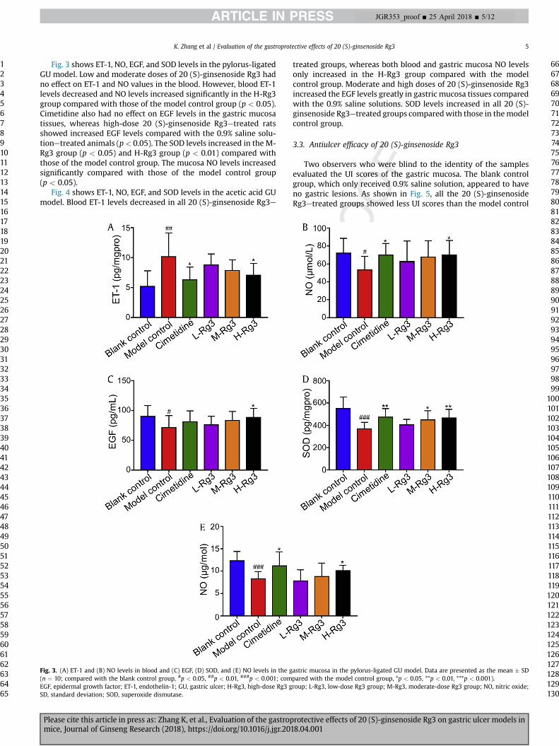
Fig. 3 shows ET-1, NO, EGF, and SOD levels in the pylorus-ligatedGU model. Low and moderate doses of 20 (S)-ginsenoside Rg3 hadno effect on ET-1 and NO values in the blood. However, blood ET-1levels decreased and NO levels increased significantly in the H-Rg3group compared with those of the model control group (p < 0.05).Cimetidine also had no effect on EGF levels in the gastric mucosatissues, whereas high-dose 20 (S)-ginsenoside Rg3etreated ratsshowed increased EGF levels compared with the 0.9% saline solu-tionetreated animals (p< 0.05). The SOD levels increased in the M-Rg3 group (p < 0.05) and H-Rg3 group (p < 0.01) compared withthose of the model control group. The mucosa NO levels increasedsignificantly compared with those of the model control group(p < 0.05).
Fig. 4 shows ET-1, NO, EGF, and SOD levels in the acetic acid GUmodel. Blood ET-1 levels decreased in all 20 (S)-ginsenoside Rg3e
Fig. 3. (A) ET-1 and (B) NO levels in blood and (C) EGF, (D) SOD, and (E) NO levels in the g(n ¼ 10; compared with the blank control group, #p < 0.05, ##p < 0.01, ###p < 0.001; comEGF, epidermal growth factor; ET-1, endothelin-1; GU, gastric ulcer; H-Rg3, high-dose Rg3 gSD, standard deviation; SOD, superoxide dismutase.
Please cite this article in press as: Zhang K, et al., Evaluation of the gastropmice, Journal of Ginseng Research (2018), https://doi.org/10.1016/j.jgr.201
treated groups, whereas both blood and gastric mucosa NO levelsonly increased in the H-Rg3 group compared with the modelcontrol group. Moderate and high doses of 20 (S)-ginsenoside Rg3increased the EGF levels greatly in gastric mucosa tissues comparedwith the 0.9% saline solutions. SOD levels increased in all 20 (S)-ginsenoside Rg3etreated groups comparedwith those in themodelcontrol group.
3.3. Antiulcer efficacy of 20 (S)-ginsenoside Rg3
Two observers who were blind to the identity of the samplesevaluated the UI scores of the gastric mucosa. The blank controlgroup, which only received 0.9% saline solution, appeared to haveno gastric lesions. As shown in Fig. 5, all the 20 (S)-ginsenosideRg3etreated groups showed less UI scores than the model control
astric mucosa in the pylorus-ligated GU model. Data are presented as the mean � SDpared with the model control group, *p < 0.05, **p < 0.01, ***p < 0.001).roup; L-Rg3, low-dose Rg3 group; M-Rg3, moderate-dose Rg3 group; NO, nitric oxide;
rotective effects of 20 (S)-ginsenoside Rg3 on gastric ulcer models in8.04.001
81828384858687888990919293949596979899
100101102103104105106107108109110111112113114115116117118119120121122123124125126127128129130

Fig. 4. (A) ET-1 and (B) NO levels in blood and (C) EGF, (D) SOD, and (E) NO levels in the gastric mucosa in the acetic acid GU model. Data are presented as the mean � SD (n ¼ 10;compared with the blank control group, #p < 0.05, ##p < 0.01, ###p < 0.001; compared with the model control group, *p < 0.05, **p < 0.01, ***p < 0.001).EGF, epidermal growth factor; ET-1, endothelin-1; GU, gastric ulcer; H-Rg3, high-dose Rg3 group; L-Rg3, low-dose Rg3 group; M-Rg3, moderate-dose Rg3 group; NO, nitric oxide;SD, standard deviation; SOD, superoxide dismutase.
J Ginseng Res 2018;-:1e126
1234567891011121314151617181920212223242526272829303132333435363738394041424344454647484950515253545556575859606162636465
66676869707172737475767778798081828384858687888990919293949596979899
100101102103104105106107108109110111112113114115116117118119120121122123124125126127128129
JGR353_proof ■ 25 April 2018 ■ 6/12
group in these three GU models. The M-Rg3 and H-Rg3 groupsgreatly decreased the UI scores when compared with the modelcontrol group (p < 0.001). The result of the UI ratios was similar tothat of UI scores. However, there were no differences in UI ratiosbetween L-Rg3 and model control groups in the alcohol GU modeland acetic acid GUmodel. In addition, the inhibition rates (%) of GUin the cimetidine, L-Rg3, M-Rg3, and H-Rg3 groups were 35.1%,16.2%, 44.1%, and 63.1% in the alcohol GU model; 52.8%, 26.4%,48.8%, and 64.8% in the pylorus-ligated GU model; and 38.2%, 8.8%,42.2%, and 62.7% in the acetic acid GU model, respectively. The GUwas inhibited better with increases in the administration of 20 (S)-ginsenoside Rg3. High-dose 20 (S)-ginsenoside Rg3 showed betterGU prevention efficacy.
Please cite this article in press as: Zhang K, et al., Evaluation of the gastropmice, Journal of Ginseng Research (2018), https://doi.org/10.1016/j.jgr.20
3.4. Effects of 20 (S)-ginsenoside Rg3 on histopathological changesof the gastric mucosa
Histopathological alterations of the gastric mucosa of differentgroups in the alcohol GU model, pylorus-ligated GU model, andacetic acid GU model are shown in Fig. 6. Normal stomach tissues,including the mucosa, submucosa, muscular layer, and serosa, wereobvious in the blank control group in all the three GU models. Theanimals in the model control group were only treated with 0.9%saline solution, and severe histopathological changes wereobserved in the gastric specimens. H&E staining of the stomachtissues of the model control group showed large ulcer-inducedmucosa lesions, large amounts of inflammation responses, gastric
rotective effects of 20 (S)-ginsenoside Rg3 on gastric ulcer models in18.04.001
130

Fig. 5. Ulcer index scores and ulcer index ratios of (A and D) the alcohol GUmodel, (B and E) pylorus-ligated GUmodel, and (C and F) acetic acid GUmodel. Data are presented as themean � SD (n ¼ 10; *p < 0.05, **p < 0.01, ***p < 0.001).GU, gastric ulcer; H-Rg3, high-dose Rg3 group; L-Rg3, low-dose Rg3 group; M-Rg3, moderate-dose Rg3 group; SD, standard deviation.
K. Zhang et al / Evaluation of the gastroprotective effects of 20 (S)-ginsenoside Rg3 7
1234567891011121314151617181920212223242526272829303132333435363738394041424344454647484950515253545556575859606162636465
66676869707172737475767778798081828384858687888990919293949596979899
100101102103104105106107108109110
JGR353_proof ■ 25 April 2018 ■ 7/12
pit cell damages, mucosa congestion and edema, and evenmuscular layer injuries. When the rats were treated with low-dose20 (S)-ginsenoside Rg3, even though the mucosa injuries were notas obvious as those of the model control group, we could stillobserve small or large mucosal lesions, mucosal congestion andedema, and inflammation responses. Similar results were observedin the cimetidine and M-Rg3 groups in which only some smallmucosa lesions with slight inflammation responses were found.High-dose 20 (S)-ginsenoside Rg3 improved these gastric alter-ations and only resulted in some very small mucosa lesions.
Fig. 6. H&E staining of the gastric mucosa of different groups in the alcohol GU model, pylGU, gastric ulcer; H&E, hematoxylin and eosin; H-Rg3, high-dose Rg3 group; L-Rg3, low-do
Please cite this article in press as: Zhang K, et al., Evaluation of the gastropmice, Journal of Ginseng Research (2018), https://doi.org/10.1016/j.jgr.201
3.5. Effects of 20 (S)-ginsenoside Rg3 on immunohistochemicalchanges of ET-1, NOS2, and EGFR
ET-1 is known to be one of the most effective materials for thecontraction of blood vessels, and it is essential for GU formation.Fig. 7A shows the immunohistochemical staining of ET-1 ofdifferent groups in the three GU models. The results indicatedthat compared with the blank control group, the model controlgroup expressed greater amounts of ET-1 (p< 0.001) (Figs. 7Be7D).The cimetidine group showed less ET-1 levels than the model
orus-ligated GU model, and acetic acid GU model (�40).se Rg3 group; M-Rg3, moderate-dose Rg3 group.
rotective effects of 20 (S)-ginsenoside Rg3 on gastric ulcer models in8.04.001
111112113114115116117118119120121122123124125126127128129130
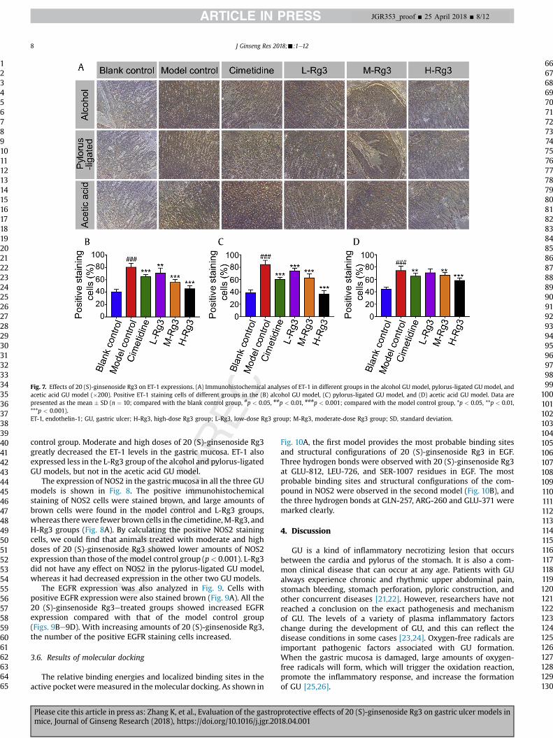
Fig. 7. Effects of 20 (S)-ginsenoside Rg3 on ET-1 expressions. (A) Immunohistochemical analyses of ET-1 in different groups in the alcohol GU model, pylorus-ligated GU model, andacetic acid GU model (�200). Positive ET-1 staining cells of different groups in the (B) alcohol GU model, (C) pylorus-ligated GU model, and (D) acetic acid GU model. Data arepresented as the mean � SD (n ¼ 10; compared with the blank control group, #p < 0.05, ##p < 0.01, ###p < 0.001; compared with the model control group, *p < 0.05, **p < 0.01,***p < 0.001).ET-1, endothelin-1; GU, gastric ulcer; H-Rg3, high-dose Rg3 group; L-Rg3, low-dose Rg3 group; M-Rg3, moderate-dose Rg3 group; SD, standard deviation.
J Ginseng Res 2018;-:1e128
1234567891011121314151617181920212223242526272829303132333435363738394041424344454647484950515253545556575859606162636465
66676869707172737475767778798081828384858687888990919293949596979899
100101102103104105106107108109110111112113114115116117118119120121122123124125126127128129130
JGR353_proof ■ 25 April 2018 ■ 8/12
control group. Moderate and high doses of 20 (S)-ginsenoside Rg3greatly decreased the ET-1 levels in the gastric mucosa. ET-1 alsoexpressed less in the L-Rg3 group of the alcohol and pylorus-ligatedGU models, but not in the acetic acid GU model.
The expression of NOS2 in the gastric mucosa in all the three GUmodels is shown in Fig. 8. The positive immunohistochemicalstaining of NOS2 cells were stained brown, and large amounts ofbrown cells were found in the model control and L-Rg3 groups,whereas therewere fewer brown cells in the cimetidine,M-Rg3, andH-Rg3 groups (Fig. 8A). By calculating the positive NOS2 stainingcells, we could find that animals treated with moderate and highdoses of 20 (S)-ginsenoside Rg3 showed lower amounts of NOS2expression than those of themodel control group (p< 0.001). L-Rg3did not have any effect on NOS2 in the pylorus-ligated GU model,whereas it had decreased expression in the other two GU models.
The EGFR expression was also analyzed in Fig. 9. Cells withpositive EGFR expression were also stained brown (Fig. 9A). All the20 (S)-ginsenoside Rg3etreated groups showed increased EGFRexpression compared with that of the model control group(Figs. 9Be9D). With increasing amounts of 20 (S)-ginsenoside Rg3,the number of the positive EGFR staining cells increased.
3.6. Results of molecular docking
The relative binding energies and localized binding sites in theactive pocketweremeasured in themolecular docking. As shown in
Please cite this article in press as: Zhang K, et al., Evaluation of the gastropmice, Journal of Ginseng Research (2018), https://doi.org/10.1016/j.jgr.20
Fig. 10A, the first model provides the most probable binding sitesand structural configurations of 20 (S)-ginsenoside Rg3 in EGF.Three hydrogen bonds were observed with 20 (S)-ginsenoside Rg3at GLU-812, LEU-726, and SER-1007 residues in EGF. The mostprobable binding sites and structural configurations of the com-pound in NOS2 were observed in the second model (Fig. 10B), andthe three hydrogen bonds at GLN-257, ARG-260 and GLU-371 weremarked clearly.
4. Discussion
GU is a kind of inflammatory necrotizing lesion that occursbetween the cardia and pylorus of the stomach. It is also a com-mon clinical disease that can occur at any age. Patients with GUalways experience chronic and rhythmic upper abdominal pain,stomach bleeding, stomach perforation, pyloric construction, andother concurrent diseases [21,22]. However, researchers have notreached a conclusion on the exact pathogenesis and mechanismof GU. The levels of a variety of plasma inflammatory factorschange during the development of GU, and this can reflect thedisease conditions in some cases [23,24]. Oxygen-free radicals areimportant pathogenic factors associated with GU formation.When the gastric mucosa is damaged, large amounts of oxygen-free radicals will form, which will trigger the oxidation reaction,promote the inflammatory response, and increase the formationof GU [25,26].
rotective effects of 20 (S)-ginsenoside Rg3 on gastric ulcer models in18.04.001
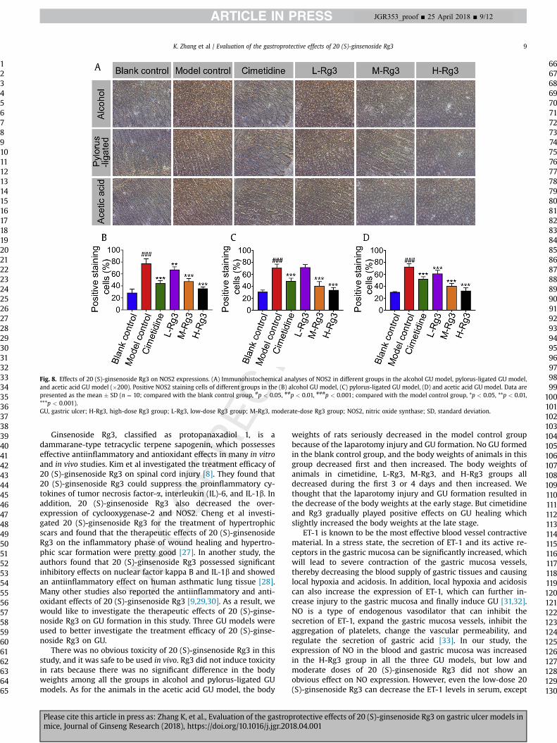
Fig. 8. Effects of 20 (S)-ginsenoside Rg3 on NOS2 expressions. (A) Immunohistochemical analyses of NOS2 in different groups in the alcohol GU model, pylorus-ligated GU model,and acetic acid GU model (�200). Positive NOS2 staining cells of different groups in the (B) alcohol GU model, (C) pylorus-ligated GU model, (D) and acetic acid GU model. Data arepresented as the mean � SD (n ¼ 10; compared with the blank control group, #p < 0.05, ##p < 0.01, ###p < 0.001; compared with the model control group, *p < 0.05, **p < 0.01,***p < 0.001).GU, gastric ulcer; H-Rg3, high-dose Rg3 group; L-Rg3, low-dose Rg3 group; M-Rg3, moderate-dose Rg3 group; NOS2, nitric oxide synthase; SD, standard deviation.
K. Zhang et al / Evaluation of the gastroprotective effects of 20 (S)-ginsenoside Rg3 9
1234567891011121314151617181920212223242526272829303132333435363738394041424344454647484950515253545556575859606162636465
66676869707172737475767778798081828384858687888990919293949596979899
100101102103104105106107108109110111112113114115116117118119120121122123124125126127128129130
JGR353_proof ■ 25 April 2018 ■ 9/12
Ginsenoside Rg3, classified as protopanaxadiol 1, is adammarane-type tetracyclic terpene sapogenin, which possesseseffective antiinflammatory and antioxidant effects in many in vitroand in vivo studies. Kim et al investigated the treatment efficacy of20 (S)-ginsenoside Rg3 on spinal cord injury [8]. They found that20 (S)-ginsenoside Rg3 could suppress the proinflammatory cy-tokines of tumor necrosis factor-a, interleukin (IL)-6, and IL-1b. Inaddition, 20 (S)-ginsenoside Rg3 also decreased the over-expression of cyclooxygenase-2 and NOS2. Cheng et al investi-gated 20 (S)-ginsenoside Rg3 for the treatment of hypertrophicscars and found that the therapeutic effects of 20 (S)-ginsenosideRg3 on the inflammatory phase of wound healing and hypertro-phic scar formation were pretty good [27]. In another study, theauthors found that 20 (S)-ginsenoside Rg3 possessed significantinhibitory effects on nuclear factor kappa B and IL-1b and showedan antiinflammatory effect on human asthmatic lung tissue [28].Many other studies also reported the antiinflammatory and anti-oxidant effects of 20 (S)-ginsenoside Rg3 [9,29,30]. As a result, wewould like to investigate the therapeutic effects of 20 (S)-ginse-noside Rg3 on GU formation in this study. Three GU models wereused to better investigate the treatment efficacy of 20 (S)-ginse-noside Rg3 on GU.
There was no obvious toxicity of 20 (S)-ginsenoside Rg3 in thisstudy, and it was safe to be used in vivo. Rg3 did not induce toxicityin rats because there was no significant difference in the bodyweights among all the groups in alcohol and pylorus-ligated GUmodels. As for the animals in the acetic acid GU model, the body
Please cite this article in press as: Zhang K, et al., Evaluation of the gastropmice, Journal of Ginseng Research (2018), https://doi.org/10.1016/j.jgr.201
weights of rats seriously decreased in the model control groupbecause of the laparotomy injury and GU formation. No GU formedin the blank control group, and the body weights of animals in thisgroup decreased first and then increased. The body weights ofanimals in cimetidine, L-Rg3, M-Rg3, and H-Rg3 groups alldecreased during the first 3 or 4 days and then increased. Wethought that the laparotomy injury and GU formation resulted inthe decrease of the body weights at the early stage. But cimetidineand Rg3 gradually played positive effects on GU healing whichslightly increased the body weights at the late stage.
ET-1 is known to be the most effective blood vessel contractivematerial. In a stress state, the secretion of ET-1 and its active re-ceptors in the gastric mucosa can be significantly increased, whichwill lead to severe contraction of the gastric mucosa vessels,thereby decreasing the blood supply of gastric tissues and causinglocal hypoxia and acidosis. In addition, local hypoxia and acidosiscan also increase the expression of ET-1, which can further in-crease injury to the gastric mucosa and finally induce GU [31,32].NO is a type of endogenous vasodilator that can inhibit thesecretion of ET-1, expand the gastric mucosa vessels, inhibit theaggregation of platelets, change the vascular permeability, andregulate the secretion of gastric acid [33]. In our study, theexpression of NO in the blood and gastric mucosa was increasedin the H-Rg3 group in all the three GU models, but low andmoderate doses of 20 (S)-ginsenoside Rg3 did not show anobvious effect on NO expression. However, even the low-dose 20(S)-ginsenoside Rg3 can decrease the ET-1 levels in serum, except
rotective effects of 20 (S)-ginsenoside Rg3 on gastric ulcer models in8.04.001

Fig. 9. Effects of 20 (S)-ginsenoside Rg3 on EGFR expressions. (A) Immunohistochemical analyses of EGFR in different groups in the alcohol GU model, pylorus-ligated GU model,and acetic acid GU model (�200). Positive NOS2 staining cells of different groups in the (B) alcohol GU model, (C) pylorus-ligated GU model, and (D) acetic acid GU model. Data arepresented as the mean � SD (n ¼ 10; compared with the blank control group, #p < 0.05, ##p < 0.01, ###p < 0.001; compared with the model control group, *p < 0.05, **p < 0.01,***p < 0.001).EGFR, epidermal growth factor receptor; GU, gastric ulcer; H-Rg3, high-dose Rg3 group; L-Rg3, low-dose Rg3 group; M-Rg3, moderate-dose Rg3 group; SD, standard deviation.
J Ginseng Res 2018;-:1e1210
1234567891011121314151617181920212223242526272829303132333435363738394041424344454647484950515253545556575859606162636465
66676869707172737475767778798081828384858687888990919293949596979899
100101102103104105106107108109110
JGR353_proof ■ 25 April 2018 ■ 10/12
in the pylorus-ligated model in which only a high dose of 20 (S)-ginsenoside Rg3 suppressed the expression of ET-1. The immu-nohistochemical analyses also indicated that there were ET-1 in-hibition effects of 20 (S)-ginsenoside Rg3 on the gastric mucosa.These results demonstrated that 20 (S)-ginsenoside Rg3, espe-cially used in high doses, can increase the expression of NO anddecrease the ET-1 levels in GU models.
Fig. 10. Docking of (A) 3RCD and (B)
Please cite this article in press as: Zhang K, et al., Evaluation of the gastropmice, Journal of Ginseng Research (2018), https://doi.org/10.1016/j.jgr.20
Even though NO can inhibit the effects of ET-1 and regulate theblood supply of the gastric mucosa, too much expression of NOcan interact with oxygen-free radicals, leading to peroxidationdamage of the cells, thus causing injuries to the gastric mucosa.Infections, endotoxins, and cytokines can stimulate the expressionof NOS2, which is a kind of precursor of NO. The increased NOS2can lead to large amounts of NO secretion and induce severe
3EAI in 20 (S)-ginsenoside Rg3.
rotective effects of 20 (S)-ginsenoside Rg3 on gastric ulcer models in18.04.001
111112113114115116117118119120121122123124125126127128129130

Q5
K. Zhang et al / Evaluation of the gastroprotective effects of 20 (S)-ginsenoside Rg3 11
1234567891011121314151617181920212223242526272829303132333435363738394041424344454647484950515253545556575859606162636465
66676869707172737475767778798081828384858687888990919293949596979899
100101102103104105106107108109110111112113114115116117118119120121122123124125126127128129130
JGR353_proof ■ 25 April 2018 ■ 11/12
damage to many kinds of tissues [8]. In our study, NOS2 levels inthe gastric mucosa were decreased nearly with all doses of 20 (S)-ginsenoside Rg3 in all the three GU models, and even the low-dose 20 (S)-ginsenoside Rg3etreated group showed less NOS2expression in the alcohol and acetic acid GU models. The molec-ular docking study also indicated that 20 (S)-ginsenoside Rg3might regulate the expression of NOS2 to treat GU, and this wasconsistent with the results of the animal study. In our opinion, thedecreased NOS2 levels ensure inhibition of the overexpression ofNO. When the animals were in GU conditions, the blood andmucosa NO levels were slightly increased in the H-Rg3 groupcompared with those of the model control group. However, therewas no significant difference in NO levels between the blankcontrol group and H-Rg3 group (p < 0.05). This finding indicatedthat 20 (S)-ginsenoside Rg3 can slightly upregulate the NO levelsand protect the gastric mucosa while decreasing the NOS2 levelsand ensuring the prevention of the overexpression of NO, whichwill lead to tissue damage.
As mentioned previously, infections and tissue injuries can in-crease the expression of oxygen-free radicals, which will lead toeven more severe tissue damage. SOD is the main antioxidantmaterial to inhibit oxygen-free radical damage in vivo. Our studyindicated that 20 (S)-ginsenoside Rg3 increased the SOD levelssignificantly in all the three GU models. This can further demon-strate that 20 (S)-ginsenoside Rg3 can decrease the contents ofoxygen-free radicals, reduce the lipid peroxidation reaction, protectthe gastric mucosa, and promote ulcer healing.
EGF is a kind of gastrointestinal nutrient peptide, which is also akind of antiulcer factor. EGF can inhibit the secretion of gastric acid,increase the blood supply to gastric mucosa, and promote epithelialproliferation and tissue repair. In this study, we investigated theEGF levels in blood and EGFR levels in the gastric mucosa.We foundthat 20 (S)-ginsenoside Rg3, especially high-dose 20 (S)-ginseno-side Rg3, can significantly increase the EGF levels in blood. This wasalso consistent with the results of molecular study which demon-strated that 20 (S)-ginsenoside Rg3might inhibit GU bymodulatingthe expression of EGF. However, the EGFR levels in the gastricmucosa were upregulated in all 20 (S)-ginsenoside Rg3etreatedgroups.
Based on these mechanisms, the treatment efficacy of 20 (S)-ginsenoside Rg3 on GU was evaluated by UI scores and H&Estaining of the gastric mucosa. UI scores and UI ratios showed thatmoderate and high doses of 20 (S)-ginsenoside Rg3 possessed asatisfactory GU inhibition effect. However, H&E staining furtherproved the gastric mucosa protection effect of 20 (S)-ginsenosideRg3.
5. Conclusions
In conclusion, this study demonstrated that 20 (S)-ginsenosideRg3 effectively inhibited GU formation and protected the gastricmucosa by decreasing NOS2 levels, slightly increasing the NOexpression, inhibiting ET-1 levels, promoting SOD expression, andstimulating EGF and EGFR expressions. As a result, this study sug-gests that 20 (S)-ginsenoside Rg3 can be a candidate for thetreatment of GU.
Conflicts of interest
The authors declare that there are no conflicts of interest.
Acknowledgment
This work was supported by Talents TeamMajor Program of JilinProvince of China (JRCBTZ. [2016] No. 3).
Please cite this article in press as: Zhang K, et al., Evaluation of the gastropmice, Journal of Ginseng Research (2018), https://doi.org/10.1016/j.jgr.201
References
[1] Shen Y, Sun J, Niu C, Yu D, Chen Z, Cong W, et al. Mechanistic evaluation ofgastroprotective effects of Kangfuxin on ethanol-induced gastric ulcer in mice.Chemico-biolog Interact 2017;273:115e24.
[2] Marotta F, Tajiri H, Safran P, Fesce E, Ideo G. Ethanol-related gastric mucosaldamage: evidence of a free radical-mediated mechanism and beneficial effectof oral supplementation with bionormalizer, a novel natural antioxidant.Digestion 1999;60(6):538e43.
[3] Goswami SK, Rand AA, Wan D, Yang J, Inceoglu B, Thomas M, et al. Pharma-cological inhibition of soluble epoxide hydrolase or genetic deletion reducesdiclofenac-induced gastric ulcers. Life Sci 2017;180:114e22.
[4] Kangwan N, Park J-M, Kim E-H, Hahm KB. Quality of healing of gastric ulcers:natural products beyond acid suppression. World J Gastrointest Pathophysiol2014;5(1):40e7.
[5] Lee CH, Kim J-H. A review on the medicinal potentials of ginseng and ginse-nosides on cardiovascular diseases. J Ginseng Res 2014;38(3):161e6.
[6] Chen QS. Pharmacological studies on notoginseng saponins isolated from thefibrous root of Panax notoginseng. Zhong Yao Tong Bao (Beijing, China : 1981)1987;12(3):45e7.
[7] Lee MR, Yun BS, Sung CK. Comparative study of white and steamed blackPanax ginseng, P. quinquefolium, and P. Notoginseng on cholinesteraseinhibitory and antioxidative activity. J Ginseng Res 2012;36(1):93e101.
[8] Kim D-K, Kweon K-J, Kim P, Kim H-J, Kim S-S, Sohn N-W, et al. GinsenosideRg3 improves recovery from spinal cord injury in rats via suppression ofneuronal apoptosis. Pro-inflamm Mediat Microglial Activ Mol 2017;22(1).
[9] Zhang L-P, Jiang Y-C, Yu X-F, Xu H-L, Li M, Zhao X-Z, et al. Ginsenoside Rg3improves cardiac function after myocardial ischemia/reperfusion via attenu-ating apoptosis and inflammation. Evid Based Complement Altern Med 2016.
[10] He B, Chen P, Yang J, Yun Y, Zhang X, Yang R, et al. Neuroprotective effect of20(R)-ginsenoside Rg(3) against transient focal cerebral ischemia in rats.Neurosci Lett 2012;526(2):106e11.
[11] Li G, Zhang X-X, Lin L, Liu X-N, Ma C-J, Li J, et al. Preparation of ginsenosideRg3 and protection against H2O2-Induced oxidative stress in human neuro-blastoma SK-N-SH cells. J Chem 2014.
[12] Singh S, Khajuria A, Taneja SC, Khajuria RK, Singh J, Johri RK, et al. The gastriculcer protective effect of boswellic acids, a leukotriene inhibitor from Bos-wellia serrata, in rats. Phytomedicine 2008;15(6e7):408e15.
[13] Xia G-Y, Yao T, Zhang B-Y, Li Y, Kang N, Cao S-J, et al. Withapubesides A-D:natural inducible nitric oxide synthase (iNOS) inhibitors from Physalispubescens. Organic Biomol Chem 2017;15(47):10016e23.
[14] Garcin ED, Arvai AS, Rosenfeld RJ, Kroeger MD, Crane BR, Andersson G, et al.Anchored plasticity opens doors for selective inhibitor design in nitric oxidesynthase. Nat Chem Biol 2008;4(11):700e7.
[15] Ishikawa T, Seto M, Banno H, Kawakita Y, Oorui M, Taniguchi T, et al. Designand synthesis of novel human epidermal growth factor receptor 2 (HER2)/epidermal growth factor receptor (EGFR) dual inhibitors bearing a Pyrrolo 3,2-d pyrimidine Scaffold. J Med Chem 2011;54(23):8030e50.
[16] Zhang N-Q, Wang C-Z, Wang Z-Z, Li Z, Sai J-Y, Meng Y, et al. Anti-myocardialischaemic effect of pseudoginsenoside F11 by inhibiting expression of beta1-adrenoceptor in rats with coronary artery ligation. J Funct Foods 2017;36:224e32.
[17] Friesner RA, Banks JL, Murphy RB, Halgren TA, Klicic JJ, Mainz DT, et al. Glide: anew approach for rapid, accurate docking and scoring. 1. Method andassessment of docking accuracy. J Med Chem 2004;47(7):1739e49.
[18] Friesner RA, Murphy RB, Repasky MP, Frye LL, Greenwood JR, Halgren TA,et al. Extra precision glide: docking and scoring incorporating a model ofhydrophobic enclosure for protein-ligand complexes. J Med Chem2006;49(21):6177e96.
[19] Halgren TA, Murphy RB, Friesner RA, Beard HS, Frye LL, Pollard WT, et al.Glide: a new approach for rapid, accurate docking and scoring. 2. Enrichmentfactors in database screening. J Med Chem 2004;47(7):1750e9.
[20] Xiong L, Qi Z, Zheng B, Li Z, Wang F, Liu J, et al. Inhibitory effect of triterpe-noids from panax ginseng on coagulation factor X. Molecules (Basel,Switzerland) 2017;22(4).
[21] Chang X, Luo F, Jiang W, Zhu L, Gao J, He H, et al. Protective activity of sali-droside against ethanol-induced gastric ulcer via the MAPK/NF-kappa Bpathway in vivo and in vitro. Int Immunopharmacol 2015;28(1):604e15.
[22] Sakatoku Y, Fukaya M, Fujieda H, Kamei Y, Hirata A, Itatsu K, et al. Trache-oesophageal fistula after total resection of gastric conduit for gastro-aorticfistula due to gastric ulcer. Surg Case Rep 2017;3.
[23] Li S-L, Zhao J-R, Ren X-Y, Xie J-P, Ma Q-Z, Rong Q-H. Increased expression ofmatrix metalloproteinase-9 associated with gastric ulcer recurrence. World JGastroenterol 2013;19(28):4590e5.
[24] Li X, Wang L, Li G, Zheng X, Duan C. Expression of miR-204 and MMP-9 inHelicobacter pylori-associated gastric ulcer. Int J Clin Exp Med 2016;9(5):7928e36.
[25] Biswas K, Bandyopadhyay U, Chattopadhyay I, Varadaraj A, Ali E, Banerjee RK.A novel antioxidant and antiapoptotic role of omeprazole to block gastriculcer through scavenging of hydroxyl radical. J Biolog Chem 2003;278(13):10993e1001.
[26] Al Batran R, Al-Bayaty F, Al-Obaidi MMJ, Abdualkader AM, Hadi HA, Ali HM,et al. In vivo antioxidant and antiulcer activity of Parkia speciosa ethanolic leafextract against ethanol-induced gastric ulcer in rats. Plos One 2013;8(5).
rotective effects of 20 (S)-ginsenoside Rg3 on gastric ulcer models in8.04.001

6
J Ginseng Res 2018;-:1e1212
12345678910
111213141516171819
JGR353_proof ■ 25 April 2018 ■ 12/12
[27] Cheng L, Sun X, Hu C, Jin R, Sun B, Shi Y, et al. In vivo early intervention andthe therapeutic effects of 20(S)-ginsenoside Rg3 on hypertrophic scar for-mation. Plos One 2014;9(12).
[28] Lee I-S, Uh I, Kim K-S, Kim K-H, Park J, Kim Y, et al. Anti-inflammatory effectsof ginsenoside Rg3 via NF-kappaB pathway in A549 cells and human asth-matic lung tissue. J Immunol Res 2016;2016. 7521601e7521601.
[29] Kang H, Hwang Y-G, Lee T-G, Jin C-R, Cho CH, Jeong H-Y, et al. Use of goldnanoparticle fertilizer enhances the ginsenoside contents andanti-inflammatoryeffects of red ginseng. J Microbiol Biotechnol 2016;26(10):1668e74.
[30] Park JY, Choi P, Kim T, Ko H, Kim H-k, Kang KS, et al. Protective effects ofprocessed ginseng and its active ginsenosides on cisplatin-induced nephro-toxicity: in vitro and in vivo studies. JAgric Food Chem 2015;63(25):5964e9.
Please cite this article in press as: Zhang K, et al., Evaluation of the gastropmice, Journal of Ginseng Research (2018), https://doi.org/10.1016/j.jgr.20
[31] Du Y, Zhao W, Lu L, Zheng J, Hu X, Yu Z, et al. Study on the antiulcer effects ofVeronicastrum axillare on gastric ulcer in rats induced by ethanol based ontumor necrosis factor-alpha (TNF-alpha) and endothelin-1 (ET-1). Asian Pa-cific J Trop Biomed 2013;3(12):925e30.
[32] Nishida T, Tsuji S, Kimura A, Tsujii M, Ishii S, Yoshio T, et al. Endothelin-1, anulcer inducer, promotes gastric ulcer healing via mobilizing gastric myofi-broblasts and stimulates production of stroma-derived factors. Am J PhysiolGastrointest Liver Physiol 2006;290(5):G1041e50.
[33] Iaquinto G, Giardullo N, Taccone W, Leandro G, Pasquale L, De Luca L, et al.Role of endogenous endothelin-1 in ethanol-induced gastric mucosal damagein humans. Dig Dis Sci 2003;48(4):663e9. Q
rotective effects of 20 (S)-ginsenoside Rg3 on gastric ulcer models in18.04.001
20