journal of pharmaceutical sciences
TRANSCRIPT

lable at ScienceDirect
Journal of Pharmaceutical Sciences 105 (2016) 786e796
Contents lists avai
Journal of Pharmaceutical Sciences
journal homepage: www.jpharmsci .org
Pharmaceutics, Drug Delivery and Pharmaceutical Technology
Constitutive Triglyceride Turnover into the Mesenteric Lymph IsUnable to Support Efficient Lymphatic Transport of a BiomimeticTriglyceride Prodrug
Sifei Han 1, Luojuan Hu 1, 2, Tim Quach 2, 3, Jamie S. Simpson 3, Natalie L. Trevaskis 1, *,Christopher J. H. Porter 1, 2, *
1 Drug Delivery, Disposition and Dynamics, Monash Institute of Pharmaceutical Sciences, Monash University, Parkville, Victoria 3052, Australia2 ARC Centre of Excellence in Convergent Bio-Nano Science and Technology, Monash Institute of Pharmaceutical Sciences, Monash University, Parkville,Victoria 3052, Australia3 Medicinal Chemistry, Monash Institute of Pharmaceutical Sciences, Monash University, Parkville, Victoria 3052, Australia
a r t i c l e i n f o
Article history:Received 31 May 2015Revised 7 September 2015Accepted 11 September 2015Available online 5 November 2015
Keywords:lymphatic transportprodrugslipids/lipoproteinsintestinal absorptionsite-specific delivery
Abbreviations used: ADA, arachidonic acid; BPF, bchylomicron; DGAT, diacylglycerol acyltransferease; Fphosphate; LA, linoleic acid; LNA, linolenic acid;glyceride; MPA, mycophenolic acid; OA, oleic acitriglyceride; VLDL, very low-density lipoprotein.* Correspondence to: Natalie L. Trevaskis (Telephon
99039853) and Christopher J. H. Porter (Telephone:399039627).
E-mail addresses: [email protected] (monash.edu (C.J.H. Porter).
http://dx.doi.org/10.1002/jps.246700022-3549/© 2016 American Pharmacists Association
a b s t r a c t
The triglyceride (TG) mimetic prodrug (1,3-dipalmitoyl-2-mycophenoloyl glycerol, 2-MPA-TG) bio-chemically integrates into intestinal lipid transport and lipoprotein assembly pathways and therebypromotes the delivery of mycophenolic acid (MPA) into the lymphatic system. As lipoprotein (LP) for-mation occurs constitutively, even in the fasted state, the current study aimed to determine whetherlymphatic transport of 2-MPA-TG was dependent on coadministered exogenous lipid. In vitro incubationof the prodrug with rat digestive fluid and in situ intestinal perfusion experiments revealed that hy-drolysis and absorption of the prodrug were relatively unaffected by the quantity of lipid in formulations.In vivo studies in rats, however, showed that the lymphatic transport of TG and 2-MPA-TG was signifi-cantly higher following administration with higher quantities of lipid and that oleic acid (C18:1) wasmore effective in promoting prodrug transport than lipids with higher degrees of unsaturation. Therecovery of 2-MPA-TG and TG in lymph correlated strongly (R2 ¼ 0.99) and more than 97% of the prodrugwas associated with chylomicrons. Inhibition of LP assembly by Pluronic L81 simultaneously inhibitedthe lymphatic transport of 2-MPA-TG and TG. In conclusion, although the TG mimetic prodrug effectivelyincorporates into TG resynthetic pathways, lipid coadministration is still required to support efficientlymphatic transport.
© 2016 American Pharmacists Association®. Published by Elsevier Inc. All rights reserved.
Introduction
Following oral administration, promotion of drug transport tothe systemic circulation via the intestinal lymphatic system, ratherthan the portal blood, has the potential to confer several pharma-cokinetic and pharmacodynamic advantages. First, lymphatic drugtransport provides a route to enhanced oral bioavailability via
ile and pancreatic fluid; CM,A, fatty acid; G3P, glycerol-3-LP, lipoprotein; MG, mono-d; PL81, Pluronic L81; TG,
e: þ61-99039718; Fax: þ61-þ61-399039649; Fax: þ61-
N.L. Trevaskis), chris.porter@
®. Published by Elsevier Inc. All ri
avoidance of hepatic first-pass metabolism and/or a reduction inenterocyte-based metabolism.1-3 Second, the lymphatic systemplays key roles in immune function,4,5 tumor metastasis,6 meta-bolic syndrome,7 and virus (e.g., HIV) replication,8 suggesting thattargeted delivery of drugs into and through the lymphatics (real-izing that delivery into the intestinal lymph will target mainly themesenteric and thoracic lymph) may improve drug treatment in arange of pathologies. Indeed, enhancing lymphatic drug transporthas been shown to improve the efficacy of vaccines,9,10 immuno-modulators,11 antitumor,10 and antiviral12 agents.
The majority of small-molecule drugs are absorbed and trans-ported to the systemic circulation via the mesenteric blood capil-laries and portal vein following oral delivery. Drug access into theintestinal lymphatics, however, can be achieved by utilizing lipo-proteins (LP) as a delivery chaperone. LP are assembled in enter-ocytes from both ingested (exogenous) and endogenous lipids andare preferentially transported from the intestine to the systemic
ghts reserved.

S. Han et al. / Journal of Pharmaceutical Sciences 105 (2016) 786e796 787
circulation via the lymphatics. Selective lymphatic access of LPoccurs, at least in part, because the blood endothelium is relativelyimpermeable to large colloidal particles such as LP, whereas thelymphatic endothelium is more permeable.13,14 Emerging evidencealso suggests that active transport of LP across the lymphaticendothelium is possible via transcellular routes.15,16 Following oralabsorption, drugs with physicochemical properties that promotepartitioning into developing LP in the enterocyte thereforeconcentrate in intestinal LP and in doing so gain access to thelymphatic system. For molecules that access the lymph in this way,coadministrationwith lipids (either from the formulation or diet) isrequired to promote LP assembly, and drug molecules must havehigh LP affinity [typically high lipophilicitydlog D > 5 at physio-logically relevant pH, solubility in long-chain triglyceride (TG) > 50mg/g1] to promote partitioning into lymph LP. Most drugs, how-ever, are not sufficiently lipophilic to enable significant lymphatictransport, as contemporary lead optimization programs commonlyseek to avoid high lipophilicity where possible, in order to promoteaqueous drug solubility and to reduce the risk of nonspecific off-target toxicity.
To promote lymphatic access for a broader range of compounds(i.e., thosewithmedium or low lipophilicity), a TGmimetic prodrugstrategy has been explored in our laboratory and shown to signif-icantly enhance the lymphatic transport of a widely used immu-nosuppressant, mycophenolic acid (MPA; Fig. 1).17 In these studies,after intraduodenal administration to rats, only 0.17% of the dose of(parent) MPA (cLog DpH7.4 0.26) was recovered in the intestinallymph, whereas the bioavailability of MPA in the systemic circu-lation was 39% (because of absorption via the portal blood). Incontrast, administration of 1,3-dipalmitoyl-2-mycophenoloyl glyc-erol (2-MPA-TG; Fig. 1), a prodrug where MPA is conjugated to the20 position of a TG backbone, enhanced the lymphatic recovery oftotal MPA-related species by 80-fold when compared withadministration of MPA alone. Lymphatic transport of the prodrugprovides potential opportunities to enhance the therapeutic effi-cacy of MPA, as the principle site of action of MPA is within lym-phocytes and lymphocytes are present in much higherconcentration within the lymphatic system than in the generalvascular circulation.18 Previous studies have confirmed that 2-MPA-TG gains access to the lymph by mimicking the catabolic/anabolicpathway of dietary TG absorption and transport.19 The first step inthis pathway is the luminal hydrolysis of 2-MPA-TG that results incleavage of the two fatty acids (FAs) in the sn-1 and sn-3 position ofthe glyceride backbone and generation of the monoglyceride (MG)equivalent of the prodrug (2-MPA-MG; Fig. 1). 2-MPA-MG is sub-sequently absorbed and re-esterified with available FA to form TGderivatives of MPA. In this way, the prodrug catabolic/anabolicpathway mimics the pathway for constitutive lipid turnover intolymph in the fasted state (as even in the fasted state, TG resynthesisoccurs, and basal levels of LP turnover through the enterocyte intolymphatics20) and also for exogenous lipid absorption postprandi-ally or after administration of a lipid-based formulation. As themass of prodrug that passes through the enterocyte duringabsorption (~1 mg/h in the current study) is lower than typical TGturnover in the fasted state (~2e3 mg/h21,22), and as the prodrug
Figure 1. Chemical structures of MPA, 2-MPA-TG, and the hydrolysis product (2-MPA-MG)Release Software (version 9.12).
biochemically integrates into lipid resynthetic pathways, theworking hypothesis underpinning the current work was that sig-nificant lymphatic transport of the prodrugmay be possible even inthe absence of coadministered exogenous lipid. This would removethe potential for food effects and variability in lymphatic transportwith different dosing strategies.
The current study has therefore examined whether coadmin-istered lipid is required to support the resynthesis process, andwhether this is dependent on the type of lipid employed. The role ofLP assembly and lipid coadministration in the lymphatic transportof 2-MPA-TG was determined via coadministration of 2-MPA-TGwith differing quantities of oleic acid (OA) to mesenteric lymph-duct-cannulated rats. Experiments were repeated in the presenceof the chylomicron (CM) assembly inhibitor Pluronic L81 (PL81).The data suggest that contrary to our initial suggestion, MPA accessto the lymph after administration of 2-MPA-TG is dependent onlymphatic lipid transport and assembly into CM. In parallel, in vitrohydrolysis and in situ intestinal perfusion studies were conductedto confirm that the extent of lymphatic transport in the presence ofvarying lipid loads did not reflect factors other than LP incorpora-tion, for example, luminal prodrug hydrolysis, absorption, orresynthesis. An in vivo study using formulations containing long-chain FA with different degrees of unsaturation was also conductedto examine the selectivity of coadministered lipids in promotinglymphatic transport of the TG mimetic prodrug.
Materials and Methods
Chemicals and Prodrug
Mycophenolic acid (>98%) was purchased from AK Scientific(Palo Alto, CA). The TGmimetic prodrug 2-MPA-TGwas synthesizedas previously described.17 OA, linoleic acid (LA), linolenic acid(LNA), arachidonic acid (ADA), and Tween 80 were purchased fromSigmaeAldrich (St. Louis, MO). PL81 was purchased from BASF(Florham Park, NJ). A922500 was purchased from AdooQ BioSci-ence (Irvine, CA). Sodium hydroxide, hydrochloric acid, and aceto-nitrile (ACN; for liquid chromatography) were purchased fromMerck Pty. Ltd. (Bayswater, Victoria, Australia). Ultrapure water wasobtained from a Milli-QTM system (EMD Millipore Corporation,Billerica, MA). All other chemicals were analytical grade or above.
Preparation of Lipid Formulations for In Vitro and In VivoExperiments
Lipid-based formulations containing 2-MPA-TG were preparedas described previously.21 Briefly, approximately 2mg of 2-MPA-TG,25 mg Tween 80, and FA (0, 4, or 40 mg of OA; or 40 mg of LA, LNA,or ADA) were mixed in a glass vial as a lipid phase (for PL81 con-taining formulations, 2 mg of PL81 was also added to the lipidphase) and incubated at 37�C for 12e18 h to equilibrate and allowthe prodrug to dissolve. An aqueous phase consisting of 5.6 mLphosphate-buffered saline (PBS; pH 7.4) was subsequently added tothe glass vial and the formulation emulsified by ultrasonicationwith a Misonix XL 2020 ultrasonic processor (Misonix,
of 2-MPA-TG with molecular weight and cLog DpH7.4 values calculated using ACD/Labs

S. Han et al. / Journal of Pharmaceutical Sciences 105 (2016) 786e796788
Farmingdale, NY) equipped with a 3.2-mm microprobe tip runningat an amplitude of 240 mm and a frequency of 20 kHz for 2 min atroom temperature. The mass of prodrug solubilized in the formu-lationwas verified on the day of dosing using HPLC-MS as describedpreviously.17
Animal Care During In Vivo Experiments
All animal experiments were approved by the local animalethics committee and were conducted in accordance with theAustralian and New Zealand Council for the Care of Animals inResearch and Teaching Guidelines. Male SpragueeDawley rats(280-320 g) were maintained on a standard diet and then fastedovernight with free access to water prior to experiments. In allexperiments, rats were anaesthetized using a combination of ke-tamine, xylazine, and acepromazine and placed on a heated pad at37�C as described previously.17 At the end of experiments, rats wereeuthanized via intraperitoneal administration of 100 mg pento-barbitone (Virbac Pty. Ltd., Milperra, New South Wales, Australia).
Impact of Lipid Formulation on Luminal Hydrolysis of 2-MPA-TG andIntestinal Absorption of Prodrug Derivatives
Luminal HydrolysisRat bile and pancreatic fluid (BPF) was collected from anes-
thetized rats via a cannula inserted into the common bile-pancreatic duct as described previously.17 Approximately 3 mL ofBPF was collected over 2 h and was used fresh (within 1 h) forin vitro prodrug hydrolysis experiments. The hydrolysis experi-ments were conducted via incubation (at 37�C) of 0.375 mL of BPFwith 0.625 mL of lipid-free formulations (0 mg OA) or high-lipiddose formulations (40 mg OA). The volume ratio of BPF to formu-lation mimicked the flow rate of BPF (~1.5 mL/h) and the infusionrate of the intraduodenal formulations (2.8 mL/h) during the in vivostudies (described in the following section). Samples (10 mL) weretaken at 2, 5, 10, 15, 30, 60, 90, 120, and 180 min, and added to 990mL of ACNewater (4:1, v/v) to stop lipolysis. Samples were thenvortexed for 1min and centrifuged at 4500g for 5min to precipitateproteins prior to analysis. The supernatant (150 mL) was assayed forresidual prodrug, and the products of prodrug hydrolysis includingthe MG derivative 2-MPA-MG and free MPA (see Fig. 1), by HPLC-MS as described previously.17
Intestinal AbsorptionThe impact of the quantity of lipid in the formulation on the
efficiency of absorption of prodrug derivatives from the GI lumeninto enterocytes was assessed via single-pass in situ perfusion ofthe lipid-free (0 mg OA) or the 40 mg OA lipid formulation (asabove) into a rat small intestine segment. The infusion conditionswere similar to those described for the lymphatic transport ex-periments and the surgical procedures were similar to thosedescribed elsewhere,23 with modifications. Briefly, a small intestinesegment (~30 cm, starting from 1e2 cm below the pylorus andbefore the entry of bile and pancreatic secretions) was cannulatedat the proximal and distal ends with sections of Teflon tubing (0.03in. i.d. proximal/inlet; Upchurch Scientific, Oak Harbor, Washing-ton; 0.0625 in. i.d. distal/outlet; Shimadzu, Kyoto, Japan). The in-testinal contents were initially flushed out with 10 mL of warmsaline. The abdominal cavity was then closedwith sutures. The inletand outlet cannulas remained exteriorized. 2-MPA-TG formulationscontaining 0 or 40 mg OA were infused via the inlet cannula at 2.8mL/h for 2 h to maintain consistency with the dosing rate duringin vivo lymphatic transport studies. After formulation infusion, sa-line was infused at 2.8 mL/h for another 1 h. The outflowing fluidwas continuously collected into a 20-mL preweighed glass vial.
At the end of the perfusion, 10 mL of warm saline was used toflush the intestinal segment and all flushing fluid was collected intothe glass vial. The total fluid volume flushed was measured gravi-metrically. Following a 1-min vortex, perfusate samples were takenfor analysis of remaining total MPA-related material by HPLC-MS asdescribed below.
Lymphatic Transport and Systemic Exposure Studies in Rats
Cannulas were inserted into the duodenum (for formulationadministration and rehydration), mesenteric lymph duct (forlymph collection), and carotid artery (for blood collection) ofanaesthetized rats as previously described.21 After surgery, ratswere rehydrated for 0.5 h via intraduodenal infusion of normalsaline at 2.8 mL/h.21 The formulations containing different quan-tities and species of FA ± PL81 or a diacylglycerol acyltransferease(DGAT) 1 inhibitor (A922500) were then infused into the duo-denum at a rate of 2.8 mL/h for 2 h, after which the infusion waschanged back to 2.8 mL/h normal saline for the remainder of theexperiment.21 Where indicated, A922500 (2 mg in 50 mL dimethylsulfoxide) was added to the lipid formulations (followed by vor-texing for 0.5 min) 5 min prior to infusion. Following initiation offormulation administration, lymph was continuously collected for8 h into preweighed eppendorf tubes containing 10 mL of 1000 IU/mL heparin. Collection tubes were changed hourly. After trans-ferring two aliquots (20 mL) of each hourly collected lymph sampleinto new eppendorf tubes (which were then stored at �80�C priorto HPLC-MS analysis), the remaining lymph samples were centri-fuged at 2000g for 5 min and the supernatant separated into tworeplicates. One replicate was kept at 4�C for less than 24 h prior toassay for TG content using a commercial enzymatic analysis kit(TR0100; Sigma-Aldrich). The other replicate was stored at �80�Cprior to separation of the TG-rich CM fraction via ultracentrifuga-tion as described previously24 with slight modifications. Briefly, 0.4mL of lymph collected between 2 and 3 h after initiation offormulation infusion was diluted 10-fold with saline, placed in a4-mL polyallomer centrifuge tube (Beckman, Brea, California), andultracentrifuged at 202,048g at 15�C for 1.5 h in a SW60Ti rotor(Beckman). The CM fraction floated to the top of the tube afterultracentrifugation. The underlying layer (i.e., under the CM su-pernatant) contained albumin and LP with higher densities, forexample, very low-density lipoprotein (VLDL), LDL, HDL, and othercomponents in the aqueous phase. This underlying layer wastransferred into a new polypropylene tube. The CM fractionremaining in the tube was reconstituted with 4 mL of saline. Ali-quots (100 mL) of the reconstituted CM, the underlying layer afterultracentrifugation and the diluted lymph before ultracentrifuga-tion were then analyzed for total 2-MPA-TG derivatives viaHPLC-MS. Blood samples (250 mL) were also collected via thecarotid artery cannula each hour for 8 h following the initiation offormulation infusion. Plasma was separated via centrifugation at2000g for 5 min prior to storage at �80�C until HPLC-MS analysis.
Sample Preparation and HPLC-MS Analysis
A variety of prodrug derivatives were present in lymphfollowing re-esterification of 2-MPA-MG in enterocytes. Analysisof total MPA-related derivatives in lymph and the lymphfractions collected following ultracentrifugation was thereforeperformed using alkaline hydrolysis to liberate MPA from the re-esterified derivatives. Total MPA was subsequently analyzed byHPLC-MS as described previously.17 In contrast, the only speciespresent in the systemic circulation after prodrug administration tolymph-duct-cannulated rats was free MPA. MPA concentrations in
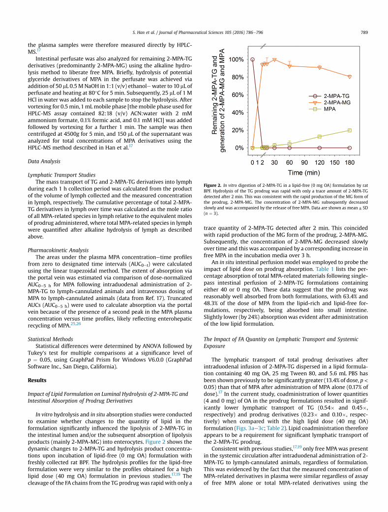
Figure 2. In vitro digestion of 2-MPA-TG in a lipid-free (0 mg OA) formulation by ratBPF. Hydrolysis of the TG prodrug was rapid with only a trace amount of 2-MPA-TGdetected after 2 min. This was consistent with the rapid production of the MG form ofthe prodrug, 2-MPA-MG. The concentration of 2-MPA-MG subsequently decreasedslowly and was accompanied by the release of free MPA. Data are shown as mean ± SD(n ¼ 3).
S. Han et al. / Journal of Pharmaceutical Sciences 105 (2016) 786e796 789
the plasma samples were therefore measured directly by HPLC-MS.17
Intestinal perfusate was also analyzed for remaining 2-MPA-TGderivatives (predominantly 2-MPA-MG) using the alkaline hydro-lysis method to liberate free MPA. Briefly, hydrolysis of potentialglyceride derivatives of MPA in the perfusate was achieved viaaddition of 50 mL 0.5 MNaOH in 1:1 (v/v) ethanolewater to 10 mL ofperfusate and heating at 80�C for 5 min. Subsequently, 25 mL of 1 MHCl in water was added to each sample to stop the hydrolysis. Aftervortexing for 0.5min,1mLmobile phase [themobile phase used forHPLC-MS assay contained 82:18 (v/v) ACN:water with 2 mMammonium formate, 0.1% formic acid, and 0.1 mM HCl] was addedfollowed by vortexing for a further 1 min. The sample was thencentrifuged at 4500g for 5 min, and 150 mL of the supernatant wasanalyzed for total concentrations of MPA derivatives using theHPLC-MS method described in Han et al.17
Data Analysis
Lymphatic Transport StudiesThe mass transport of TG and 2-MPA-TG derivatives into lymph
during each 1 h collection period was calculated from the productof the volume of lymph collected and the measured concentrationin lymph, respectively. The cumulative percentage of total 2-MPA-TG derivatives in lymph over time was calculated as the mole ratioof all MPA-related species in lymph relative to the equivalent molesof prodrug administered, where total MPA-related species in lymphwere quantified after alkaline hydrolysis of lymph as describedabove.
Pharmacokinetic AnalysisThe areas under the plasma MPA concentrationetime profiles
from zero to designated time intervals (AUC0et) were calculatedusing the linear trapezoidal method. The extent of absorption viathe portal vein was estimated via comparison of dose-normalizedAUC0e5 h for MPA following intraduodenal administration of 2-MPA-TG to lymph-cannulated animals and intravenous dosing ofMPA to lymph-cannulated animals (data from Ref. 17). TruncatedAUCs (AUC0e5 h) were used to calculate absorption via the portalvein because of the presence of a second peak in the MPA plasmaconcentration versus time profiles, likely reflecting enterohepaticrecycling of MPA.25,26
Statistical MethodsStatistical differences were determined by ANOVA followed by
Tukey's test for multiple comparisons at a significance level ofp ¼ 0.05, using GraphPad Prism for Windows V6.0.0 (GraphPadSoftware Inc., San Diego, California).
Results
Impact of Lipid Formulation on Luminal Hydrolysis of 2-MPA-TG andIntestinal Absorption of Prodrug Derivatives
In vitro hydrolysis and in situ absorption studies were conductedto examine whether changes to the quantity of lipid in theformulation significantly influenced the lipolysis of 2-MPA-TG inthe intestinal lumen and/or the subsequent absorption of lipolysisproducts (mainly 2-MPA-MG) into enterocytes. Figure 2 shows thedynamic changes to 2-MPA-TG and hydrolysis product concentra-tions upon incubation of lipid-free (0 mg OA) formulation withfreshly collected rat BPF. The hydrolysis profiles for the lipid-freeformulation were very similar to the profiles obtained for a highlipid dose (40 mg OA) formulation in previous studies.17,19 Thecleavage of the FA chains from the TG prodrug was rapid with only a
trace quantity of 2-MPA-TG detected after 2 min. This coincidedwith rapid production of the MG form of the prodrug, 2-MPA-MG.Subsequently, the concentration of 2-MPA-MG decreased slowlyover time and this was accompanied by a corresponding increase infree MPA in the incubation media over 3 h.
An in situ intestinal perfusion model was employed to probe theimpact of lipid dose on prodrug absorption. Table 1 lists the per-centage absorption of total MPA-related materials following single-pass intestinal perfusion of 2-MPA-TG formulations containingeither 40 or 0 mg OA. These data suggest that the prodrug wasreasonably well absorbed from both formulations, with 63.4% and48.3% of the dose of MPA from the lipid-rich and lipid-free for-mulations, respectively, being absorbed into small intestine.Slightly lower (by 24%) absorptionwas evident after administrationof the low lipid formulation.
The Impact of FA Quantity on Lymphatic Transport and SystemicExposure
The lymphatic transport of total prodrug derivatives afterintraduodenal infusion of 2-MPA-TG dispersed in a lipid formula-tion containing 40 mg OA, 25 mg Tween 80, and 5.6 mL PBS hasbeen shownpreviously to be significantly greater (13.4% of dose, p<0.05) than that of MPA after administration of MPA alone (0.17% ofdose).17 In the current study, coadministration of lower quantities(4 and 0 mg) of OA in the prodrug formulations resulted in signif-icantly lower lymphatic transport of TG (0.54� and 0.45�,respectively) and prodrug derivatives (0.23� and 0.10�, respec-tively) when compared with the high lipid dose (40 mg OA)formulation (Figs. 3ae3c; Table 2). Lipid coadministration thereforeappears to be a requirement for significant lymphatic transport ofthe 2-MPA-TG prodrug.
Consistent with previous studies,17,19 only free MPAwas presentin the systemic circulation after intraduodenal administration of 2-MPA-TG to lymph-cannulated animals, regardless of formulation.This was evidenced by the fact that the measured concentration ofMPA-related derivatives in plasma were similar regardless of assayof free MPA alone or total MPA-related derivatives using the

Table 1Absorption of Total MPA-Related Derivatives After In Situ Single-Pass Perfusion (over 3 h) of 2-MPA-TG Formulations Containing 0 or 40 mg OA Through an Upper SmallIntestine Segment (~30 cm) in Rats
Formulation Used for Intestinal Perfusion Absorbed Fraction Measured in Individual Ratsa Mean ± SD
Lipid-free formulation (OA 0 mg, n ¼ 5) 38.3% 56.7% 55.1% 41.6% 50.0% 48.3 ± 8.1%b
High lipid dose formulation (OA 40 mg, n ¼ 5) 58.1% 62.4% 50.5% 65.9% 80.3% 63.4 ± 12.6%
a The absorbed fraction (%) was calculated by (Mpre �Mpost)/Mpre � 100%, whereMpre was the moles of the prodrug in the original perfusate before perfusion andMpost wasthe moles of prodrug derivatives remained post the single-pass perfusion.Mpost was calculated from the product of the volume of the outflowing perfusate and the measuredconcentrations of total MPA-related derivatives in the fluid.
b Statistically significant difference (p < 0.05) in rats-administered formulations containing 0 mg OA when compared with formulations containing 40 mg OA.
S. Han et al. / Journal of Pharmaceutical Sciences 105 (2016) 786e796790
alkaline hydrolysis method19 (data not shown). In contrast, theprimary MPA-related material in lymph was re-esterified MPA. Re-esterified MPAwas thus transported into lymph, whereas free MPAliberated from the prodrug in the GI lumen or during absorptionappeared to be transported to the systemic circulation via theportal vein. It remains possible, however, that 2-MPA-MG or re-esterified derivatives thereof were absorbed into the blood andrapidly hydrolyzed to MPA such that only MPA was quantifiable inblood.
In lymph-cannulated animals, the systemic exposure of MPA(AUC0e5 h) was not significantly changed by increasing lipid
Figure 3. Rate of (a) lymphatic transport of TG (mg/h) and (b) total MPA-related derivativrivatives (% of prodrug dose), and (d) dose-normalized (to 3 mg/kg equivalent dose of MPA) p(over 2 h) of 2-MPA-TG formulations to anaesthetized, mesenteric lymph-duct-cannulated rTween 80, and 5.6 mL PBS, in the absence or presence of the CM formation inhibitor PL81 (n ¼ 3e6 rats. The data for the “OA 40 mg” group (filled circles) are reproduced from Han e
quantities in the formulations, although the 4-mg OA formulationshowed a trend toward higher MPA plasma concentrations at latertime points and the 0-mg OA formulation seemed to reduce plasmaexposure slightly when compared with administrationwith 4 or 40mg OA (Fig. 3d; Table 2). In contrast, coadministration of theresynthesis inhibitor A922500 with 2-MPA-TG administered in0 mg OA resulted in a significant increase in plasma concentrationsof MPA (AUC0e5h 2.37 vs. 0.64 mg�h/mL). This enhanced MPAexposure in the plasma suggests efficient absorption of the pro-drug, even in the absence of OA, but redirection to the blood in theabsence of incorporation into glyceride resynthetic pathways.
es (% of prodrug dose/h), (c) cumulative lymphatic transport of total MPA-related de-lasma concentrations of free MPA versus time (mg/mL) following intraduodenal infusionats. Formulations contained 2 mg of 2-MPA-TG dispersed in OA (0, 4, or 40 mg), 25 mg2 mg) or the DGAT-1 inhibitor A922500 (2 mg). Data are presented as mean ± SEM fort al.17 with permission from Elsevier for comparative purposes.

Table 2Summary of Lymphatic Transport of Total MPA-Related Derivatives and TG, Dose-Normalized Plasma Exposure (AUC0e5h) of MPA, and Estimated MPA Absorption into PortalBlood Following Intraduodenal Infusion (over 2 h) of 2-MPA-TG in Formulations Containing Different Quantities of OA (in the Presence or Absence of 2 mg of the DGAT-1Inhibitor A922500 or the CM Assembly Inhibitor PL81) to Anaesthetized, Mesenteric Lymph-Duct-Cannulated Rats
Absorption/Transport Parameters OA 40 mga
(n ¼ 5)OA 0 mg(n ¼ 4)
OA 0 mg þ A922500(n ¼ 6)
OA 4 mg(n ¼ 3)
OA 40 mg þ PL81(n ¼ 4)
Transport of total MPA derivatives in lymph (% of dose over 8 h) 13.4 ± 3.8%b 1.3 ± 0.3% 0.35 ± 0.23% 3.1 ± 1.3% 4.2 ± 1.9%TG transport in lymph over 8 h (mg, equivalent of triolein) 56 ± 11b 25 ± 5 23 ± 5 (n ¼ 4) 30 ± 1 41 ± 5c
Dose-normalized plasma AUC0e5h of MPA (mg�h/mL) 1.21 ± 0.65 0.64 ± 0.16 2.37 ± 1.22d 1.82 ± 0.72 1.23 ± 0.54Estimated portal blood absorptione 8.3 ± 4.4% 4.4 ± 1.1% 16.1 ± 8.3%d 12.4 ± 4.9% 8.3 ± 3.7%
Doses are normalized to a 3-mg/kg equivalent MPA dose and data are presented as mean ± SD.a Data reproduced from a previous study17 with permission.b Significantly greater (p < 0.05) in rats-administered formulations containing 40 mg OAwhen compared with formulations containing 0 mg OA, 0 mg OA þ A922500, 4 mg
OA, and 40 mg OA þ PL81.c Significantly greater (p < 0.05) in rats-administered formulations containing 40 mg OA þ PL81 when compared with formulations containing 0 mg OA or 0 mg OA þ
A922500.d Significantly greater (p < 0.05) in rats-administered formulations containing 0 mg OA þ A922500 when compared with formulations containing 0 mg OA alone.e Percentage of drug dose absorbed into portal blood was estimated as the ratio of dose-normalized (to 3 mg/kg) plasma AUC0e5h of free MPA following intraduodenal
dosing of 2-MPA-TG versus the plasma AUC0e5h following i.v. dosing of MPA (for which the plasma AUC0e5h was 14.7 mg�h/mL, as reported previously17). Absolutebioavailability could not be calculated because of second peaks in the plasma concentration profiles as described in the Materials and Methods section.
S. Han et al. / Journal of Pharmaceutical Sciences 105 (2016) 786e796 791
Prodrug Association with Lipoproteins in the Intestinal Lymph
Chylomicrons, the largest TG-rich LP species, have previouslybeen shown to play a significant role in the lymphatic transport oflipophilic drugs and vitamins.27,28 In the current studies, associa-tion of prodrug derivatives with CM was examined in order toprobe the significance of CM in the lymphatic transport of MPAprodrug derivatives formed within enterocytes. Following ultra-centrifugation of intestinal lymph samples collected 2e3 h afterinitiation of infusion of the 40-mg OA formulation, 97.6 ± 2.2% (n ¼4) of total MPA-related materials were recovered in the CM fractionof lymph.
Coadministration of an inhibitor of CM formatione PL81 (2 mg)with the 2-MPA-TG prodrug formulation containing 40 mg OA hadno effect on the plasma exposure of MPA (Fig. 3d), but significantlyreduced the lymphatic transport of both TG and total 2-MPA-TGderivatives. The effect was most obvious between 1 and 3 h afterinitiation of formulation infusion (Figs. 3ae3c; Table 2). The cu-mulative lymphatic transport of TG and prodrug derivatives was0.73� lower (41 vs. 56 mg, p < 0.05) and 0.31� lower (4.2% vs. 13.4%dose, p < 0.05), respectively, in PL81-dosed rats (Table 2). Thesedata suggest that CM is a critical carrier for transport of the re-esterified prodrug into the lymph.
The Impact of FA Type on Lymphatic Transport and SystemicExposure
To evaluate the impact of lipid type on lymphatic transport of 2-MPA-TG, formulations were also coadministered with LA, LNA, andADA to provide a comparison to previous data with OA. Coadmin-istration of the same quantity (40 mg) of LA and LNA, as thatemployed for OA, resulted in similar lymphatic transport of TG,whereas coadministration of ADA resulted in significantly reduced(by 46%, p < 0.05) recovery of TG in the lymph (Fig. 4a; Table 3). Incontrast, there were no statistically significant differences betweenthe cumulative (over 8 h) lymphatic transport of prodrug de-rivatives following coadministration of any of the FA (Table 3). Thepeak rate of prodrug transport following coadministration of theOA formulation (6.2 ± 1.5% dose/h), however, was significantlygreater (p < 0.05) when compared with the LA (3.4 ± 0.9% dose/h),LNA (3.1 ± 0.8% dose/h), and ADA (2.6 ± 1.1% does/h) formulations.The plasma MPA concentrationetime profiles following coadmin-istration of 40 mg of OA, LA, or LNA were almost identical (Fig. 4d;Table 3). In contrast, coadministration of ADA resulted in a
significant increase in plasma concentrations of MPA. The increasein plasma concentration of MPA after coadministration with ADAoccurred in spite of limited changes to lymphatic transport. As such,a larger proportion of absorbedMPAwas transported into the bloodwhen compared with the lymph. These changes are consistent withan alteration in the metabolic fate of the MPA prodrug derivatives,reducing the efficiency of lymphatic transport and increasing theefficiency of MPA liberation into the blood.
Discussion
Previous studies, using intrinsically highly lipophilic drugs(rather than prodrugs), have shown a positive correlation betweenthe extent of intestinal lymphatic drug transport and the coad-ministered lipid dose or the mass of lipid recovered in the intestinallymphatics.29-32 In these studies, the increase in lymphatic drugtransport stimulated by the coadministration of higher quantitiesof lipid (derived from either formulation or dietary lipids) wassuggested to reflect enhanced partitioning of lipophilic compoundsinto lymph-directing LP, the secretion of which is stimulated bylipid coadministration.1,33 For TG mimetic, lymph-directing pro-drugs, however, the role of lipid coadministration and LP associa-tion in stimulating lymphatic transport has not been explored. Inrecent studies, we have shown that the TG mimetic prodrug 2-MPA-TG is hydrolyzed in the GI lumen to a MG-equivalentconstruct, absorbed and then resynthesized back to the TG deriv-ative. The TG derivative of the prodrug is then incorporated into LPassembly pathways and transported into lymph.19 Unlike tradi-tional lymphatic transport pathways for lipophilic drugs, this pro-cess dictates that the prodrug forms a critical part of the LP via ametabolic integration process. Under these circumstances, it is notclear whether exogenous lipid is required to boost LP assembly, orwhether the constitutive turnover of lipids and LP through theenterocyte, even under fasting conditions, is sufficient to promoteprodrug lymphatic transport. The current study therefore aimed toevaluate whether the quantities and type of coadministered exog-enous lipid influence prodrug digestion, absorption, resynthesis,and lymphatic transport; whether resynthesized TG prodrug de-rivatives associate with LPs; and whether this association is criticalfor efficient lymphatic transport. An understanding of the mecha-nisms by which the TG mimetic prodrugs are transported in lymphand the importance of coadministered exogenous lipid is critical forthe design of the most appropriate dosing strategies for TGmimeticprodrugs.

Figure 4. Rate of (a) lymphatic transport of TG (mg/h) and (b) total MPA-related derivatives (% of prodrug dose/h), (c) cumulative lymphatic transport of total MPA-related de-rivatives (% of prodrug dose), and (d) dose-normalized (to 3 mg/kg equivalent dose of MPA) plasma concentrations (mg/mL) of free MPA versus time following intraduodenal infusion(over 2 h) of 2-MPA-TG formulations to anaesthetized, mesenteric lymph-duct-cannulated rats. Formulations contained 2 mg of 2-MPA-TG dispersed in 25 mg Tween 80, 5.6 mLPBS, and 40 mg of different FA with increasing degree of unsaturationdOA (C18:1), LA (C18:2), LNA (C18:3), and ADA (C20:4) acid. Data are presented as mean ± SEM for n ¼ 3e5rats. The data for the “OA 40 mg” group (filled circles) are reproduced from Han et al.17 with permission from Elsevier for comparative purposes.
S. Han et al. / Journal of Pharmaceutical Sciences 105 (2016) 786e796792
Lipolysis, Absorption, and Resynthesis of 2-MPA-TG Prior toIncorporation Into Intestinal LP Are Largely Unaffected by Changesto Lipid Quantities in the Formulation
In addition to effects on lymphatic transport, previous studieswith small-molecule lipophilic drugs have shown that changes tothe quantity of coadministered lipid may influence factors such asintestinal solubilization and the fraction absorbed.34 This might be
Table 3Summary of Lymphatic Transport of Total MPA-Related Derivatives and TG, Dose-NormaBlood Following Intraduodenal Infusion (over 2 h) of 2-MPA-TG in Formulations Contain
Absorption/Transport Parameters OA 40 m(n ¼ 5)
Transport of total MPA derivatives in lymph (% of dose over 8 h) 13.4 ± 3TG transport in lymph over 8 h (mg, equivalent of triolein) 56 ± 1Dose-normalized plasma AUC0e5h of MPA (mg�h/mL) 1.21 ± 0Estimated portal blood absorptiond 8.3 ± 4
Doses are normalized to a 3-mg/kg equivalent MPA dose and data are presented as meaa Data reproduced from a previous study17 with permission.b Significantly lower (p < 0.05) in rats-administered formulations containing 40 mg Ac Significantly greater (p < 0.05) in rats-administered formulations containing 40 mgd Percentage of drug dose absorbed into portal blood was estimated as the ratio of d
dosing of 2-MPA-TG versus the plasma AUC0e5h following i.v. dosing ofMPA (for which thecould not be calculated because of second peaks in the plasma concentration profiles as
expected to indirectly influence the proportion of the administereddose that is subsequently recovered in lymph.33 In the currentstudies, experiments were therefore first conducted to explore theinfluence of lipid dose on the events (hydrolysis, absorption, and re-esterification) that occur prior to LP association in the lymphatictransport pathway of the TG mimetic prodrug.
Interestingly, luminal hydrolysis, absorption, and resynthesis ofthe TG prodrug were relatively insensitive to the quantity of
lized Plasma Exposure (AUC0e5h) of MPA and Estimated MPA Absorption Into Portaling Different FAs to Anaesthetized, Mesenteric Lymph-Duct-Cannulated Rats
ga LA 40 mg(n ¼ 4)
LNA 40 mg(n ¼ 3)
ADA 40 mg(n ¼ 3)
.8% 8.7 ± 2.9% 7.8 ± 3.1% 7.4 ± 2.4%1 56 ± 10 52 ± 7 30 ± 8b
.65 1.12 ± 0.88 1.24 ± 0.87 2.95 ± 0.60c
.4% 7.6 ± 6.0% 8.4 ± 5.9% 20.0 ± 4.1%c
n ± SD.
DA when compared with formulations containing 40 mg OA or LA.ADA, compared with formulations containing 40 mg OA, LA, or LNA.ose-normalized (to 3 mg/kg) plasma AUC0e5h of free MPA following intraduodenalplasma AUC0e5h was 14.7 mg�h/mL, reported previously17). Absolute bioavailabilitydescribed in the Materials and Methods section.

S. Han et al. / Journal of Pharmaceutical Sciences 105 (2016) 786e796 793
coadministered lipid. Thus, in vitro hydrolysis of 2-MPA-TG in thelipid-free (OA 0 mg) formulation (Fig. 2) was consistent with thedata obtained previously for the high lipid (OA 40 mg) formula-tion.17,19 First, it seems likely, therefore, that the formulation hadlittle effect on the 2-MPA-TG to 2-MPA-MG transition in the GIlumen. Second, the intestinal perfusion data (Table 1) suggest thatthe absorption of 2-MPA-MG from the OA-free and OA-rich for-mulationswas efficient in both cases. Absorption from the lipid-freeformulation was slightly (24%) lower than that from the high lipidformulation; however, the fraction absorbed (~50%) over a relativelyshort intestine segment (30 cm) suggests that absorption was un-likely to be a major limitation to the bioavailability of the prodrugfrom formulationswith either high or low lipid loads. Finally, the re-esterification of 2-MPA-MG back to the TG analog in enterocytesappeared to occur efficiently, regardless of the quantities of lipid inthe coadministered formulation. The latter conclusionwas based onthe fact that the prodrug was well absorbed from low and high lipiddose formulations, plasma levels of free (i.e., nonesterified) MPA(particularly at early time points, e.g., 1e3 h) were low regardless oflipid dose, and that free MPA was the only MPA-related speciespresent in plasma. In contrast, in the absence of efficient re-esterification, nonesterified material (either MPA-MG or liberatedMPA)was expected to be absorbed through the portal blood into thesystemic circulation. Indeed, previous studies19 suggest that a lackof enterocyte-based resynthesis leads to liberation of free MPA andincreases in MPA exposure in the blood. Consistent with thesesuggestions, in the current studies, MPA plasma concentrationswere significantly enhanced when a TG resynthesis inhibitor(A922500) was coadministered with the lipid free formulation(Fig. 3d). Thus, the data suggest that lipolysis, absorption, and re-esterification of 2-MPA-TG prior to incorporation into LP are notmarkedly affected by changes in the quantities of lipid in thecoadministered formulation, although small increases in total ab-sorption are evident. Instead, the highly significant reductions inlymphatic transport (but small changes in plasma exposure) in thegroups administered no lipid or the low lipid dose (i.e., 4 mg) mostlikely reflect a reduction in LP incorporation.
The extent of absorption, estimated from loss from the perfusatein the in situ intestinal perfusion studies, was greater than the sumof lymphatic drug transport and calculated plasma exposure, in thebioavailability studies in lymph-cannulated rats. A completeexplanation for these differences in not evident at this stage butmayreflect (1) the use of truncated plasma AUCs (0e5 h) of MPA tocalculate plasma exposure in the bioavailability studies because ofenterohepatic recycling at later time points. This underestimatesthe extent of portal drug absorption: (2) intestinal or hepatic first-pass metabolism of MPA in the bioavailability studies. This re-duces bioavailability in vivowhen compared with fraction absorbedin the in situ perfusion experiments (where absorption was calcu-lated via loss from the perfusate): (3) effective MPA absorption andre-esterification in enterocytes (resulting in significant absorptionin the in situ studies) but accumulation of re-esterified 2-MPA-TG inenterocytes over time periods of up to hours (as has been suggestedto occur for dietary TG35,36) without complete transport into eitherthe portal blood or the lymphatics. The latter speculation is sup-ported by the fact that the TG resynthesis inhibitor A922500significantly increased plasma MPA exposure following coadmin-istration of the 0-mg lipid formulation, suggesting that theMPAmaybe temporarily stored in enterocytes in an esterified form.
Incorporation of Prodrug Derivatives Into CM Is Critical forLymphatic Transport
Following administration of 2-MPA-TG in a lipid formulationcontaining 40 mg of OA, 13.4% of the MPA dose was recovered in
mesenteric lymph (Table 2) and the majority of lymphaticallytransported MPAwas present in lymph as re-esterified glycerides.17
In the current study, the distribution of MPA-related materialsacross intestinal lymph species was examined and more than 97%of the prodrug derivatives were recovered in the CM fraction oflymph. This suggests that CMs (the largest LP particles produced inenterocytes) serve as highly effective carriers and facilitate thelymphatic transport of re-esterified 2-MPA-TG from the enterocyteinto the underlying lymphatics. This is consistent with previousstudies where the affinity of highly lipophilic (non-TG mimetic)compounds for CM has been shown to correlate with lymphaticdrug transport.27,37 The importance of CM in promoting lymphatictransport of the prodrug derivatives was confirmed here by ex-periments using PL81, an inhibitor of CM formation.38,39 In thesestudies, coadministration of PL81 significantly reduced (mostobviously during the 1e3 h postdose period) the lymphatic re-covery of prodrug derivatives. This occurred in parallel with inhi-bition of TG transport into the lymph (Fig. 3; Table 2).
For highly lipophilic drugs (i.e., in contrast to the prodrugsexamined here), the extent of lymphatic drug transport correlateswell with the affinity of drugs for CM and lymphatic drug transportdecreases significantly when the quantity of coadministeredTGs (the source of CM) is reduced.27,32 In the case of TG mimeticprodrugs, which appear to behave similarly to TG with respectto biotransformation, we initially hypothesized that lymphatictransport would be relatively insensitive to the quantities ofcoadministered lipid. This hypothesis was based on the assumptionthat even in the absence of exogenous lipid (when CM production islimited), TG synthesis and LP assembly continues as endogenouslipids are transported in lymph in the fasted state in the form ofsmall LPs of a size consistent with VLDL (30e80 nm in diameter).Output of these smaller LP in lymph occurs constitutively, includingin the fasted state and is relatively unresponsive to lipid intake.20
The data obtained, however, were not consistent with this hy-pothesis and the recovery of 2-MPA-TG derivatives in the lymphwas only significant when coadministered with a larger quantity ofOA (40 mg). The lymphatic transport of TG and prodrug derivativeswas low in the 4-mg OA or 0-mg OA group. In these groups, smallerVLDL-sized LP, derived from endogenous lipids, are expected to bethe primary LP that are assembled in enterocytes and transportedin lymph (Fig. 3; Table 2).38 Data following administration of thedifferent formulations revealed a strong correlation (Fig. 5) be-tween the lymphatic transport of prodrug derivatives and TG. Boththe peak rate (1e2 h post the initiation of formulation infusion;Fig. 5a) and cumulative quantity (over 8 h; Fig. 5b) of prodrugoutput correlated well (R2 ¼ 0.99) with the peak rate of TG outputin lymph (1e2 h). The data suggest that exogenous lipid-inducedCM, rather than smaller LP that are primary synthesized fromendogenous lipids, are the main drivers of lymphatic transport ofre-esterified 2-MPA-TG. The correlation between cumulativequantities of prodrug and TG transport (R2 ¼ 0.90; Fig. 5c) wasslightly weaker, and coadministration of PL81 resulted in propor-tionally lower prodrug recovery when compared with TG transportover 8 h. This suggests that although PL81 disrupts lymphatictransport of prodrug and lipid, prodrug transport is more signifi-cantly reduced. A complete explanation for this anomaly is not clearat this time, although it is possible that in reducing and delayinglymphatic lipid transport PL81 shifts the patterns of TG transportthrough the enterocyte to later time points, reducing the oppor-tunity for colocalization of CMs and prodrug in the enterocyteduring the period of prodrug absorption.
There are two pathways for TG resynthesis in the enterocyte. Inthe fasted state, TG is formed primarily via the glycerol-3-phosphate(G3P) pathway.40,41 In contrast, in the fed state, TG is assembled from2-MG and FA via the MG pathway.42,43 Which of these pathways is

Figure 5. Correlation of 2-MPA-TG versus TG transport in lymph following prodrug administration in formulations containing varying quantities and types of FA. (a) Rate of 2-MPA-TG transport (% of dose per hour) versus rate of TG transport (mg/h) in lymph between 1 and 2 h after the initiation of formulation infusion (when the peak rate of prodrug transportappeared). (b) Cumulative 2-MPA-TG transport (% of dose) over 8 h versus rate of TG transport (mg/h) in lymph between 1 and 2 h after the initiation of formulation infusion. (c andd) Cumulative lymphatic transport 2-MPA-TG (% of dose) and TG (mg) over 8 h after the initiation of formulation infusion. Data are presented as mean ± SEM for n ¼ 3e6 rats. Thedata for the “OA 40 mg” group are reproduced from Han et al.17 with permission from Elsevier for comparative purposes.
S. Han et al. / Journal of Pharmaceutical Sciences 105 (2016) 786e796794
active in the fasted state after administration of a lipid formulation,however, is unknown and likely to depend on the quantity of lipidadministered. The G3P pathway is believed to provide the primarysource of TG for VLDL-sized LP assembly in the fasted state, whereasthe 2-MG pathway provides TG to form the core of CM followingingestion of food.20 The current data are consistent with thisdistinction and suggest that incorporation of exogenousMGmimeticprodrug digestion products into LP assembly pathways requiresintegration into the 2-MG pathway that drives CM production andthat this is not activated in the absence of moderate quantities ofexogenous lipid. In contrast, the G3P pathway sources the glycerolbackbone of TG fromG3P (and not 2-MG) and transfers activated FAsto that backbone to form the TG core of VLDL-sized LP. As such,exogenousMG substrate (such as theMGeprodrug derivatives here)aremore likely to provide the building blocks for TG synthesis via the2-MG pathway in the fed state.
Specificity of FA Species in Promoting Lymphatic Transport
Subsequent studies explored the importance of the type of FAemployed, and specifically the degree of unsaturation of the lipid,in enhancing the lymphatic transport of 2-MPA-TG. Previous liter-ature suggests differences in lymphatic transport profiles of longchain FA with differing degrees of unsaturation; however, whetherthis influences the transport and metabolism of TG mimetic
prodrugs was unknown prior to the conduct of the current studies.Here, long-chain FAs (C18e20) with increasing degrees of unsatu-ration (D from 1 to 4) were employed to reveal different patterns ofmetabolism and transport of TG, and the prodrug, after coadmin-istration with the different lipids. The recovery of TG in themesenteric lymph was reduced and delayed after coadministrationwith lipids with increasing degrees of unsaturation (Fig. 4a).However, statistical differences were only attained for the ADAgroup. This trend is consistent with previous studies that havedemonstrated delayed lymphatic transport of polyunsaturatedlipids when compared with lipids with lower degrees of unsatu-ration (e.g., palmitic acid, OA, and LA).44,45 Unlike the strong cor-relation between lymphatic transport of prodrug and TG followingadministration of differing quantities of OA (Figs. 5aec), thelymphatic transport of prodrug-related materials was less wellcorrelated with TG recovery after coadministration of differentlipids (Fig. 5d). This correlation remained poor even when corre-lating TG and prodrug transport over early time points (when CMoutput appears to be the most important driver of prodruglymphatic transport) (data not shown). An explanation for thesedata is not clear at this stage but may reflect differences in the ef-ficiency of incorporation of prodrug derivatives resynthesized withdifferent FA into CM enriched with different lipids. For example,prodrugs resynthesized with LA may be less able to incorporateinto CM than those resynthesized with OA.
S. Han et al. / Journal of Pharmaceutical Sciences 105 (2016) 786e796 795
The plasma MPA concentrationetime profiles obtained for OA-,LA-, and LNA-dosed rats were almost identical (and low), sug-gesting efficient enterocyte-based re-esterification of 2-MPA-MG(Fig. 4d; Table 3). In contrast, coadministration of ADA led to anincrease in MPA redirection into the portal blood (Fig. 4d; Table 3;2.7-fold higher blood vs. lymph transport). This suggests a differentmetabolic fate of the prodrug or prodrug derivatives in the pres-ence of the highly unsaturated FA, such that a larger proportion ofnonlymphatically transported MPA derivatives were hydrolyzed torelease free MPA. Although increased blood transport of MPA oc-curs on both administration of ADA and after coadministration ofthe DGAT-1 inhibitor A922500 (Figs. 3d and 4d), it seems likely thatthe mechanisms driving the increase of MPA in the blood isdifferent in each case. Thus, after coadministration of A922500, re-esterification of prodrug derivatives is blocked, resulting in almostcompletely abolished lymphatic transport of MPA glycerides and animmediate increase in free MPA release into portal blood, pre-sumably because of hydrolysis of 2-MPA-MG (and/or diglyceridederivatives). In contrast, in the presence of ADA, MPA derivativeswere presumably re-esterified relatively efficiently as they weretransported into the lymph in proportion to the (albeit low) levelsof TG transport (Fig. 5d). Instead it seemsmore likely that the lowerefficiency of lymphatic transport of re-esterified ADA reduced thecapacity to support lymphatic prodrug transport and therefore aproportion of the re-esterified prodrug derivatives remained inenterocytes for longer periods, increasing availability for hydrolysisto release MPA. This suggestion is also supported by the delayedand sustained release of MPA into plasma after coadministrationwith ADA (Fig. 4d). The alternative scenario where coadministra-tion of ADA simply reduces re-esterification of 2-MPA-MG wouldbe expected to have resulted in rapid release of MPA from 2-MPA-MG over similar timescales to that seen in the presence of A922500(Fig. 3d). It is also possible that coadministrationwith ADA resultedin changes to MPA absorption via changes to drug solubilization, orthat ADAmay have resulted in changes to lipid or drug metabolism.
Conclusions
The lymphatic transport of 2-MPA-TG is dependent on prodrugintegration into 2-MG resynthetic pathways and is enhanced bycoadministrationwithmoderate quantities of exogenous lipids thatpromote CM assembly. Constitutive assembly of TG from endoge-nous lipids in the fasted state and turnover of endogenous lipidsinto the lymph as VLDL-sized LP appears less able to support sig-nificant lymphatic transport of 2-MPA-TG. Even though 2-MPA-TGis likely to be incorporated into the TG-rich core of developing LPs,coadministration with exogenous lipid is therefore required tomaximally stimulate lymphatic transport. Furthermore, mono-unsaturated lipids such as OA appear to more effectively supportprodrug lymphatic transport than polyunsaturated lipids, at least atthe higher lipid doses where lymph transport is most effective. CMassembly and secretion was the primary lipid-dependent step inlymphatic transport as lipolysis, absorption, and resynthesis ofprodrug derivatives were largely unaffected by lipid load. The datasuggest that coadministration with food or with an appropriatelipid-based formulation that promotes CM assembly may berequired to maximally stimulate the lymphatic transport of TGmimetic prodrugs.
Acknowledgment
This work was financially supported by the National Health andMedical Research Council and the Australian Research Council ofAustralia.
References
1. Porter CJH, Charman WN. Intestinal lymphatic drug transport: An update. AdvDrug Deliv Rev. 2001;50(1e2):61-80.
2. Trevaskis NL, Porter CJH, Charman WN. An examination of the interplay be-tween enterocyte-based metabolism and lymphatic drug transport in the rat.Drug Metab Dispos. 2006;34(5):729-733.
3. Choo EF, Boggs J, Zhu C, Lubach JW, Catron ND, Jenkins G, Souers AJ, Voorman R.The role of lymphatic transport on the systemic bioavailability of the Bcl-2protein family inhibitors Navitoclax (ABT-263) and ABT-199. Drug Metab Dis-pos. 2014;42(2):207-212.
4. Swartz MA. The physiology of the lymphatic system. Adv Drug Deliv Rev.2001;50(1e2):3-20.
5. Swartz MA, Hirosue S, Hubbell JA. Engineering approaches to immunotherapy.Sci Transl Med. 2012;4(148):148rv9.
6. Cao YH. OpiniondEmerging mechanisms of tumour lymphangiogenesis andlymphatic metastasis. Nat Rev Cancer. 2005;5(9):735-743.
7. Chakraborty S, Zawieja S, Wang W, Zawieja DC, Muthuchamy M. Lymphaticsystem: A vital link between metabolic syndrome and inflammation. Ann N YAcad Sci. 2010;1207:E94-E102.
8. Fletcher CV, Staskus K, Wietgrefe SW, Rothenberger M, Reilly C, Chipman JG,Beilman GJ, Khoruts A, Thorkelson A, Schmidt TE, Anderson J, Perkey K,Stevenson M, Perelson AS, Douek DC, Haase AT, Schacker TW. Persistent HIV-1replication is associated with lower antiretroviral drug concentrations inlymphatic tissues. Proc Natl Acad Sci USA. 2014;111(6):2307-2312.
9. Irvine DJ, Swartz MA, Szeto GL. Engineering synthetic vaccines using cues fromnatural immunity. Nat Mater. 2013;12(11):978-990.
10. Liu H, Moynihan KD, Zheng Y, Szeto GL, Li AV, Huang B, Van Egeren DS, Park C,Irvine DJ. Structure-based programming of lymph-node targeting in molecularvaccines. Nature. 2014;507(7493):519-522.
11. Trevaskis NL, Charman WN, Porter CJH. Targeted drug delivery to lymphocytes:A route to site-specific immunomodulation? Mol Pharm. 2010;7(6):2297-2309.
12. Freeling JP, Ho RJY. Anti-HIV drug particles may overcome lymphatic druginsufficiency and associated HIV persistence. Proc Natl Acad Sci USA.2014;111(25):E2512-E2513.
13. Omorchoe CCC, Omorchoe PJ. Differences in lymphatic and blood capillarypermeability: Ultrastructural-functional correlations. Lymphology. 1987;20(4):205-209.
14. Leak LV. Permeability of lymphatic capillaries. J Cell Biol. 1971;50(2):300-323.15. Reed AL, Rowson SA, Dixon JB. Demonstration of ATP-dependent, transcellular
transport of lipid across the lymphatic endothelium using an in vitro model ofthe lacteal. Pharm Res. 2013;30(12):3271-3280.
16. Dixon JB, Raghunathan S, Swartz MA. A tissue-engineered model of the in-testinal lacteal for evaluating lipid transport by lymphatics. Biotechnol Bioeng.2009;103(6):1224-1235.
17. Han S, Quach T, Hu L, Wahab A, Charman WN, Stella VJ, Trevaskis NL,Simpson JS, Porter CJH. Targeted delivery of a model immunomodulator to thelymphatic system: Comparison of alkyl ester versus triglyceride mimetic lipidprodrug strategies. J Control Release. 2014;177:1-10.
18. Trepel F. Number and distribution of lymphocytes in mandCritical analysis.Klin Wochenschr. 1974;52(11):511-515.
19. Han S, Hu L, Quach T, Simpson JS, Trevaskis NL, Porter CJ. Profiling the role ofdeacylation-reacylation in the lymphatic transport of a triglyceride-mimeticprodrug. Pharm Res. 2015;32(5):1830-1844.
20. Kindel T, Lee DM, Tso P. The mechanism of the formation and secretion ofchylomicrons. Atheroscler Suppl. 2010;11(1):11-16.
21. Trevaskis NL, Porter CJH, CharmanWN. Bile increases intestinal lymphatic drugtransport in the fasted rat. Pharm Res. 2005;22(11):1863-1870.
22. Porter CJH, Charman SA, Charman WN. Lymphatic transport of halofantrine inthe triple-cannulated anesthetized rat model: Effect of lipid vehicle dispersion.J Pharm Sci. 1996;85(4):351-356.
23. Yeap YY, Trevaskis NL, Quach T, Tso P, Charman WN, Porter CJH. Intestinal bilesecretion promotes drug absorption from lipid colloidal phases via induction ofsupersaturation. Mol Pharm. 2013;10(5):1874-1889.
24. Trevaskis NL, Shanker RM, Charman WN, Porter CJH. The mechanism oflymphatic access of two cholesteryl ester transfer protein inhibitors(CP524,515 and CP532,623) and evaluation of their impact on lymph lipo-protein profiles. Pharm Res. 2010;27(9):1949-1964.
25. Yau W-P, Vathsala A, Lou H-X, Zhou S, Chan E. Mechanism-based enterohepaticcirculation model of mycophenolic acid and its glucuronide metabolite:Assessment of impact of cyclosporine dose in Asian renal transplant patients.J Clin Pharmacol. 2009;49(6):684-699.
26. Saitoh H, Kobayashi M, Oda M, Nakasato K, Kobayashi M, Tadano K. Charac-terization of intestinal absorption and enterohepatic circulation of mycophe-nolic acid and its 7-O-glucuronide in rats. Drug Metab Pharmacokinet.2006;21(5):406-413.
27. Gershkovich P, Hoffman A. Uptake of lipophilic drugs by plasma derived iso-lated chylomicrons: Linear correlation with intestinal lymphatic bioavailability.Eur J Pharm Sci. 2005;26(5):394-404.
28. Dahan A, Hoffman A. Evaluation of a chylomicron flow blocking approach toinvestigate the intestinal lymphatic transport of lipophilic drugs. Eur J PharmSci. 2005;24(4):381-388.
29. Caliph SM, Charman WN, Porter CJH. Effect of short-, medium-, and long-chain fatty acid-based vehicles on the absolute oral bioavailability and in-

S. Han et al. / Journal of Pharmaceutical Sciences 105 (2016) 786e796796
testinal lymphatic transport of halofantrine and assessment of mass balancein lymph-cannulated and non-cannulated rats. J Pharm Sci. 2000;89(8):1073-1084.
30. Trevaskis NL, Caliph SM, Nguyen G, Tso P, Charman WN, Porter CJH. A mousemodel to evaluate the impact of species, sex, and lipid load on lymphatic drugtransport. Pharm Res. 2013;30(12):3254-3270.
31. Hauss DJ, Fogal SE, Ficorilli JV, Price CA, Roy T, Jayara AA, Keirns JJ. Lipid-based delivery systems for improving the bioavailability and lymphatictransport of a poorly water-soluble LTB4 inhibitor. J Pharm Sci. 1998;87(2):164-169.
32. White KL, Nguyen G, Charman WN, Edwards GA, Faassen WA, Porter CJH.Lymphatic transport of methylnortestosterone undecanoate (MU) and thebioavailability of methylnortestosterone are highly sensitive to the mass ofcoadministered lipid after oral administration of MU. J Pharmacol Exp Ther.2009;331(2):700-709.
33. Trevaskis NL, McEvoy CL, McIntosh MP, Edwards GA, Shanker RM,Charman WN, Porter CJH. The role of the intestinal lymphatics in the absorp-tion of two highly lipophilic cholesterol ester transfer protein inhibitors(CP524,515 and CP532,623). Pharm Res. 2010;27(5):878-893.
34. Williams HD, Trevaskis NL, Charman SA, Shanker RM, Charman WN,Pouton CW, Porter CJH. Strategies to address low drug solubility in discoveryand development. Pharmacol Rev. 2013;65(1):315-499.
35. Fielding BA, Callow J, Owen RM, Samra JS, Matthews DR, Frayn KN.Postprandial lipemia: The origin of an early peak studied by specific di-etary fatty acid intake during sequential meals. Am J Clin Nutr. 1996;63(1):36-41.
36. Robertson MD, Parkes M, Warren BF, Ferguson DJP, Jackson KG, Jewell DP,Frayn KN. Mobilisation of enterocyte fat stores by oral glucose in humans. Gut.2003;52(6):834-839.
37. Gershkovich P, Qadri B, Yacovan A, Amselem S, Hoffman A. Different impacts ofintestinal lymphatic transport on the oral bioavailability of structurally similarsynthetic lipophilic cannabinoids: Dexanabinol and PRS-211,220. Eur J PharmSci. 2007;31(5):298-305.
38. Tso P, Balint JA, Bishop MB, Rodgers JB. Acute inhibition of intestinal lipidtransport by pluronic L-81 in the rat. Am J Physiol. 1981;241(6):G487-G497.
39. Fatma S, Yakubov R, Anwar K, Hussain MM. Pluronic L81 enhances tri-acylglycerol accumulation in the cytosol and inhibits chylomicron secretion.J Lipid Res. 2006;47(11):2422-2432.
40. Levy E, Mehran M, Seidman E. Caco-2 cells as a model for intestinal lipoproteinsynthesis and secretion. FASEB J. 1995;9(8):626-635.
41. Lehner R, Kuksis A. Biosynthesis of triacylglycerols. Prog Lipid Res. 1996;35(2):169-201.
42. Mansbach CM, Siddiqi SA. The biogenesis of chylomicrons. Annu Rev Physiol.2010;72:315-333.
43. Iqbal J, Hussain MM. Intestinal lipid absorption. Am J Physiol Endocrinol Metab.2009;296(6):E1183-E1194.
44. Degrace P, Caselli C, Rayo JM, Bernard A. Intestinal lymph absorption of butter,corn oil, cod liver oil, menhaden oil, and eicosapentaenoic and docosahexae-noic acid ethyl esters in rats. Lipids. 1996;31(4):405-414.
45. McDonald GB, Weidman M. Partitioning of polar fatty-acids into lymph andportal vein after intestinal absorption in the rat. Q J Exp Physiol Cogn Med Sci.1987;72(2):153-159.