kir6.2 is not the mitochondrial katp channel but is ... · required for cardioprotection by...
TRANSCRIPT
doi:10.1152/ajpheart.00972.2012 304:H1439-H1445, 2013. First published 12 April 2013;Am J Physiol Heart Circ Physiol
Keith Nehrke and Paul S. BrookesAndrew P. Wojtovich, William R. Urciuoli, Shampa Chatterjee, Aron B. Fisher,preconditioningrequired for cardioprotection by ischemic
channel but isATPKir6.2 is not the mitochondrial K
You might find this additional info useful...
42 articles, 21 of which can be accessed free at:This article cites /content/304/11/H1439.full.html#ref-list-1
including high resolution figures, can be found at:Updated information and services /content/304/11/H1439.full.html
can be found at:AJP - Heart and Circulatory Physiologyabout Additional material and information http://www.the-aps.org/publications/ajpheart
This information is current as of September 5, 2013.
ISSN: 0363-6135, ESSN: 1522-1539. Visit our website at http://www.the-aps.org/.Physiological Society, 9650 Rockville Pike, Bethesda MD 20814-3991. Copyright © 2013 by the American Physiological Society. intact animal to the cellular, subcellular, and molecular levels. It is published 12 times a year (monthly) by the Americanlymphatics, including experimental and theoretical studies of cardiovascular function at all levels of organization ranging from the
publishes original investigations on the physiology of the heart, blood vessels, andAJP - Heart and Circulatory Physiology
on Septem
ber 5, 2013D
ownloaded from

CALL FOR PAPERS Mitochondria in Cardiovascular Physiology and Disease
Kir6.2 is not the mitochondrial KATP channel but is requiredfor cardioprotection by ischemic preconditioning
Andrew P. Wojtovich,1 William R. Urciuoli,2 Shampa Chatterjee,3 Aron B. Fisher,3 Keith Nehrke,1
and Paul S. Brookes2
1Department of Medicine, University of Rochester Medical Center, Rochester, New York; 2Department of Anesthesiology,University of Rochester Medical Center, Rochester, New York; and 3Institute for Environmental Medicine, University ofPennsylvania Medical Center, Philadelphia, Pennsylvania
Submitted 28 December 2012; accepted in final form 9 April 2013
Wojtovich AP, Urciuoli WR, Chatterjee S, Fisher AB, NehrkeK, Brookes PS. Kir6.2 is not the mitochondrial KATP channel but isrequired for cardioprotection by ischemic preconditioning. Am JPhysiol Heart Circ Physiol 304: H1439–H1445, 2013. First publishedApril 12, 2013; doi:10.1152/ajpheart.00972.2012.—ATP-sensitiveK� (KATP) channels that contain K� inward rectifier subunits of the6.2 isotype (Kir6.2) are important regulators of the cardiac response toischemia-reperfusion (I/R) injury. Opening of these channels is im-plicated in the cardioprotective mechanism of ischemic precondition-ing (IPC), but debate surrounds the contribution of surface KATP
(sKATP) versus mitochondrial KATP (mKATP) channels. While re-sponses to I/R injury and IPC have been examined in Kir6.2�/� micebefore, breeding methods and other technical obstacles may haveconfounded interpretations. The aim of this study was to elucidate therole of Kir6.2 in cardioprotection and mKATP activity, using conven-tionally bred Kir6.2�/� mice with wild-type littermates as controls.We found that perfused hearts from Kir6.2�/� mice exhibited anormal baseline response to I/R injury, were not protected by IPC, andshowed a blunted response to the IPC mimetic drug diazoxide. Thesedata suggest that the loss of IPC in Kir6.2�/� hearts is not due to anunderlying difference in I/R sensitivity. Furthermore, mKATP channelactivity was identical in cardiac mitochondria isolated from wild-typeversus Kir6.2�/� mice, suggesting no role for Kir6.2 in the mKATP.Collectively, these data indicate that Kir6.2 is required for the fullresponse to IPC or diazoxide but is not involved in mKATP formation.
mitochondria; ischemic preconditioning; KATP channel; diazoxide
THE HEART AND OTHER ORGANS can be protected against ischemia-reperfusion (I/R) injury via ischemic preconditioning (IPC),wherein short periods of I/R can engage protective signalingpathways to reduce the impact of a prolonged ischemic event(23). The protective effects of IPC can be mimicked by openersof ATP-sensitive potassium (KATP) channels. Since changes incardiomyocyte bioenergetics are known to occur during IPC(11), the metabolic-sensing role of these channels has driveninterest in their potential role in cardioprotective signaling(13, 24).
KATP channels comprise octamers of four inward-rectifyingpotassium channel subunits (Kir6.1 or -6.2) and four sulfonyl-urea receptors 1, 2A, or 2B (SUR1, -2A, or -2B), with differentKir/SUR combinations giving rise to unique cellular roles,locations, and pharmacological profiles (13). For example, the
cardiac surface KATP (sKATP) is Kir6.2/SUR2A. It plays a rolein cardiomyocyte volume regulation and stress responses (28)and is activated by cromakalim but not diazoxide (DZX) (11,33, 42). In contrast, the pancreatic sKATP is Kir6.2/SUR1,which plays a role in insulin secretion and is activated by DZXbut not cromakalim (13).
Despite there being no effect of DZX on cardiac sKATP
channels (4, 9), DZX is known to mimic IPC and protect theheart against I/R injury. This led to speculation on the exis-tence of another DZX-sensitive KATP channel in the heart,namely, the mitochondrial KATP (mKATP). However, previousstudies using Kir6.2�/� mice concluded that there was no rolefor Kir6.2 in the formation of the mKATP, although channelactivity was determined only by the surrogate measurement ofmitochondrial flavoprotein oxidation rather than by direct mea-surements of ion transport (33).
Furthermore, to date cardiac studies on Kir6.2 �/� mice (10,11, 31–33) have used a pure-line breeding strategy [knockout �knockout, purchasing or breeding a separate line of wild-type(WT) controls]. Thus, while it was previously shown thatKir6.2�/� mice cannot be protected by IPC, it is not clearwhether this is represents a true failure of IPC or is simply dueto a reported greater sensitivity of the knockout animals tobaseline I/R injury (vs. WT controls) (32, 33). Several factorscan conspire to alter the sensitivity of mouse hearts to I/Rinjury (strain, vendor, diet, sex, shipping and handling, andseason) (12). Thus we sought to use a conventional breedingstrategy (heterozygous � heterozygous, with Mendelian off-spring ratios, tail-clip PCR genotyping of all animals) todetermine whether Kir6.2 is truly required for IPC. Further-more, we directly assayed mKATP channel activity in mito-chondria isolated from WT and Kir6.2�/� littermate mice todetermine the role of this Kir in the mKATP.
The results herein show that Kir6.2�/� mouse hearts exhib-ited identical sensitivity to baseline injury, compared with WTlittermate controls. Furthermore, both IPC and DZX-mediatedprotection were blunted in Kir6.2�/�, indicating a role for thechannel in cardioprotective signaling. Notably, mKATP channelactivity was identical between WT and Kir6.2�/�-derivedcardiac mitochondria, indicating no role for this Kir in formingthe mKATP.
MATERIALS AND METHODS
Animals. All animals were maintained in an Association for Ac-creditation of Laboratory Animal Care-accredited pathogen-free bar-
Address for reprint requests and other correspondence: P. S. Brookes, Dept.of Anesthesiology, Box 604, Univ. of Rochester Medical Ctr., 601 ElmwoodAve., Rochester, NY 14642 (e-mail: [email protected]).
Am J Physiol Heart Circ Physiol 304: H1439–H1445, 2013.First published April 12, 2013; doi:10.1152/ajpheart.00972.2012.
0363-6135/13 Copyright © 2013 the American Physiological Societyhttp://www.ajpheart.org H1439
on Septem
ber 5, 2013D
ownloaded from
rier facility with food and water available ad libitum. All procedureswere in accordance with the National Institutes of Health’s Guide forthe Care and Use of Laboratory Animals and were approved by anInstitutional Animal Care and Use Committee. Mice harboring dele-tion of the KCNJ11 gene that encodes Kir6.2 were obtained withpermission from their originator Dr. Susumu Seino (Kobe University,Japan) (20). These mice were reported to be backcrossed to theC57BL/6 background for five generations and were backcrossedherein for an additional two generations onto C57BL/6J (JAX, BarHarbor ME). Mice were conventionally bred (heterozygous Kir6.2�/� �Kir6.2�/�, Mendelian offspring ratios), and resulting male WT andKir6.2�/� littermates (age, 8–10 wk) were used in all experiments.Mice were genotyped by tail biopsy PCR using a mouse genotypingkit (Kapa Biosystems, Woburn MA) according to manufacturer’sprotocol. Briefly, 2-mm3 tail fragments were digested in 10 �l of 10�KAPA Express Extract Buffer plus 2 �l of 1 U/�l KAPA ExpressEnzyme, brought to 100 �l with PCR-grade water and incubated at75°C for 10 min, followed by 5 min at 95°C. Samples were centri-fuged for 1 min at 2,000 g, and 70-�l supernatant were transferred tofresh PCR tubes. The DNA amplification protocol (as per KAPAmouse genotyping kit) was 95°C, 3 min; (95°C, 15 s; 62°C, 15 s; and72°C, 30 s) � 35 cycles; 72°C, 3 min; and 4°C hold. The followingprimers were used and yielded products of 390 bp for Kir6.2�/� and223 bp for WT: forward F1, TCC CTG AGG AAT ATG TGC TGACC; reverse, AGG AAG GAC ATG GTG AAA ATG AGC; and Neo,TCT GCA CGA GAC TAG TGA GAC G.
Isolated mitochondria, mKATP thallium flux assay. Mitochondriawere isolated from WT and Kir6.2�/� mouse hearts using differentialcentrifugation in sucrose-based media, as previously described (37).During the procedure, mitochondria were loaded with benzothiazolecoumarin-acetyoxymethyl ester (Invitrogen, Carlsbad, CA) for 10 minat 25°C, as previously described (37, 39). Following isolation, ben-zothiazole coumarin-acetyoxymethyl ester-loaded mitochondria weresubject to mKATP thallium flux (Tl� flux) assay, whereby Tl� servesas surrogate for K� and where Tl� entry into mitochondria results indye fluorescence (�ex, 488 nm, and �em, 525 nm). mKATP activity wasmonitored as a change in fluorescence following addition of Tl2SO4,as previously described (37, 39).
Ex vivo perfused heart. Mice were anesthetized with Avertin (100mg/kg ip), and an EKG reading was obtained. Corrected QT intervalwas calculated as QT/(RR interval/100)0.5 (17). The aorta was can-nulated in situ and rapidly transferred to a perfusion apparatus andperfused without pacing in constant flow mode (4 ml·min�1·100mg�1) with Krebs-Henseleit buffer (KH, in mM) 118 NaCl, 4.7 KCl,25 NaHCO3, 10 glucose, 1.2 MgSO4, 1.2 KH2PO4, and 2.5 CaCl2,gassed with 95% O2-5% CO2 at 37°C, as previously described (37). A
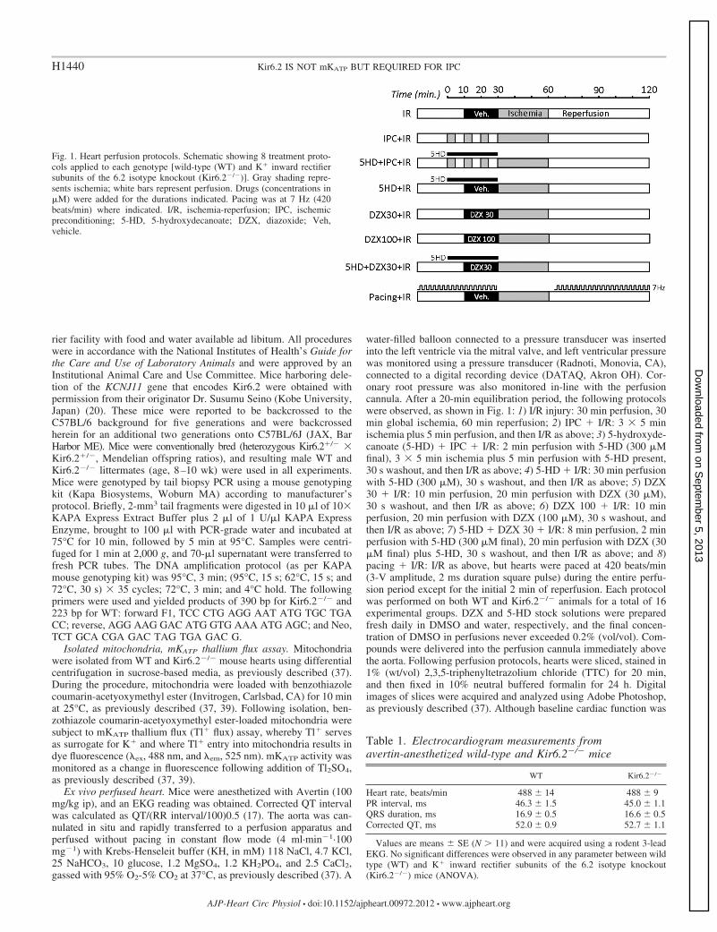
water-filled balloon connected to a pressure transducer was insertedinto the left ventricle via the mitral valve, and left ventricular pressurewas monitored using a pressure transducer (Radnoti, Monovia, CA),connected to a digital recording device (DATAQ, Akron OH). Cor-onary root pressure was also monitored in-line with the perfusioncannula. After a 20-min equilibration period, the following protocolswere observed, as shown in Fig. 1: 1) I/R injury: 30 min perfusion, 30min global ischemia, 60 min reperfusion; 2) IPC � I/R: 3 � 5 minischemia plus 5 min perfusion, and then I/R as above; 3) 5-hydroxyde-canoate (5-HD) � IPC � I/R: 2 min perfusion with 5-HD (300 �Mfinal), 3 � 5 min ischemia plus 5 min perfusion with 5-HD present,30 s washout, and then I/R as above; 4) 5-HD � I/R: 30 min perfusionwith 5-HD (300 �M), 30 s washout, and then I/R as above; 5) DZX30 � I/R: 10 min perfusion, 20 min perfusion with DZX (30 �M),30 s washout, and then I/R as above; 6) DZX 100 � I/R: 10 minperfusion, 20 min perfusion with DZX (100 �M), 30 s washout, andthen I/R as above; 7) 5-HD � DZX 30 � I/R: 8 min perfusion, 2 minperfusion with 5-HD (300 �M final), 20 min perfusion with DZX (30�M final) plus 5-HD, 30 s washout, and then I/R as above; and 8)pacing � I/R: I/R as above, but hearts were paced at 420 beats/min(3-V amplitude, 2 ms duration square pulse) during the entire perfu-sion period except for the initial 2 min of reperfusion. Each protocolwas performed on both WT and Kir6.2�/� animals for a total of 16experimental groups. DZX and 5-HD stock solutions were preparedfresh daily in DMSO and water, respectively, and the final concen-tration of DMSO in perfusions never exceeded 0.2% (vol/vol). Com-pounds were delivered into the perfusion cannula immediately abovethe aorta. Following perfusion protocols, hearts were sliced, stained in1% (wt/vol) 2,3,5-triphenyltetrazolium chloride (TTC) for 20 min,and then fixed in 10% neutral buffered formalin for 24 h. Digitalimages of slices were acquired and analyzed using Adobe Photoshop,as previously described (37). Although baseline cardiac function was
Table 1. Electrocardiogram measurements fromavertin-anesthetized wild-type and Kir6.2�/� mice
WT Kir6.2�/�
Heart rate, beats/min 488 � 14 488 � 9PR interval, ms 46.3 � 1.5 45.0 � 1.1QRS duration, ms 16.9 � 0.5 16.6 � 0.5Corrected QT, ms 52.0 � 0.9 52.7 � 1.1
Values are means � SE (N � 11) and were acquired using a rodent 3-leadEKG. No significant differences were observed in any parameter between wildtype (WT) and K� inward rectifier subunits of the 6.2 isotype knockout(Kir6.2�/�) mice (ANOVA).
Fig. 1. Heart perfusion protocols. Schematic showing 8 treatment proto-cols applied to each genotype [wild-type (WT) and K� inward rectifiersubunits of the 6.2 isotype knockout (Kir6.2�/�)]. Gray shading repre-sents ischemia; white bars represent perfusion. Drugs (concentrations in�M) were added for the durations indicated. Pacing was at 7 Hz (420beats/min) where indicated. I/R, ischemia-reperfusion; IPC, ischemicpreconditioning; 5-HD, 5-hydroxydecanoate; DZX, diazoxide; Veh,vehicle.
H1440 Kir6.2 IS NOT mKATP BUT REQUIRED FOR IPC
AJP-Heart Circ Physiol • doi:10.1152/ajpheart.00972.2012 • www.ajpheart.org
on Septem
ber 5, 2013D
ownloaded from
not different between the entire cohort of WT and Kir6.2�/� hearts(N � 40, see Table 2), individual subgroups with N � 6–9 weresomewhat noisier; hence, cardiac functional data in Figs. 2 and 3 werepresented as percentages, relative to the initial value for each heart.
Statistics. Statistical significance (P 0.05) between multiplegroups was determined using analysis of variance (ANOVA) withBonferroni correction.
RESULTS
Blunted protective effect of IPC in Kir6.2�/� mouse hearts.Following anesthesia (Avertin) and before heart isolation,EKG was recorded on anesthetized mice. No significant dif-ferences in measured parameters were observed between WTand Kir6.2�/� (Table 1). Upon heart perfusion, all heartsconformed to recently described criteria for perfused heartstudies (2), and no differences were observed in baselinecardiac functional parameters (Table 2), consistent with previ-ous reports (33). Upon exposure to global ischemia, Kir6.2�/�
hearts entered ischemic hypercontracture significantly fasterthan WT and exhibited a greater degree of contracture (Table2), consistent with previous reports (10).
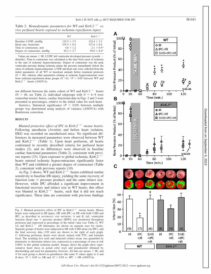
As Fig. 2 shows, WT and Kir6.2�/� hearts exhibited similarsensitivity to baseline I/R injury, yielding the same recovery offunction (rate � pressure product) and the same infarct size.However, while IPC afforded a significant improvement infunctional recovery and infarct size in WT hearts, this effectwas blunted in Kir6.2�/� hearts, such that it did not reachsignificance. These data are consistent with previous findings
Table 2. Hemodynamic parameters for WT and Kir6.2�/� exvivo perfused hearts exposed to ischemia-reperfusion injury
WT Kir6.2�/�
Baseline LVDP, mmHg 124.5 � 3.5 124.4 � 3.2Heart rate, beats/min 335.5 � 8.4 327.6 � 8.6Time to contracture, min 4.6 � 1.2 2.1 � 0.5*Degree of contracture, mmHg 45.2 � 3.7 59.0 � 8.4*
Values are means � SE. LVDP, left ventricular developed pressure (systolic �diastolic). Time to contracture was calculated as the time from onset of ischemiato the start of ischemic hypercontracture. Degree of contracture was the peakventricular pressure during ischemia minus the pressure immediately before theonset of ischemic hypercontracture. LVDP and heart rate were collected from theinitial parameters of all WT or knockout animals before treatment protocols(N � 40), whereas other parameters relating to ischemic hypercontracture werefrom ischemia-reperfusion-alone groups (N �6). *P 0.05 between WT andKir6.2�/� hearts (ANOVA).
Fig. 2. Blunted protective effect of IPC in Kir6.2�/� mouse hearts. Mousehearts were subjected to I/R injury, I/R with IPC, or I/R with both 5-HD andIPC, as described in MATERIALS AND METHODS. A and B, left: ventricularfunction [heart rate � pressure product (RPP)] was monitored throughoutperfusion and expressed as percentage of the initial value (see Table 2). WT(A) and Kir6.2�/� (B) littermates are shown on separate axes for clarity.Separate groups of hearts were subjected to I/R with 5-HD alone (no IPC), andthe final recovery data (120 min) are shown to the right of each graph.C: following perfusion, hearts were sliced, stained with TTC, and formalinfixed. The resulting live (red) and infarcted (white) tissue was quantified byplanimetry to determine infarct size, expressed as a percentage of area at risk(100% in this global ischemia model). Images above the graph show repre-sentative heart slices in actual color (top) and pseudocolor obtained bythresholding and used for planimetry (bottom). All data are means � SE, andN for each group is shown in parentheses (the same N values apply to A andB also). *P 0.05 vs. I/R and †P 0.05 vs. IPC � I/R (ANOVA).
H1441Kir6.2 IS NOT mKATP BUT REQUIRED FOR IPC
AJP-Heart Circ Physiol • doi:10.1152/ajpheart.00972.2012 • www.ajpheart.org
on Septem
ber 5, 2013D
ownloaded from

in vivo (33). Furthermore, the protective effect of IPC wasblocked by the mKATP antagonist 5-HD in both WT andKir6.2�/� hearts, indicating that the remaining mild cardiopro-tection afforded in the absence of Kir6.2 was likely due to amKATP channel. 5-HD alone was without effect on baseline I/Rinjury (functional recovery: WT, 15 � 3%, and Kir6.2�/�, 22 � 7%;infarct size: WT, 41 � 5%, and Kir6.2�/�, 37 � 5%; data shown tothe right of graphs in Fig. 2).
Blunted protective effect of DZX in Kir6.2�/� mouse hearts.The antidiabetic drug DZX is a KATP channel opener and hasbeen extensively used to study the role of KATP channels incardioprotection. At appropriate concentrations (10–30 �M), itis somewhat selective for the mKATP over the cardiac sKATP
(Kir6.2/SUR2A) (8, 18), although at higher concentrations ithas off-target effects such as inhibition of mitochondrial com-plex II (18, 26). As shown in Fig. 3, we found that 30 �M DZXwas protective in WT hearts (improved functional recovery andreduced infarct size), but this protection was blunted inKir6.2�/� hearts. Overall, results with 30 �M DZX (loss ofcardioprotection in Kir6.2�/�) were not as impressive as thoseseen with IPC (Fig. 2) but trended in the same direction.Importantly, the protective effects of 30 �M DZX wereblocked by the mKATP inhibitor 5-HD in both WT andKir6.2�/� hearts (Fig. 3), suggesting that the mitochondrialchannel is the bona fide target of low concentrations of DZX.As we previously reported (35), 5-HD alone was without effecton basal I/R injury.
When a higher concentration of DZX (100 �M) was used,WT hearts exhibited no further improvement in post-I/R func-tional recovery and even a slightly worse infarct size, com-pared with hearts with 30 �M DZX. However, in Kir6.2�/�
hearts, 100 �M DZX elicited a significant improvement inpost-I/R functional recovery and a slight reduction in infarctsize, compared with 30 �M DZX (Fig. 3). Thus, in theKir6.2�/� heart, a higher concentration of DZX (associatedwith off-target effects) is required to elicit cardioprotection.
mKATP channel activity is independent of Kir6.2. Despiterecent advances in this area (5), the molecular identity of themKATP remains unclear, and pharmacological evidence hassuggested it may be derived from a canonical KATP involvingKir6.2 (3, 7, 26). However, mKATP channel activity has neverbeen directly measured in Kir6.2�/� mice. Herein we used amitochondrial Tl� flux assay, which follows K� channel-mediated electrophoretic uptake of Tl� as a surrogate for K�
(37, 39) to investigate mKATP activity in mitochondria isolated
Fig. 3. Blunted protective effect of diazoxide in Kir6.2�/� mouse hearts.Mouse hearts were subjected to I/R injury in the presence of 30 or 100 �MDZX, or 30 �M DZX plus 300 �M 5-HD, as described in MATERIALS AND
METHODS (Fig. 1). A and B: left ventricular function (RPP) was monitoredthroughout perfusion and expressed as percentage of the initial value (seeTable 2). WT (A) and Kir6.2�/� (B) littermates are shown on separate axes forclarity. For comparative purposes, functional recovery data for I/R injury alone(from Fig. 2) are shown to the right of each graph. C: following perfusion,hearts were sliced, stained with TTC, and formalin fixed. The resulting live(red) and infarcted (white) tissue was quantified by planimetry to determineinfarct size, expressed as a percentage of area at risk (100% in this globalischemia model). Images above the graph show representative heart slices inactual color (top) and pseudocolor obtained by thresholding and used forplanimetry (bottom). All data are means � SE; N for each group is shown inparentheses. *P 0.05 vs. I/R (Fig. 1) and †P 0.05 vs. 30 DZX � I/R(ANOVA).
H1442 Kir6.2 IS NOT mKATP BUT REQUIRED FOR IPC
AJP-Heart Circ Physiol • doi:10.1152/ajpheart.00972.2012 • www.ajpheart.org
on Septem
ber 5, 2013D
ownloaded from
from WT and Kir6.2�/� hearts. This assay has been exten-sively validated in mammalian and nematode mitochondria,with genetic deletion of candidate proteins leading to ablationof specific pharmacophore-stimulated Tl� fluxes (36, 37, 39).
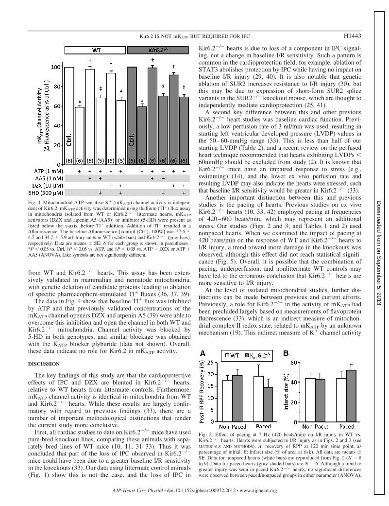
The data in Fig. 4 show that baseline Tl� flux was inhibitedby ATP and that previously validated concentrations of themKATP channel openers DZX and atpenin A5 (39) were able toovercome this inhibition and open the channel in both WT andKir6.2�/� mitochondria. Channel activity was blocked by5-HD in both genotypes, and similar blockage was obtainedwith the KATP blocker glyburide (data not shown). Overall,these data indicate no role for Kir6.2 in mKATP activity.
DISCUSSION
The key findings of this study are that the cardioprotectiveeffects of IPC and DZX are blunted in Kir6.2�/� hearts,relative to WT hearts from littermate controls. Furthermore,mKATP channel activity is identical in mitochondria from WTand Kir6.2�/� hearts. While these results are largely confir-matory with regard to previous findings (33), there are anumber of important methodological distinctions that renderthe current study more conclusive.
First, all cardiac studies to date on Kir6.2�/� mice have usedpure-bred knockout lines, comparing these animals with sepa-rately bred lines of WT mice (10, 11, 31–33). Thus it wasconcluded that part of the loss of IPC observed in Kir6.2�/�
mice could have been due to a greater baseline I/R sensitivityin the knockouts (33). Our data using littermate control animals(Fig. 1) show this is not the case, and the loss of IPC in
Kir6.2�/� hearts is due to loss of a component in IPC signal-ing, not a change in baseline I/R sensitivity. Such a pattern iscommon in the cardioprotection field; for example, ablation ofSTAT3 abolishes protection by IPC while having no impact onbaseline I/R injury (29, 40). It is also notable that geneticablation of SUR2 increases resistance to I/R injury (30), butthis may be due to expression of short-form SUR2 splicevariants in the SUR2�/� knockout mouse, which are thought toindependently mediate cardioprotection (25, 41).
A second key difference between this and other previousKir6.2�/� heart studies was baseline cardiac function. Previ-ously, a low perfusion rate of 3 ml/min was used, resulting instarting left ventricular developed pressure (LVDP) values inthe 50–60-mmHg range (33). This is less than half of ourstarting LVDP (Table 2), and a recent review on the perfusedheart technique recommended that hearts exhibiting LVDPs 60mmHg should be excluded from study (2). It is known thatKir6.2�/� mice have an impaired response to stress (e.g.,swimming) (14), and the lower ex vivo perfusion rate andresulting LVDP may also indicate the hearts were stressed, suchthat baseline I/R sensitivity would be greater in Kir6.2�/� (33).
Another important distinction between this and previousstudies is the pacing of hearts. Previous studies on ex vivoKir6.2�/� hearts (10, 33, 42) employed pacing at frequenciesof 420–600 beats/min, which may represent an additionalstress. Our studies (Figs. 2 and 3; and Tables 1 and 2) usednonpaced hearts. When we examined the impact of pacing at420 beats/min on the response of WT and Kir6.2�/� hearts toI/R injury, a trend toward more damage in the knockouts wasobserved, although this effect did not reach statistical signifi-cance (Fig. 5). Overall, it is possible that the combination ofpacing, underperfusion, and nonlittermate WT controls mayhave led to the erroneous conclusion that Kir6.2�/� hearts aremore sensitive to I/R injury.
At the level of isolated mitochondrial studies, further dis-tinctions can be made between previous and current efforts.Previously, a role for Kir6.2�/� in the activity of mKATP hadbeen precluded largely based on measurements of flavoproteinfluorescence (33), which is an indirect measure of mitochon-drial complex II redox state, related to mKATP by an unknownmechanism (19). This indirect measure of K� channel activity
Fig. 4. Mitochondrial ATP-sensitive K� (mKATP) channel activity is indepen-dent of Kir6.2. mKATP activity was determined using thallium (Tl�) flux assayin mitochondria isolated from WT or Kir6.2�/� littermate hearts. mKATP
activators [DZX and atpenin A5 (AA5)] or inhibitor (5-HD) were present aslisted below the x-axis, before Tl� addition. Addition of Tl� resulted in afluorescence. The baseline fluorescence [control (Ctrl), 100%] was 37.6 �4.7 and 34.7 � 5.9 arbitrary units in WT (white bars) and Kir6.2�/� (gray bars),respectively. Data are means � SE; N for each group is shown in parentheses .*P 0.05 vs. Ctrl, †P 0.05 vs. ATP, and ‡P 0.05 vs. ATP � DZX or ATP �AA5 (ANOVA). Like symbols are not significantly different.
Fig. 5. Effect of pacing at 7 Hz (420 beats/min) on I/R injury in WT vs.Kir6.2�/� hearts. Hearts were subjected to I/R injury as in Figs. 2 and 3 (seeMATERIALS AND METHODS). A: recovery of RPP at 120 min time point, aspercentage of initial. B: infarct size (% of area at risk). All data are means �SE. Data for nonpaced hearts (white bars) are reproduced from Fig. 2 (N � 8to 9). Data for paced hearts (gray-shaded bars) are N � 6. Although a trend togreater injury was seen in paced Kir6.2�/� hearts, no significant differenceswere observed between paced/nonpaced groups in either parameter (ANOVA).
H1443Kir6.2 IS NOT mKATP BUT REQUIRED FOR IPC
AJP-Heart Circ Physiol • doi:10.1152/ajpheart.00972.2012 • www.ajpheart.org
on Septem
ber 5, 2013D
ownloaded from
was further complicated by the use of a high concentration ofDZX (100 �M), which may have mKATP independent effects[e.g., uncoupling or complex II inhibition (18, 26)]. We mea-sured mitochondrial Tl� flux, which is a more direct measureof actual mKATP activity, and used a low dose of DZX (10 �M)or the potent mKATP agonist atpenin A5 (1 nM). This enablesa greater degree of confidence in the conclusion that Kir6.2�/�
is not part of the mKATP channel.The molecular origin of the small amount of cardioprotec-
tion still present in Kir6.2�/� hearts exposed to IPC or 30 �MDZX is unclear. It is known that Kir6.2�/� mice exhibit largechanges in the metabolic proteome (1) and IPC is known tomodulate energy metabolism (11). In addition loss of Kir6.2has been reported to elicit a shortened action potential durationin cardiac myocytes (33), which may alter responses to I/Rinjury. Thus, long term effects of Kir6.2 loss may upregulateother protective signaling pathways that can be recruited byIPC or DZX in the absence of the channel. Furthermore, as Fig.3 shows, higher concentrations of DZX were able to overridethe lack of protection, possibly via recruitment of nonspecificpathways. Notably, the fact that we saw a blunted cardiopro-tective responses in Kir6.2�/� using a blood-free perfusedheart (Fig. 2) suggests the segment of protection that is attrib-utable to Kir6.2 is a cardiac phenomenon and does not involveleukocytes or other factors absent from the perfused heart.
Examining the data in our study altogether, an importantparadox arises: Kir6.2�/� is not part of the mKATP channel andis not a target of DZX in isolated mitochondria, but DZX-mediated protection (at appropriate concentrations) is stillblunted in Kir6.2�/� hearts. The off-target effects of DZX suchas inhibition of mitochondrial complex II (18, 26) are generallythought to occur at high concentrations (100 �M), whereaslower concentrations (e.g., 30 �M) are thought to be specificfor mKATP (4, 9). Notably, however, the sKATP blockerHRM1098 can prevent DZX-induced cardioprotection in themouse (32) and can partially block DZX-induced cardiopro-tection in the rabbit (34). While this might suggest sKATP
(which undoubtedly contains Kir6.2) is a target of DZX, thefact that 100 �M DZX was protective in Kir6.2�/� heartssuggests this is not the case. It is also possible that the differenteffects of low versus high doses of DZX in WT versusKir6.2�/� hearts may represent a simple difference in the DZXdose response.
The present conclusions that Kir6.2 is not involved inmKATP and only minimally involved in IPC- or low-doseDZX-mediated protection are somewhat consistent with a re-cent report that the mKATP channel is derived from Kir 1.1 (therenal outer medullary K� channel, ROMK) (5). However, theassignment of ROMK as the mKATP conflicts with pharmaco-logical data on the sensitivity of either channel to ATP andfluoxetine (15, 16, 22, 38, 39).
Regarding the possibility that the mKATP channel maycontain the 6.1 isotype of Kir, although this channel is DZXsensitive, several pieces of evidence rule down this protein asa mKATP channel candidate, including studies on the pharma-cological properties of the mKATP (39), dominant-negativegene transfer models (27) and indirect measures of channelactivity (21). Moreover, commonly used antibodies that orig-inally used to implicate Kir 6.1 as the mKATP were recentlyshown to recognize off-target mitochondrial proteins (6). Fur-thermore, we found that mKATP activity and IPC are indepen-
dent of all isotypes of Kir channel in Caenorhabditis elegans(36), which suggests the mKATP might not contain a Kir at all.Clearly, cardiac-specific knockouts of ROMK and other K�
channels may aid in elucidating the identity of mKATP and itsrole in cardioprotection.
In conclusion, Kir6.2 plays a role in cardioprotection byIPC, but not by forming the mKATP channel. Furthermore,Kir6.2 appears to play only a partial role in cardioprotection bylow doses of DZX, with the remainder of DZX-mediatedcardioprotection being accounted for by other Kir6.2-indepen-dent mechanisms, which are as yet unknown. Recent advancesin the genetic identification of mKATP components (5, 36)suggest that future approaches should explore both Kir andnon-Kir genes as diverse candidates for both mKATP activityand cardioprotection.
GRANTS
This work was supported in part by an American Heart Association,Founder’s Affiliate Postdoctoral Fellowship award 11POST7290028 (to A. P.Wojtovich) and by National Institute of General Medical Sciences GrantGM-087483 (to P. S. Brookes and K. Nehrke).
DISCLOSURES
No conflicts of interest, financial or otherwise, are declared by the author(s).
AUTHOR CONTRIBUTIONS
A.P.W. and W.R.U. performed experiments; A.P.W. interpreted results ofexperiments; A.P.W. prepared figures; A.P.W. drafted manuscript; S.C.,A.B.F., K.N., and P.S.B. conception and design of research; S.C., A.B.F.,K.N., and P.S.B. edited and revised manuscript; K.N. and P.S.B. analyzed data;P.S.B. approved final version of manuscript.
REFERENCES
1. Arrell DK, Zlatkovic LJ, Yamada S, Terzic A. K(ATP) channel-dependent metaboproteome decoded: systems approaches to heart failureprediction, diagnosis, and therapy. Cardiovasc Res 90: 258–266, 2011.
2. Bell RM, Mocanu MM, Yellon DM. Retrograde heart perfusion: theLangendorff technique of isolated heart perfusion. J Mol Cell Cardiol 50:940–950, 2011.
3. Facundo HT, Fornazari M, Kowaltowski AJ. Tissue protection medi-ated by mitochondrial K� channels. Biochim Biophys Acta 1762: 202–212, 2006.
4. Faivre JF, Findlay I. Effects of tolbutamide, glibenclamide and diazoxideupon action potentials recorded from rat ventricular muscle. BiochimBiophys Acta 984: 1–5, 1989.
5. Foster DB, Ho AS, Rucker J, Garlid AO, Chen L, Sidor A, Garlid KD,O=Rourke B. Mitochondrial ROMK channel is a molecular component ofmitoKATP. Circ Res 111: 446–454, 2012.
6. Foster DB, Rucker JJ, Marban E. Is Kir6.1 a subunit of mitoK(ATP)?Biochem Biophys Res Commun 366: 649–656, 2008.
7. Garlid KD, Halestrap AP. The mitochondrial K(ATP) channel—fact orfiction? J Mol Cell Cardiol 52: 578–583, 2012.
8. Garlid KD, Paucek P, Yarov-Yarovoy V, Murray HN, Darbenzio RB,D’Alonzo AJ, Lodge NJ, Smith MA, Grover GJ. Cardioprotective effectof diazoxide and its interaction with mitochondrial ATP-sensitive K�
channels. Possible mechanism of cardioprotection. Circ Res 81: 1072–1082, 1997.
9. Garlid KD, Paucek P, Yarov-Yarovoy V, Sun X, Schindler PA. Themitochondrial KATP channel as a receptor for potassium channel openers.J Biol Chem 271: 8796–8799, 1996.
10. Gumina RJ, O’Cochlain DF, Kurtz CE, Bast P, Pucar D, Mishra P,Miki T, Seino S, Macura S, Terzic A. KATP channel knockout worsensmyocardial calcium stress load in vivo and impairs recovery in stunnedheart. Am J Physiol Heart Circ Physiol 292: H1706–H1713, 2007.
11. Gumina RJ, Pucar D, Bast P, Hodgson DM, Kurtz CE, Dzeja PP, MikiT, Seino S, Terzic A. Knockout of Kir6.2 negates ischemic precondition-ing-induced protection of myocardial energetics. Am J Physiol Heart CircPhysiol 284: H2106–H2113, 2003.
H1444 Kir6.2 IS NOT mKATP BUT REQUIRED FOR IPC
AJP-Heart Circ Physiol • doi:10.1152/ajpheart.00972.2012 • www.ajpheart.org
on Septem
ber 5, 2013D
ownloaded from
12. Guo Y, Flaherty MP, Wu WJ, Tan W, Zhu X, Li Q, Bolli R. Geneticbackground, gender, age, body temperature, and arterial blood pH have amajor impact on myocardial infarct size in the mouse and need to becarefully measured and/or taken into account: results of a comprehensiveanalysis of determinants of infarct size in 1,074 mice. Basic Res Cardiol107: 288, 2012.
13. Hibino H, Inanobe A, Furutani K, Murakami S, Findlay I, Kurachi Y.Inwardly rectifying potassium channels: their structure, function, andphysiological roles. Physiol Rev 90: 291–366, 2010.
14. Kane GC, Behfar A, Yamada S, Perez-Terzic C, O’Cochlain F, ReyesS, Dzeja PP, Miki T, Seino S, Terzic A. ATP-sensitive K� channelknockout compromises the metabolic benefit of exercise training, resultingin cardiac deficits. Diabetes 53, Suppl 3: S169–S175, 2004.
15. Kobayashi T, Washiyama K, Ikeda K. Inhibition of G protein-activatedinwardly rectifying K� channels by fluoxetine (Prozac). Br J Pharmacol138: 1119–1128, 2003.
16. Kobayashi T, Washiyama K, Ikeda K. Modulators of G protein-acti-vated inwardly rectifying K� channels: potentially therapeutic agents foraddictive drug users. Ann NY Acad Sci 1025: 590–594, 2004.
17. Korte T, Fuchs M, Guener Z, Bonin J, de Sousa M, Niehaus M,Tebbenjohanns J, Drexler H. In-vivo electrophysiological study in micewith chronic anterior myocardial infarction. J Interv Card Electrophysiol6: 121–132, 2002.
18. Kowaltowski AJ, Seetharaman S, Paucek P, Garlid KD. Bioenergeticconsequences of opening the ATP-sensitive K� channel of heart mito-chondria. Am J Physiol Heart Circ Physiol 280: H649–H657, 2001.
19. Liu Y, Sato T, O’Rourke B, Marban E. Mitochondrial ATP-dependentpotassium channels: novel effectors of cardioprotection? Circulation 97:2463–2469, 1998.
20. Miki T, Nagashima K, Tashiro F, Kotake K, Yoshitomi H, TamamotoA, Gonoi T, Iwanaga T, Miyazaki J, Seino S. Defective insulin secretionand enhanced insulin action in KATP channel-deficient mice. Proc NatlAcad Sci USA 95: 10402–10406, 1998.
21. Miki T, Suzuki M, Shibasaki T, Uemura H, Sato T, Yamaguchi K,Koseki H, Iwanaga T, Nakaya H, Seino S. Mouse model of Prinzmetalangina by disruption of the inward rectifier Kir6.1. Nat Med 8: 466–472,2002.
22. Mironova GD, Negoda AE, Marinov BS, Paucek P, Costa AD, Grig-oriev SM, Skarga YY, Garlid KD. Functional distinctions between themitochondrial ATP-dependent K� channel (mitoKATP) and its inwardrectifier subunit (mitoKIR). J Biol Chem 279: 32562–32568, 2004.
23. Murry CE, Jennings RB, Reimer KA. Preconditioning with ischemia: adelay of lethal cell injury in ischemic myocardium. Circulation 74:1124–1136, 1986.
24. Nichols CG. KATP channels as molecular sensors of cellular metabolism.Nature 440: 470–476, 2006.
25. Pu JL, Ye B, Kroboth SL, McNally EM, Makielski JC, Shi NQ.Cardiac sulfonylurea receptor short form-based channels confer a gliben-clamide-insensitive KATP activity. J Mol Cell Cardiol 44: 188–200,2008.
26. Schafer G, Wegener C, Portenhauser R, Bojanovski D. Diazoxide, aninhibitor of succinate oxidation. Biochem Pharmacol 18: 2678–2681,1969.
27. Seharaseyon J, Ohler A, Sasaki N, Fraser H, Sato T, Johns DC,O’Rourke B, Marban E. Molecular composition of mitochondrial ATP-sensitive potassium channels probed by viral Kir gene transfer. J Mol CellCardiol 32: 1923–1930, 2000.
28. Sellitto AD, Al-Dadah AS, Schuessler RB, Nichols CG, Lawton JS. Anopen sarcolemmal adenosine triphosphate-sensitive potassium channel isnecessary for detrimental myocyte swelling secondary to stress. Circula-tion 124: S70–S74, 2011.
29. Smith RM, Suleman N, Lacerda L, Opie LH, Akira S, Chien KR, SackMN. Genetic depletion of cardiac myocyte STAT-3 abolishes classicalpreconditioning. Cardiovasc Res 63: 611–616, 2004.
30. Stoller D, Kakkar R, Smelley M, Chalupsky K, Earley JU, Shi NQ,Makielski JC, McNally EM. Mice lacking sulfonylurea receptor 2(SUR2) ATP-sensitive potassium channels are resistant to acute cardio-vascular stress. J Mol Cell Cardiol 43: 445–454, 2007.
31. Suzuki M, Li RA, Miki T, Uemura H, Sakamoto N, Ohmoto-Sekine Y,Tamagawa M, Ogura T, Seino S, Marban E, Nakaya H. Functionalroles of cardiac and vascular ATP-sensitive potassium channels clarifiedby Kir6.2-knockout mice. Circ Res 88: 570–577, 2001.
32. Suzuki M, Saito T, Sato T, Tamagawa M, Miki T, Seino S, Nakaya H.Cardioprotective effect of diazoxide is mediated by activation of sarcolem-mal but not mitochondrial ATP-sensitive potassium channels in mice.Circulation 107: 682–685, 2003.
33. Suzuki M, Sasaki N, Miki T, Sakamoto N, Ohmoto-Sekine Y, Tama-gawa M, Seino S, Marban E, Nakaya H. Role of sarcolemmal K(ATP)channels in cardioprotection against ischemia/reperfusion injury in mice. JClin Invest 109: 509–516, 2002.
34. Tanno M, Miura T, Tsuchida A, Miki T, Nishino Y, Ohnuma Y,Shimamoto K. Contribution of both the sarcolemmal K(ATP) and mito-chondrial K(ATP) channels to infarct size limitation by K(ATP) channelopeners: differences from preconditioning in the role of sarcolemmalK(ATP) channels. Naunyn Schmiedebergs Arch Pharmacol 364: 226–232, 2001.
35. Wojtovich AP, Brookes PS. The complex II inhibitor atpenin A5 protectsagainst cardiac ischemia-reperfusion injury via activation of mitochondrialKATP channels. Basic Res Cardiol 104: 121–129, 2009.
36. Wojtovich AP, DiStefano P, Sherman T, Brookes PS, Nehrke K.Mitochondrial ATP-sensitive potassium channel activity and hypoxicpreconditioning are independent of an inwardly rectifying potassiumchannel subunit in Caenorhabditis elegans. FEBS Lett 586: 428–434,2012.
37. Wojtovich AP, Sherman TA, Nadtochiy SM, Urciuoli WR, BrookesPS, Nehrke K. SLO-2 is cytoprotective and contributes to mitochondrialpotassium transport. PLoS One 6: e28287, 2011.
38. Wojtovich AP, Smith CO, Haynes CM, Nehrke KW, Brookes PS.Physiological consequences of complex II inhibition for aging, disease,and the mKATP channel. Biochim Biophys Acta 1827: 598–611, 2013.
39. Wojtovich AP, Williams DM, Karcz MK, Lopes CM, Gray DA,Nehrke KW, Brookes PS. A novel mitochondrial KATP channel assay.Circ Res 106: 1190–1196, 2010.
40. Xuan YT, Guo Y, Han H, Zhu Y, Bolli R. An essential role of theJAK-STAT pathway in ischemic preconditioning. Proc Natl Acad Sci USA98: 9050–9055, 2001.
41. Ye B, Kroboth SL, Pu JL, Sims JJ, Aggarwal NT, McNally EM,Makielski JC, Shi NQ. Molecular identification and functional charac-terization of a mitochondrial sulfonylurea receptor 2 splice variant gener-ated by intraexonic splicing. Circ Res 105: 1083–1093, 2009.
42. Zingman LV, Hodgson DM, Bast PH, Kane GC, Perez-Terzic C,Gumina RJ, Pucar D, Bienengraeber M, Dzeja PP, Miki T, Seino S,Alekseev AE, Terzic A. Kir6.2 is required for adaptation to stress. ProcNatl Acad Sci USA 99: 13278–13283, 2002.
H1445Kir6.2 IS NOT mKATP BUT REQUIRED FOR IPC
AJP-Heart Circ Physiol • doi:10.1152/ajpheart.00972.2012 • www.ajpheart.org
on Septem
ber 5, 2013D
ownloaded from