knockdown of the c. elegans kinome identifies kinases required for
TRANSCRIPT

Lehmann et al. Cell Communication and Signaling 2013, 11:71http://www.biosignaling.com/content/11/1/71
RESEARCH Open Access
Knockdown of the C. elegans Kinome identifiesKinases required for normal protein Homeostasis,Mitochondrial network structure, and Sarcomerestructure in muscleSusann Lehmann, Joseph J Bass and Nathaniel J Szewczyk*
Abstract
Background: Kinases are important signalling molecules for modulating cellular processes and major targets ofdrug discovery programs. However, functional information for roughly half the human kinome is lacking. Weconducted three kinome wide, >90%, RNAi screens and epistasis testing of some identified kinases against knownintramuscular signalling systems to increase the functional annotation of the C. elegans kinome and expand ourunderstanding of kinome influence upon muscle protein degradation.
Results: 96 kinases were identified as required for normal protein homeostasis, 74 for normal mitochondrialnetworks and 50 for normal sarcomere structure. Knockdown of kinases required only for normal proteinhomeostasis and/or mitochondrial structure was significantly less likely to produce a developmental or behaviouralphenotype than knockdown of kinases required for normal sarcomere structure and/or other sub-cellular processes.Lastly, assessment of kinases for which knockdown produced muscle protein degradation against the knownregulatory pathways in C. elegans muscle revealed that close to half of kinase knockdowns activated autophagy in aMAPK dependent fashion.
Conclusions: Roughly 40% of kinases studied, 159 of 397, are important in establishing or maintaining muscle cellhealth, with most required for both. For kinases where decreased expression triggers protein degradation,autophagy is most commonly activated. These results increase the annotation of the C. elegans kinome to roughly75% and enable future kinome research. As 33% of kinases identified have orthologues expressed in human muscle,our results also enable testing of whether identified kinases function similarly in maintaining human musclehomeostasis.
Keywords: Autophagy, C. elegans, Kinome, MAPK, Mitochondria, Muscle, Protein degradation, Proteostasis,Sarcomere
BackgroundKinases are enzymes that alter proteins and lipids byphosphorylation, the addition of a phosphate group. Thismodification can influence a protein’s steric structureand cause changes in protein-protein binding affinitiesand enzyme activities [1]. Kinase encoding genes consti-tute 2% of the human genome making kinases one of
* Correspondence: [email protected] Research Council/Arthritis Research UK Centre for MusculoskeletalAgeing Research, University of Nottingham, Royal Derby Hospital, DerbyDE22 3DT, England
© 2013 Lehmann et al.; licensee BioMed CentCommons Attribution License (http://creativecreproduction in any medium, provided the or
the largest protein families, which is termed the kinome.Kinases also appear to play a major role in modulatingcellular processes as roughly 30% of intracellular proteinsare phosphate bound at any given time [2]. Accordingly,kinase inhibitors account for a large part of drug discoveryprograms in the pharmaceutical industry. Roughly 150inhibitors of 42 well validated kinase targets are cur-rently being tested in clinical trials [3]. These 42 targetsconstitute only about 8% of the kinome. Although fur-ther progress is being made in identifying the functionof already well-known kinases in the regulation of
ral Ltd. This is an Open Access article distributed under the terms of the Creativeommons.org/licenses/by/2.0), which permits unrestricted use, distribution, andiginal work is properly cited.

Lehmann et al. Cell Communication and Signaling 2013, 11:71 Page 2 of 17http://www.biosignaling.com/content/11/1/71
specific cellular processes, 50% of the kinome remainslargely uncharacterized [3]. Thus, we lack an under-standing of the complexity of process modulation by aconsiderable part of the kinome. In this study, we took amulti-level functional genomics approach to identifykinases required for normal function of individual andmultiple processes within a single tissue in vivo.To accomplish this work we employed the soil nematode
Caenorhabditis elegans as it is a convenient multicellularorganism for systems biology research [4]. The C. eleganskinome contains 438 kinases which have been assigned to168 subfamilies [5]. Of these subfamilies, 153 are sharedwith the human kinome [5]. This conservation suggeststhat 81% of human kinases have homologues in C. elegans.Despite knowledge of these kinase encoding genes, severalfunctional genomic screens looking at the developmentaland behavioural effects of RNA interference againsteach gene in the genome [6,7], and the effort to knockout every gene in the genome [4], the C. elegans kinomestill appears understudied. A search of the C. elegansdatabase, www.wormbase.org [8], reveals that only roughly60% of all kinase-encoding genes have been assigned agenetic or RNAi phenotype.As several past studies failed to detect a developmental
or behavioural effect of RNAi against kinase encodinggenes, we studied the effect of kinase knockdown byRNAi on subcellular processes within muscle. We chosemuscle as it is a highly regulated, adaptable tissue thatresponds to environmental inputs such as use and nutri-tion in a balanced fashion in order to maintain whole bodyhomeostasis. Additionally, identification of new therapeutictargets for modulating muscle homeostasis is desirable asinability to maintain muscle can become a major healthconcern. Severe wasting of muscle is associated with condi-tions such as disuse, starvation, several diseases, and inevit-ably occurs in the elderly [9].To study the kinome requirement for establishing and
maintaining cellular homeostasis, we picked two pro-cesses that occur in all cell types, protein homeostasisand mitochondrial dynamics, and one process that isspecific to muscle, sarcomere assembly and maintenance.We obtained previously utilized RNAi constructs and ex-amined the effect of knockdown of each kinase upon eachprocess in muscle. We established that 159 kinase-encoding genes, 40% of the C. elegans kinome screened[5], appear to influence sub-cellular processes withinmuscle. Of these 159 genes, 64% appear to be required tomaintain homeostasis of fully differentiated adult muscle,32% appear to be required for multiple sub-cellular pro-cesses, and 50% have identified human orthologues [5,10]of which 53 are reported to be expressed in human skel-etal muscle [11] (Additional file 1). This quantifies thekinome requirement for normal development and main-tenance of a single tissue in vivo and assigns RNAi
phenotypes to 51 kinases for which no phenotype was pre-viously assigned by genetic or RNAi approaches. Similarto a past study of genes known to influence muscle func-tion [12], we found that individual kinases were most fre-quently required for proper protein homeostasis and leastfrequently required for proper sarcomere structure. Thissuggests that past studies aimed at understanding genomiccontrol of sarcomere structure [13,14] have only begun touncover the complexity of genomic control of muscle.To better understand the nature of the kinome re-
quirement for maintenance of muscle homeostasis, weperformed epistasis tests with kinases whose knockdowntriggered muscle protein degradation against known pro-teolytic signalling mechanisms in C. elegans muscle.While knockdown of individual kinases triggered differ-ent types of protein degradation, mitogen activated pro-tein kinase MPK-1 dependent autophagy was triggeredin close to half of the knockdowns that triggered degrad-ation. Our results not only contribute to the global un-derstanding of the kinome, they may also lead to thediscovery of new therapeutic targets for the modulationof muscle homeostasis.
ResultsKinases required for normal protein synthesis anddegradation in muscleModulation of global protein synthesis, degradation, orboth, can lead to either cellular hypertrophy or atrophy. Inmulticellular organisms these processes must be regulatedsuch that adjacent cells can grow or shrink together. Forexample, adult C. elegans muscle and hypodermis undergocoordinated hypertrophy [15]. Similarly, regulation is re-quired so that one tissue does not receive inordinate nutri-ents. For example, tumours circumvent such regulation. Inman, several extramuscular signals are known to affectprotein homeostasis in muscle and are thought to do so viaa number of mechanisms, rather than via a single mech-anism [16]; however in many cases the transducing sig-nals or mechanisms are not known or are inadequatelyunderstood.To study the effects of altered kinase signalling on
cytosolic protein homeostasis, termed proteostasis, weused a well-established C. elegans model [17,18]. Inthis model a myosin heavy chain gene promoter andenhancer drive the expression of a lacZ reporter trans-gene to report on alterations in muscle protein synthesis.As β-galactosidase synthesis stops when animals reachadulthood and the β-galactosidase remains stable for atleast 72 hours [17], loss of β-galactosidase in responseto acute interventions indicates that degradation, not areduction in protein synthesis, is occurring in fullydifferentiated adult muscles. Chronic RNAi knockdownof 96 genes resulted in altered levels of β-galactosidaseactivity, suggesting that these genes are required for
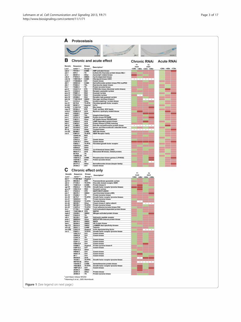
Figure 1 (See legend on next page.)
Lehmann et al. Cell Communication and Signaling 2013, 11:71 Page 3 of 17http://www.biosignaling.com/content/11/1/71

(See figure on previous page.)Figure 1 Kinases that appear to be required for normal muscle protein synthesis and/or degradation. (A) Sample images of normalβ-galactosidase reporter staining (dark blue) in control, non-RNAi treated, PD55 animal (left) and RNAi treated animals showing decreasedβ-galactosidase reporter staining. RNAi treatments are indicated in the lower right corner. Scale bars represent 100 μm. (B) Kinases identified asrequired for normal lack of protein degradation in muscle. Indicated RNAi treatments were conducted chronically and followed up acutely andscored for decreased β-galactosidase reporter staining, see materials and methods. Displayed are scoring data for each time point (n = 20-30).Green indicates staining not appreciably different from controls. Red indicates that at least 25% of scored animals displayed lighter/diffuse blue orlack of blue staining versus controls. Dark red indicates that at least 50% of scored animals displayed light/diffuse blue or lack of blue stainingversus controls. (C) Kinases identified as required for normal protein homeostasis in muscle. RNAi treatments were conducted and scored as in(B) however the listed treatments produced a defect in the chronic screen but not the acute screen. – Indicates lack of data for a given timepoint. ‡ Indicates a treatment was classed as producing a lack of progeny in this screen.
Lehmann et al. Cell Communication and Signaling 2013, 11:71 Page 4 of 17http://www.biosignaling.com/content/11/1/71
proper regulation of protein synthesis and/or degrad-ation (Figure 1). At least some of these 96 kinases arelikely to be required for normal protein synthesis duringdevelopment, for example EGL-15 which is required forproper muscle development [19], but further experi-ments are required to confirm to what extent genesidentified in the chronic screen may regulate synthesis,degradation, or both. To determine if decreased levelsof β-galactosidase activity could be accounted for by in-creased protein degradation alone, we knocked downthe identified genes acutely. We also acutely knockeddown genes for which a lack of progeny prohibited fullanalysis in the chronic screen. Using this approach weidentified 48 kinases that appear to be required to pre-vent abnormal muscle protein degradation (Figure 1).Two of the 48 kinases, pat-4 and unc-82, have recentlybeen shown to be negative regulators of muscle proteindegradation in C. elegans [15] and two, gsk-3 [20] andsgk-1 [21], are known to interact with insulin signallingwhich is also a known regulator of muscle protein deg-radation in C. elegans [22]. Thus, some of the resultsfrom the screen appear to validate our approach. Asexpected, several kinases without known phenotypes inmutants or in response to RNAi were found to be re-quired for normal protein synthesis and/or degradationor required to prevent abnormal protein degradation(Additional file 1). Lastly, the identification of ire-1 andpek-1, which are well conserved regulators of the endo-plasmic reticulum unfolded protein response [23] demon-strates that, as expected, some of the identified kinases actto maintain protein homeostasis not just in muscle, but inmost tissues.
Kinases required for normal mitochondrial networkstructureIt is now widely accepted that mitochondrial networks aredynamic, undergoing morphological changes to maintainorganelle homeostasis or to respond to the metabolicchanges within the cell [24]. Advances have been made indetermining the mechanisms of mitochondrial organellarquality control; it is however still unclear how mitochon-dria integrate multiple cellular signals into fission and
fusion processes and to what extent mitochondria areself-regulated.In order to gain insight into the kinome requirement
for normal mitochondrial structure in muscle, we exam-ined mitochondrial morphology in C. elegans containingmitochondrial localized GFP [25] (Figure 2). ChronicRNAi knockdown of 74 kinase encoding genes induceda fragmented mitochondrial network suggesting thatthese kinases are required for proper establishment and/or maintenance of the mitochondrial network in muscle.Included within these 74 genes is pink-1, which encodesPTEN-induced putative kinase 1, and which when mu-tated in Drosophila melanogaster is known to inducemitochondrial morphology defects in muscle as well asother tissues [26]. Utilizing a pink-1 knockout allele, seematerials and methods, we confirmed that loss of func-tion of pink-1 results in fragmentation of the mitochon-drial network in muscle. To determine if disruptedmitochondrial morphology could be attributed to a kin-ase requirement for maintenance of the mitochondrialnetwork in muscle we knocked down the identifiedkinase encoding genes acutely in adults. We also acutelyknocked down genes for which a lack of progenyprohibited full analysis in the chronic screen. Using thisapproach we identified 44 kinases that appear to berequired for proper maintenance of the mitochondrialnetwork in muscle (Figure 2). Within this set of 44 genesis kin-1, which is already known to be involved in themaintenance of mitochondrial networks [27]. As was thecase with the screen for kinases required for proteinhomeostasis, the identification of known regulators ofmitochondrial dynamics appears to support our ap-proach and, as expected, several kinases without knownphenotypes in mutants or in response to RNAi werefound to be required for normal mitochondrial networkstructure (Additional file 1).
Kinases required for normal sarcomere assembly andmaintenanceIn addition to studying two processes that occur inmost tissues, we studied the kinome requirement for amuscle specific process. For this we chose sarcomereassembly and maintenance, as the most recognized
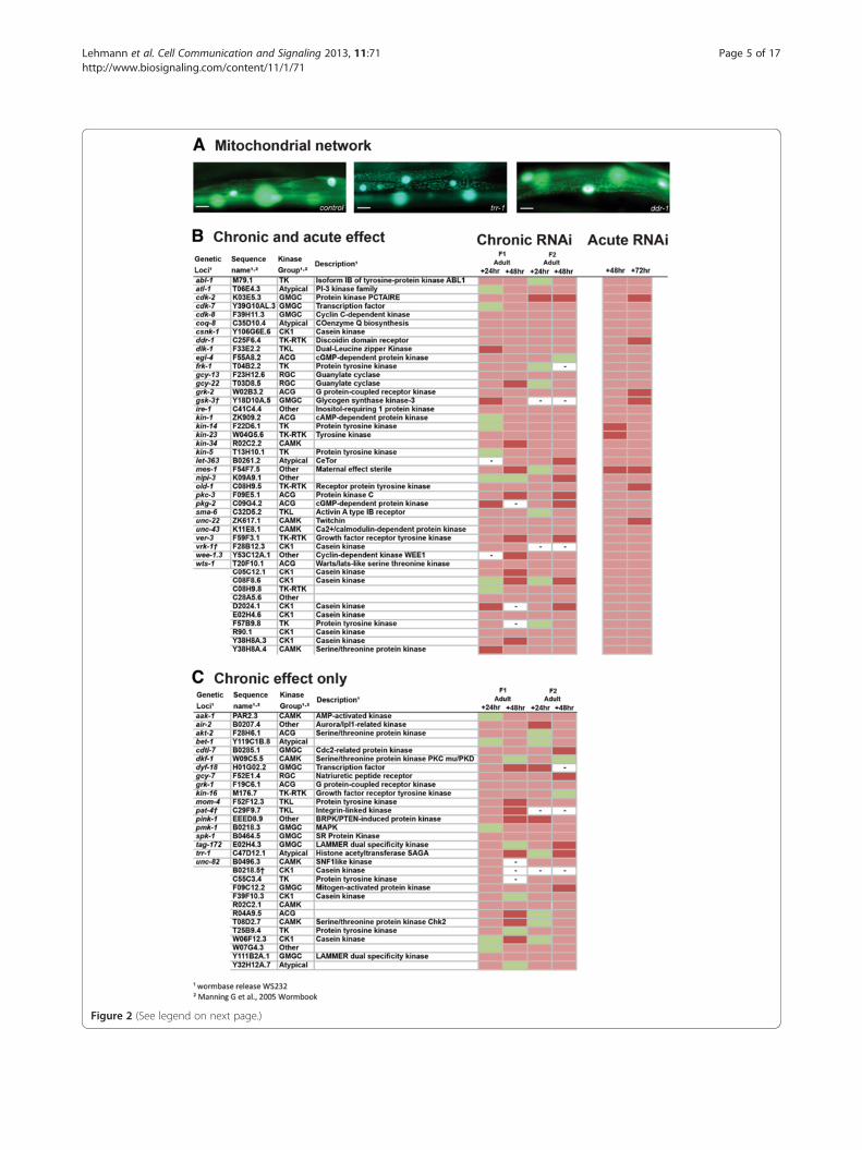
Figure 2 (See legend on next page.)
Lehmann et al. Cell Communication and Signaling 2013, 11:71 Page 5 of 17http://www.biosignaling.com/content/11/1/71

(See figure on previous page.)Figure 2 Kinases that appear to be required for normal mitochondrial network structure. (A) Sample images of GFP labelledmitochondrial networks, and nuclei (large over exposed circles), showing normal mitochondrial network structure in control, non-RNAi treated,CB5600 animal (left) and RNAi treated animals showing disrupted network structure. RNAi treatments are indicated in the lower right corner.Scale bars represent 20 μm. (B) Kinases identified as required for normal maintenance of mitochondrial network structure in adult muscle.Indicated RNAi treatments were conducted chronically and followed up acutely and scored for loss of mitochondrial network organization, seematerials and methods. Displayed are scoring data for each time point (n = 20-30). Green indicates networks not appreciably different fromcontrols. Red indicates that at least 25% of scored animals displayed disrupted mitochondrial network structure versus controls. Dark red indicatesthat at least 50% of scored animals displayed disrupted mitochondrial network structure versus controls. (C) Kinases identified as required fornormal mitochondrial network structure. RNAi treatments were conducted and scored as in (B) however the listed treatments produced a defectin the chronic screen but not the acute screen. – Indicates lack of data for a given time point. † Indicates a treatment was classed as producing alack of progeny in this screen.
Lehmann et al. Cell Communication and Signaling 2013, 11:71 Page 6 of 17http://www.biosignaling.com/content/11/1/71
function of skeletal muscle is to enable body move-ment through the contraction and relaxation of manyhighly organized sarcomeric units. Additionally, as sarco-mere structure is conserved from C. elegans through highermetazoans, C. elegans has become a well-established andvalidated model for the study of sarcomere structure andassembly [28,29].To examine the effects of kinase knockdown on sarco-
mere structure, we used animals expressing a myosin heavychain GFP that localizes to the M-line of sarcomeres [30].Chronic RNAi knockdown of 50 kinase-encoding genes in-duced disorganization or tears in the sarcomeric structure(Figure 3) suggesting that these kinases are required fornormal sarcomere assembly or maintenance. 12% of these50 genes are already known to be involved in the regulationof sarcomere structure. For example, pat-4, which encodesintegrin-linked kinase, and which is part of the muscle at-tachment complex to the basement membrane [31] andunc-89 and unc-22 which encode the structural proteinsobscurin [32] and twitchin [33], and which play a majorrole in the structural integrity of the sarcomeres. To deter-mine if disrupted sarcomere structure could be accountedfor by a kinase requirement for normal maintenance of sar-comeres, we knocked down these 50 kinase-encoding genesacutely in adults. We also acutely knocked down genes forwhich a lack of progeny prohibited full analysis in thechronic screen. Using this approach we identified 34 ki-nases that appear to be required for proper maintenance ofthe sarcomeres (Figure 3). As was the case with our othertwo screens for kinome requirement for sub-cellularprocesses within muscle, the results of this screen confirmprevious observations and identify several genes for whichno phenotype was previously assigned (Additional file 1).
Multiple subcellular defects are more likely to produce adevelopmental or behavioural phenotypeComparison of single vs. multiple defects within musclefollowing RNAi (Figure 4) revealed that kinases appear tobe most frequently required to maintain proteostasis andleast frequently required for normal sarcomere structure.This result is similar to a past RNAi screen of 159 genespreviously known to influence muscle contraction [12].
However, in contrast to this past study, which found anoverrepresentation of genes required for normal proteinhomeostasis, mitochondrial network structure and sarco-mere structure, we found that the distribution of kinases re-quired to maintain multiple processes within muscle wasnot significantly different from a normal distribution (χ2,GraphPad Prism). However, there was significant enrich-ment (p < .05, χ2, GraphPad Prism) of developmental or be-havioural phenotypes amongst the genes for which RNAiproduced defects in sarcomere structure and proteinhomeostasis and/or mitochondrial network structure(Figure 4, bottom three clusters of genes). Taken together,the results from our study and the past study [12] suggestthat past genetic screens aimed at understanding genesregulating muscle function, usually contraction, are morelikely to have identified genes that disrupt multiple sub-cellular processes within muscle than genes that disrupt asingle process (NB 9 of the 10 kinases we identified in allthree screens are well studied). The results also imply thatthe genomic control of the metabolic functions of muscle,such as protein homeostasis and mitochondrial energy pro-duction, are likely underestimated by past studies aimed atunderstanding the genomic control of sarcomere structureor function [13,14].
Lack of enrichment of any kinase group amongst kinasesidentifiedWe assessed which kinase groups were represented bythe genes identified in each of our three screens. A com-parison of the number of kinases identified from eachgroup versus the total number of kinases screened ineach group revealed no statistically significant (χ2,GraphPad Prism) enrichment of any kinase group. Thislack of enrichment was observed in each screen and forthe total set of kinases identified from all three screens(not shown).
Epistasis testing of potential degradation-regulatingkinases versus known signalsWe conducted epistasis tests to gain further functionalinsight into how some of the identified kinases are act-ing. Because there are well defined signals regulating

Figure 3 (See legend on next page.)
Lehmann et al. Cell Communication and Signaling 2013, 11:71 Page 7 of 17http://www.biosignaling.com/content/11/1/71

(See figure on previous page.)Figure 3 Kinases that appear to be required for normal sarcomere structure. (A) Sample images of GFP labelled sarcomeres showingnormally aligned sarcomeres in control, non-RNAi treated, PJ727 animal (left) and RNAi treated animals showing sarcomere structure defects.RNAi treatments are indicated in the lower right corner. Scale bars represent 20 μm. (B) Kinases identified as required for maintenance of normalsarcomere structure in adult muscle. Indicated RNAi treatments were conducted chronically and followed up acutely and scored for loss ofsarcomere organization, see materials and methods. Displayed are scoring data for each time point (n = 20-30). Green indicates sarcomeres notappreciably different from controls. Red indicates that at least 25% of scored animals displayed disrupted sarcomere structure versus controls.Dark red indicates that at least 50% of scored animals displayed disrupted sarcomere structure versus controls. (C) Kinases identified as requiredfor normal sarcomere structure. RNAi treatments were conducted and scored as in (B) however the listed treatments produced a defect in thechronic screen but not the acute screen. – Indicates lack of data for a given time point. * Indicates a treatment was classed as producing a lackof progeny in this screen.
Lehmann et al. Cell Communication and Signaling 2013, 11:71 Page 8 of 17http://www.biosignaling.com/content/11/1/71
protein degradation within C. elegans muscle [18] wefocused on epistasis testing the genes identified in theprotein degradation screen (Figure 1) against the knownsignalling pathways. In C. elegans muscle, presumptiveautophagic degradation is controlled by the balance be-tween constitutive, autocrine fibroblast growth factorreceptor-Ras-Raf-MAPK signalling [34,35] and insulingrowth factor receptor-PI3K-Akt-Raf signalling [22]. Aswith past studies we used unc-51, which encodesAutophagy-related1 (Atg1) [36], mutants to block au-tophagic protein degradation and we also used muta-tions in mpk-1, which encodes mitogen activated proteinkinase (MAPK) [37], and daf-18, which encodes phos-phatase and tensin homolog kinase [38], to map kinasesrequired to prevent cytosolic protein degradation toactivation of the autophagic signalling pathways in C.elegans. Proteasomal degradation appears to be con-trolled by plasma membrane polarization [12] and in-creased degradation by this system can be observed inresponse to starvation [17], denervation [39], andneurodegeneration [40]; as with past studies we usedthe proteasome inhibitor MG132 to block any degrad-ation which required proteasomal activity [39].For 21 of 48 kinases, β-galactosidase degradation was
suppressed in the unc-51 and mpk-1 mutant strainssuggesting that close to half of the kinases for whichknockdown triggered muscle protein degradation arecausing increased MPK-1 mediated autophagic proteindegradation (Figure 5). To confirm autophagy wastriggered by knockdown of each of the 21 kinases, weexamined the accumulation of autophagic vesicles in re-sponse to knockdown in animals expressing GFP fusedto the autophagic vesicle marker LGG-1 [36]. As shownin Figure 6, a significant increase in autophagic vesicleswas observed in response to knockdown of each of these21 genes (p < .0001, one way ANOVA, GraphPad Prism).These results suggest that autophagic protein degrad-ation is the proteolytic mechanism most commonlytriggered by decreased expression of an individual kin-ase. Consistent with this, knockdown of only 6 kinasesappeared to trigger proteasome-mediated degradation asevidenced by lack of degradation in the MG132 treat-ment condition. Two of these kinases, IRE-1 and PEK-1,
are well conserved regulators of the endoplasmaticreticulum unfolded protein response which modulatesboth autophagic and proteasomal degradation. For 21of the 48 kinases, β-galactosidase degradation wassuppressed in daf-18 animals suggesting that knock-down of these kinases alters signalling in an insulin-mediated pathway. The majority of these kinases alsorequire MPK-1 and UNC-51, which is therefore consistentwith past studies of signalling networks controlling proteindegradation in C. elegans muscle. Included in this groupare sgk-1 [21], gsk-3 [20] and the gene that encodes AMP-activated protein kinase [41], each of which is alreadyknown to be part of insulin-mediated control of proteinhomeostasis in other species. Additionally, 10 kinasesappeared to trigger an as yet unidentified proteolyticmechanism as β-galactosidase degradation upon RNAiknockdown was not suppressed in the mutants or inMG132 treated animals. One of these genes is pat-4 forwhich calpains have recently been demonstrated to be theregulated protease [15]. While future study may uncoverthe details of the other potentially novel mechanisms, it isinteresting that autophagy was triggered in response toroughly half of the kinase knockdowns in C. elegans.
DiscussionWe have used RNAi to knock down the vast majority ofthe C. elegans kinome to examine the kinome require-ment for establishing and maintaining sub-cellular pro-cesses within muscle. The use of C. elegans and RNAihas allowed us to define a preliminary in vivo functionalkinome requirement for muscle which currently remainstechnically and economically challenging to establish inrodents and infeasible in human subjects. The evolution-ary distance between the C. elegans and the humankinome suggests that not all results obtained in thisstudy will be relevant to man and that there are someimportant kinases that we have not studied as they donot exist in C. elegans. However, our identification ofkinases as required for normal subcellular processes inC. elegans muscle that are already known to regulate thesame subcellular process(es) in mammals, suggests thatsome, if not many, of our results will be relevant tohigher metazoans. Similarly, while our use of RNAi by

Figure 4 (See legend on next page.)
Lehmann et al. Cell Communication and Signaling 2013, 11:71 Page 9 of 17http://www.biosignaling.com/content/11/1/71

(See figure on previous page.)Figure 4 Kinases that appear to be required for multiple subcellular processes to be normal. (A) Kinases identified as required for normalmuscle protein homeostasis and mitochondrial structure (e.g. kinases identified in both the proteostasis screen and mitochondrial structurescreen in Figures 1 and 2), kinases identified as required for normal muscle protein homeostasis and sarcomere structure (e.g. from theproteostasis and sarcomere screens), kinases identified as required for normal mitochondrial and sarcomere structure (e.g. from the mitochondrialand sarcomere screens), and kinases identified as required for normal muscle protein homeostasis and mitochondrial and sarcomere structure(e.g. from all three screens). ‡, †, * Indicates a treatment was classed as producing a lack of progeny in the proteostasis, mitochondria, orsarcomere screen, respectively. Kinases listed on the left relate to colour coded affected subcellular processes and presence of a developmentalor behavioural phenotype on the right as described in inset legend. (B) Venn diagrams displaying number of kinases which are required for asingle process to be normal (data extracted from Figures 1, 2 and 3) and number of kinases required for multiple processes to be normal (grey)during muscle development (left) and/or in adult muscle (right). Colour codes match inset legend. Diameter of circles relate to total number ofkinases which are required for this process to be normal.
Lehmann et al. Cell Communication and Signaling 2013, 11:71 Page 10 of 17http://www.biosignaling.com/content/11/1/71
feeding has allowed us to define a preliminary in vivofunctional kinome requirement for muscle, our use ofthis single method will almost certainly have caused usto overlook certain kinases. For example, RNAi by feed-ing does not produce a reproducible quantitative knock-down from animal to animal [42,43]. Thus, negativeresults and quantitative differences in defects in re-sponse to RNAi against different genes are not interpret-able, as it is experimentally challenging to demonstratequantitative knockdown in the same worm that is scoredfor subcellular defects and economically not feasible onthe scale of the work reported here. Similarly, as theknockdowns occur in the whole animal it is not possibleto definitively conclude if it is the knockdown of thetargeted gene or another gene that is producing the ob-served defect nor is it possible to conclude if it is theknockdown in muscle, another tissue, or both that isproducing the observed defect. Clearly future studies inkinase gene mutants are required, as are demonstrationsof the tissue(s) in which each kinase is required for es-tablishment and/or maintenance of muscle homeostasis.The recent availability of knockouts for most of the C.elegans kinome [44] should facilitate such future studies.Our approach of observing several sub-cellular pro-
cesses allowed us to analyse differences in the kinomerequirement for multiple subcellular processes. For eachof the processes studied there appear to be many morespecific kinases required than kinases required for mul-tiple processes to be normal within muscle. Thus, it doesnot appear to be the case that for the majority of thekinome disruption of any one individual kinase resultsin complete loss of cellular homeostasis. Our data alsoshow that the two general cellular processes, cytosolicprotein homeostasis and mitochondrial dynamics, ap-pear to be more frequently affected by kinase RNAiknockdown than the muscle specific process of assemblyand maintenance of sarcomeres. This raises the questionwhether there has been more frequent evolutionary se-lection for kinases required for general, metabolic pro-cesses over those required for other, more specializedcellular processes. Similarly, our observations raise thequestion whether processes more heavily impacted by
kinome knockdown, such as protein homeostasis, aremore tolerant of dysregulation without catastrophic fail-ure. If so, this might explain why only 31% of defects incytosolic protein homeostasis resulted in overall defectsin behaviour or development in comparison to 48% ofdefects in sarcomeres. Together these observations sug-gest that further analysis of sub-cellular phenotypes orconditional phenotypes should allow assignment of puta-tive function to the entire kinome of C. elegans.Our use of chronic and acute RNAi screens revealed
that the majority of kinases appeared to be required forthe same subcellular processes during development as interminally differentiated cells. This observation suggeststhat a large number of kinases that influence developmentcontinue to have an important function in the biology offully differentiated cells; it may be that cellular regulatorynetworks are established during development. While itremains to be seen if the kinases regulate developmentand physiology and do so via identical mechanisms, theobservation of conserved effects of knockdown duringdevelopment and in fully differentiated muscle suggeststhat candidate drug targets could be selected based uponknown roles of genes in the development of tissues ofinterest.It remains to be seen if our findings are general fea-
tures of the kinome in tissues outside of muscle but ouridentification of kinases that were already known to ubi-quitously control protein homeostasis and mitochondrialdynamics suggests that at least some of the kinases iden-tified are likely to affect these processes in other tissues.Additionally, as insulin/insulin growth factor receptor isa general controller of cell size, presumably as the resultof MAPK dependent control of the overall rate of trans-lation [45], our observation that functional MAPK is re-quired to produce protein the degradation observed inresponse to knockdown of most kinases suggests thatour observations may be not specific to muscle.
ConclusionsWe identified 159 kinases for which RNAi knockdown in-duced defects in single or multiple sub-cellular processesin muscle. Some of the identified genes were already

Figure 5 (See legend on next page.)
Lehmann et al. Cell Communication and Signaling 2013, 11:71 Page 11 of 17http://www.biosignaling.com/content/11/1/71

(See figure on previous page.)Figure 5 Epistasis testing of kinases apparently required to prevent protein degradation against known pathways. (A) The 48 kinases thatwere identified as required to prevent protein degradation (Figure 1B) were examined for potential interaction with known proteolytic signallingpathways in C. elegans muscle [22,34,39]. For these experiments the acute RNAi screen, see materials and methods, was rerun on a control set of PD55animals (not shown) and on PJ1009 (unc-51(e369), which has been shown to block autophagic degradation [22]), PD55 treated with MG132 (which hasbeen shown to block proteasomal degradation [39]), PJ1103 (mpk-1(n2521), which has been shown to block degradation resulting from excessive FGFRor insufficient IGFR signalling [22]), and PJ1132 (daf-18(e1375), which has been shown to block degradation resulting from excessive FGFR or insufficientIGFR signalling[22]). At least two independent experiments per gene per strain were performed, with a third and/or more run in case of discrepantresults. (B) Kinase knockdowns identified as requiring autophagy, the proteasome, or another proteolytic system to induce degradation are indicatedby the columns on the left. Kinase knockdowns identified as requiring MPK-1 and/or DAF-18 for muscle protein degradation are indicated by thecolumns on the right. The observation, or lack of observation, of protein degradation caused by knockdown of the indicated kinase (middle columns),in at least 2 experiments, in each of the test conditions is indicated by a colour code for which an inset legend is provided.
Lehmann et al. Cell Communication and Signaling 2013, 11:71 Page 12 of 17http://www.biosignaling.com/content/11/1/71
known to regulate the distinct sub-cellular processes thatwe also identified and therefore support the validity of thisstudy. Most of the genes identified are new requirementsfor subcellular processes examined. For 51 of the identi-fied genes no behavioural or developmental phenotypehad previously been reported in functional genomicsscreens. Thus, our results provide measurable phenotypesto enable more detailed analyses of specific kinases thatpreviously could not be studied in vivo and bring the por-tion of the C. elegans kinome that has a known functionclose to 75%. Lastly, our results may have application toclinical conditions associated with loss of musclehomeostasis. For example, 33% of kinases identified asrequired for C. elegans muscle homeostasis have humanorthologues expressed in muscle (Additional file 1).Thus, some of these kinases may serve as the intramuscu-lar transducers of known extramuscular factors that con-trol muscle homeostasis. In this context, it is interesting tonote that MAPK appears to be a central regulator ofmuscle protein degradation in C. elegans and the classicalMAPK, extracellular-signal regulated kinase (ERK) isknown to be functional in adult human muscle [46].
Materials and methodsNematode handling and strains utilizedNematode strains were maintained and grown at 20°Cusing Escherichia coli strain OP50 as a food source. Strainsused were CB5600 (ccIs4251 I; him-8(e1489) IV), CC25(pink-1 (tm1779) II), CC46 (ccIs4251 I; pink-1(tm1779) II;him-8(e1489) IV), PD55 (ccIs55 V), PJ727 (jIs01; ccIs55 V),PJ1009 (unc-51(e369), ccIs55 V), PJ1103 (mpk-1 (n2521) III;him-8 (e1489) cha-1(1182ts) IV; ccIs55 V), PJ1132 (daf-18(e1375) IV; ccIs55 V), and KAG146 (kagEx12 (pKG169(pdyc-1S::gfp::lgg-1) + pCFJ190(pmyo-2::mcherry) + pBSC)).pink-1 mutant strains were constructed using standardtechniques [47], with the presence of pink-1 homozygotesconfirmed by PCR (primers forward 5′ tcattaggatctcgcttgag;reverse 5' agcctcgggcttattaagga).
Identification and source of RNAi clones utilizedThe global list of C. elegans kinases [5] was used to searchfor RNAi bacterial feeding clones previously utilized to
determine the effect of knockdown of roughly each gene inthe genome upon development and behaviour [6,7]. Theseidentified clones were obtained from Source BioScience(Nottingham, UK). Additionally, a clone against let-363 wasobtained from the University of Colorado [48]. After se-quence verifying all positive results from our screen, weidentified that previously utilized RNAi constructs wereavailable for 397 kinase-encoding genes, which comprised91% of the C. elegans kinome; clone names beginning witha roman numeral arise from the Ahringer C. elegans RNAilibrary [6] while clones names beginning with an Arabicnumber arise from the Vidal ORF RNAi library [7](Additional file 1).
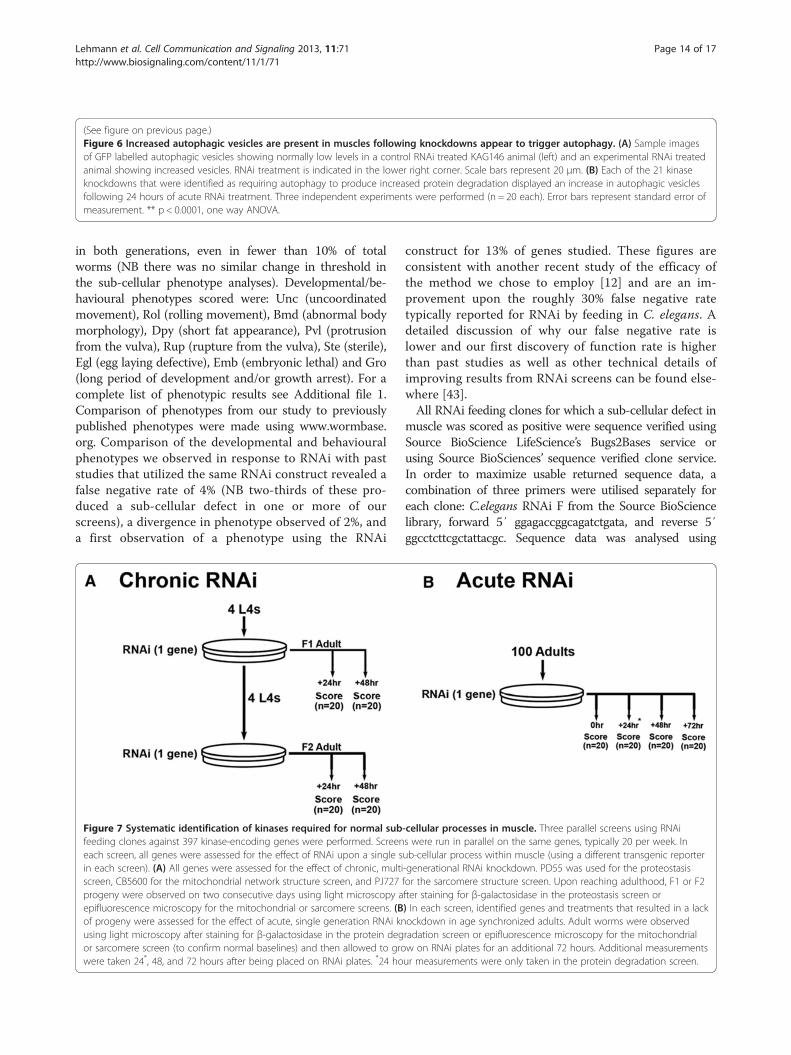
Quality control of our RNAi screensA schematic of how the three screens were performed isprovided (Figure 7). RNAi using bacterial clones grownas described [6] was performed with both chronic andacute RNAi experiments as described previously [12]. Adetailed technical description of the strengths, limita-tions, and caveats to interpretation of results from thisscreening methodology is available elsewhere [43].We used RNAi against unc-112 as a positive control as
RNAi unc-112 has been shown to produce a develop-mental, behavioural, muscle protein degradation, mito-chondrial, and sarcomere defect [15]. Each of the threescreens was run in parallel such that the same geneswere assessed in the same week, typically 20 genes/week.In cases where the positive control did not provide anabnormal phenotype in each of the screens the entirebatch of genes was rerun with an unc-112 positivecontrol.We used previously utilized RNAi constructs so that
we could directly compare our results for developmentalphenotypes with those of others who previously utilizedthe same RNAi construct as a quality control measure.The false positive rate for developmental and behaviouralphenotypes observed in response to RNAi by feeding inC. elegans is <1%, thus thresholds were set for ease ofscoring. Developmental phenotypes were recorded if atleast 20% of worms on the RNAi seeded plate showed aphenotype and also if the same phenotype was observed

Figure 6 (See legend on next page.)
Lehmann et al. Cell Communication and Signaling 2013, 11:71 Page 13 of 17http://www.biosignaling.com/content/11/1/71
(See figure on previous page.)Figure 6 Increased autophagic vesicles are present in muscles following knockdowns appear to trigger autophagy. (A) Sample imagesof GFP labelled autophagic vesicles showing normally low levels in a control RNAi treated KAG146 animal (left) and an experimental RNAi treatedanimal showing increased vesicles. RNAi treatment is indicated in the lower right corner. Scale bars represent 20 μm. (B) Each of the 21 kinaseknockdowns that were identified as requiring autophagy to produce increased protein degradation displayed an increase in autophagic vesiclesfollowing 24 hours of acute RNAi treatment. Three independent experiments were performed (n = 20 each). Error bars represent standard error ofmeasurement. ** p < 0.0001, one way ANOVA.
Lehmann et al. Cell Communication and Signaling 2013, 11:71 Page 14 of 17http://www.biosignaling.com/content/11/1/71
in both generations, even in fewer than 10% of totalworms (NB there was no similar change in threshold inthe sub-cellular phenotype analyses). Developmental/be-havioural phenotypes scored were: Unc (uncoordinatedmovement), Rol (rolling movement), Bmd (abnormal bodymorphology), Dpy (short fat appearance), Pvl (protrusionfrom the vulva), Rup (rupture from the vulva), Ste (sterile),Egl (egg laying defective), Emb (embryonic lethal) and Gro(long period of development and/or growth arrest). For acomplete list of phenotypic results see Additional file 1.Comparison of phenotypes from our study to previouslypublished phenotypes were made using www.wormbase.org. Comparison of the developmental and behaviouralphenotypes we observed in response to RNAi with paststudies that utilized the same RNAi construct revealed afalse negative rate of 4% (NB two-thirds of these pro-duced a sub-cellular defect in one or more of ourscreens), a divergence in phenotype observed of 2%, anda first observation of a phenotype using the RNAi
Figure 7 Systematic identification of kinases required for normal subfeeding clones against 397 kinase-encoding genes were performed. Screeneach screen, all genes were assessed for the effect of RNAi upon a single sin each screen). (A) All genes were assessed for the effect of chronic, multiscreen, CB5600 for the mitochondrial network structure screen, and PJ727progeny were observed on two consecutive days using light microscopy aepifluorescence microscopy for the mitochondrial or sarcomere screens. (Bof progeny were assessed for the effect of acute, single generation RNAi knusing light microscopy after staining for β-galactosidase in the protein degor sarcomere screen (to confirm normal baselines) and then allowed to growere taken 24*, 48, and 72 hours after being placed on RNAi plates. *24 ho
construct for 13% of genes studied. These figures areconsistent with another recent study of the efficacy ofthe method we chose to employ [12] and are an im-provement upon the roughly 30% false negative ratetypically reported for RNAi by feeding in C. elegans. Adetailed discussion of why our false negative rate islower and our first discovery of function rate is higherthan past studies as well as other technical details ofimproving results from RNAi screens can be found else-where [43].All RNAi feeding clones for which a sub-cellular defect in
muscle was scored as positive were sequence verified usingSource BioScience LifeScience’s Bugs2Bases service orusing Source BioSciences’ sequence verified clone service.In order to maximize usable returned sequence data, acombination of three primers were utilised separately foreach clone: C.elegans RNAi F from the Source BioSciencelibrary, forward 5′ ggagaccggcagatctgata, and reverse 5′ggcctcttcgctattacgc. Sequence data was analysed using
-cellular processes in muscle. Three parallel screens using RNAis were run in parallel on the same genes, typically 20 per week. Inub-cellular process within muscle (using a different transgenic reporter-generational RNAi knockdown. PD55 was used for the proteostasisfor the sarcomere structure screen. Upon reaching adulthood, F1 or F2fter staining for β-galactosidase in the proteostasis screen or) In each screen, identified genes and treatments that resulted in a lackockdown in age synchronized adults. Adult worms were observedradation screen or epifluorescence microscopy for the mitochondrialw on RNAi plates for an additional 72 hours. Additional measurementsur measurements were only taken in the protein degradation screen.
Lehmann et al. Cell Communication and Signaling 2013, 11:71 Page 15 of 17http://www.biosignaling.com/content/11/1/71
4Peaks (version 1.7.2) software and entered into NCBI C.elegans BLAST for confirmation of correct sequence.
Assessment of proteostasis, proteolysis, mitochondrialnetwork structure, and sarcomere structure via transgenicreporter proteinsMuscle-specific protein homeostasis and protein degrad-ation was assessed using transgene ccIs55 (unc-54::lacZ),with histochemical staining for β-galactosidase activityas described [17]. The protein product of ccIs55 is con-tinually synthesized throughout development and re-mains stable (e.g. is neither synthesized nor degraded) inthe cytosol for the first 72–96 hours post adulthoodin wild-type animals [15,17,22,35,39]. Thus, alterationsin β-galactosidase activity observed in response tochronic RNAi indicate alterations in protein synthesisand/or degradation whereas alterations in response toacute RNAi applied to fully developed adults indicate ac-tivation of protein degradation alone.Muscle-specific mitochondria and nuclei were assessed
using transgene ccIs4251(Pmyo-3::MitGFP; Pmyo-3::NLS::GFP-lacZ), with epifluorescence microscopy as described[12]. Note that observations and images were taken of live,non-immobilized animals. This was achieved by capturingimages while the animal was still.Sarcomere structures were assessed using transgene
jIs01(myo-3::GFP) which produces a translational fusionof the full-length MYO-3 (myosin heavy chain A) geneto GFP, with epifluorescence microscopy as described[12]. Note that observations and images were taken oflive, non-immobilized animals. This was achieved bycapturing images while the animal was still.
Scoring criteria and procedure for each of the RNAiscreensSub-cellular phenotypes scored in each of the threescreens were: Cytosolic protein content (normal, abnor-mal), mitochondrial morphology (normal, abnormal), andsarcomere morphology (normal, abnormal); see Figures 1,2 and 3, respectively, for examples of normal and abnor-mal sub-cellular phenotypes. In all cases abnormalitiesdeemed minor (e.g. not appreciably different from RNAicontrol) were scored as normal. Observations were scoredas abnormal if defects were observed in at least 25% ofworms on the slide (NB this is a 5% increase in thresholdfrom the study [12] upon which this protocol is based).Defects, within an individual worm, were classed using thesame thresholds as from the study upon which this proto-col is based [12], as follows: i) cytosolic protein content: atleast a 30% loss of stain (e.g. intensity viewed as light blueor absent in contrast to dark blue); ii) mitochondrialmorphology: loss of at least 30% of the mitochondrialnetwork in at least two muscle cells (e.g. loss of linearnetworks that was significant enough to be noticeable and
different from control animals). iii) sarcomere morph-ology: at least 2 disorganized or broken arrays of sarco-meres in at least two muscle cells (e.g. loss of linear arraysof sarcomeres that was significant enough to be noticeableand different from control animals). All scoring was donemanually using a Nikon H600L microscope.For chronic RNAi exposures (Figure 7A), a defect had
to be observed in any two of the four time points exam-ined to be scored as positive. Genes for which lack of pro-geny prevented scoring were also scored as giving a defectfor the purposes of further examination. Genes that wereidentified as affecting cytosolic protein content, mitochon-drial structure, or sarcomere structure in chronicallytreated animals were then examined for effects of acuteRNAi exposure.For acute RNAi exposures (Figure 7B), a defect had to
be observed at any two time points after introduction toRNAi, with particular attention to progressive loss ofcytosolic protein between the 48 and 72 hour timepoints. The criteria for a positive score at a single timepoint are identical to those described for the chronicRNAi screens.For all observations, 4–6 representative images were
captured on a Nikon H600L microscope with a NikonDigital Sight DS-Fi1 digital camera and proprietary soft-ware. At the end of all of the screens, these images wereused to confirm the results from the screen. Addition-ally, at the end of all screens all images were reviewed byan independent observer to confirm or correct initialscoring. In cases of a discrepancy between the first andsecond individual scoring the images the last authormanually reviewed the data and the two sets of scoringin order to reach a final scoring.
Epistasis testing of identified genes against knownprotein degradation pathwaysMutants and MG132 used for clustering identified genesto known proteolytic pathways/mechanisms were as de-scribed [12]. Acute RNAi experiments with these mu-tants were performed as described [12]. Degradation wasscored as in acute RNAi treatments in two independentexperiments; in case of discrepancy a third experimentwas run.
Assessment of autophagic vesicles via transgenic reporterproteinMuscle-specific autophagic vesicles were assessed usingtransgene kagEx12(pdyc-1S::gfp::lgg-1), with epifluores-cence. Transgenic animals were synchronised and sub-ject to the acute RNAi protocol with scoring occurringat 24 hours post introduction to RNAi clone or a controlclone lacking a targeted sequence. Autophagic vesicleswere quantified within the two body wall muscles thatvisually appeared to contain the greatest number of

Lehmann et al. Cell Communication and Signaling 2013, 11:71 Page 16 of 17http://www.biosignaling.com/content/11/1/71
vesicles. This was repeated in a total of 60 animals perRNAi treatment with 20 animals per experiment andeach experiment run three times. Two or three RNAiclones with a separate control clone were analysed atonce.
StatisticsAll statistical analysis was undertaken utilizing GraphPadPrism (GraphPad Software, Inc., La Jolla, CA, USA). Forassessment of distributions of data being significantly dif-ferent from a normal distribution, χ2 analysis was used.For assessment of tested RNAi clone autophagic vesicledata being significantly different from control RNAi clone,one-way ANOVA with Dunnett’s multiple comparison testwas used.
Additional file
Additional file 1: The following additional data are available withthe online version of the paper. A comprehensive list of genesscreened, clones used, and results. Sheet “All genes screened” providesinformation and results for 397 genes screened including the bacterialclone used in this study, subcellular defects observed as indicated bycolour coding (see legend at bottom) and developmental phenotypesobserved, known human orthologues along with data on expression inhuman muscle. Sheet “no clones available” displays all genes for whichan existing bacterial clone in the C. elegans RNAi library/Ahringer and C.elegans ORF-RNAi library/Vidal libraries was not available or for whichsequencing suggests it may contain an incorrect target sequence.
AbbreviationsC. elegans: Caenorhabditis elegans; ERK: Extracellular-signal regulated kinase;GFP: Green fluorescent protien; MAPK: Mitogen activated protein kinase;PI3K: Phosphatidylinositide 3 Kinase; RNAi: RNA interference.
Competing interestsThe authors declare that they have no competing interests.
Authors’ contributionsSL, JJB and NJS designed the studies and analyzed the data. SL and JJBconducted the RNAi experiments. JJB and NJS conducted the sequenceanalysis. SL and NJS wrote the paper. All authors read and approved the finalmanuscript.
AcknowledgementsOur thanks to L.A. Jacobson (University of Pittsburgh) for useful discussions,the Mitani laboratory (Tokyo Women’s Medical University) for providing thepink-1(tm1779) mutant strain, and the Gieseler laboratory (Université ClaudeBernard Lyon 1) for making and providing strain KAG146 prior to publication.This work was funded by the US NIH- NIAMS (AR-054342). J.J.B. was fundedby an MRC doctoral training award (J500495).
Received: 19 February 2013 Accepted: 15 September 2013Published: 23 September 2013
References1. Jin J, Pawson T: Modular evolution of phosphorylation-based signalling
systems. Philos Trans R Soc Lond B Biol Sci 2012, 367(1602):2540–2555.2. Cohen P: The regulation of protein function by multisite
phosphorylation–a 25 year update. Trends Biochem Sci 2000,25(12):596–601.
3. Fedorov O, Muller S, Knapp S: The (un)targeted cancer kinome. Nat ChemBiol 2010, 6(3):166–169.
4. Piano F: C. elegans network biology: a beginning (August 21, 2006). InWormBook. Edited by The C. elegans Research Community, WormBook.doi:10.1895/wormbook.1.118.1. http://www.wormbook.org.
5. Manning G: Genomic overview of Protein Kinases (December 13, 2005).In WormBook. Edited by The C. elegans Research Community, WormBook.doi:10.1895/wormbook.1.60.1. http://www.wormbook.org.
6. Kamath RS, Fraser AG, Dong Y, Poulin G, Durbin R, Gotta M, Kanapin A, LeBot N, Moreno S, Sohrmann M, et al: Systematic functional analysis of theCaenorhabditis elegans genome using RNAi. Nature 2003,421(6920):231–237.
7. Rual JF, Ceron J, Koreth J, Hao T, Nicot AS, Hirozane-Kishikawa T,Vandenhaute J, Orkin SH, Hill DE, van den Heuvel S, et al: Towardimproving Caenorhabditis elegans phenome mapping with anORFeome-based RNAi library. Genome research 2004, 14(10B):2162–2168.
8. Chen N, Harris TW, Antoshechkin I, Bastiani C, Bieri T, Blasiar D, Bradnam K,Canaran P, Chan J, Chen CK, et al: WormBase: a comprehensive dataresource for Caenorhabditis biology and genomics. Nucleic Acids Res2005, 33(Database issue):D383–D389.
9. Glass D, Roubenoff R: Recent advances in the biology and therapy ofmuscle wasting. Ann N Y Acad Sci 2010, 1211:25–36.
10. Shaye DD, Greenwald I: OrthoList: a compendium of C. elegans geneswith human orthologs. PLoS One 2011, 6(5):e20085.
11. Uhlen M, Oksvold P, Fagerberg L, Lundberg E, Jonasson K, Forsberg M,Zwahlen M, Kampf C, Wester K, Hober S, et al: Towards a knowledge-based Human Protein Atlas. Nat Biotechnol 2010, 28(12):1248–1250.
12. Shephard F, Adenle AA, Jacobson LA, Szewczyk NJ: Identification andfunctional clustering of genes regulating muscle protein degradationfrom amongst the known C. elegans muscle mutants. PLoS One 2011,6(9):e24686.
13. Meissner B, Warner A, Wong K, Dube N, Lorch A, McKay SJ, Khattra J,Rogalski T, Somasiri A, Chaudhry I, et al: An integrated strategy to studymuscle development and myofilament structure in Caenorhabditiselegans. PLoS Genet 2009, 5(6):e1000537.
14. Schnorrer F, Schonbauer C, Langer CC, Dietzl G, Novatchkova M,Schernhuber K, Fellner M, Azaryan A, Radolf M, Stark A, et al: Systematicgenetic analysis of muscle morphogenesis and function in Drosophila.Nature 2010, 464(7286):287–291.
15. Etheridge T, Oczypok EA, Lehmann S, Fields BD, Shephard F, Jacobson LA,Szewczyk NJ: Calpains Mediate Integrin Attachment ComplexMaintenance of Adult Muscle in Caenorhabditis elegans.PLoS Genet 2012, 8(1):e1002471.
16. Sandri M: Signaling in muscle atrophy and hypertrophy.Physiology (Bethesda) 2008, 23:160–170.
17. Zdinak LA, Greenberg IB, Szewczyk NJ, Barmada SJ, Cardamone-Rayner M,Hartman JJ, Jacobson LA: Transgene-coded chimeric proteins as reportersof intracellular proteolysis: starvation-induced catabolism of a lacZfusion protein in muscle cells of Caenorhabditis elegans. J Cell Biochem1997, 67(1):143–153.
18. Lehmann S, Shephard F, Jacobson LA, Szewczyk NJ: Integrated control ofprotein degradation in muscle. Worm 2012, 1(3):141–150.
19. Lo TW, Bennett DC, Goodman SJ, Stern MJ: Caenorhabditis elegansfibroblast growth factor receptor signaling can occur independently ofthe multi-substrate adaptor FRS2. Genetics 2010, 185(2):537–547.
20. McColl G, Killilea DW, Hubbard AE, Vantipalli MC, Melov S, Lithgow GJ:Pharmacogenetic analysis of lithium-induced delayed aging inCaenorhabditis elegans. J Biol Chem 2008, 283(1):350–357.
21. Hertweck M, Gobel C, Baumeister R: C. elegans SGK-1 is the criticalcomponent in the Akt/PKB kinase complex to control stress responseand life span. Dev Cell 2004, 6(4):577–588.
22. Szewczyk NJ, Peterson BK, Barmada SJ, Parkinson LP, Jacobson LA: Opposedgrowth factor signals control protein degradation in muscles ofCaenorhabditis elegans. EMBO J 2007, 26(4):935–943.
23. Shen X, Ellis RE, Lee K, Liu CY, Yang K, Solomon A, Yoshida H, Morimoto R,Kurnit DM, Mori K, et al: Complementary signaling pathways regulate theunfolded protein response and are required for C. elegans development.Cell 2001, 107(7):893–903.
24. Chan DC: Fusion and Fission: Interlinked Processes Critical forMitochondrial Health. Annu Rev Genet 2012, 46:265–287.
25. Fire A, Xu S, Montgomery MK, Kostas SA, Driver SE, Mello CC: Potent andspecific genetic interference by double-stranded RNA in Caenorhabditiselegans. Nature 1998, 391(6669):806–811.
Lehmann et al. Cell Communication and Signaling 2013, 11:71 Page 17 of 17http://www.biosignaling.com/content/11/1/71
26. Clark IE, Dodson MW, Jiang C, Cao JH, Huh JR, Seol JH, Yoo SJ, Hay BA, GuoM: Drosophila pink1 is required for mitochondrial function and interactsgenetically with parkin. Nature 2006, 441(7097):1162–1166.
27. Cribbs JT, Strack S: Reversible phosphorylation of Drp1 by cyclic AMP-dependent protein kinase and calcineurin regulates mitochondrialfission and cell death. EMBO Rep 2007, 8(10):939–944.
28. Qadota H, Benian GM: Molecular structure of sarcomere-to-membraneattachment at M-Lines in C. elegans muscle. J Biomed Biotechnol 2010,2010:864749.
29. Sparrow J, Hughes SM, Segalat L: Other model organisms for sarcomericmuscle diseases. Adv Exp Med Biol 2008, 642:192–206.
30. Fostel JL, Benner Coste L, Jacobson LA: Degradation of transgene-codedand endogenous proteins in the muscles of Caenorhabditis elegans.Biochem Biophys Res Commun 2003, 312(1):173–177.
31. Mackinnon AC, Qadota H, Norman KR, Moerman DG, Williams BD: C.elegans PAT-4/ILK functions as an adaptor protein within integrinadhesion complexes. Curr Biol 2002, 12(10):787–797.
32. Benian GM, Tinley TL, Tang X, Borodovsky M: The Caenorhabditis elegansgene unc-89, required fpr muscle M-line assembly, encodes a giantmodular protein composed of Ig and signal transduction domains.J Cell Biol 1996, 132(5):835–848.
33. Benian GM, Kiff JE, Neckelmann N, Moerman DG, Waterston RH: Sequenceof an unusually large protein implicated in regulation of myosin activityin C. elegans. Nature 1989, 342(6245):45–50.
34. Szewczyk NJ, Peterson BK, Jacobson LA: Activation of Ras and themitogen-activated protein kinase pathway promotes proteindegradation in muscle cells of Caenorhabditis elegans. Mol Cell Biol 2002,22(12):4181–4188.
35. Szewczyk NJ, Jacobson LA: Activated EGL-15 FGF receptor promotesprotein degradation in muscles of Caenorhabditis elegans. EMBO J 2003,22(19):5058–5067.
36. Melendez A, Talloczy Z, Seaman M, Eskelinen EL, Hall DH, Levine B:Autophagy genes are essential for dauer development and life-spanextension in C. elegans. Science 2003, 301(5638):1387–1391.
37. Lackner MR, Kornfeld K, Miller LM, Horvitz HR, Kim SK: A MAP kinasehomolog, mpk-1, is involved in ras-mediated induction of vulval cellfates in Caenorhabditis elegans. Genes Dev 1994, 8(2):160–173.
38. Ogg S, Ruvkun G: The C. elegans PTEN homolog, DAF-18, acts in theinsulin receptor-like metabolic signaling pathway. Mol Cell 1998,2(6):887–893.
39. Szewczyk NJ, Hartman JJ, Barmada SJ, Jacobson LA: Genetic defects inacetylcholine signalling promote protein degradation in muscle cells ofCaenorhabditis elegans. J Cell Sci 2000, 113(Pt 11):2003–2010.
40. Estevez AO, Mueller CL, Morgan KL, Szewczyk NJ, Teece L, Miranda-VizueteA, Estevez M: Selenium induces cholinergic motor neuron degenerationin Caenorhabditis elegans. Neurotoxicology 2012, 33(5):1021–1032.
41. Apfeld J, O’Connor G, McDonagh T, DiStefano PS, Curtis R: The AMP-activated protein kinase AAK-2 links energy levels and insulin-likesignals to lifespan in C. elegans. Genes Dev 2004, 18(24):3004–3009.
42. Min K, Kang J, Lee J: A modified feeding RNAi method for simultaneousknock-down of more than one gene in Caenorhabditis elegans.Biotechniques 2010, 48(3):229–232.
43. Lehmann S, Shephard F, Jacobson LA, Szewczyk NJ: Using MultiplePhenotype Assays and Epistasis Testing to Enhance the Reliability ofRNAi Screening and Identify Regulators of Muscle Protein Degradation.Genes (Basel) 2012, 3(4):686–701.
44. Consortium TCeDM: Large-Scale Screening for Targeted Knockouts in theCaenorhabditis elegans Genome. G3 (Bethesda) 2012, 2(11):1415–1425.
45. Pause A, Belsham GJ, Gingras AC, Donze O, Lin TA, Lawrence JC Jr,Sonenberg N: Insulin-dependent stimulation of protein synthesis byphosphorylation of a regulator of 5′-cap function. Nature 1994,371(6500):762–767.
46. Drummond MJ, Fry CS, Glynn EL, Dreyer HC, Dhanani S, Timmerman KL,Volpi E, Rasmussen BB: Rapamycin administration in humans blocks the
contraction-induced increase in skeletal muscle protein synthesis.J Physiol 2009, 587(Pt 7):1535–1546.
47. Brenner S: The genetics of Caenorhabditis elegans. Genetics 1974,77(1):71–94.
48. Henderson ST, Bonafe M, Johnson TE: daf-16 protects the nematodeCaenorhabditis elegans during food deprivation. J Gerontol A Biol Sci MedSci 2006, 61(5):444–460.
doi:10.1186/1478-811X-11-71Cite this article as: Lehmann et al.: Knockdown of the C. elegans Kinomeidentifies Kinases required for normal protein Homeostasis,Mitochondrial network structure, and Sarcomere structure in muscle.Cell Communication and Signaling 2013 11:71.
Submit your next manuscript to BioMed Centraland take full advantage of:
• Convenient online submission
• Thorough peer review
• No space constraints or color figure charges
• Immediate publication on acceptance
• Inclusion in PubMed, CAS, Scopus and Google Scholar
• Research which is freely available for redistribution
Submit your manuscript at www.biomedcentral.com/submit