lab 1 tissue culture -...
TRANSCRIPT
Lab 1 Introduction to animal tissue culture
BIO-331 Cell biology – Lab 1 Animal Tissue Culture 1
Purpose The goal of this lab is to introduce novice cytology students to both the utility and the limitations of modern tissue culture techniques. Proficiency in tissue culture requires expertise can only be gained through experience (especially with respect to the maintenance of aseptic conditions). Therefore students should strive to achieve competency in the following foundational techniques: 1. operation and maintenance of vertical laminar flow culture hoods 2. operation and maintenance of CO2 incubators
3. tissue dissection and enzymatic disaggregation 4. cell staining and basic identification by morphology
Background & introduction History of tissue culture Tissue culture was first demonstrated by R.G. Harrison over 100 years ago1, as a method for studying the behavior of animal cells free of systemic physiological variations and influence. Harrison’s initial discovery using frog tissue has been adapted to accommodate the specific conditions necessary to sustain a wide range of animal tissue (including human) in vitro. Tissue culture has rapidly become one of the most important tools available for biomedical research and the biotech industry. To illustrate this point, a January 2010 PubMed search for the term “tissue culture” returned 145,013 research articles. Currently the most widely used sources of “primary” tissue for research are the chick and mouse animal models. Tissue from embryonic stages of development have proved especially amenable for cell culture. Utility of tissue culture Tissue culture offers three significant advantages over whole-animal models:
1. precise control of the chemical environment (pH, temperature, osmotic pressure, etc…)
2. moderate control of the physiological conditions (culture media supplemented with animal serum may exhibit variations in hormone concentrations from different batches and/or suppliers)
3. increased accessibility for manipulation and characterization of living tissue (inverted microscopy and vital dye labeling). Forms of tissue culture Cell culture normally refers to the culture of dispersed and clonally derived and long established (sometimes called “immortal”) cell lines. “Primary” cell culture refers to the use of cells taken directly from animals in contrast to more established cell “lines” that may have been propagated over long periods of time in vitro. Tissue culture when strictly used refers to the maintenance of dissected pieces (explants) of tissue or even whole organs out side of an animal (in vitro). It should be noted that for some cytologists “cell culture” is considered one specific type of “tissue culture” and for others these terms may be used almost interchangeably. Primary tissue culture For experiments where animal models do not allow for sufficient control of chemical or physiological variables and suitable immortalized cell lines are not available, primary tissue culture may suffice. There are three main options for initiating a primary tissue culture: organ
1 Harrison, R.G. (1907) Observations on the living developing nerve fiber. Proc. Cos. Exp. Biol. Med. 4: 140-143.
Lab 1 Introduction to animal tissue culture
BIO-331 Cell biology – Lab 1 Animal Tissue Culture 2
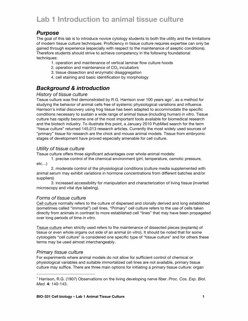
culture, explant culture, and primary cell culture. In organ culture entire embryonic organs are excised and suspended at a gas-liquid media interface to retain the original tissue architecture. In primary explant culture, a fragment of excised animal tissue is placed at a solid-liquid interface and subsequently allowed to attach and migrate on the glass or plastic substrate. In primary cell culture dissected tissue is mechanically or enzymatically dispersed to obtain individual cells to be cultured on a solid substrate in monolayer. See Figure 1 for a summary of these methods.
Figure 1. Illustration of methods used to obtain primary cells, explants or organs for
culture (from Culture of Animal Cells, 4th Ed., by R.I. Freshney p. 7) Adhesion & proliferation of cultured cells While some specialty cell lines are cultured in suspension (blood and lymph cells), the vast majority of animal cell lines thrive only when attached directly to a solid surface. Therefore culture substrates that facilitate adequate cell adhesion are an important consideration in experimental design. It is well established that culture flasks or dishes treated to provide a slight negative charge are far superior for adherent cell culture than uncharged substrates. It is thought that the negative charge attracts key extracellular matrix (ECM) proteins and proteoglycans secreted by the cultured cells. These ECM molecules then interface with membrane bound integrin proteins. Adhesion of some cell types may alos be assisted by coating the culture surface with extracellular matrix molecules such as fibronectin, laminin, collagen, derivatives such as gelatin, or other charged polymers such as poly-D-lysine. Culture media A common basal media for cell culture is Dulbecco’s modified Eagle’s Media or DMEM2. This media is buffered at pH 7.4 and is supplemented with phenol red as a visual indicator of pH. Phenol red turns more yellow at acidic pH (below 7.0) and purple in base (above 7.8). To buffer pH, precise atmospheric CO2 concentration must be maintained (usually at 5.0%). Most cultured cells have a surprisingly large tolerance for a range of osmotic pressure. However, to maintain optimal growing conditions, DMEM is osmotically balanced with salt. A humid culture environment should be maintained to minimize changes in osmotic pressure due to the evaporation of media. Temperature should be maintained close to the body temperature of the
2 Dulbecco, R., & Freeman, G. (1959) Plaque production by the polyoma virus. Virology. 8: 396-397.
Lab 1 Introduction to animal tissue culture
BIO-331 Cell biology – Lab 1 Animal Tissue Culture 3
host organism. For most warm-blooded mammals this is approximately 37oC (note: avian body temperature may be slightly warmer at ~38.5oC). Glucose is included in most media (including DMEM) as a source of energy. Essential amino acids are present in DMEM, and non-essential amino acids are supplemented for some sensitive cell lines. DMEM contains water soluble vitamins (the B-group, choline, folic acid, inositol, and nicotinamide), and other vitamins are supplied through the addition of serum to the media. The addition of serum also provides hormones and growth factors (although many of these have not been well characterized or defined). Antibiotics are sometimes added to reduce the frequency of contamination, however the maintenance of cultured tissue in antibiotics may merely slow contaminant growth resulting in delayed detection.
Materials 1. Fertile chick eggs: Order from Sunny-Side Hatchery (Beaver Dam, WI contact Sue at 920-887-1290) at least one week in advance. Store at 15oC to inhibit development and incubate at 38oC with high humidity for 10 days to obtain suitably sized chick embryos. Avoid incubation times greater than 10 days as midgestation vertebrates are considered sentient and would therefore require institutional animal use and care committee (IACUC) approval as directed in the Animal Welfare Act (AWA) governing the use of vertebrate models for education and research. Furthermore embryonic tissue is easier to disaggregate and yields more proliferating cells.
2. HBSS: Hanks’ Balanced Salt Solution with antibiotics (Sigma H6136-10x1L $15.40)
To make add the following to autoclaved 1x Hanks’ BSS: Penicillin (250U/mL) Streptomycin (250ug/mL) Kanamycin (100ug/mL)
3. Trypsin tissue disaggregation solution: (store frozen stocks at -20oC) 0.25% Trypsin and 0.38g/L EDTA in HBSS (Invitrogen #25200-072 = $28.50)
4. Collagenase/Dispase tissue disaggregation solution: (store frozen stocks at -20oC) Dilute 1mg/mL collagenase and dispase (Roche #11-097-113-001) in DMEM.
5. Growth medium: DMEM:F12 Medium ATCC #30-2006 (440mL), 10% fetal bovine serum (50mL) (Atlanta Biologicals S11150), 2% antibiotic/antimycotic (10mL) (Sigma A5955), 1.25mM Putrescine (a growth factor polyamine which stimulates cell division in some cells) (0.1g). Store the medium at 4oC. Warm to 37oC prior to use.
6. Sterile plasticware: 100mm bacteriological Petri dishes and cell-culture-grade 24-well electrostatically charged tissue culture dishes
7. 1mg/mL gelatin solution: 0.5g gelatin in 500mL dH2O (autoclave, store RT)
8. 50µg/mL Poly-D-Lysine solution: Add 100mL of sterile tissue culture grade water to 5mg poly-D-lysine (Sigma P6407) Mix by pipetting several times. Store at -20oC
9. 70% ethanol with cotton swabs
10. Sterile forceps, scissors, and scalpels

Lab 1 Introduction to animal tissue culture
BIO-331 Cell biology – Lab 1 Animal Tissue Culture 4
11. Sterile pipettes and pipette aid
12. Conical centrifuge tubes (15mL and 50mL sizes) 13. Optional cell strainers, 40µM mesh size (BD Biosciences #352340) 14. Optional fibroblast inhibitor: Hydroxyurea (Sigma #H8627-1G $21.60) may be added to the growth media to inhibit fibroblast growth. To prepare 400x stock, add 0.76g / 50ml (0.2M) hydroxyurea in PBS (Hagiwara & Ozawa, 1994). Supplement growth media with 2.5µL of 400x stock solution in PBS to inhibit fibroblast growth.
15. Optional neuronal growth supplement3 (all reagents from Sigma): Individual stock solutions (store at -20oC) Selenium dioxide (204315-10g) 37.5µg/mL PBS
(a 20x prestock 0.75mg/mL may be prepared first by dissolving 0.0375g into 50mL PBS)
Putrescine dihydrochloride 16.1mg/mL PBS (0.161g into 10mL PBS) BSA 10mg/mL PBS (0.1g into 10mL PBS) Partially iron-saturated human 10mg/mL PBS Holo-transferrin (T0665-100mg) (dissolve entire vial in 10mL PBS) Insulin (10516-5mL) 10mg/mL - Tri-iodothyronine (T6397-100mg) 200µg/mL 0.1N NaOH (add 2g NaOH to 500mL water then dissolve entire vial) Progesterone (P8783-1g) 0.63mg/mL 100% EtOH (dissolve 0.031g into 50mL 100% EtOH) Corticosterone (27840-100mg) 2mg/mL 100% EtOH (Dissolve 0.1g into 50mL 100% EtOH) β-estradiol (E2758-250mg) 100µg/mL 100% EtOH (Dissolve 0.005g into 50mL) 7S Nerve growth factor (N0513-.1mg) 100µg/mL PBS (Dissolve entire vial into 1mL)
To prepare 100mL of 10x neuronal growth supplement add the following stock solutions to 80mL of M199. (store at -20oC)
Selenium dioxide 2mL Putrescine dihydrochloride 2mL BSA 2mL Transferrin 10mL Insulin 2mL Tri-iodothyronine 100µL Progesterone 100µL Corticosterone 100µL
β-estradiol 1mL Nerve growth factor 100µL
16. Tissue fixative: a 1:1 mixture of PBS and methanol. Store at -20oC.
3 Hollenbeck, J., & Bamburg, J. (2003) Neurons: Methods and Applications for the Cell Biologist. p61.
Lab 1 Introduction to animal tissue culture
BIO-331 Cell biology – Lab 1 Animal Tissue Culture 5
17. Optional crystal violet stain: Dissolve 0.5g crystal violet in 500mL water. Filter through Whatman #1 before use.
18. Stereomicroscopes, magnifing lamps or surgical loupes for microdissection, tungsten dissection needle, mouth-pipette with capillary needle
Methods Cell culture dish preperation Few cells adhere readily to untreated plastic (bacteriological grade) plastic dishes. Most animal cell types require some sort of substrate for adherance. The electrostatic charge given to “tissue-culture” grade dishes is sufficient for some cells (i.e., fibroblasts), but other cells benefit from other forms of matrix such as gelatin (i.e., myoblasts) or poly-D-Lysine (i.e. neurons).
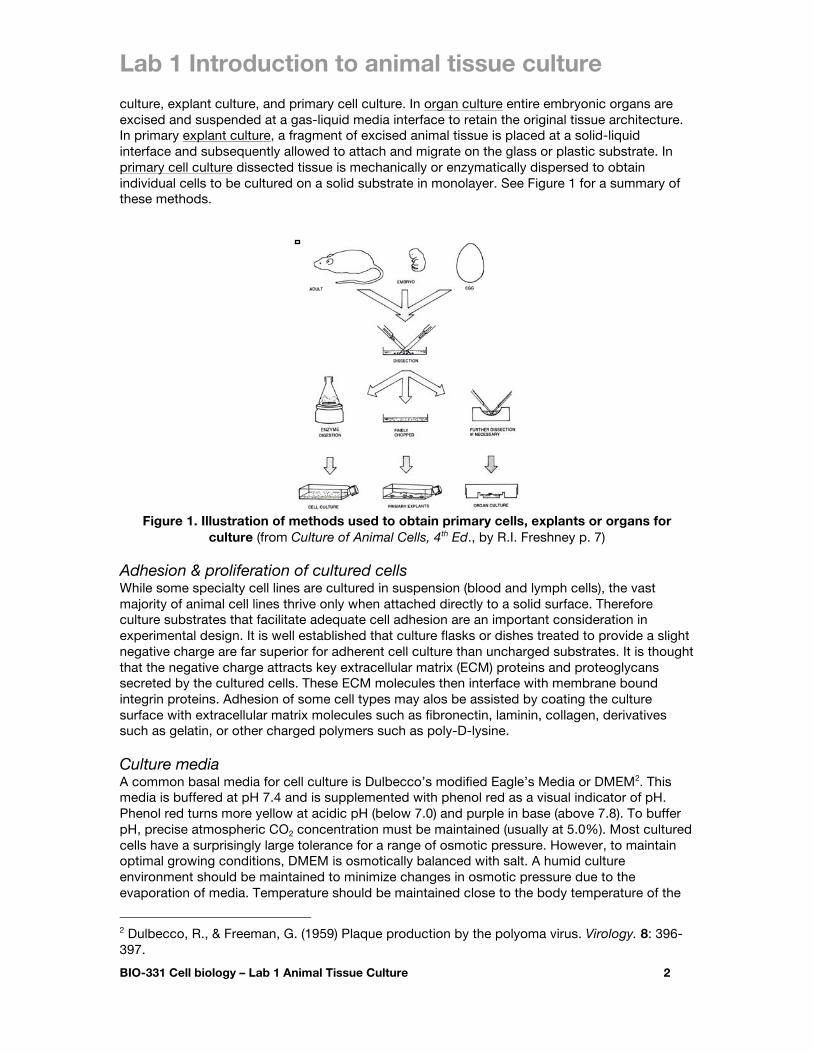
1. Obtain an label a 24-well tissue culture grade dish (as shown in Figure 2). Do not open this dish outside of the laminar flow hood. To maintain sterility of the dish. 2. Working under the hood, add enough matrix solution (gelatin in columns 1 & 2 and poly D-lysine solution in column 5) to barely cover the slides.
1mg/mL gelatin = 0.5g gelatin in 500mL dH2O (store @ RT) 50µg/mL Poly-D-Lysine (Sigma P6407) = (store @ -20oC)
3. Incubate slides 1-24h at 37oC. 4. Using a sterile pasteur pipette, aspirate the gelatin or poly-D-lysine solution. 5. Wash with sterile water or PBS. The plate is now ready for tissue culture.
Figure 2. Suggested arrangement for preparing the tissue culture dish for cell culture. Obviously, the research should customize the arrangement as he or she deems appropriate.
Egg Candeling
1. Shine a bright light (something akin to a flashlight) through the egg. A dark embryo and a root-like vascular network will be revealed in the fertile and viable eggs. Eggs that appear clear should be discarded. Gross dissection
1. Swab the egg with 70% ethanol and place it blunt end up in a beaker. This will reduce the amount of bacteria on the shell.
Lab 1 Introduction to animal tissue culture
BIO-331 Cell biology – Lab 1 Animal Tissue Culture 6
2. Crack the top of the shell and peel it back to the air sack using sterile forceps.
3. Sterilize the forceps (ie dip in ethanol, burn the alcohol off, cool the forceps) then use the forceps to peel off the white shell membrane to reveal the vascularized chorioallantoic membrane (CAM).
4. Pierce the CAM with sterile curved forceps, and lift out the embryo by gently grasping it under the head. Do not close the forceps completely or the neck will sever.
5. Transfer the embryo to a sterile Petri dish (non-tissue-culture grade) filled with 10-20mL HBSS
6. Decapitate the embryo quickly.
7. Isolate, culture, and document six of the tissue detailed below (see Appendix A for an
abbreviated list of potential tissues and see Figure 3 for stereomicrographs of the dissected chick tissue).
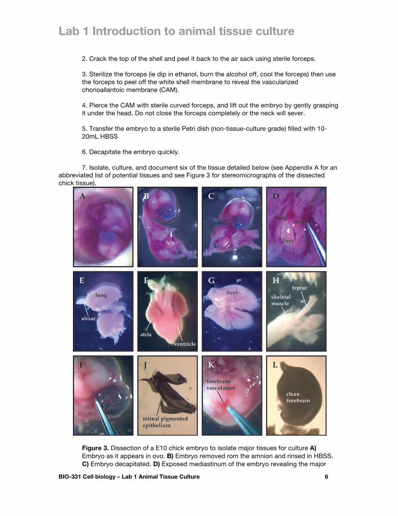
Figure 3. Dissection of a E10 chick embryo to isolate major tissues for culture A) Embryo as it appears in ovo. B) Embryo removed rom the amnion and rinsed in HBSS. C) Embryo decapitated. D) Exposed mediastinum of the embryo revealing the major
Lab 1 Introduction to animal tissue culture
BIO-331 Cell biology – Lab 1 Animal Tissue Culture 7
internal organs. E) Isolated lungs notice the ringed trachea and translucent airsacs. F) The isolated heart – notice the cone-like ventricle and the thin-walled atria. G) The fetal liver. H) The chick embryo thigh with the skin pulled back to reveal skeletal muscle and the femur. I) Separation of the eye from the orbit. J) A sheet-like layer of retinal pigmented epithelial cells. K) The dissected forebrain with the meninges intact. L) The forebrain with the meninges properly peeled away. Note the lack of vasculature in the properly dissected forebrain.
Skeletal muscle tissue
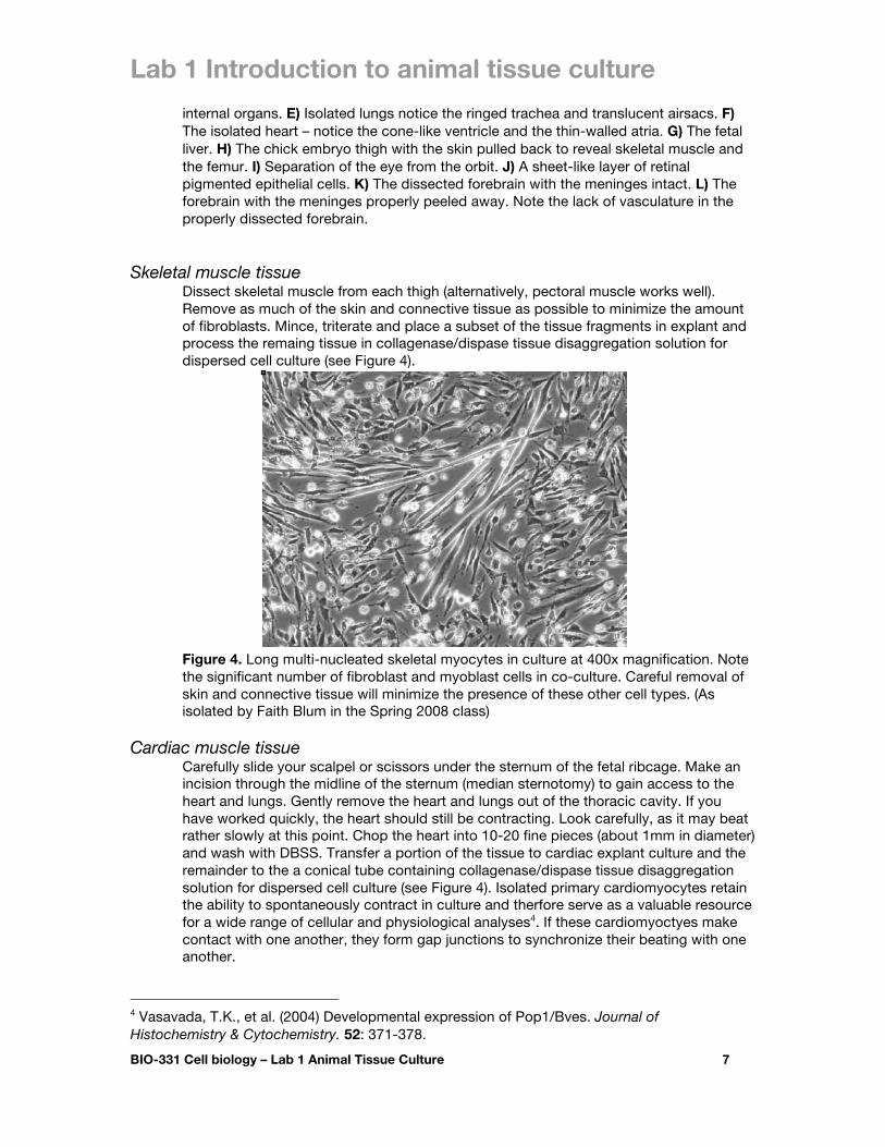
Dissect skeletal muscle from each thigh (alternatively, pectoral muscle works well). Remove as much of the skin and connective tissue as possible to minimize the amount of fibroblasts. Mince, triterate and place a subset of the tissue fragments in explant and process the remaing tissue in collagenase/dispase tissue disaggregation solution for dispersed cell culture (see Figure 4).
Figure 4. Long multi-nucleated skeletal myocytes in culture at 400x magnification. Note the significant number of fibroblast and myoblast cells in co-culture. Careful removal of skin and connective tissue will minimize the presence of these other cell types. (As isolated by Faith Blum in the Spring 2008 class)
Cardiac muscle tissue
Carefully slide your scalpel or scissors under the sternum of the fetal ribcage. Make an incision through the midline of the sternum (median sternotomy) to gain access to the heart and lungs. Gently remove the heart and lungs out of the thoracic cavity. If you have worked quickly, the heart should still be contracting. Look carefully, as it may beat rather slowly at this point. Chop the heart into 10-20 fine pieces (about 1mm in diameter) and wash with DBSS. Transfer a portion of the tissue to cardiac explant culture and the remainder to the a conical tube containing collagenase/dispase tissue disaggregation solution for dispersed cell culture (see Figure 4). Isolated primary cardiomyocytes retain the ability to spontaneously contract in culture and therfore serve as a valuable resource for a wide range of cellular and physiological analyses4. If these cardiomyoctyes make contact with one another, they form gap junctions to synchronize their beating with one another.
4 Vasavada, T.K., et al. (2004) Developmental expression of Pop1/Bves. Journal of Histochemistry & Cytochemistry. 52: 371-378.

Lab 1 Introduction to animal tissue culture
BIO-331 Cell biology – Lab 1 Animal Tissue Culture 8
Figure 5. Rectangular-shaped cardiomyoctyes in culture at 600x magnification. (As isolated by Alexis Rwatambuga in the Spring 2010 class)
Lung tissue
The lungs are among the last internal organs to develop in the fetus, and therefore are rather small at mid-gestation. Since the tissue remains fragile at this stage, it may be easily dispersed by triteration or in trypsin tissue disaggregation solution.
Figure 6. Pancake-shaped lung epithelial cells (pneumocytes) in culture at 400x magnification. (As isolated by Emily Fischer in the Spring 2010 class)
Liver tissue
The liver is located just below the diaphragm that separates the thoracic cavity from the abdominal cavity. At mid-gestation, the fetal liver is visible as a pink organ beneath the ventral surface of the skin. If you create a transverse section through the trunk of the fetus posterior to the diaphragm, the liver will be revealed. Gently remove the liver from among the folds of the gut. Separate the liver from the gut. Chop the liver into 10-20 fine pieces and transfer a subset of the pieces to the culture dish and the remaining pieces into trypsin tissue disaggregation solution for hepatocyte culture (see Figure 7).
Lab 1 Introduction to animal tissue culture
BIO-331 Cell biology – Lab 1 Animal Tissue Culture 9
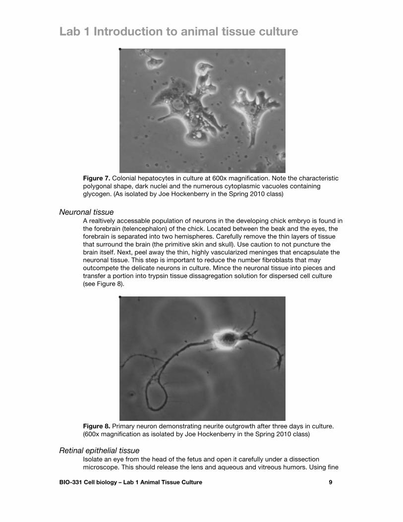
Figure 7. Colonial hepatocytes in culture at 600x magnification. Note the characteristic polygonal shape, dark nuclei and the numerous cytoplasmic vacuoles containing glycogen. (As isolated by Joe Hockenberry in the Spring 2010 class)
Neuronal tissue
A realtively accessable population of neurons in the developing chick embryo is found in the forebrain (telencephalon) of the chick. Located between the beak and the eyes, the forebrain is separated into two hemispheres. Carefully remove the thin layers of tissue that surround the brain (the primitive skin and skull). Use caution to not puncture the brain itself. Next, peel away the thin, highly vascularized meninges that encapsulate the neuronal tissue. This step is important to reduce the number fibroblasts that may outcompete the delicate neurons in culture. Mince the neuronal tissue into pieces and transfer a portion into trypsin tissue dissagregation solution for dispersed cell culture (see Figure 8).
Figure 8. Primary neuron demonstrating neurite outgrowth after three days in culture. (600x magnification as isolated by Joe Hockenberry in the Spring 2010 class)
Retinal epithelial tissue
Isolate an eye from the head of the fetus and open it carefully under a dissection microscope. This should release the lens and aqueous and vitreous humors. Using fine

Lab 1 Introduction to animal tissue culture
BIO-331 Cell biology – Lab 1 Animal Tissue Culture 10
forceps, gently peel the sheet of pigmented retinal epithelium off the neural retina and connective tissue. Chop the pigmented retinal epithelium into 10-20 fine pieces and transfer some of the pieces to the 24-well tissue culture dish for primary explant culture and the remaining pieces to the trypsin tissue dissagregation solution for epithelial primary cell culture (see Figure 9).
Figure 9. Chick primary pigmented retinal epithelial cells in culture 400x magnification (As isolated by Alexis Rwatambuga in the Spring 2010 class)
Skin epithelial tissue
Epithelial tissue from the skin may be obtained by striping the skin off the back of the chick embryo. Collect and place some in explant and process other for dispersed cell culture in trypsin or collagenase/dispase solution (see Figure 10).
Figure 10. Chick primary keratinocytes and skin fibroblasts in culture at 400x magnification. (As isolated by Faith Blum in the Spring 2008 class)
Tissue dissaggregation to visualize single cells
1. Wash the dissected tissue in HBSS. 2. Centrifuge tissue and aspirate excess HBSS.
Lab 1 Introduction to animal tissue culture
BIO-331 Cell biology – Lab 1 Animal Tissue Culture 11
3. Select one or more of the following options for enzymatic dissaggregation of tissue: A. Trypsinization (the fastest and most common method for dissaggregation is the use of the protease trypsin to degrade extracellular matrix combined with EDTA to chelate Ca2+ required for cadherins): incubate in trypsin/EDTA tissue dissaggregation solution with aggitation for at least 30 minutes at 37oC. B. Trypsinization with cold preexposure (prolonged exposure to warm trypsin may damage some cell types, therefore a preincubation of cold trypsin may allow for full penetration of the trypsin into the tissue while minimizing damaging effects on the cells): incubate tissue in trypsin/EDTA tissue dissaggregation solution for 6-24h at 4oC, then raise the temperature of the trypsin to 37oC for 15-30 minutes.
C. Collagenase and Dispase treatement (trypsinization may be damaging (e.g., epithelial) or ineffective (e.g., connective & muscle) for some tissue types. In this case, alternative proteases may be used). Incubate in collagenase/dispase tissue dissaggregation solution for at least 30 minutes at 37oC.
4. Following digestion the solution should appear cloudy due to the release of cells from the tissue into suspension. Large tissue aggregates may be broken up with gentle triteration and/or removed with an optional polypropylene filter (BD Biosciences 40µM mesh #352340) or simply allowed to settle to the bottom before removing the cloudy dispersed cells at the top. 5. Quench the digestion reaction with 3mL of growth media. Allow large pieces to settle, pipette and transfer 250µL of diluted suspension plus 250µL of additional growth medium per well to create a 50% concetration. And then add 100µL of diluted suspension plus 400µL of additional growth medium per well to create a 10% concetration. 6. Add neuronal growth supplement (optional): neurons benefit from the supplementation of selenium dioxide, putrescine, BSA, transferrin, insulin, tri-iodothyronine, progesterone, corticosterone, β-estradiol (E2758-250mg), and nerve growth factor in M199 medium5. All of these are included in the 10x stock neuronal growth supplement solution stored at -20oC. Add 50µL of the 10x stock neuronal growth supplement solution to each well containing neurons. 7. Add fibroblast inhibitor (optional): Many tissues contain fibroblasts (stromal connective tissue cells) that frequently outgrow other more specialized cell types (e.g., neurons and myocytes). To inhibit fibroblast growth add 2.5µL of 400x stock hydroxyurea solution per 1mL of growth media6. Alternatively, differential plating techniques may be used as fibroblast typically adhere to culture substrate much faster than other cell types (typically <1h).
General analysis & documentation After two to five days in culture, contracting cells may be observed in the heart tissue cultures, colonies of pigmented retinal epithelial cells may be spreading in colonies across the dish, immature skeletal myotubes may be present, and neurons may have spread neurite extensions across the dish. Document these and other observations by obtaining digital micrographs on an
5 Hollenbeck, J., & Bamburg, J. (2003) Neurons: Methods and Applications for the Cell Biologist. p61. 6 Hagiwara, Y. & Ozawa, E. (1994) A new method for fibroblast-less primary skeletal muscle cell culture by the use of hydroxyurea. Develop. Growth & Differ. 36: 141-148.
Lab 1 Introduction to animal tissue culture
BIO-331 Cell biology – Lab 1 Animal Tissue Culture 12
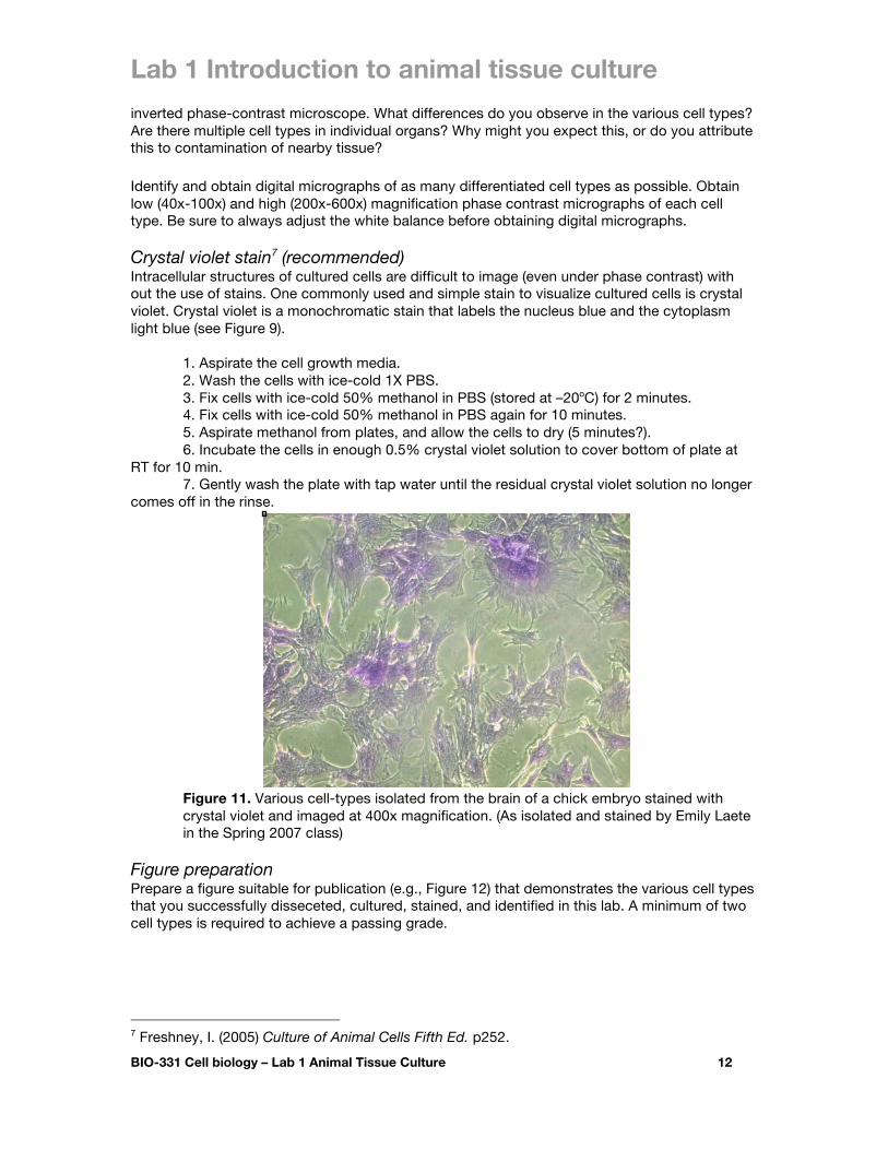
inverted phase-contrast microscope. What differences do you observe in the various cell types? Are there multiple cell types in individual organs? Why might you expect this, or do you attribute this to contamination of nearby tissue? Identify and obtain digital micrographs of as many differentiated cell types as possible. Obtain low (40x-100x) and high (200x-600x) magnification phase contrast micrographs of each cell type. Be sure to always adjust the white balance before obtaining digital micrographs. Crystal violet stain7 (recommended) Intracellular structures of cultured cells are difficult to image (even under phase contrast) with out the use of stains. One commonly used and simple stain to visualize cultured cells is crystal violet. Crystal violet is a monochromatic stain that labels the nucleus blue and the cytoplasm light blue (see Figure 9).
1. Aspirate the cell growth media. 2. Wash the cells with ice-cold 1X PBS. 3. Fix cells with ice-cold 50% methanol in PBS (stored at –20oC) for 2 minutes. 4. Fix cells with ice-cold 50% methanol in PBS again for 10 minutes. 5. Aspirate methanol from plates, and allow the cells to dry (5 minutes?). 6. Incubate the cells in enough 0.5% crystal violet solution to cover bottom of plate at
RT for 10 min. 7. Gently wash the plate with tap water until the residual crystal violet solution no longer
comes off in the rinse.
Figure 11. Various cell-types isolated from the brain of a chick embryo stained with crystal violet and imaged at 400x magnification. (As isolated and stained by Emily Laete in the Spring 2007 class)
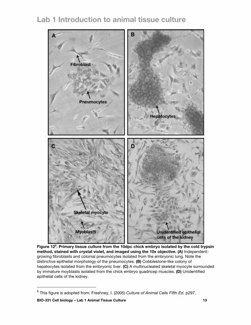
Figure preparation Prepare a figure suitable for publication (e.g., Figure 12) that demonstrates the various cell types that you successfully disseceted, cultured, stained, and identified in this lab. A minimum of two cell types is required to achieve a passing grade.
7 Freshney, I. (2005) Culture of Animal Cells Fifth Ed. p252.
Lab 1 Introduction to animal tissue culture
BIO-331 Cell biology – Lab 1 Animal Tissue Culture 13
Figure 128. Primary tissue culture from the 10dpc chick embryo isolated by the cold trypsin method, stained with crystal violet, and imaged using the 10x objective. (A) Independent-growing fibroblasts and colonial pneumocytes isolated from the embryonic lung. Note the distinctive epithelial morphology of the pneumocytes. (B) Cobblestone-like colony of hepatocytes isolated from the embryonic liver. (C) A multinucleated skeletal myocyte surrounded by immature moyblasts isolated from the chick embryo quadricep muscles. (D) Unidentified epithelial cells of the kidney. 8 This figure is adopted from: Freshney, I. (2005) Culture of Animal Cells Fifth Ed. p297.
Lab 1 Introduction to animal tissue culture
BIO-331 Cell biology – Lab 1 Animal Tissue Culture 14
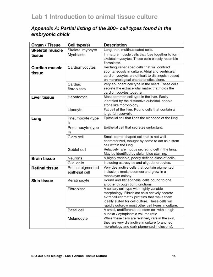
Appendix A: Partial listing of the 200+ cell types found in the embryonic chick Organ / Tissue Cell type(s) Description Skeletal muscle tissue
Skeletal myocyte Long, thin, multinucleated cells.
Myoblasts Immature muscle cells that fuse together to form skeletal myocytes. These cells closely resemble fibroblasts.
Cardiac muscle tissue
Cardiomyocytes Rectangular-shaped cells that will contract spontaneously in culture. Atrial and ventricular cardiomyocytes are difficult to distinguish based on morphological characteristics alone.
Cardiac fibroblasts
Very abundant cell type in the heart. These cells secrete the extracellular matrix that holds the cardiomyocytes together.
Liver tissue Hepatocyte Most common cell type in the liver. Easily identified by the distinctive cuboidal, cobble-stone like morphology.
Lipocyte Fat cell of the liver. Round cells that contain a large fat reservoir.
Lung Pneumocyte (type I)
Epithelial cell that lines the air space of the lung.
Pneumocyte (type II)
Epithelial cell that secretes surfactant.
Clara cell Small, dome-shaped cell that is not well characterized, thought by some to act as a stem cell within the lung.
Goblet cell Relatively rare mucus secreting cell in the lung. May be identified by alcian blue staining.
Brain tissue Neurons A highly variable, poorly defined class of cells.
Glial cells Including astrocytes and oligodendrocytes.
Retinal tissue Retinal pigmented epithelial cell
Very destinctive cells that contain pigmented inclusions (melanosomes) and grow in a monolayer colony.
Skin tissue Keratinocyte Round and flat epithelial cells bound to one another through tight junctions.
Fibroblast A solitary cell type with highly variable morphology. Fibroblast cells actively secrete extracellular matrix proteins that make them ideally suited for cell culture. These cells will rapidly outgrow most other cell types in culture.
Basal cell A small, undifferentiated stem cell with a high nucelar / cytoplasmic volume ratio.
Melanocyte While these cells are relatively rare in the skin, they are very distinctive in culture (branched morphology and dark pigmented inclusions).