larynx anatomy - sborl.es · glottis: vocal folds + space between them - the narrowest portion of...
TRANSCRIPT
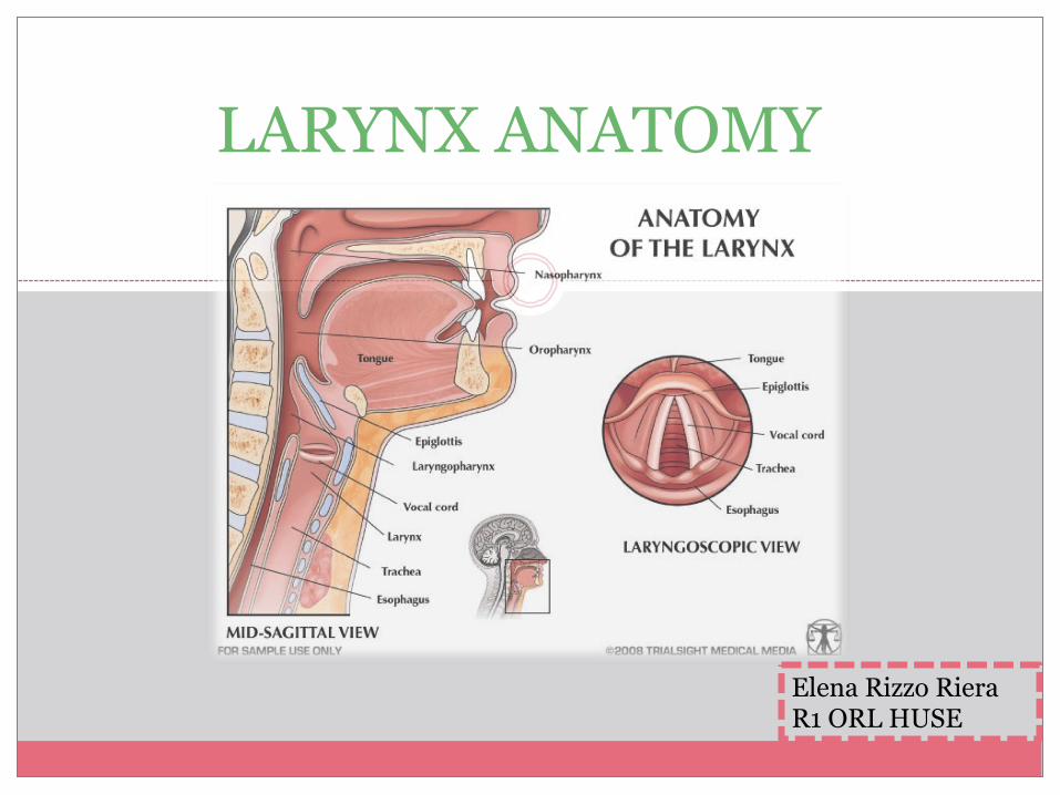
LARYNX ANATOMY
Elena Rizzo Riera R1 ORL HUSE
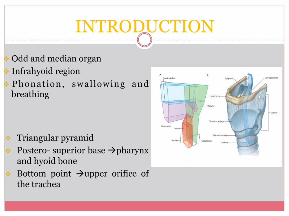
INTRODUCTION v Odd and median organ v Infrahyoid region v Phonat ion, swal lowing and
breathing v Triangular pyramid v Postero- superior base àpharynx
and hyoid bone v Bottom point àupper orifice of
the trachea
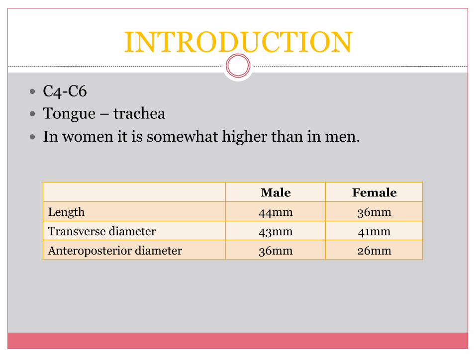
INTRODUCTION � C4-C6 � Tongue – trachea � In women it is somewhat higher than in men.
Male Female Length 44mm 36mm Transverse diameter 43mm 41mm Anteroposterior diameter 36mm 26mm
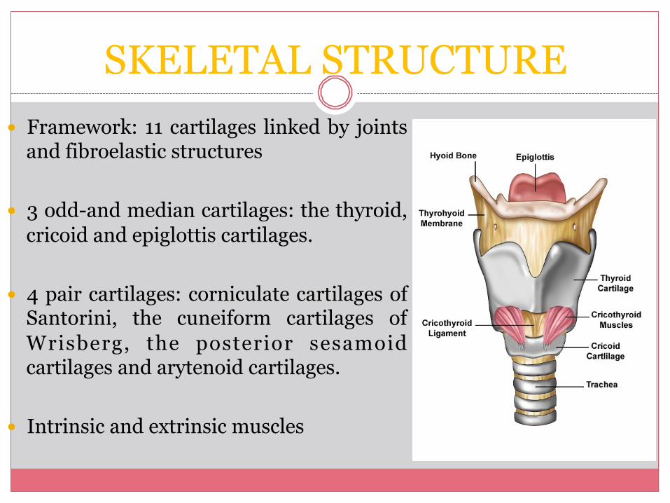
SKELETAL STRUCTURE � Framework: 11 cartilages linked by joints
and fibroelastic structures � 3 odd-and median cartilages: the thyroid,
cricoid and epiglottis cartilages. � 4 pair cartilages: corniculate cartilages of
Santorini, the cuneiform cartilages of Wrisberg, the posterior sesamoid cartilages and arytenoid cartilages.
� Intrinsic and extrinsic muscles
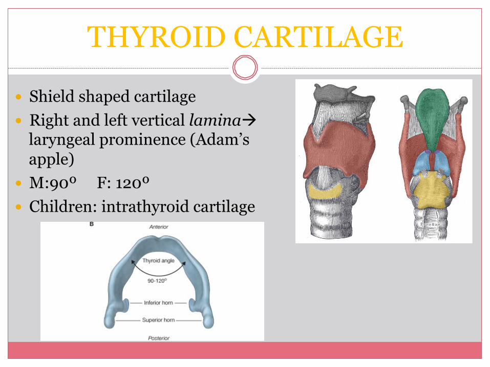
THYROID CARTILAGE � Shield shaped cartilage � Right and left vertical laminaà
laryngeal prominence (Adam’s apple)
� M:90º F: 120º � Children: intrathyroid cartilage
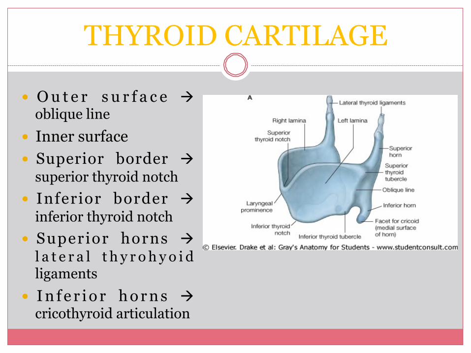
� O u t e r s u r f a c e à oblique line
� Inner surface � Superior border à
superior thyroid notch � Inferior border à
inferior thyroid notch � Superior horns à
l a t e r a l t h y r o h y o i d ligaments
� I n f e r i o r h o r n s à cricothyroid articulation
THYROID CARTILAGE
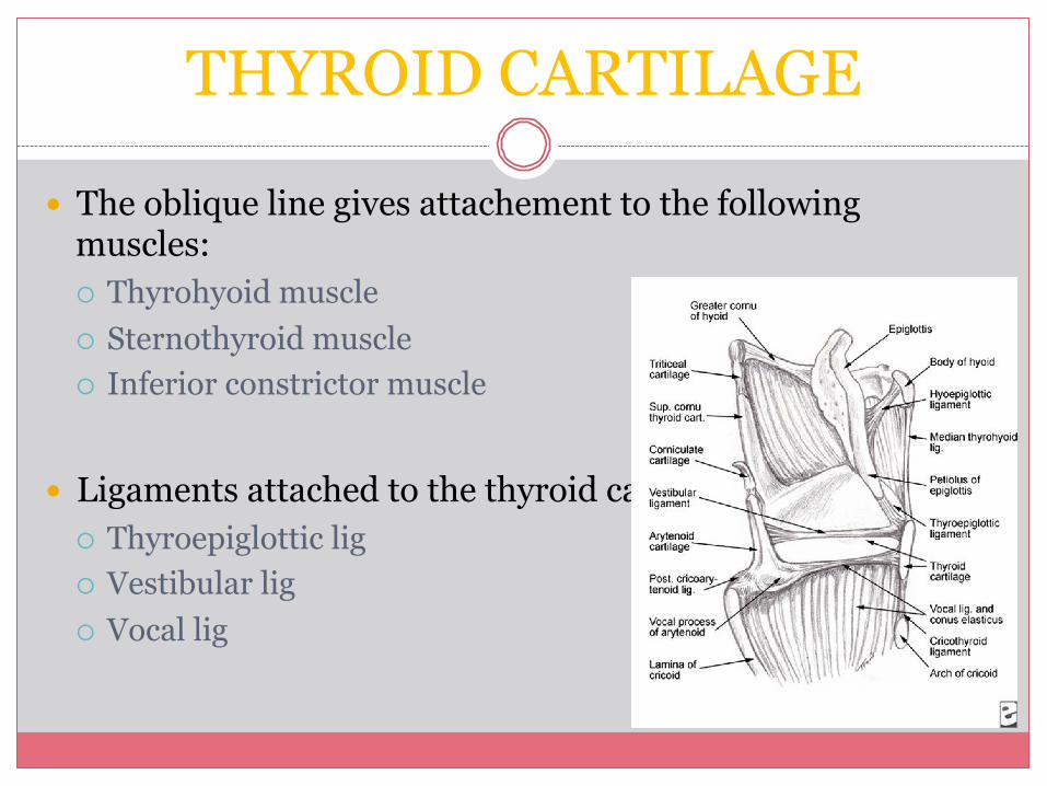
� The oblique line gives attachement to the following muscles: ¡ Thyrohyoid muscle ¡ Sternothyroid muscle ¡ Inferior constrictor muscle
� Ligaments attached to the thyroid cartilage ¡ Thyroepiglottic lig ¡ Vestibular lig ¡ Vocal lig
THYROID CARTILAGE
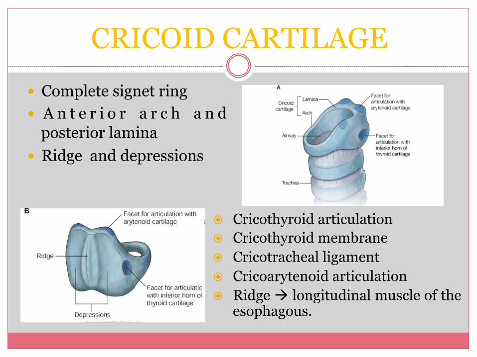
CRICOID CARTILAGE � Complete signet ring � A n t e r i o r a r c h a n d
posterior lamina � Ridge and depressions
� Cricothyroid articulation � Cricothyroid membrane � Cricotracheal ligament � Cricoarytenoid articulation � Ridge à longitudinal muscle of the
esophagous.
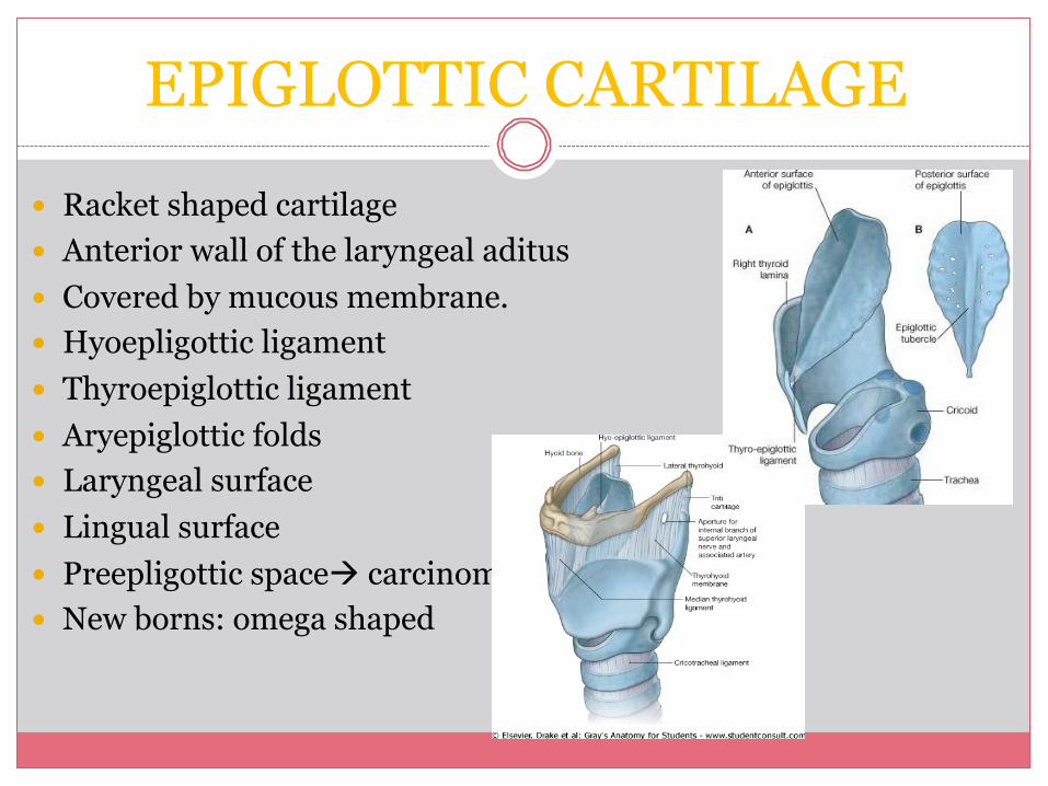
EPIGLOTTIC CARTILAGE � Racket shaped cartilage � Anterior wall of the laryngeal aditus � Covered by mucous membrane. � Hyoepligottic ligament � Thyroepiglottic ligament � Aryepiglottic folds � Laryngeal surface � Lingual surface � Preepligottic spaceà carcinoma � New borns: omega shaped
EPIGLOTTIC CARTILAGE
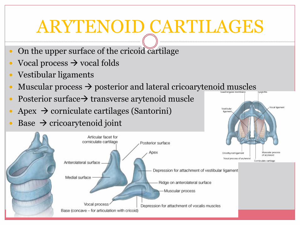
ARYTENOID CARTILAGES � On the upper surface of the cricoid cartilage � Vocal process à vocal folds � Vestibular ligaments � Muscular process à posterior and lateral cricoarytenoid muscles � Posterior surfaceà transverse arytenoid muscle � Apex à corniculate cartilages (Santorini) � Base à cricoarytenoid joint
OTHER CARTILAGES
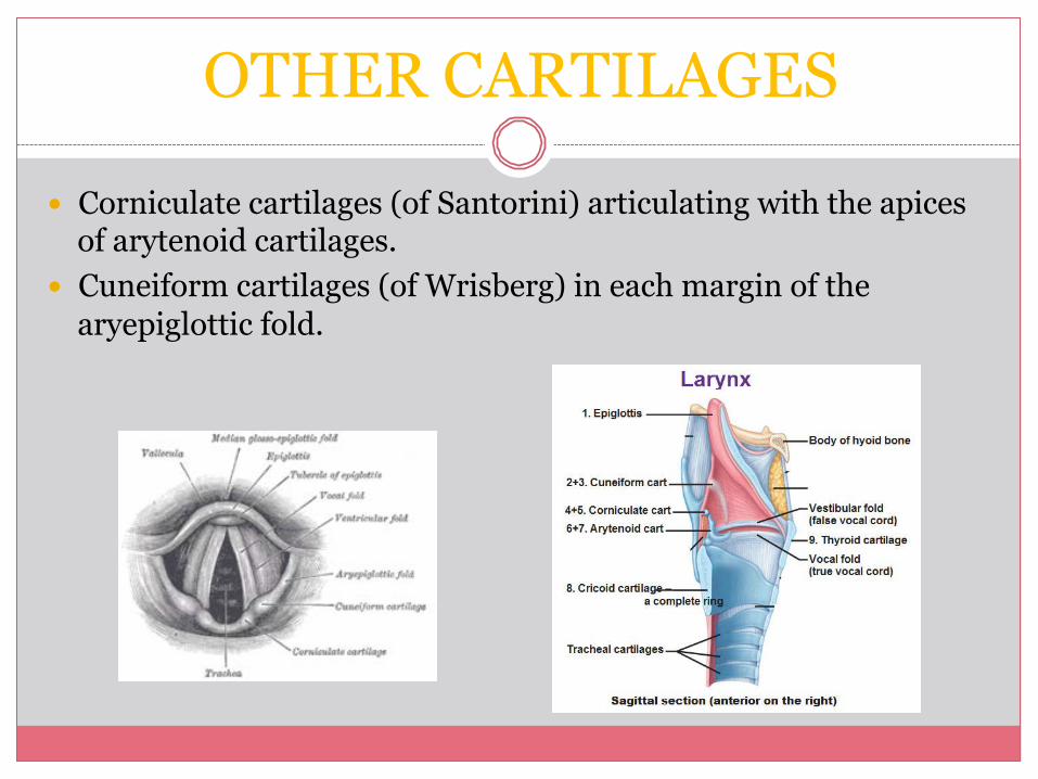
� Corniculate cartilages (of Santorini) articulating with the apices of arytenoid cartilages.
� Cuneiform cartilages (of Wrisberg) in each margin of the aryepiglottic fold.
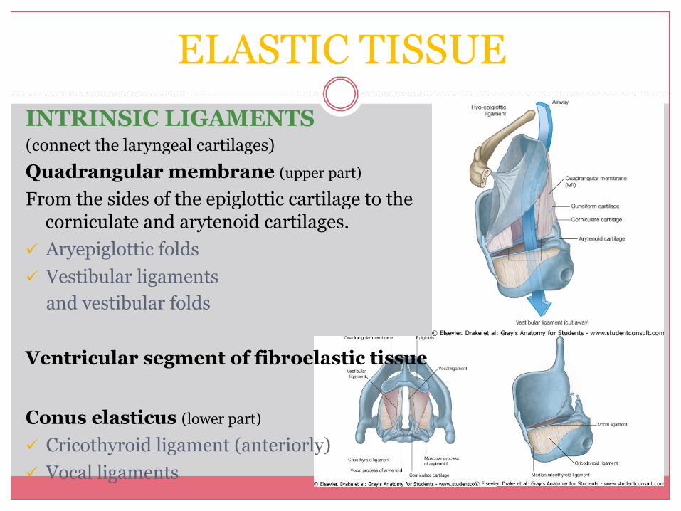
ELASTIC TISSUE INTRINSIC LIGAMENTS (connect the laryngeal cartilages) Quadrangular membrane (upper part)
From the sides of the epiglottic cartilage to the corniculate and arytenoid cartilages.
ü Aryepiglottic folds ü Vestibular ligaments
and vestibular folds Ventricular segment of fibroelastic tissue Conus elasticus (lower part)
ü Cricothyroid ligament (anteriorly) ü Vocal ligaments
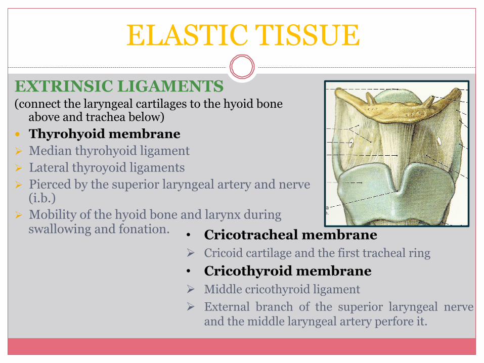
EXTRINSIC LIGAMENTS (connect the laryngeal cartilages to the hyoid bone
above and trachea below) � Thyrohyoid membrane Ø Median thyrohyoid ligament Ø Lateral thyroyoid ligaments Ø Pierced by the superior laryngeal artery and nerve
(i.b.) Ø Mobility of the hyoid bone and larynx during
swallowing and fonation. • Cricotracheal membrane Ø Cricoid cartilage and the first tracheal ring • Cricothyroid membrane Ø Middle cricothyroid ligament Ø External branch of the superior laryngeal nerve
and the middle laryngeal artery perfore it.
ELASTIC TISSUE
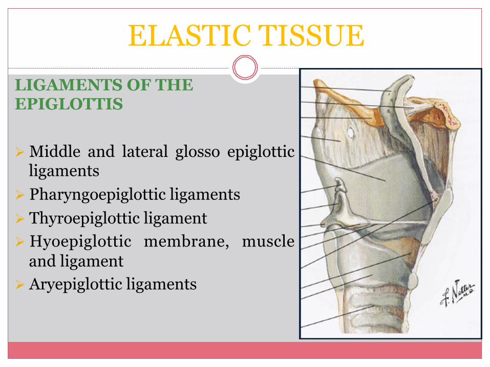
LIGAMENTS OF THE EPIGLOTTIS Ø Middle and lateral glosso epiglottic
ligaments Ø Pharyngoepiglottic ligaments Ø Thyroepiglottic ligament Ø Hyoepiglottic membrane, muscle
and ligament Ø Aryepiglottic ligaments
ELASTIC TISSUE
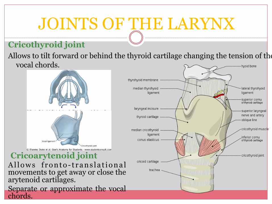
JOINTS OF THE LARYNX Cricothyroid joint Allows to tilt forward or behind the thyroid cartilage changing the tension of the
vocal chords.
Cricoarytenoid joint A l l o w s f r o n t o - t r a n s l a t i o n a l movements to get away or close the arytenoid cartilages. Separate or approximate the vocal chords.
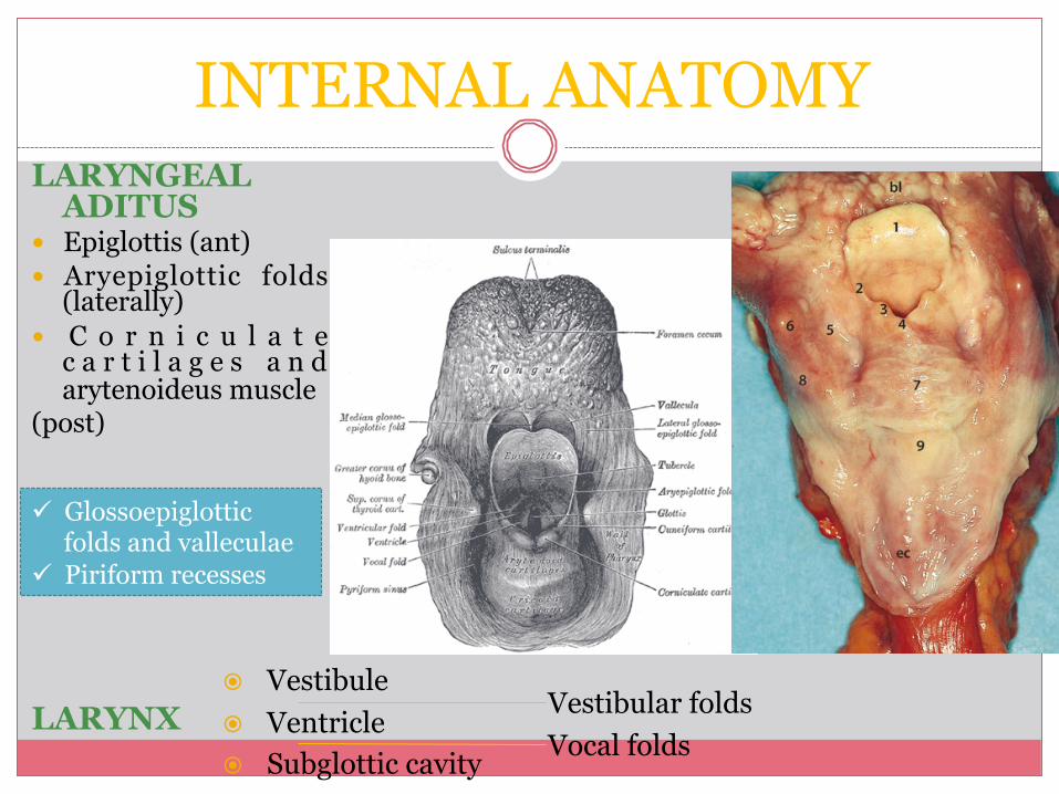
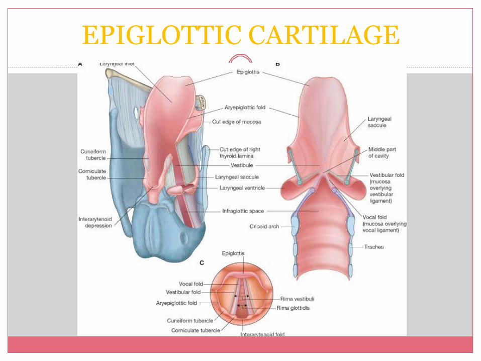
INTERNAL ANATOMY LARYNGEAL
ADITUS � Epiglottis (ant) � Aryepiglottic folds
(laterally) � C o r n i c u l a t e
c a r t i l a g e s a n d arytenoideus muscle
(post) LARYNX
� Vestibule � Ventricle � Subglottic cavity
Vestibular folds Vocal folds
ü Glossoepiglottic folds and valleculae
ü Piriform recesses
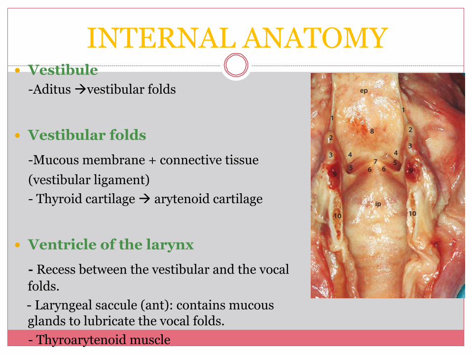
� Vestibule -Aditus àvestibular folds
� Vestibular folds
-Mucous membrane + connective tissue (vestibular ligament) - Thyroid cartilage à arytenoid cartilage
� Ventricle of the larynx
- Recess between the vestibular and the vocal folds.
- Laryngeal saccule (ant): contains mucous glands to lubricate the vocal folds. - Thyroarytenoid muscle
INTERNAL ANATOMY
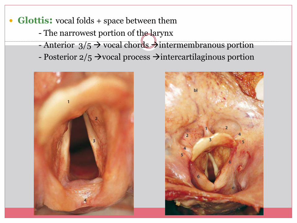
� Glottis: vocal folds + space between them - The narrowest portion of the larynx - Anterior 3/5 à vocal chords àintermembranous portion - Posterior 2/5 àvocal process àintercartilaginous portion
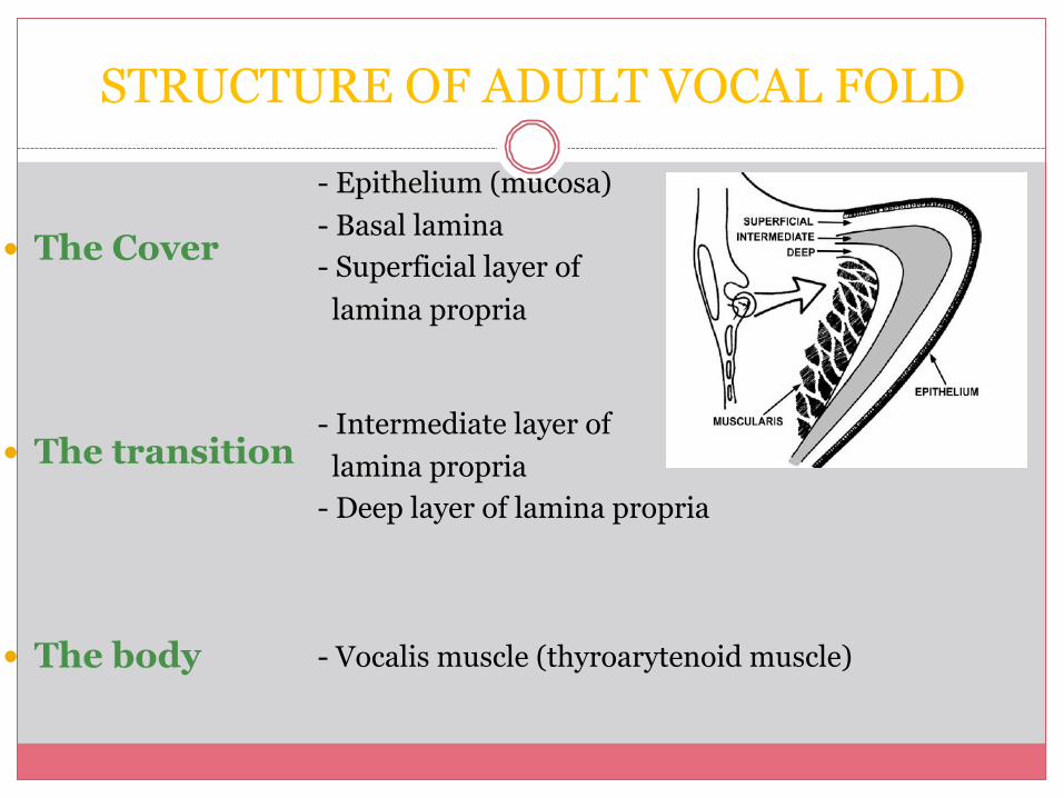
STRUCTURE OF ADULT VOCAL FOLD
� The Cover � The transition � The body
- Epithelium (mucosa) - Basal lamina - Superficial layer of lamina propria
- Intermediate layer of lamina propria - Deep layer of lamina propria
- Vocalis muscle (thyroarytenoid muscle)
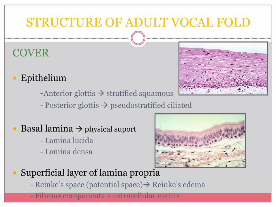
COVER � Epithelium
-Anterior glottis à stratified squamous - Posterior glottis à pseudostratified ciliated
� Basal lamina à physical suport
- Lamina lucida - Lamina densa
� Superficial layer of lamina propria - Reinke’s space (potential space)à Reinke’s edema - Fibrous components + extracellular matrix
STRUCTURE OF ADULT VOCAL FOLD
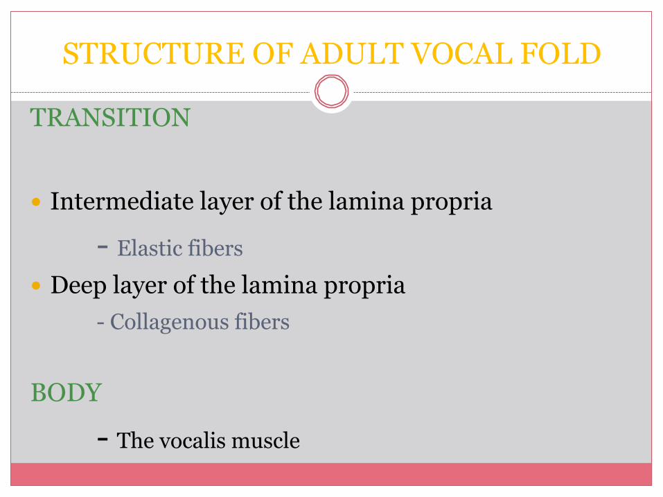
TRANSITION
� Intermediate layer of the lamina propria
- Elastic fibers � Deep layer of the lamina propria
- Collagenous fibers BODY - The vocalis muscle
STRUCTURE OF ADULT VOCAL FOLD
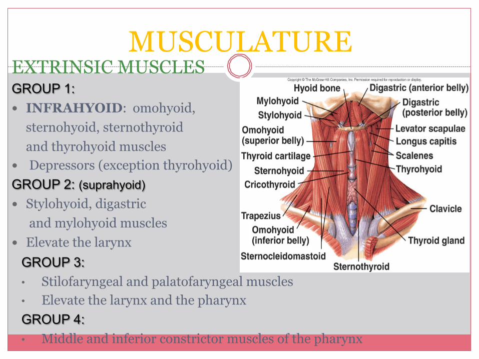
MUSCULATURE EXTRINSIC MUSCLES GROUP 1: � INFRAHYOID: omohyoid,
sternohyoid, sternothyroid and thyrohyoid muscles
� Depressors (exception thyrohyoid) GROUP 2: (suprahyoid) � Stylohyoid, digastric
and mylohyoid muscles � Elevate the larynx
GROUP 3: • Stilofaryngeal and palatofaryngeal muscles • Elevate the larynx and the pharynx GROUP 4: • Middle and inferior constrictor muscles of the pharynx
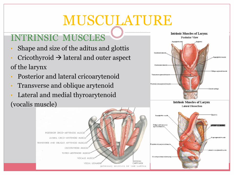
MUSCULATURE INTRINSIC MUSCLES • Shape and size of the aditus and glottis • Cricothyroid à lateral and outer aspect of the larynx • Posterior and lateral cricoarytenoid • Transverse and oblique arytenoid • Lateral and medial thyroarytenoid (vocalis muscle)
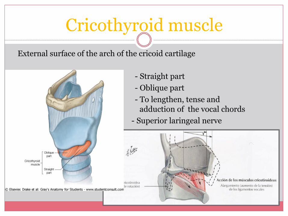
Cricothyroid muscle External surface of the arch of the cricoid cartilage
- Straight part - Oblique part - To lengthen, tense and
ad adduction of the vocal chords - Superior laringeal nerve
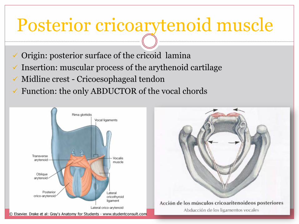
Posterior cricoarytenoid muscle ü Origin: posterior surface of the cricoid lamina ü Insertion: muscular process of the arythenoid cartilage ü Midline crest - Cricoesophageal tendon ü Function: the only ABDUCTOR of the vocal chords
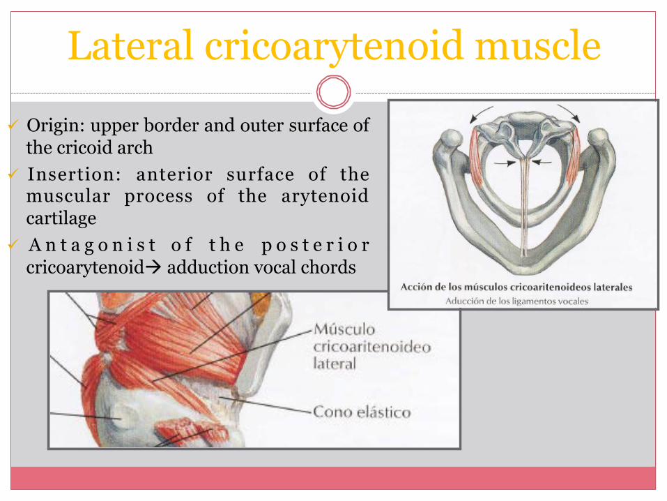
Lateral cricoarytenoid muscle
ü Origin: upper border and outer surface of the cricoid arch
ü Insertion: anterior surface of the muscular process of the arytenoid cartilage
ü A n t a g o n i s t o f t h e p o s t e r i o r cricoarytenoidà adduction vocal chords
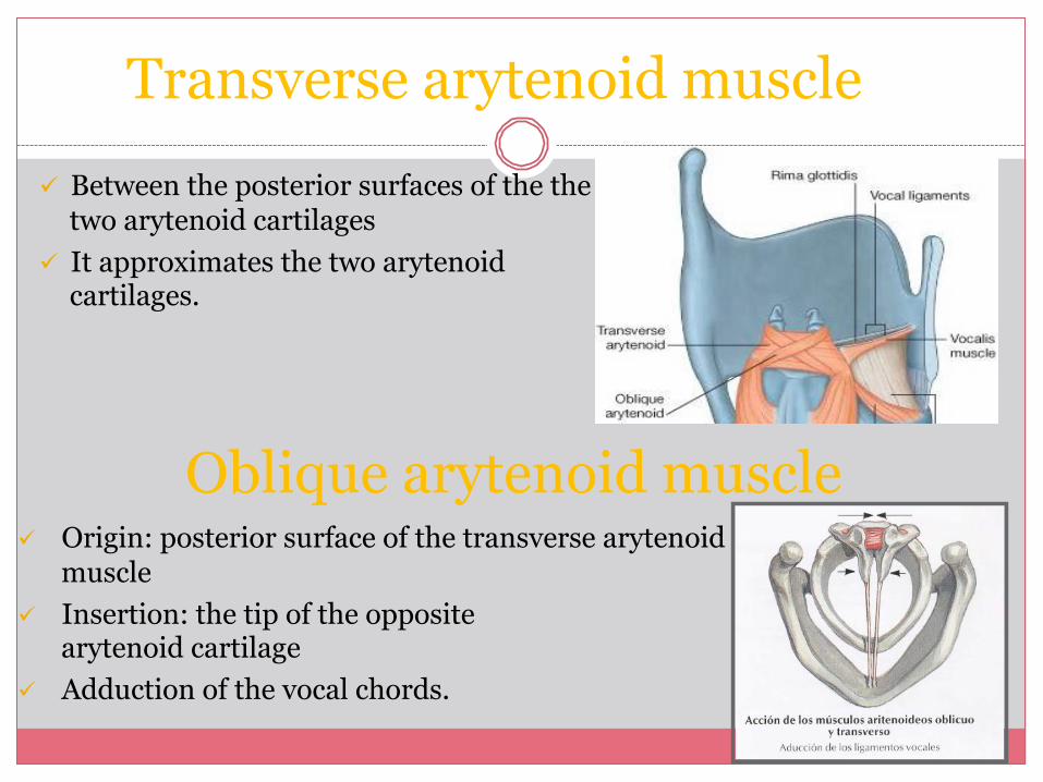
Transverse arytenoid muscle ü Between the posterior surfaces of the the
two arytenoid cartilages ü It approximates the two arytenoid
cartilages.
ü Origin: posterior surface of the transverse arytenoid muscle
ü Insertion: the tip of the opposite arytenoid cartilage
ü Adduction of the vocal chords.
Oblique arytenoid muscle
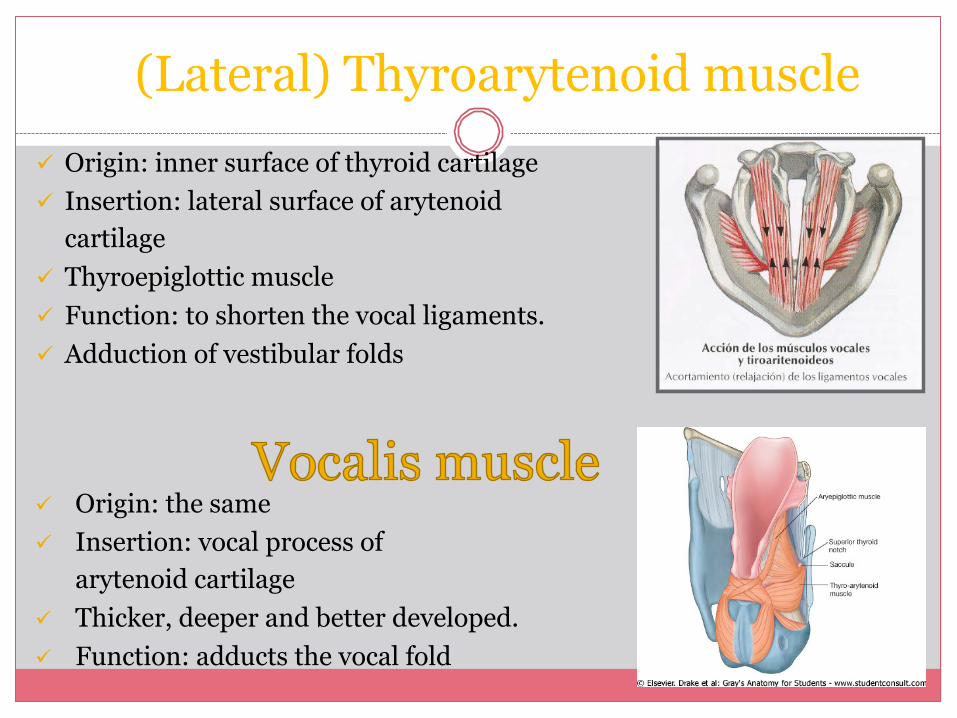
(Lateral) Thyroarytenoid muscle ü Origin: inner surface of thyroid cartilage ü Insertion: lateral surface of arytenoid
cartilage ü Thyroepiglottic muscle ü Function: to shorten the vocal ligaments. ü Adduction of vestibular folds
ü Origin: the same ü Insertion: vocal process of
arytenoid cartilage ü Thicker, deeper and better developed. ü Function: adducts the vocal fold
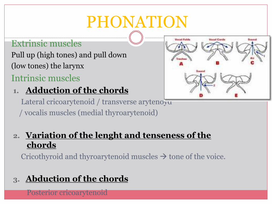
PHONATION Extrinsic muscles Pull up (high tones) and pull down (low tones) the larynx
Intrinsic muscles 1. Adduction of the chords
Lateral cricoarytenoid / transverse arytenoyd / vocalis muscles (medial thyroarytenoid)
2. Variation of the lenght and tenseness of the chords
Cricothyroid and thyroarytenoid muscles à tone of the voice.
3. Abduction of the chords Posterior cricoarytenoid
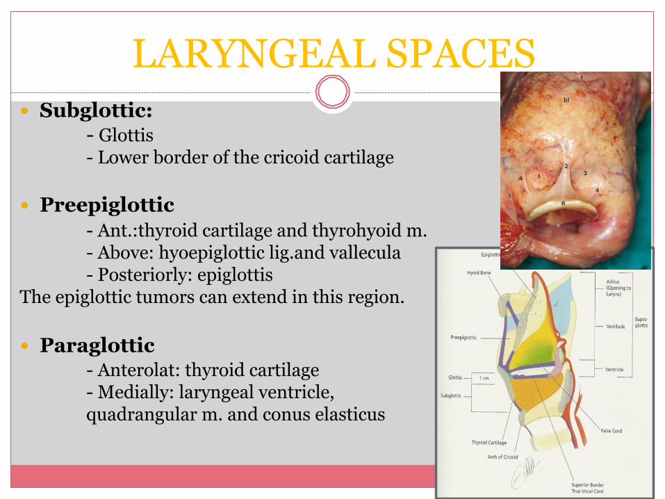
LARYNGEAL SPACES � Subglottic:
- Glottis - Lower border of the cricoid cartilage
� Preepiglottic
- Ant.:thyroid cartilage and thyrohyoid m. - Above: hyoepiglottic lig.and vallecula - Posteriorly: epiglottis
The epiglottic tumors can extend in this region. � Paraglottic
- Anterolat: thyroid cartilage - Medially: laryngeal ventricle, quadrangular m. and conus elasticus
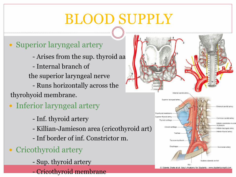
BLOOD SUPPLY � Superior laryngeal artery
- Arises from the sup. thyroid aa - Internal branch of the superior laryngeal nerve - Runs horizontally across the
thyrohyoid membrane. � Inferior laryngeal artery
- Inf. thyroid artery - Killian-Jamieson area (cricothyroid art) - Inf border of inf. Constrictor m.
� Cricothyroid artery - Sup. thyroid artery - Cricothyroid membrane
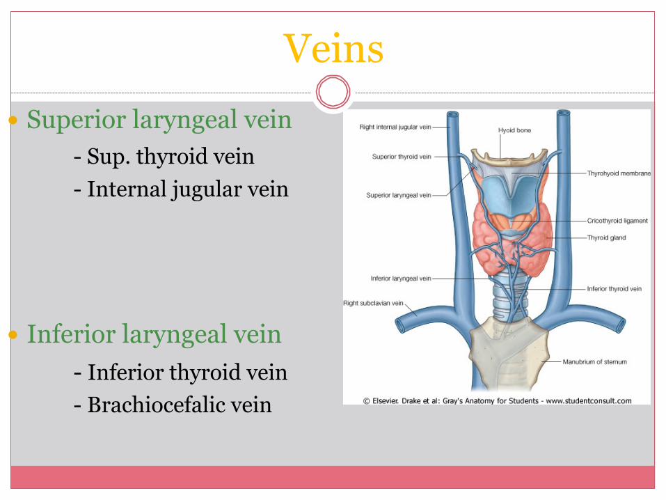
Veins � Superior laryngeal vein
- Sup. thyroid vein - Internal jugular vein
� Inferior laryngeal vein
- Inferior thyroid vein - Brachiocefalic vein
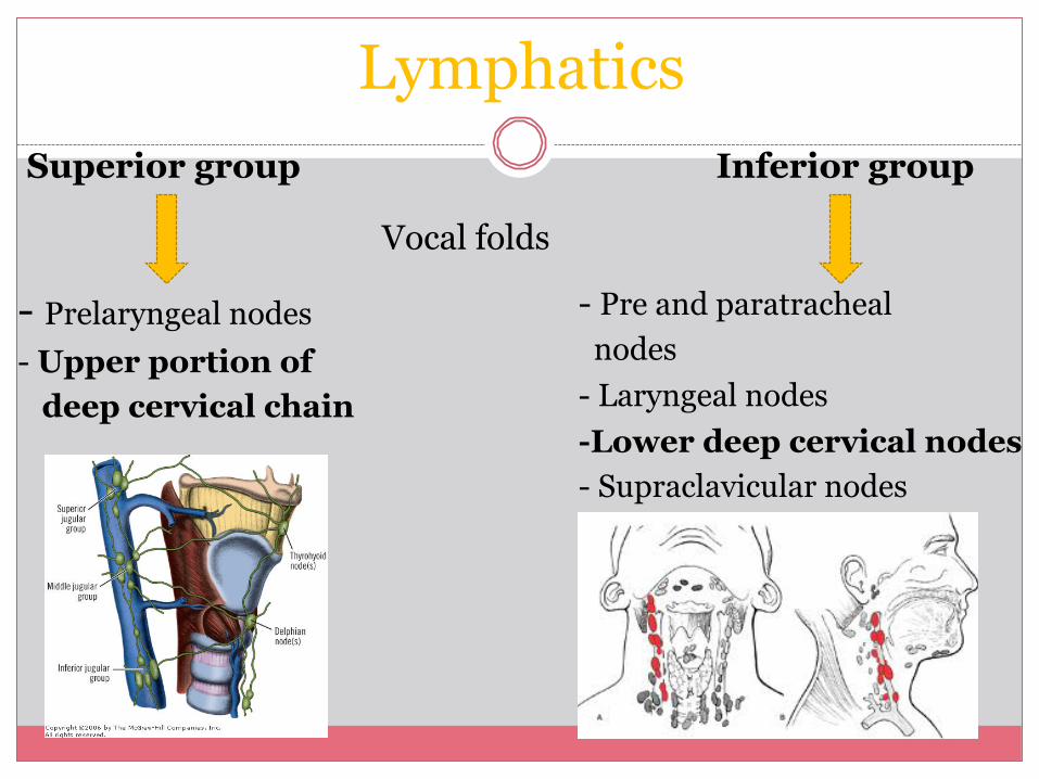
Lymphatics Superior group
Vocal folds
Inferior group
- Prelaryngeal nodes - Upper portion of deep cervical chain
- Pre and paratracheal nodes - Laryngeal nodes -Lower deep cervical nodes - Supraclavicular nodes
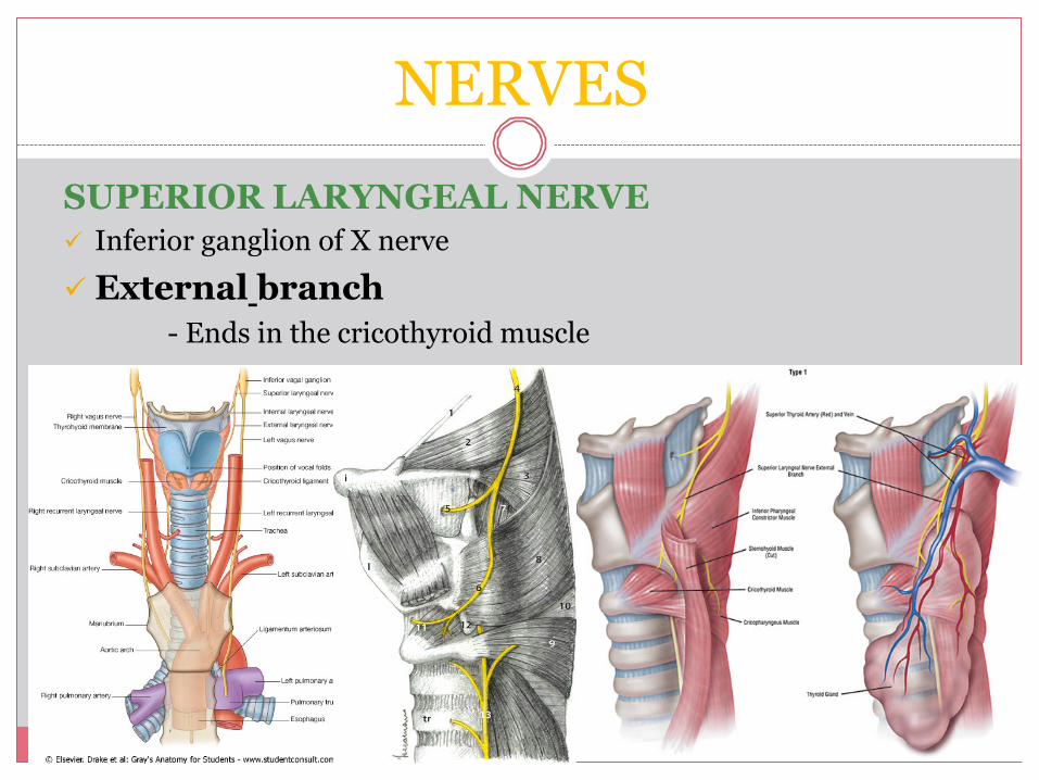
NERVES SUPERIOR LARYNGEAL NERVE ü Inferior ganglion of X nerve ü External branch
- Ends in the cricothyroid muscle
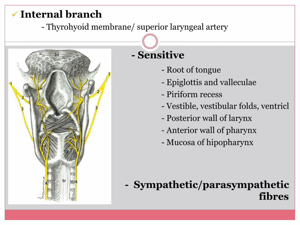
ü Internal branch - Thyrohyoid membrane/ superior laryngeal artery
- Sensitive - Root of tongue - Epiglottis and valleculae - Piriform recess - Vestible, vestibular folds, ventricl - Posterior wall of larynx - Anterior wall of pharynx - Mucosa of hipopharynx
- Sympathetic/parasympathetic fibres
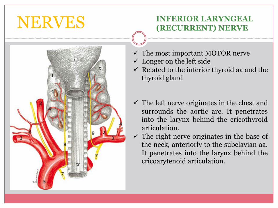
NERVES INFERIOR LARYNGEAL (RECURRENT) NERVE
ü The most important MOTOR nerve ü Longer on the left side ü Related to the inferior thyroid aa and the
thyroid gland
ü The left nerve originates in the chest and surrounds the aortic arc. It penetrates into the larynx behind the cricothyroid articulation.
ü The right nerve originates in the base of the neck, anteriorly to the subclavian aa. It penetrates into the larynx behind the cricoarytenoid articulation.
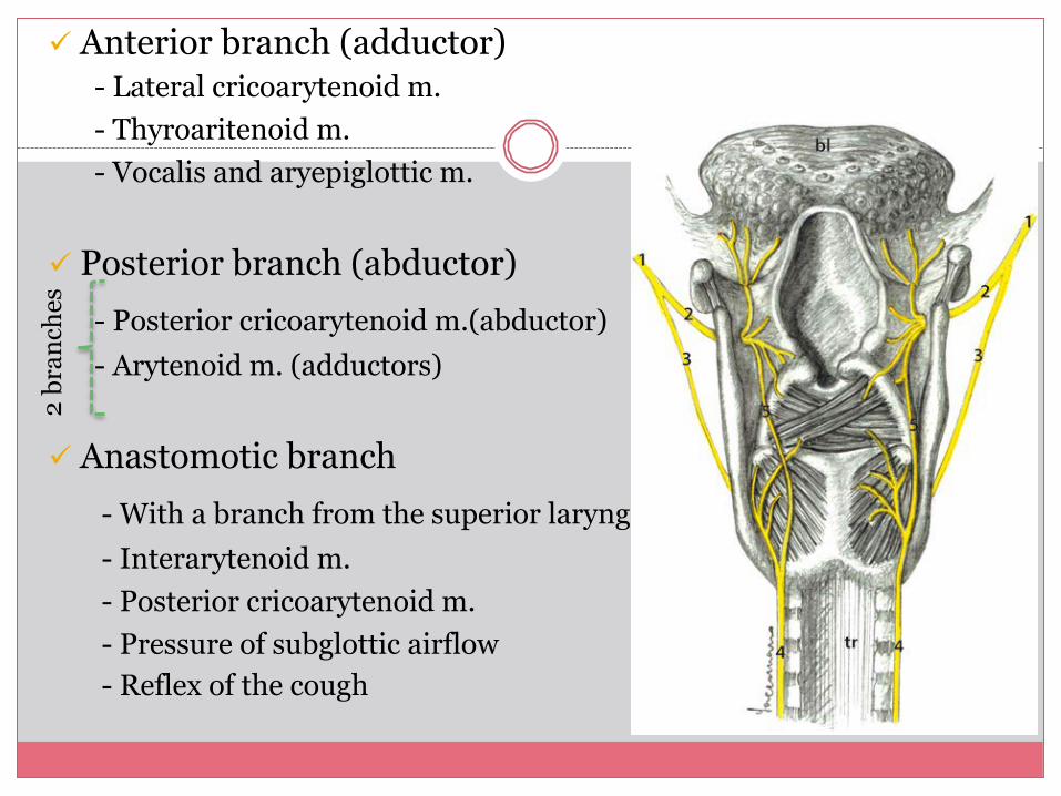
ü Anterior branch (adductor) - Lateral cricoarytenoid m. - Thyroaritenoid m. - Vocalis and aryepiglottic m.
ü Posterior branch (abductor)
- Posterior cricoarytenoid m.(abductor) - Arytenoid m. (adductors)
ü Anastomotic branch
- With a branch from the superior laryngeal - Interarytenoid m. - Posterior cricoarytenoid m. - Pressure of subglottic airflow - Reflex of the cough
2 br
anch
es
� Surgical anatomy of the head and neck. P. Janfaza, J. B. Nadol, R. J. Galla, R. L. Fabian, W. W. Montgomery.
� Otorrinolaringología. W. Becker, H. H. Naumann, C. R. Pfaltz.
� Otorhinolaryngology, Head & Neck Surgery. M. Anniko, M. Bernal- Sprekelsen, V. Bonkowsky, P. Bradley, S. Iurato
� Cummings otolaryngology head and neck surgery. Paul W. Flint, K. Thomas Robbins, Bruce H. Haughey, J. Regan Thomas et al.
BIBLIOGRAPHY