laser dentistry - zwp online · vivek et al.24 treated 28 patients with histologi-cally diagnosed...
TRANSCRIPT

laserinternational magazine of laser dentistry2
2012
i s sn 2193-4665 Vol. 4 • Issue 2/2012
| researchThe antibacterial effects of lasers in endodontics
| overviewLaser treatment of dentine hypersensitivity
| case reportA novel technique of Er:YAG laser-enhancedearly implant stability

©2012
Breaks thespeed Barrier
enaBles BiOlOGiCallY FriendlY dentistrY
prOvides Great
return On investment
ilase™
940nm diOde laserdOCkinG statiOn
• Adds dual wavelength versatility and convenience
• First totally wireless dental laser• 5 Watts peak power with
ComfortPulse™
• Battery operated with finger switch activation
• Exclusive bendable tips in many diameters & lengths
intuitive & intelliGent GraphiCal user interFaCe
• No micro-fractures or thermal damage • More precise, minimally invasive
• Increases treatment acceptance ofday-to-day restorative cases
• Attracts new patients • Increases productivity and enables
new procedures
• ЯR™ powered laser delivers 100 pulses/sec. forsuperior soft-tissue cutting
• Patented laser technology delivers 10 watts of power• Enables multi-quadrant same-day procedures
For example, performing a Class i Cavity prepwith the iplus™ is as easy as 1,2,3...
Step 1 Select “Restorative” from the first screenStep 2 Choose “Class I” from the next screen
that appears automaticallyStep 3 Specify any other concerns such as patient
sensitivity or bond strength that’s it! step on the foot pedal, and
start working.
R R
2011
TOP 50Technology Products
| www.BiolaSe.com | Toll-free 888-424-6527
The Dual Wavelength Waterlase iPlusAdvancing Laser Technology to Its Ultimate
iPlus_LaserInternational_050912.indd 1 5/9/12 2:20 PM

editorial I
I 03laser2_2012
_Have you been following the coverage of dental congresses in the past few weeks? If so,you might have felt just the same positive sensation as I have when I came across the fact thatscientific contributions on laser applications in implantology have gained a high rank in the pastcongress season. By the way, the same holds true for scientific texts on implantology in dentalspecialist publications.
The growing impact of laser applications on both congresses and scientific literature does in-deed pose a snapshot of the current status of laser in dental therapies and might even express arecent trend. This trend, in my view, bears various notable facets:
Many of the numerous implantological congresses and symposia intersecting with laser den-tistry have included reports on the application of monochromatic light into their programs.Moreover, whole sessions are dedicated to laser in both implantological and periodontal con-gresses and symposia. On such occasions, the high value of atraumatic laser incisions with sig-nificantly reduced hemorrhage is highlighted, along with the lack of alternatives to laser sur-face decontamination in the treatment of periimplantitis.
The antibacterial effects of lasers in endodontics and the advantages of laser therapy of oralhaemangiomas contribute to the wide range of applications of laser in dentistry.
As you can see, we are provided with a sufficient (and evidence-based) number of oppor -tunities to pursue our passion for monochromatic light in dental therapy. It follows that our résumé be “No (more) dentistry without laser”!_
With best regards,
Dr Georg Bach
Laser (in)dispen sable
in dentistry?Dr Georg Bach

I content _ laser
04 I laser2_2012
I editorial
03 Laser (in)dispen sable in dentistry?| Dr Georg Bach
I research
06 Laser in oral surgery and medicine—Part II| Antonio Batista-Cruzado et al.
12 Evaluation of com bined Nd:YAG laser treatment ofmoderate periodontitis| Dr Anna-Maria Yiannikou-Loucaidou
16 The antibacterial effects of lasers in endodontics| Dr Selma Cristina Cury Camargo
I overview
24 Laser treatment of dentine hypersensitivity| Dr Ute Botzenhart et al.
I case report
28 A novel technique of Er:YAG laser-enhanced early implant stability| Dr Kenneth Luk et al.
32 Laser therapy of oral haemangiomas| Friedrich Müller et al.
I industry report
34 Lasers in oral Implantology| Dr Ilay Maden et al.
I education
40 “Laser Supported Dentistry” in Turkey| Prof Dr Aslihan Usumez et al.
42 Innovation and information at IDS 2013| VDDI
I meetings
43 International events 2012
44 DGL annual conference in Leipzig, Germany| Jürgen Isbaner
46 “Scientifically, laser has now reached its highest standard”| Dajana Mischke
I news
38 Manufacturer News
48 News
I about the publisher
50 | imprint
page 42 page 44 page 48
page 16 page 24 page 34
Cover image courtesy of Biolase Technology Inc.,
www.biolase.com.
Original Background: ©Excellent backgrounds
Artwork by Sarah Fuhrmann, OEMUS MEDIA AG.

FASCINATION OFLASER DENTISTRYFOR EXPERTS AND BEGINNERS
FURTHERINFORMATION
laser 2/12
OFFICE STAMPPLEASE FAX THIS FORM+49 341 48474-390
Further information about:
❏ LASER START UP 2012 ❏ 21st ANNUAL CONGRESS OF THE DGL e.V
September 7–8, 2012, Leipzig, Germany
NAME/E-MAIL
ORGANIZATION/REGISTRATIONDeutsche Gesellschaft für Laserzahnheilkunde e.V. (German Society for Laserdentistry)Klinik für Zahn erhaltung, Parodontologie und PräventiveZahnheilkunde Universitätsklinikum der RWTH Aachen,Pauwelsstraße 30, 52074 Aachen, GermanyFon: +49 241 8088-164Fax: +49 241 [email protected]
OEMUS MEDIA AGHolbeinstraße 2904229 Leipzig, GermanyFon: +49 341 48474-308Fax: +49 341 48474-390event@oemus-media.dewww.oemus.comwww.dgl-jahrestagung.dewww.startup-laser.de
MAIN SPONSOR
SEPTEMBER 7–8, 2012LEIPZIG
HOTEL THE WESTINLEIPZIG, GERMANY

_The laser has been used in the field of oral sur-gery for a wide range of indications. In this article, wefocus on its surgical uses. The success of the surgicaltreatment of lesions in the oral cavity depends greatlyon knowledge of the aetiology and histology of the le-sion. There are pathologies that can be treated withlaser, such as cancer sores and hyperkeratosis. Others,like candidiasis, cannot be treated with laser. Further-more, laser has quickly become a predictable andfavourable treatment modality for leukoplakia, hae-mangioma and epulis.
In the last issue of laser, the authors gave anoverview on in vitro studies and in vivo animal studiesin this field. They continued by giving examples of invivo studies on humans on the subjects of wound andbone healing supported by laser treatment. Finally, theauthors analysed soft-tissue surgery and examples ofcancer treatment via CO2 laser and photodynamictherapy under the headline of “clinical studies”. Theycontinue this chapter in the present issue of laser bydiscussing, among others, leukoplakia treatment, be-nign diseases and frenectomy. In the conclusion, theysum up the positive effects of laser on oral surgery.
Leukoplakia treatmentLeukoplakia is a premalignant lesion associated
with excessive consumption of alcohol and tobacco.
Although there is no specific treatment to prevent itsrecurrence, abandoning these habits can decreasethe probability of recurrence, as well as the transfor-mation into malignant tumours.
Vivek et al.24 treated 28 patients with histologi-cally diagnosed leukoplakia in order to study effi-cacy, safety and acceptability of lasers, particularlythe Nd:YAG laser. After laser treatment, post-opera-tive complications associated with ablation were as-sessed. They recorded only mild to moderate pain,with slight swelling up to 72 hours post-treatment.A follow-up study was initiated three years later. Ap-proximately 92 per cent of the patients were foundto have been cured. Therefore, the authors regardedNd:YAG laser as an effective tool for the treatment ofthis pathology.
There are also studies that recommend CO2 laserfor the excision of leukoplakia. For example, Reddiand Shafer25 found the CO2 laser to be of great suc-cess in the excision of leukoplakia in their study. Theyalso applied laser to the treatment of erythroplasiaand lichen planus.
Treatment of lichen planus Owing to its inflammatory effects, lichen planus
can be painful both in atrophic and erosive forms.
06 I laser2_2012
Laser in oral surgeryand medicine—Part IIAuthors_Antonio Batista-Cruzado, Daniel Torres-Lagares, Blanca Moreno-Manteca, Gerd Volland,
Patricia Bargiela-Perez, Martin Jorgen & Jose-Luis Gutierrez-Perez, Spain
I research
[BACKGROUND: ©PERFECTIONIST]

research I
The traditional treatment, therefore, makes use oftopical corticosteroids.
Cafaro et al.26 conducted a prospective cohortstudy of 13 patients with lichen planus in order to in-vestigate the effectiveness of LLLT. Patients weregiven biostimulation by diode laser (904 nm, pulsedmode). In general, a decrease in the size of the lesionsand pain, and overall stable results were observed.The authors therefore recommend LLLT as a possibletreatment for patients with lichen planus, but rec-ommend that future studies be done with a largergroup of patients in order to corroborate their results.
Aphthous stomatitisLLLT has also been used in the treatment of recur-
rent aphthous stomatitis. The study by De Souza etal.27 employed LLLT not as an inhibitor of the process,but for its modulating and healing effect on tissues.The authors assessed the effect of LLLT on aphthousstomatitis in 20 patients divided into two groups.Group I was treated with topical corticoids (triamci-nolone acetonide) and group II was treated via diodelaser (670 nm, 50 mW). Patients reported a decreasein pain already directly after laser treatment. Fourdays post-treatment, the lesion had receded com-pletely in group I, compared with complete recessionseven days post-treatment in group II.
Benign diseasesIn this section, pathological entities treated with
laser in recent years are discussed. Attention is paidto the technique applied, as well as frequency and im-pact of the laser used for the respective oral surgery.
Owing to the high frequency of pyogenic granu-loma in the oral cavity, especially during pregnancy,Jafarzadeh et al.28 reviewed this disease and consid-ered treatments and new approaches. Possible treat-ment options are, among others, resection by meansof a scalpel, cryotherapy, the use of corticosteroids, orthe use of an Nd:YAG or CO2 laser. The authors statethat laser treatment can help control bleeding, doesnot result in adverse effects and is therefore consid-
ered a successful treatment method with high ac-ceptance by patients.
Actinic cheilitis is another medical condition thatcan be treated with laser, since results show a highclinical resolution and low recurrence. Its successfultreatment is based on the removal of epitheliumwhile avoiding the resulting scarred tissue. De GodoyPeres et al.29 compared two protocols of low morbid-ity clinico-histologically in which CO2 laser was usedwith different parameters. A biopsy was done beforeand after laser treatment. In both groups, a signifi-cant reduction in epithelial dysplasia was achieved.Therefore, the authors recommend the use of lasersin cases of mild to moderate dysplasia.
Adipose tissue tumours are found frequently inthe maxillo-facial region, for example on the lips andbuccal mucosa. Although these tumours have tradi-tionally been treated with a scalpel, laser can be avalid alternative. Suture is not necessary, and there isonly minimal tissue scarring. Capodiferro et al.30 is aninsightful study on this topic.
HyperkeratosisAbnormal thickening of the stratum corneum
caused by an increase of keratin is known as hyper -keratosis. The biological behaviour of this lesion is re-lated to different histopathological changes. Varioustherapies, such as the use of scalpel, electrocautery,cryotherapy, PDT and topical medications have beenproposed for its removal. Owing to advances in theuse of laser in the oral environment, laser therapy ap-pears a promising method for treating hyperkerato-sis.
Santos et al.31 sought to verify the advantages ofCO2 lasers (10,600 nm) and removed lesions by fo-cusing the beam of light around each lesion. The re-moved tissue was then sent for histopathological ex-
I 07laser2_2012
[PICTURE: ©CHRIS HARVEY]
I research
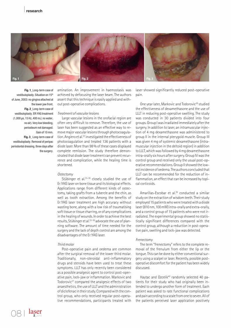
Fig. 1_Long-term case of
vestibuloplasty. Situation on 15th
of June, 2003: no gingiva attached at
the lower jaw front.
Fig. 2_Long-term case of
vestibuloplasty. ER:YAG treatment
(1,000 µs, 15 Hz, 400 mJ, no water,
no air). Very low bleeding,
periosteum not damaged.
Gain of 10 mm.
Fig. 3_ Long-term case of
vestibuloplasty. Removal of peripac
periodontal dressing, three days after
the surgery.
amination. An improvement in haemostasis wasachieved by defocusing the laser beam. The authorsassert that this technique is easily applied and with-out post-operative complications.
Treatment of vascular lesionsLarge vascular lesions in the orofacial region are
often very difficult to remove. Therefore, the use oflaser has been suggested as an effective way to re-move major vascular lesions through photocoagula-tion. Angiero et al.32 investigated the effectiveness ofphotocoagulation and treated 136 patients with adiode laser. More than 98 % of these cases displayedcomplete remission. The study therefore demon-strated that diode laser treatment can prevent recur-rence and complication, while the healing time isshortened.
OstectomyStübinger et al.33–36 closely studied the use of
Er:YAG laser on bone tissue and its biological effects.Applications range from different kinds of ostec-tomy, taking grafts from a tubercle and the chin, aswell as tooth extraction. Among the benefits ofEr:YAG laser treatment are high accuracy withoutwasting bone, along with a low risk of traumatisingsoft tissue or tissue charring, or of any complicationsin the healing of wounds. In order to achieve the bestresults, Stübinger et al.33–36 advocate the use of plan-ning software. The amount of time needed for thesurgery and the lack of depth control are among thedisadvantages of the Er:YAG laser.
Third molarPost-operative pain and oedema are common
after the surgical removal of the lower third molar.Traditionally, non-steroidal anti-inflammatorydrugs and steroids have been used to treat thesesymptoms. LLLT has only recently been consideredas a possible analgesic agent to control post-oper-ative pain, lock-jaw or inflammation. Markovic andTodorovic37 compared the analgesic effects of twoanaesthetics, the use of LLLT and the administrationof diclofenac in their study. Compared with the con-trol group, who only received regular post-opera-tive recommendations, participants treated with
laser showed significantly reduced post-operativepain.
One year later, Markovic and Todorovic38 studiedthe effectiveness of dexamethasone and the use ofLLLT in reducing post-operative swelling. The studywas conducted in 30 patients divided into fourgroups. Group I was irradiated immediately after thesurgery. In addition to laser, an intramuscular injec-tion of 4 mg dexamethasone was administered togroup II in the internal pterygoid muscle. Group IIIwas given 4 mg of systemic dexamethasone (intra-muscular injection in the deltoid region) in additionto LLLT, which was followed by 4 mg dexamethasoneintra-orally six hours after surgery. Group IV was thecontrol group and received only the usual post-op-erative recommendations. Group II showed the low-est incidence of oedema. The authors concluded thatLLLT can be recommended for the reduction of in-flammation, an effect that can be increased by topi-cal corticoids.
Amarillas-Escobar et al.39 conducted a similarstudy on the extraction of wisdom teeth. Their studyemployed 15 patients who were treated with a diodelaser (810 nm, 100 mW) intra-orally and extra-orally,and a control group of 15 patients who were not ir-radiated. The experimental group showed no statis-tically significant differences compared with thecontrol group, although a reduction in post-opera-tive pain, swelling and lock-jaw was detected.
FrenectomyThe term “frenectomy” refers to the complete re-
moval of the frenulum from either the lip or thetongue. This can be done by either conventional sur-gery using a scalpel or laser. Recently, possible post-operative discomfort for the patient has been widelydiscussed.
Haytac and Ozcelik40 randomly selected 40 pa-tients for their study who had originally been in-tended to undergo another form of treatment. Eachpatient was asked to rate functional complicationsand pain according to a scale from one to seven. All ofthe patients perceived laser application positively
08 I laser2_2012
Fig. 2 Fig. 3Fig. 1

Master of Science (M.Sc.) in Lasers in Dentistry
Become a part of the International Dentists‘ Elite! The first accredited M.Sc. programme in dentistry in Germany Career-accompanying postgraduate programme of two years at the University of Excellence RWTH Aachen Combination of lectures, skill training sessions, live ops, tutorials, and practical workshops Internationally accepted and accredited by the German Government, the European Union,
the Washington Accord and the Bologna Process Science-based and practice-orientated on highest national and international level
More information:
AALZ GmbH · Pauwelsstrasse 17 · 52074 Aachen · GermanyTel. +49 - 2 41 - 47 57 13 10 · Fax +49 - 2 41 - 47 57 13 29www.aalz.de · [email protected]
The next Master programme starts on 24 September 2012 in Aachen
Lay the Foundations to yoursuccessful Future now!
AALZ_DGL_A4_lasereng112.pdf 1AALZ_DGL_A4_lasereng112.pdf 1 05.03.12 16:1205.03.12 16:12

I research
Fig. 4_ Long-term case of
vestibuloplasty. Fibrin cover seven
days after surgery.
Patient free of pain.
Fig. 5_ Long-term case of
vestibuloplasty. Situation on 28th of
July, 2003: healing complete.
Gain of 7 mm.
10 I laser2_2012
and experienced reduced discomfort with laser. Seefor example a long-term case of vestibuloplasty from2003 by Prof. Dr. Gerd Volland, where no gingiva wasattached at the lower jaw front (Fig. 1). After treat-ment with Er:YAG laser (1,000 µs, 15 Hz, 400 mJ, nowater, no air), only very low bleeding occurred and again of 10 mm was noted (Fig. 2). Three days after thesurgery, the periodontal dressing was removed (Fig.3), and the patient was free of pain seven days afterthe surgery (Fig.4). The healing was completed sixweeks after the surgery and a gain of 7 mm was ob-served (Fig. 5). Three years later, the final gain was setat 5,5 mm and no scarring occurred (Fig. 6). A follow-up in 2009 showed no recessions and stable results(Fig. 7).
Pathology of the salivary glandsMucoceles, ranulas or sialolithiasis can result in
obstructive salivary-gland pathologies. Mucocelesare produced by an accumulation of mucin from aruptured salivary-gland duct, usually caused by lo-cal trauma. They are characterised by a high per-centage of relapse. Two approaches to removingmucoceles have been suggested in the literature: re-section by either scalpel or CO2 laser. Yagüe-García etal.41 compared the effectiveness of using a scalpelwith that of a CO2 laser in removing mucoceles intheir study. They treated 38 patients using a scalpeland 30 patients using a CO2 laser (5–7 W). The resultsshowed a repetition rate of 8.8 % for the conven-tional scalpel ablation. In 13.2 % of the cases, com-plications such as fibrous scars arose. In the lasergroup, a follow-up study at 12 months showed nocomplications or recurrence. The authors thereforerecommend laser treatment, since its results aremore predictable and its recurrence rate is lowerthan that of the traditional treatment. Furthermore,fewer complications occur. Huang et al.43 con-tributed to this line of argumentation in reporting onlaser vaporisation, a procedure that they recom-mend for children and non-cooperative patients es-pecially.
Ranulas are due to an accumulation of mucincaused by the obstruction of a salivary-gland duct(generally that of the sublingual and submandibular
glands), which is usually the result of previous localtrauma. Marsupialisation, the removal of the ranulawith or without the sublingual gland, laser splitting,and vaporisation of the ranula have been proposedas possible treatments. Lai and Poon44 present a se-ries of three cases in which ranulas were removedand the injuries vaporised using CO2 laser. The au-thors state that this treatment can be recommendedbecause of the precision of excision, a clear and ster-ilised operating field and the low risk of damage tothe Wharton’s duct and the gingival nerve. Further-more, CO2 laser treatment results only in minimal orno recurrence. Zola et al.45 present an alternativemethod for removing ranulas. They used anEr,Cr:YSGG laser (1.5 W). The authors found theirtreatment to offer advantages similar to those foundby Lai and Poon.44
Sialolithiasis is the mechanical obstruction ofsalivary glands or their excretory ducts owing to theformation of concretions. It accounts for 30 % ofsalivary gland pathologies and mainly affects thesubmaxillary glands (83–94 %), followed by theparotid (4–10 %) and sublingual glands (1–7 %).Yang and Chen46 present 19 clinical cases entailingthe removal of stones from the Wharton’s duct intheir article. All of the patients were treated with aCO2 laser (4–6 W). Their success rate was 95 % andonly very few complications occurred. For this rea-son, the authors advocate CO2 laser treatment as thefirst technique to be used to treat this pathology.
Bisphosphonates The clinical scope of avascular necrosis caused by
bisphosphonates ranges from a single fistula tolarge areas of exposed necrotic bone tissue. Addi-tional symptoms are paraesthesia, pus, swelling,pain and even fracture. The treatment and manage-ment of avascular necrosis resulting from bisphos-phonates has proven to be challenging, as no treat-ments have been effective in the long term. Depend-ing on the patient’s health, possible treatments arethe temporary or permanent suspension of bisphos -phonate use, use of local or systemic antibiotics orhyperbaric oxygen, and surgical debridement of thelesions. The combination of these therapies maybring about more predictable results.
The use of LLLT has been increasingly favoured asan alternative for treating this type of pathology. Intheir 2010 review of the treatment of avascularnecrosis by LLLT, Vescovi and Nammour47 explain theeffects of the laser on the healing process. Laserstimulation increases organic bone matrix, osteo -blast proliferation and capillary growth. Owing to itsstrong affinity to water and hydroxyapatite, theEr:YAG laser can be easily applied to both soft andbone tissue. Necrotic bone is vaporised in the courseFig. 5Fig. 4

research I
Fig. 6_Long-term case of
vestibuloplasty. Situation on 15th of
May, 2006: no scars.
Final gain of 5,5 mm.
Fig. 7_Long-term case of
vestibuloplasty. Situation on 25th of
November, 2009. Stable results, no
recessions.
I 11laser2_2012
of conservative surgery until healthy bone isreached. Another advantage of Er:YAG laser treat-ment is its bactericidal action, which increases thehealing of bone tissue. Er:YAG laser treatment there-fore appears to be a promising technique, since it isregarded as safe, well tolerated by patients and al-lows minimally invasive treatment of the disease inthe early stages.
In a study in 2008, Vescovi et al.48 present theirclinical results of the treatment of 28 patients af-fected by osteonecrosis. They treated the four groupsof patients with an Nd:YAG laser in combination withmedical and surgical treatment. Group I was treatedmedically only, for example via antibiotics and anti-septics. Group II was treated medically and surgically.Group III was treated medically and via LLLT. Finally,group IV was treated medically, surgically and usingLLLT. Twelve of the 14 patients treated with LLLTshowed significant clinical improvement and reduc-tion in symptoms, nine patients exhibited completeclinical success. The authors state that while the re-sults of their study were not conclusive, the results in-dicate that Nd:YAG laser treatment has significantpotential to treat lesions caused by bisphosphonate-associated osteonecrosis.
In 2010, Vescovi et al.49 published the results of asimilar study. Between 2004 and 2008, 91 patientsunderwent stomatological observation and 55 sitesaffected by osteonecrosis were examined. Thesewere divided into four groups and different thera-peutic modalities were studied. Group I comprised13 lesions that were treated medically (1 g amoxi-cillin three times a day and 250 mg metronidazoletwice a day, orally) for a minimum of two weeks.Group II consisted of 17 lesions that were treatedmedically and via LLLT using an Nd:YAG laser(1,064 nm) once a week for two months. Group IIIconsisted of 13 cases of avascular necrosis treatedsurgically by the removal of necrotic bone, debride-ment, alveolar removal and corticotomy. Finally,group IV comprised 12 lesions treated using anEr:YAG laser (2,040 nm) in combination with LLLT us-ing an Nd:YAG laser.
All of the lesions treated with the Er:YAG lasershowed a clinical improvement of 100 % and com-plete healing in 87.5 % of the cases. The group IV re-sults differed significantly from those of the othergroups. The authors suggest that the reason for thisis increased accessibility to both soft and bone tissueusing the Er:YAG laser. They therefore highlight therole of the Er:YAG laser in the treatment of os-teonecrosis and conservative surgery. Consequently,a surgical approach combined with LLLT can be con-sidered the most efficient treatment method for bis-phosphonate-associated osteonecrosis.
_Conclusion
In the last 20 years, lasers have become an excel-lent tool in oral surgery. Especially in soft-tissue sur-gery, laser enables the practitioner to excise tumorsof different types in a safer and more precise mannerthan with conventional techniques using a scalpel orelectrotome.
Modern laser application is based on our knowl-edge about absorption and other aspects of workingwith a laser beam. Over the past ten years, 980 nm and810 nm diode lasers have evolved in particular. Theyare relatively inexpensive and provide a good com-promise between superficial visible absorption andpenetration, in favor of achieving optimal coagula-tion without necrosis in the depths of the tissue.
As a consequence, fibromas, papillomas or lipo-mas can be removed even from sites like the lips andthe cheek with a clear operating field and predictableresults. In addition, sutures can be reduced to a min-imum and scar formation is also reduced. For hard tis-sues, erbium lasers appear to be the best choice be-cause of their high absorption in water. Their effect isbased on thermomechanical principles, unlike diodelasers, which interact thermally. Therefore, waterspray is essential. This way, bone can be removedwithout inhibiting healing owing to thermal necro-sis. Thus applied, laser can increase the positive ef-fects of oral surgery by providing reliability for thesurgeon and comfort for the patient._
Fig. 7Fig. 6
Prof Dr MSc mult Gerd Volland
Facultad de Odontología
Cirurgia bucal
Universidad de Sevilla
C/ Avicena s/n Sevilla, Spain
_contact laser

I research
_Introduction
One of the main goals of dentistry is the preventionof disease. Minimally invasive methods of treatmentare preferred. For this reason, the concept of treatmentin periodontology has radically changed over the pastdecades. While in the early days, extensive surgical in-terventions used to be the centre of attention, todaymore conservative treatment is the focus.
Treatment procedures recently transitioned fromsurgical to non-surgical, after the potential of scalingand root planning (SRP) to eliminate inflammationand arrest progression of periodontal disease was suc-cessfully demonstrated in a number of clinical trials(Axelsson & Lindhe 1978; Badersten et al. 1984;Hirschfeld & Wasserman 1978; Lovdal et al. 1961). Re-searchers debate whether there is a significant reduc-tion in the depth of the periodontal pocket when theNd:YAG laser is applied as an adjuvant therapy.
The balance of evidence seems to favour the im-provement of the pocket depth with the use of Nd:YAGas an additional tool for the periodontal treatment, butmore research still is needed in this area in order toevaluate the effectiveness of laser treatment with dif-ferent settings.
_Aims of the present study
The objective of this study is to examine whetherthe use of Nd:YAG laser as an adjunct to traditional SRPimproves the results of traditional therapy, especiallyconcerning the bleeding index and the depth of the pe-riodontal pocket. Furthermore, the present study is
aimed at providing insight into the existing debate inscientific literature regarding the bleeding index andthe depth of the periodontal pocket.
The hypothesis of this study is that the applicationof the Nd:YAG laser as an adjunct tool to local, non-surgical SRP therapy will result in a significantlygreater reduction of the bleeding index and the peri-odontal pocket depth than the traditional mechanicaltreatment of periodontitis alone. Consequently, thenull hypothesis states that there will be no significantdifferences between the two test groups on the twoclinical parameters of bleeding index and pocketdepth.
_Materials and methods
A total of 20 healthy patients (twelve women, eightmen), aged between 35 and 55, with mild periodonti-tis (pockets of a depth of 4 to 6 mm) participated in thisstudy. Patients were excluded from this study accord-ing to the following criteria: smokers, pregnantwomen or nursing mothers, type I and type II diabet-ics, patients currently under antibiotic treatment andpatients who had taken antibiotics within threemonths prior to their selection for the study, patientssuffering from cardiovascular disease (high-risk heartdisorder) and patients with contagious diseases (ElYazami et al. 2004).
The patients were randomly selected to be dividedinto two groups of ten persons each. Group 1 was cho-sen to be the test group. Patients were treated accord-ing to the protocol of AALZ (Aachen Dental Laser Cen-ter) with SRP, using manual instruments combinedwith the Nd:YAG laser. Group 2 was assigned to be the
Evaluation of com binedNd:YAG laser treatmentof moderate periodontitisA randomised controlled clinical study
Author_Dr Anna-Maria Yiannikou-Loucaidou
12 I laser2_2012

control group and the patients were treated with SRPusing manual instruments. Additionally, all of the pa-tients were given instructions for oral hygiene rou-tines and methods.
Clinical assessments of the BOP (bleeding on prob-ing) index and mean PD (periodontal pocket depth)were recorded prior to phase 1, immediately afterphase 2 and three months after phase 3.
_Pre-treatment examination
Every patient was initially assessed by taking his orher medical history (Armitage 2004; Raffetto 2004).The patients underwent a clinical and radiographicalexamination prior to the treatment. Their X-rays weretaken with the bitewing technique with the Planmecadixi 3 digital intra-oral digital imaging system. Two pe-riodontal parameters were registered and charted:BOP and PD. Measurements were taken for six aspectsof each tooth: mesiobuccal (mb), buccal (b), distobuc-cal (db), mesiolingual (ml), lingual (l), and distolingual(dl) using calibrated periodontal probes.
_Initial therapy
Initial therapy entailed removing plaque and pol-ishing the teeth, as well as giving instructions and en-couraging the patients.
_Closed curettage
Closed curettage was carried out with mechanicalroot planning using hand instruments—Graceycurettes # 1/2, 3/4, 7/8, 11/12, and 13/14 for both of thegroups (Schwarz et al. 2003). The average amount ofinstrumentation in each group was nine minutes forsingle-rooted teeth and ten minutes for multi-rootedteeth.
_Laser treatment
De-epithelisation of the sulcus was performed inone session, one week after the cleaning of the lastquadrant with a 2,940 nm Er:YAG laser. The followingsettings were applied: frequency 20 Hz, energy100mJ, average output 2 W, with the aid of an RO7handpiece, without water, only with the use of air, andpulse duration 750–950 µs. The laser was used for thede-epithelisation of the sulcus, effectively removingthe epithelium of the sulcus. This treatment was exe-cuted by continuously moving the tip of the RO7handpiece back and forth from the gingival crest.
During this procedure, the surface of the sulcus ap-peared to be a whitish colour and the tip of the RO7handpiece became covered in the removable epithe-lium cells. These cells on the handpiece and the sur-
face of the sulcus were frequently wiped off with a cot-ton roll or wet gauze (Harris et al. 2002). Scientific lit-erature shows that the concept of de-epithelisationencompasses the promotion of reattachment and theformation of new connective tissue.
One week after the de-epithelisation of the sulcus,pocket sterilisation (Fig. 1) was performed with a 1,064nm Nd:YAG (output power 2 W, frequency 20 Hz, withthe aid of a 300 µm fibre, pulse duration 75 to 100 µs).Before the use of the Nd:YAG laser in the pockets, thearea was dried with air. With the aid of a 300 µm thickquartz fibre placed on the bottom of the pocket, thepocket was irradiated circularly for 30 to 40 secondsparallel to the surface of the root, maintaining contactwith the tissue. This procedure was performed in theentire mouth without anaesthesia, only with the ap-plication of topical anaesthetic gel. When signs ofbleeding occurred, the fibre was applied to the nextpocket. The procedure was repeated three times in in-tervals of four to seven days.
It is important to note that this interval time be-tween the treatments must be strictly kept. If the pa-tients are treated earlier than four days, more tissuewill be removed, the wound will be larger and shrink-ing will occur. A treatment later than seven days canresult in recolonisation of the periodontal bacteria.
research I
Fig. 1_Mean PD values (mm) during
the three phases of treatment for
both groups.
Fig. 2_Mean BOP (%) for both groups
throughout the treatment.
I 13laser2_2012
Fig. 2
Fig. 3
I research
The researcher chose the setting parameters above be-cause it was reported that Nd:YAG laser irradiationwith a setting of 100 mJ, 20 Hz, 2 W for 30 seconds onlyinhibits the DNA metabolism and the cell division rate(Gutknecht et al. 1998). In this case, a safe soft-tissuelaser treatment can be performed. White et al. (1994)examined in vitro the changes of intra-pulpal temper-atures during Nd:YAG laser irradiation of root surfacesat 0.3 to 3.0 W (30 to 150 mJ/pulse, 10 or 20 Hz). Theyreported that within the parameters outlined in theirstudy, pulsed Nd:YAG laser energy should not causeany devitalising rise in the intra-pulpal temperaturewhen it is applied to root surfaces with adequate re-maining dentine thickness (Aoki et al. 2004).
_Statistical analysis
The data collected was tested for normality by Q-Qplots and Kolmogorov–Smirnov tests. The data wasfound to be normally distributed and parametric testswere conducted to examine significant differencesbetween the mean values. Dependent t-tests wereused to check for significant differences between thesame subjects before, during and at the end of thetreatment, and three months post-treatment. Thisprocedure was carried out in both groups. In addition,independent t-tests were used to check for significantdifferences between the two groups at each phase,that is, pre-treatment, at the end of the treatment andthree months post-treatment.
_Results
According to the statistical analysis, there were sig-nificant differences (p < 0.05) for each group betweenphases 1 to 2 and phases 2 to 3 for BOP and PD. Morespecifically, the mean PD value decreased in the laser-combined SRP therapy group from 1.28 ± 0.54 mm(p<0.05) at the end of the therapy, to 0.25 ± 0.32 mm(p<0.05) after three months and in the SRP groupfrom 1.03 ± 0.81 mm (p<0.05) at the end of the treat-ment, to 0.54 ± 0.38 mm (p<0.05) three months post-treatment (Fig. 2).
Furthermore, the BOP mean value decreased in thegroup under laser-combined SRP therapy from 21.6 ±9.5% (p<0.05) at the end of the therapy to 7.3 ±6.03% (p<0.05) three months post-treatment and inthe SRP group from 30.07 ± 20.65% (p<0.05) at theend of the treatment to 7.06 ± 8.66% (p<0.05) threemonths post-treatment, showing a statistically sig-nificant decrease (p<0.05) between phases 1 and 2and phases 2 and 3 (Fig. 3).
No significant difference (p>0.05) in the PD meanvalues occurred in the comparisons of the two groups(pre-treatment: t = 0.2, p = 0.845; end of treatment: t = - 0.6, p = 0.56; three months post-treatment:
t = 0.4, p = 0.72). When the two treatment groups werecompared for mean differences in BOP values (%), nostatistically significant differences emerged (p>0.05)at any phase of the treatment.
_Discussion
Two parameters of periodontal disease were inves-tigated in this randomised controlled study, the prob-ing depth and the sulcus haemorrhage. The aim of thepresent study was to compare the clinical results ofthese parameters after non-surgical periodontaltreatment to those of SRP via hand instruments orNd:YAG laser as an adjunct tool to the conventionalmechanical instrumentation. The results have demon-strated that non-surgical periodontal treatment withboth of the two treatment modalities leads to a signif-icant reduction in PD and BOP. However, when the twotreatment groups—test and control—were comparedfor mean differences in BOP values and in PD values ateach phase of the treatment, there were no significantdifferences.
_Conclusion
At this stage and within the framework of the pres-ent study, it appears that the use of Nd:YAG combinedwith non-surgical periodontal treatment improvesthe clinical outcome of an initial periodontal therapy.The findings should be confirmed by a study of a largernumber of patients, a longer follow-up period, differ-ent treatment-planning protocols and different en-ergy settings. Furthermore, basic and clinical studiesare required in order to clarify the application of theNd:YAG laser as a complementary therapy in peri-odontal therapy.
A bright future lies ahead for laser applications inperiodontal procedures. Laser-assisted therapy is asuccessful treatment option that can effectively helpthe patient to maintain optimal periodontal health._
Editorial note: A complete list of references is available
from the publisher.
14 I laser2_2012
Dr Anna-Maria Yiannikou-Loucaidou
RWTH Aachen University
Yiannikou Dental Polyclinic
8 Alkeous and Pindarou St.
1060 Nicosia, Cyprus
Tel.: +357 22 764765
Fax: +357 22 756160
_contact laser

DTSC_A4.pdf 1DTSC_A4.pdf 1 23.05.12 19:0723.05.12 19:07
I research
16 I laser2_2012
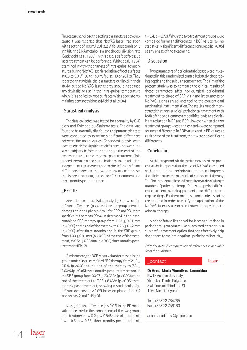
_Endodontic infection
Endodontic treatment can attain success rates ofbetween 85 and 97%.1 Adequate treatment proto-cols, knowledge and infection control are essential to achieving such rates (Figs. 1a–d).2 It is well knownthat apical periodontitis is caused by the communi-cation of root-canal micro-organisms and their by-products to the surrounding periodontal structures.Exposure of dental pulp directly to the oral cavity orvia accessory canals, open dentinal tubules or peri-odontal pockets is the most probable route of the endodontic infection.2,3
Clinically, apical periodontitis is not evident as long as the necrotic tissue is not infected with micro-organisms.4–6 There are up to 40 isolated species ofbacteria present in the root canal. Cocci, rods, filaments,spirochaetes, anaerobic and facultative anaerobic mi-croorganisms are frequently identified in primary in-fections. Fungus can also be isolated.2,7 Endodonticmicrobiota can be found suspended in the main rootcanal, attached to the canal walls and deep in thedentinal tubules at a depth of up to 300µm (Figs.2a–c). The absence of cementum dramatically in-creases bacterial penetration into dentinal tubules.8–11
It has been shown that bacteria can also beenfound outside the root-canal system, located at theapical cementum and as an external biofilm on theapex.12–15 Following conventional endodontic treat-ment, 15 to 20% of non-vital teeth with apical peri-odontitis fail.16–18 The presence of bacteria after thedecontamination phase or the inability to seal rootcanals after treatment are reasons for failure.2 The remaining contamination in endodontically treatedteeth continues the infectious disease process in theperiapical tissue.
Retreatments are the first choice for failed rootcanals. The microbiota found in persistent infectionsdiffer from that in primary infection (Figs. 3a–c). Fac-ultative anaerobic Gram-positive (G+) and -negative(G-) micro-organisms and fungi are common.19–21
Special attention is given to Enterococcus faecalis, a resistant facultative anaerobic G+ cocci, identifiedin a much higher incidence in failed root canals.22–25
Bacterial control plays a significant role in endodon-tic success. Adequate and effective disinfection of theroot-canal system is necessary.
Fig. 1_Success in endodontic
treatment: apical radiolucency repair.
The antibacterial effects of lasers inendodonticsAuthor_ Dr Selma Cristina Cury Camargo, Brazil
Fig. 1

research I
_Endodontic therapy
The bacterial flora of the root canal must be ac-tively eliminated through a combination of debride-ment and antimicrobial chemical treatment. Me-chanical instrumentation eliminates more than 90%of the microbial amount.26 An important point to noteis the adequate shaping of the root canal. Evaluatingthe antibacterial efficacy of mechanical preparationitself, Dalton et al.27 conclude that instrumentation toan apical size of #25 resulted in 20% of canals free ofcultivable bacteria. When shaped to a size of #35,60% showed negative results.
An Irrigating solution has been used with me-chanical instrumentation to facilitate an instru-ment’s cutting efficiency, remove debris and thesmear layer, dissolute organic matter, clean inacces-sible areas and act against micro-organisms. Sodiumhypochlorite is the most common irrigant used in en-dodontics.28 It has an excellent cleansing ability, dis-solves necrotic tissue, has a potential antibacterial ef-fect and, depending on the concentration, is well tol-erated by biological tissues. When accompanied bymechanical instrumentation, it reduces the numberof infected canals by 40 to 50%.
Other irrigating solutions can also be used during endodontic preparation. EDTA, a chelating agent usedprimarily to remove the smear layer and facilitate theremoval of debris from the canal, has no antibacterialeffect.29 Chlorhexidine gluconate has a strong anti-bacterial effect on an extensive number of bacterialspecies, even the resistant E. faecalis, but it does notbreak down proteins and necrotic tissue as sodiumhypochlorite does.30
As mechanical instrumentation and irrigating solutions are not able to eliminate bacteria from thecanal system totally—a requirement for root-canalfilling—additional substances and medicaments havebeen tested in order to address the gap in standard endodontic protocols. The principal goal of dressingthe root canal between appointments is to ensuresafe antibacterial action with long-lasting effects.31 Agreat number of medicaments have been used asdressing material, such as formocresol, camphoratedparachlorophenol, eugenol, iodine-potassium iodide,antibiotics, calcium hydroxide and chlorhexidine.
Calcium hydroxide has been used in endodontictherapy since 1920.31 With a high pH at saturation (pHabove 11), it induces mineralisation, reduces bacteria
I 17laser2_2012
Fig. 2_Primary infection: black
pigmented strains and G-rods.
Fig. 3_Persistent infection.
Figs. 4a & b_Nd:YAG laser
intra-canal irradiation.
Fig. 5_Nd:YAG laser irradiation,
deep penetration.
Fig. 4a Fig. 4b Fig. 5
Fig. 3Fig. 2

I research
and dissolves tissue. For extended antibacterial effec-tiveness, the pH must be kept high in the canal and inthe dentine as well. Sustaining the pH depends on thediffusion through dentinal tubules.32
Although most micro-organisms are destroyed at pH of 9.5, a few can survive a pH of 11 or higher, suchas E. faecalis and Candida.21 Because of the resistanceof some micro-organisms to conventional treatmentprotocols—and the direct relation between the pres-ence of viable bacteria in the canal system and the reduced rate of treatment success—additional efforthas to be made to control canal system infection.
_Lasers in endodontics
Lasers were introduced to endodontics as a com-plementary therapy to conventional antibacterialtreatment. The antibacterial action of Nd:YAG,diodes, Er:YAG and photoactivated disinfection(PAD) have been explored by a number of investiga-tors. In the following section, each laser is evaluatedwith the aim of selecting an adequate protocol witha high probability of success in teeth with apical pe-riodontitis.
Nd:YAG laser
The Nd:YAG laser was one of the first lasers tested in endodontics. It is a solid-state laser. The active medium is usually yttrium aluminium garnet(Y3Al5O12), where some Y3+ ions are replaced byNd3+ions. It is a four-level energy system operating in a continuous wave or pulsed mode. It emits a1,064nm infra-red wavelength. Thus, this laserneeds a guide light for clinical application. Flexible fi-bres with a diameter between 200 and 400µm areused as delivery systems. The laser can be used on in-tra-canal surfaces, in contact mode (Figs. 4a & b).
The typical morphology of root-canal wallstreated with the Nd:YAG laser shows melted dentinewith a globular and glassy appearance, and few areasare covered by a smear layer. Some areas show denti-nal tubules sealed by fusion of the dentine and de-posits of mineral components.33,34 This morphologi-cal modification reduces dentine permeability sig-nificantly.35,36 However, because the emission of thelaser beam from the optical fibre is directed along theroot canal and not laterally, not all root-canal wallsare irradiated, which gives more effective action atthe apical areas of the root.37 Undesirable morpho-logical changes, such as carbonisation and cracks,are seen only when high energy parameters are used.
One of the major problems regarding intra-canallaser irradiation is the temperature increase at the external surface of the root. Laser light exerts a ther-mal effect when it reaches tissue. The heat is directlyassociated with the energy used as well as time andirradiation mode. An increase in temperature levelsabove 10°C per minute can cause damage to peri-odontal tissues, such as necrosis and anchylosis.
Lan38 evaluated in vitro the temperature increaseon the external surface of the root after irradiationwith a Nd:YAG laser under the following energy parameters: 50, 80 and 100mJ at 10, 20 and 30 pulsesper second. The increase of temperature was less than 10°C. The same results were obtained by Bach-man et al.39, Kimura et al.40 and Gutknecht et al.41
In contrast to the external surface, the intra-canaltemperature rises dramatically at the apical area,
18 I laser2_2012
Figs. 6a & b_Diode 980 nm
intra-canal irradiation.
Fig. 6a
Fig. 6b


I research
20 I laser2_2012
promoting effective action against bacterial con -tamination. For the Nd:YAG laser, 1.5W and 15Hz, aresafe energy parameters for temperature and mor-phological changes.33,41
The primary use of the Nd:YAG laser in endodon-tics is focused on elimination of micro-organisms in the root-canal system. Rooney et al.42 evaluated the antibacterial effect of Nd:YAG lasers in vitro. Bacterial reduction was obtained considering energy parameters. The researchers developed dif-ferent in- vitro models simulating the organisms expected in non-vital, contaminated teeth. Nd:YAGirradiation was effective for Bacillus stearother-
mophilus,43,44 Streptococcus faecalis, Escherichia
coli,45 Streptococcus mutans,46 Streptococcus
sanguis, Prevotella intermedia47 and a specific mi-cro-organism resistant to conventional endodontictreatment, E. faecalis.48–50 Nd:YAG has an anti -bacterial effect in dentine at a depth of 1,000µm (Fig. 5).50
Histological models were also developed in orderto evaluate periapical tissue response after intra-canal Nd:YAG laser irradiation. Suda et al.51 demon-strated in dog models that Nd:YAG irradiation at
100mJ/30 pulses per second for 30 seconds was safeto surrounding root tissues. Maresca et al.,52 using human teeth indicated for apical surgery, corrobo-rated Suda et al.’s51 and Ianamoto et al.’s53 results.Koba et al.54 analysed histopathological inflamma-tory response after Nd:YAG irradiation in dogs at 1and 2W. Results showed significant inflammatoryreduction at four and eight weeks compared withthe non-irradiated group.
Clinical reports published in the literature confirmthe benefits of intra-canal Nd:YAG irradiation. In1993, Eduardo et al.55 published a successful clinicalcase that combined conventional endodontic treat-ment with Nd:YAG irradiation for retreatment, apicalperiodontitis, acute abscess and perforation. Clinicaland radiographic follow-up showed complete heal-ing after six months.
Similar results were shown by Camargo et al.56
Gutknecht et al.57 reported a significant improve-ment in the healing of laser-treated infected canals,when compared with non-irradiated cases.
Camargo et al.58 compared in vivo the antibacter-ial effects of conventional endodontic treatment and the conventional protocol associated with theNd:YAG laser. Asymptomatic teeth with apical radi-olucency and necrotic pulps were selected and di-vided into two groups: conventional treatment andlaser-irradiated. Microbiological samples were takenbefore canal instrumentation, after canal prepara-tion and/or laser irradiation and one week after treat-ment. The results showed a significant antibacterialeffect in the laser group compared to conventionaltreatment. When no other bactericidal agent wasused, it was assumed that the Nd:YAG laser played aspecific role in the bacterial reduction for endodon-tic treatment in patients.
Diodes
The diode laser is a solid-state semiconductor laser that uses a combination of gallium, arsenide,aluminium and/or indium as the active medium. Theavailable wavelength for dental use ranges between800 and 1,064nm and emits in continuous wave and gated pulsed mode using an optical fibre as thedelivery system (Figs. 6a & b). Diode lasers havegained increasing importance in dentistry owing totheir compactness and affordable cost. A combina-tion of smear layer removal, bacterial reduction andreduced apical leakage are advantages of this laserand make it viable for endodontic treatment. Theprincipal laser action is photo-thermal.
The thermal effect on tissue depends on the irra-diation mode and settings. Wang et al.59 irradiatedroot canals in vitro and demonstrated a maximum
Fig. 7_Er:YAG laser. Fig. 7

research I
I 21laser2_2012
temperature increase of 8.1°C using 5W for sevenseconds. Similar results were obtained by Da CostaRibeiro.60 Gutknecht et al.61 evaluated intra-canaldiode irradiation with an output of 1.5W and ob-served a temperature increase of 7°C in the externalsurface of the root using a 980nm diode laser at apower setting of 2.5W at a continuous and choppedmode, and found that the temperature increase never exceeded 47°C, which is considered safe for periodontal structures.41
Morphological changes at the apical portion ofthe root after intra-canal diode irradiation were ob-served in clean intra-canal dentinal surfaces withsealed dentinal tubules, indicating melting and re-crystallisation.62 In general, near infra-red wave-lengths, such as 1,064 and 980nm, promote fusionand recrystallisation on the dentinal surface, sealingdentinal tubules.
The apparent consensus is that diode laser irradi-ation has a potential antibacterial effect. In mostcases, the effect is directly related to the amount ofenergy delivered. In a comparative study by Gut -knecht et al.,63 an 810nm diode was able to reducebacterial contamination by up to 88.38% with a distal output of 0.6W in continuous wave mode. A 980nm diode laser has an efficient antibacterial effect of an average of between 77 to 97% in rootcanals contaminated with E. faecalis. Energy out-puts of 1.7, 2.3 and 2.8W were tested. Efficiency wasdirectly related to the amount of energy and dentinethickness.64
Er:YAG laser
Er:YAG lasers are solid-state lasers with a lasingmedium of erbium-doped yttrium aluminium garnet(Er:Y3Al5O12). Er:YAG lasers typically emit light with a wavelength of 2,940nm, which is infra-red light.Unlike Nd:YAG lasers, the output of an Er:YAG laser is strongly absorbed by water because of atomic res-onances. The Er:YAG wavelength is well absorbed byhard dental tissue. This laser was approved for den -tal procedures in 1997. Smear layer removal, canalpreparation and apicoectomy are indications for endodontic use (Fig. 7).
The morphology of a dentinal surface irradiatedwith an Er:YAG laser is characterised by clean areas,showing open dentinal tubules free of a smear layer,in a globular surface. Bacterial reduction using theEr:YAG was observed by Moritz et al.65
Stabholz et al.37 describe a new endodontic tip that can be used with an Er:YAG laser system. The tipallows lateral emission of the radiation rather than direct emission through a single opening at the farend. It emits through a spiral tip located along thelength of the tip. Examining the efficacy of the spiral tip in removing the smear layer, Stabholz et al.66
found clean intra-canal dentinal walls free of a smear layer and debris under SEM evaluation.
Photoactivated disinfection
PAD is another method of disinfection in endo -dontics and is based on the principle that photoacti-vated substances, which are activated by light of aparticular wavelength, bind to target cells. Free radi-cals are formed, producing a toxic effect to bacteria.
Fig. 9_Intra-canal laser irradiation,
molar.
Fig. 8
Fig. 8_Therapeutic plan.

I research
22 I laser2_2012
Toluidine blue and methylene blue are examples ofphotoactivated substances. Toluidine blue is able tokill most oral bacteria. In in vitro studies, PAD has aneffective action against photosensitive bacteria suchas E. faecalis, Fusobacterium nucleatum, P. interme-
dia, Peptostreptococcus micros and Actino-
mycetemcomitans.67,68 On the other hand, Souza etal.,69 evaluating PAD antibacterial effects as a sup-plement to instrumen tation/irrigation of canals in-fected with E. faecalis, did not prove a significant ef-fect regarding intra-canal disinfection. Further ad-justments to the PAD protocols and comparative research models may be required before recommen-dations can be made regarding clinical usage.
_Discussion and conclusion
There are good reasons to focus the treatment ofnon-vital contaminated teeth on the destruction ofbacteria in the root canal. The possibility of afavourable treatment outcome is significantlyhigher if the canal is free from bacteria when it is ob-turated. If, on the other hand, bacteria persist at thetime of root filling, there is a higher risk of treatmentfailure. Therefore, the prime objective of treatment isto achieve complete elimination of all bacteria fromthe root-canal system.2,31
Today, the potential antibacterial effect of laser irradiation associated with the bio-stimulation ac-tion and accelerated healing process is well known.Research has supported the improvement of endo -dontic protocol. Laser therapy in endodontic treat-ment offers benefits to conventional treatment, such as minimal apical leakage, effective actionagainst resistant micro-organisms and external api-cal biofilm, and an increase in periapical tissue repair.
For this reason, laser procedures have been incorpo-rated into conventional therapeutic concepts to improve endodontic therapy (Figs. 8a–d).
Clinical studies have proven the benefits of an endodontic laser protocol in apical periodontitistreatment. For endodontic treatment, the protocolentails standard treatment strategies for cleaningand shaping the root canal to a minimum of #35, irrigating solutions with antibacterial properties andintra-canal laser irradiation using controlled energyparameters. Ideal sealing of the root canal and ade-quate coronal restoration are needed for an optimalresult.
In practice, little additional time is required forlaser treatment. Irradiation is simple when flexibleoptical fibres of 200µm in diameter are used. The fibre can easily reach the apical third of the rootcanal, even in curved molars (Fig. 9). The releasedlaser energy has an effect in dentine layers and be-yond the apex in the periapical region. The laser’s ef-fect extends to inaccessible areas, such as externalbiofilm at the root apex.
The irradiation technique must adhere to the fol-lowing basic principles. A humid root canal is re-quired and rotary movements from the coronal por-tion to the apex should be carried out, as well as scan-ning the root canal walls in contact mode (Figs.10a–c). The power settings and irradiation mode de-pend on one’s choice of a specific wavelength.
Nd:YAG, diodes of different wavelengths,Er:YAG, and low-power lasers can be used for dif-ferent procedures with acceptable results. Lasertechnology in dentistry is a reality. The developmentof specific delivery systems and the evolution oflasers combined with a better understanding oflaser–tissue interaction increase the opportunitiesand indications in the endodontic field._
Editorial note: A complete list of references is available
from the publisher.
Fig. 10_Intra-canal laser irradiation,
technique.
Dr Selma Camargo
University of São PauloRua Pinto Gonçalves, 85/54 PerdizesSão Paulo, SP 05005-010Brazil
_contact laser
Fig. 10

FOR MORE INFORMATION:Greater New York Dental Mee ng®
570 Seventh Avenue - Suite 800New York, NY 10018 USA
Tel: (212) 398-6922 / Fax: (212) 398-6934E-mail: [email protected]
Scien c Mee ng: Friday - Wednesday,
November 23 - 28
Exhibit Dates: Sunday - Wednesday,
November 25 - 28
MARK YOUR CALENDAR
A A N CNever a pre-registra on fee at the Greater New York Dental Mee ng
M H 600 EXH BJacob K. Javits Conven on Center 11th Ave.
between 34-39th Streets (Manha an)
Q New York Marrio Marquis otel
L V D A - N T
L D T H S A V
M TH 350 S PSeminars, ands-on Workshops, Essays & Scien c Poster Sessions as well as
Specialty and Auxiliary Programs
E P V
S P H E F
E N Y C B H V
H !
Sponsored by the New York County Dental Society and the Second District Dental Society
GNYDM_2012_A4_eng.pdf 1GNYDM_2012_A4_eng.pdf 1 31.01.12 17:3931.01.12 17:39

I overview
Fig. 1_Laser wavelength and
absorption spectrum of the different
laser types used for desensitization in
human tissues.
_Introduction
More than two decades ago, laser applications inthe treatment of dentine hypersensitivity were intro-duced to dentistry. Many clinical studies using differ-ent laser types have been published since. Thisoverview summarises the basic and clinical aspects,including treatment protocols.
In the last issue of laser, conventional approachestowards the treatment of dentine hypersensitivitywere discussed with regard to a set of criteria for asuccessful treatment as proposed by L. I. Grossman(1935). The authors came to the conclusion that, sofar, no conventional therapy has been able to meet allthe criteria. The authors then moved on to studies onlaser treatment. Studies on GaAIAs laser and He-Nelasers were introduced and analysed. Part I of this ar-ticle was finished by a comparison between He-Nelasers and Nd:YAG lasers.
Part II in this issue of laser continues with studieson the application of Nd:YAG lasers in dental thera-pies as well as an overview on Er:YAG lasers.
_Middle-output power lasers: Nd:YAG laser
The Nd:YAG laser with a wavelength of 1,064 nmbelongs to the group of middle-output power lasers.Matsumoto et al. (1985b) referred to the application ofthis laser type in the therapy of dentine hypersensitiv-ity. Since then, it has been established for the therapyof dentine hypersensitivity in a number of studies (Dil-siz et al. 2009; Yonaga et al. 1999; Kobayashi et al.1999; Gutknecht et al. 1997; Lan et al. 1996; Renton-Harper et al. 1992; Gelsky et al. 1992; White et al. 1990;Goodis et al. 1989; Matsumoto et al. 1985b).
The energy level used ranges from 0.3 to 10 W, withthe most frequent use of 1 or 2 W (Kimura et al. 2000b).The methods of application are highly dependent onthe laser energy used and vary according to this, from0.3 W for 90 seconds in non-contact mode up to 2 Wfor 0.5 seconds with the application of an absorber incontact mode (Kimura et al. 2000b). In different stud-ies, the efficiency of therapy ranges from 5 to 100%(Kimura et al. 2000b). Among the middle-outputpower lasers, the Nd:YAG laser is regarded as exceed-ingly effective (Dilsiz et al. 2010b; Birang et al. 2007; DeMagalhaes et al. 2004; Yonaga et al. 1999). In a com-parative in vitro study of the ability of Er,Cr:YSGG,Nd:YAG, CO2 and diode lasers to melt dentinal tubules,all laser types had a statistically significant ability toseal perpendicular dentine and occlude dentinaltubules partially or totally, but the highest reduction inmean tubule diameter resulted from the Nd:YAG laserwith 53% effectiveness (Gholami et al. 2011). Compa-rable results were also found by Dilsiz et al. (2009),comparing immediate and late therapeutic effects af-ter Nd:YAG (1 W, 10 Hz, 60 seconds, non-contact mode,without cooling) and diode-laser application (25 mW,9 Hz, 100 seconds). Both were effective in reducingdentine hypersensitivity, but there was a higher suc-
Laser treatment of dentine hypersensitivityAn overview Part II
Author_Dr Ute Botzenhart, Dr Andreas Braun & Prof Matthias Frentzen, Germany
24 I laser2_2012
Fig. 1

overview I
cess rate after Nd:YAG laser application, especially af-ter immediate application and 60 days after conclud-ing the treatment.
Abed et al. (2011) compared the sealing ability ofNd:YAG laser application (1 W, 10 Hz, 60 seconds, non-contact mode without cooling) to that of a resin (Seal& Protect®, DENTSPLY DeTrey) applied to exposed hu-man dentinal tubules in vitro. Compared with the con-trol group, laser application showed a homogeneousdentinal surface with less exposed tubules and a re-duction in the diameter of the exposed tubules of50%. Nevertheless, in this study, the resin was muchbetter, with a 90% sealing ability compared with thecontrol group (Abed et al. 2011).
Effects of Nd:YAG laser application
When using Nd:YAG laser light and black ink as anabsorption amplifier, it is recommended that deeppenetration of laser light through enamel and dentinebe evaded so that excessive harmful effects on the pul-pal tissue can be avoided (Launay et al. 1987) and sur-ficial sealing effects can be enhanced (Morioka et al.1984; Gelsky et al. 1993; Yonaga et al. 1999; Kobayashiet al. 1999). The closure or narrowing of dentinaltubules (Lan & Liu 1995, 1996) and direct nerve anal-gesia are assumed to be mechanisms of Nd:YAG laserlight action (Whitters et al. 1995). In an in vitroSEM ex-amination, the melting of dentinal tubule openingsand the solidification of the dentine surface with apenetration depth varying between 1 to 7 µm were ob-served, depending on irradiation parameters (30 mJ,0.3 W, 7 Hz; 40 mJ, 0.4 W, 7 Hz for 43 seconds with aten-second interval; De Magalhaes et al. 2004).
In a study by Moriyama et al. (2004), morphologi-cal and chemical changes in human dentine surfacesresulting from Nd:YAG laser irradiation with differentpulse durations were observed. SEM analysis con-firmed a melting and resolidification with larger reso-lidification structures and a smoother surface afterusing long pulses. An increased concentration of calcium and phosphorous was also found in all irradi-ated samples compared with the control group, possi-bly making it less susceptible to acid dissolution(Moriyama et al. 2004). Laser of a wavelength of1,064nm also demonstrates effects upon microcircu-lation (Funato et al. 1991; Zennyu et al. 1996).
Causes of the analgesic effects of Nd:YAG lasers
A multitude of theories on the way in which theNd:YAG laser induces its analgesic effect have beensuggested (Kimura et al. 2000b). For example, laser en-ergy is thought to interfere with the sodium pump andalter the cell membrane permeability and/or affect theendings of sensory axons (Myers et al. 1991). Not onlycan Nd:YAG laser application block off the depolarisa-tion of very slow C-fibre afferences, but it can also af-
fect fast-conducting A-ß-fibres (Orchardson et al.1997). It is also thought that a desensitising effect canbe achieved by denaturing the odontoblast processand by overheating dentinal fluid (White et al. 1990;Goodis et al. 1989).
A glazing of the dentine was described after the ap-plication of Nd:YAG laser light to the exposed toothneck (Birang et al. 2007; Lan et al. 2004; Dederich et al.1984; Halket et al. 1996). The result was a glazed, non-cavernous surface with closure of the exposed tubulesand without surface cracks. When using an Nd:YAGlaser, the sealing depth is dependent on the chosen pa-rameters and the optical properties of dentine. Withthe use of 30 mJ/pulse and 10 Hz, the sealing depth isless than 4 µm (Liu et al.1997). In an in vivo study byGutknecht et al. (1997), different laser parameters forNd:YAG laser application were tested. Roughly thesame success rates were detected in each case. It wasconcluded that Nd:YAG laser light must have been ef-fective even with a low-energy dose. Manton et al.(1992) compared the effect of the Nd:YAG laser withthat of an untreated control group. Directly before andafter laser application, as well as after three and 28 days, the time it took for teeth to become painful af-ter cold stimuli was measured. Nd:YAG laser light ap-plication showed a statistically significant clinical im-provement over a period of 28 days. These results wereconfirmed by a study by Yonaga et al. (1999). Recrude-scence occurred after more than two months. After afollow-up period of one year, the effectiveness ofNd:YAG laser application was 85.4% effective com-pared with 90% immediately after the application(Zhang 1990).
Hu (2004) analysed the effect of a pulsed Nd:YAGlaser in the therapy of dentine hypersensitivity com-pared with a control group treated with NaF. The ef-fectiveness immediately after laser application, andone month and six months after was significantly bet-ter compared with the control group (Hu 2004). Cia-
Fig. 2_SEM examination of a human
cervical dentinal surface after
removal of the smear layer with 50 %
citric acid for one minute to open up
the dentinal tubules and simulate
hypersensitive dentine and
application of Bifluorid 12®
(combination of sodium fluoride and
calcium fluoride, 1,000 x-
magnification). Dentinal tubules are
totally or partially closed by
fluoride-containing covering layer.
I 25laser2_2012
Fig. 2

I overview
ramicoli et al. (2003) also confirmed the results of thestudies described above. The effect of Nd:YAG laser ir-radiation (2 W, 100 mJ, 20 Hz, 60 seconds, with air cool-ing) compared with conventional fluoridation (Biflu-orid 12®, VOCO, Fig. 2) immediately after and one, two,three and four weeks after application showed a sig-nificant improvement in VAS scores to air blast imme-diately after and one week after laser treatment (Karaet al. 2009). Whereas at weeks two, three and four inthe fluoridated group, VAS scores decreased up tonearly 75 to 85% of the baseline scores, VAS scores re-mained nearly unchanged in the laser group. Fluoridewas applied in three consecutive visits in this study, butlaser was only applied once. Nd:YAG laser irradiationthus appears to be a suitable tool for the immediateand successful reduction of pain within a shortertreatment time.
Recurring dentine hypersensitivity
The mechanism of the recurrence of dentine hy-persensitivity is unknown (Yonaga et al. 1999), but it isassumed that after laser treatment, a reappearance ofsymptoms depends on the initial intensity of the den-tine hypersensitivity before the application (Yonaga etal. 1999). The irradiation of the cervical region withNd:YAG laser light under the use of an absorber en-hances the efficiency—with this method, a recurrenceof the symptoms can be delayed (Yonaga et al. 1999).According to this study, morphological aspects, in ad-dition to analgesic effects, are assumed to be impor-tant for the sustainability of treatment effects (Yon-aga et al. 1999).
Side-effects
The thermal impacts of laser light on the pulpal tis-sue constitute a problem for using Nd:YAG laser in
vivo. Compared with other laser types, the Nd:YAGlaser beam has deep penetration into dentine, boneand soft tissue (Dederich 1993; Zennyu et al. 1996). Ex-posure of dentine beyond the safety threshold cancause thermal damage to the pulpal tissue (Yonaga etal. 1999; Zhang 1990; Matsumoto et al. 1988; Zach etal. 1965). However, clinical studies state that despitethe danger of thermal damage, no side-effects werefound (Yonaga et al. 1999).
Other clinical studies examined a partial oxygensaturation of pulpal blood in anterior hypersensitiveteeth after Nd:YAG laser irradiation (1 W, 10 Hz, 60 sec-onds, non-contact mode, without cooling) as a possi-ble indicator of pulpal damage. A slight but significantincrease in the oxygen saturation of the pulpal bloodwas observed immediately and one week after laserapplication compared with the control group. How-ever, partial oxygen saturation of pulpal blood in laser-treated teeth had gained its pretreatment level at fol-low-up measurement after one month, thus main-taining the teeth vitality and indicating no irreversible
damage in the dental pulp after laser applicationwithin the limit of desensitisation parameters (Biranget al. 2008).
The additional use of an absorber defines the deptheffect of Nd:YAG laser light and reduces the possibil-ity of side-effects. Zapletalova et al. (2007) tested dif-ferent dye solutions for topical application in combi-nation with Nd:YAG laser energy and found erythrosinto be the best agent to avoid damage to the dentinalstructure. Sealing of tubules occurred after four dosesof 30 mJ pulses (total energy density 33 J/cm2).
Concerning efficiency and simplicity, the applica-tion of Nd:YAG laser light is a relevant treatmentmethod for clinical practice (Yonaga et al. 1999). Thegreat variances in the different studies can be ex-plained by morphological differences that inevitablyoccur on account of intra- and inter-individual varia-tion of the dentine structure and depend, among oth-ers, on the age and clinical history of the teeth(Moriyama et al. 2004). For example, in their SEM ex-amination of the occluding ability of Nd:YAG laser onexposed human dentine compared with a resin, Abedet al. (2011) discovered that the number and diameterof dentinal tubules vary significantly from tooth totooth even for different sections of the same tooth.This confirms most of the clinical findings morpho-logically.
Combined treatment with Nd:YAG laser and fluoride
The clinical application of the Nd:YAG laser and NaFvarnish, as well as Nd:YAG laser light alone, showedsignificant improvement in dentine hypersensitivity ineach case (Kumar et al. 2005). However, the combina-tion of the Nd:YAG laser and NaF varnish showed agreater efficiency compared with either of these usedalone (Kumar et al. 2005). SEM examinations confirmthe clinical results. A reduction in the number of opentubules was combined with an improvement in the ef-ficiency of the therapy (Kumar et al. 2005). The studyby Ciaramicoli et al. (2003) supports the above results.Hsu et al. (2006) also hypothesised the improved de-sensitising effect of a combined treatment. A fluoride-containing dentine desensitiser was first applied andthis was followed by Nd:YAG laser irradiation (1,062 nm, 33 mJ, 50 pulses/second [pps] for two min-utes in slight contact) resulted in long-lasting acid andbrushing resistance. The posttreatment was carriedout very briefly and the integration of fluoride into thedentinal surface was not confirmed by structuralanalysis. This confines the validity of this study. Qual-itative microanalysis of ions and ultrastructuralchanges in dentine exposed to Nd:YAG laser (1.5 W,100 mJ, 15 Hz, 60 seconds, energy density 125 mJ/cm2,with black ink as dye solution) and flouride solutions(10% SnF2 for 30 minutes; 10% SrCl2 for 30 minutes)proved the capability of the Nd:YAG laser to alter the
26 I laser2_2012

overview I
I 27laser2_2012
dentinal structure by melting and resolidification ofthe surface with an occluding effect on exposed den-tine and additionally altering the absorption of ions,leading to a better infiltration into the dentinal struc-ture (Glauche et al. 2005). After the combined use ofthe Nd:YAG laser and 10% SnF2, Sn2+ could be de-tected up to 250 µm in EDX analysis compared with100 µm without laser application. The combined use ofthe Nd:YAG laser and 10% SrCl2 resulted in an uptakeof Sr2+ up to 500 µm compared with 23 µm withoutlaser irradiation (Glauche et al. 2005).
_Middle-output power lasers: Er:YAG laser
Today, there are only a few clinical studies on theapplication of the Er:YAG laser (2,940 nm) in the ther-apy of dentine hypersensitivity. A possible explanationfor the desensitising effect of the Er:YAG laser is itshigh absorption in water; thereby an evaporation ofdentinal fluid and the retention of the smear layer witha deposition of insoluble salts in the exposed dentinaltubules are assumed (Moritz et al. 2006). Another pos-sible explanation for the Er:YAG laser effects in thetreatment of dentine hypersensitivity is an analgesiceffect on the pulpal nerves, which would explain theimmediate effect and the progressive increase ofsymptoms after irradiation over time (Badran et al.2011).
SEM analysis of human dentine after Er:YAG laser ir-radiation (300 mJ, 10 pps, ten seconds; up to 700 mJ, 10 pps, ten seconds, with and without water cooling)demonstrated that laser energy of 500 mJ/pulse at 10 pps for ten seconds was sufficient for inducing melt-ing and recrystallisation of dentine crystals (Lee et al.2004). At irradiation parameters of 60 mJ, 2 Hz, with-out air/water-cooling on human dentine in vitro for 30,60 or 120 seconds, a partial reduction in tubule diame-ter (30 seconds), an almost complete obliteration of ex-posed tubules with visible signs of melting (60 seconds)and complete occlusion with a rugous melted dentinalsurface were observed (Badran et al. 2011). A decreasein dentine permeability in 26.05% was also achieved at60 mJ and 2 Hz for four applications of 20 seconds invitro (Aranha et al. 2005). The use of water-cooling isimportant for the reduction of thermal effects (Lee etal. 2004). Firoozmand et al. (2008) proved that the invitro use of an Er:YAG laser (250 mJ, 4 Hz, 80 s,19 mJ/cm2, non-contact mode, with constant water-cooling) for cavity preparation on bovine dentine didnot exceed the critical temperature of 5.5 °C.
Clinical investigations of Er:YAG laser application(100 mJ, 3 Hz, two applications of 60 seconds) havefound an acceptable therapeutic effect with reductionin pain over a period of up to six months (Birang et al.2007).
Comparative studies of Er:YAG lasers
Schwarz et al. (2002) compared the efficiency ofEr:YAG laser application (80 mJ/pulse and 3 Hz) and aconventional treatment and observed a significantimprovement in symptoms directly after the applica-tion of both of the two treatments and after six monthscompared with the control group. However, at two andsix months, the group treated with fluoride varnishshowed an increase in dentine hypersensitivity com-pared with the group treated with laser. The latter hadthe same level as directly after the laser treatment.
Another comparative in vivo study evaluating theeffect of the Er:YAG and CO2 lasers and the combina-tion of these laser types with fluoride application (NaF)proved the efficiency of the two treatment options.With the use of Er:YAG laser light at energy levels of 60 mJ at 30 Hz for 10 seconds without water/air sprayalone or in combination with fluoride, the clinical im-provement in discomfort determined by a cold-airblast and VAS score was significantly reduced oneweek, one month and six months after treatment com-pared with a control group treated solely with fluori-dation. No significant differences between Er:YAGlaser application and CO2 laser application as a singledose or as a combined treatment were observed (Ipciet al. 2009).
Similar results in an in vitro study that comparedthe occluding ability of the Er:YAG laser (60 mJ, 30 Hz,ten seconds) and CO2 laser (1 W, cw, one second) ap-plication alone or in combination with 2% NaF gel cor-roborated the clinical findings mentioned above. Amelted appearance along with the occlusion of thedentinal tubules could be found in all irradiatedgroups, but in terms of number and diameter of opentubules, no significant differences between the laseronly and the combination group were found (Cakar etal. 2008)._
Editorial note: To be continued in our next issue of laser. A
complete list of references is available from the publisher.
Dr Ute Botzenhart
Department of Orthodontics
Centre of Dentistry and Oral Health
Ernst Moritz Arndt University of Greifswald
Germany
Dr Andreas Braun/Prof Matthias Frentzen
Department of Periodontology, Operative
and Preventive Dentistry
University of Bonn
Germany
_contact laser
Scan QR-Code for part I of this article with your smart phone.

I case report
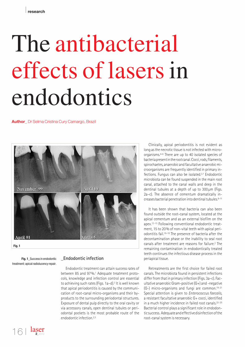
Fig. 1_IO360 tip. (Courtesy of
elexxion AG)
Fig. 2_Representation of the beam
profile of the IO360 tip at 1 W
emission. (Courtesy of elexxion AG)
Fig. 3_SEM image of sample 1,
the SDS-prepared site, showing
smear layer in bovine bone.
_Introduction
Over the years, developments in implant dentistryhave concentrated on implant design and surfacetreatment of titanium,1–11 and successful osseointegra-tion12 is now considered the minimum standard for allimplants.
To avoid bone necrosis, preparation of the os-teotomy site is commonly achieved with a series ofdrills and copious water irrigation. Studies show thaterbium lasers with water spray can also be used to pre-pare the site.13–17However, little attention has been paidto the possibilities of laser conditioning after drillpreparation of the implant site.
The Er:YAG laser has a wavelength of 2,940 nm. Itfalls within the mid-infra-red region (invisible to thehuman eye) and is well absorbed by water moleculesand hydroxyapatite. Since water is one of the main con-stituents of all oral hard and soft tissues, the Er:YAGlaser is therefore used in the ablation of tooth, bone anddental soft tissue.18–33
Er:YAG laser osteotomy and drill osteotomy havebeen compared in animal studies.16, 17, 34, 35 In these in-
vestigations, laser osteotomy yielded comparable orbetter results than drill osteotomy in terms of bonehealing, and some of these studies14, 16, 35 showedfavourable laser results for bone-to-implant contact.Romanos et al.13 demonstrate that Er:YAG laser irradi-ation appears to stimulate the secretion of platelet-de-rived growth factor when preparing osteotomy sites ina rat model. The study postulates that laser irradiationcould improve the healing of those sites. Aleksic et al.36
demonstrate the low-level laser effect of Er:YAG laserirradiation with the enhancement of osteoblast prolif-eration.
_Objective
The authors thus instituted a study to investigatethe possibility of using an Er:YAG laser to condition anosteotomy site prepared according to a standarddrilling sequence (SDS). They utilised a newly designedtip, performed a simple in vitro experiment, and thenproceeded to test the technique with two clinicalcases. The aim of this study was to compare the stabil-ity values of implant placement with and withoutEr:YAG laser conditioning of the bone surface follow-ing an SDS. The maxillary region was chosen for bothpatients.
A novel technique ofEr:YAG laser-enhancedearly implant stabilityAuthor_Dr Kenneth Luk & Dr Ryan S. K. Seto, China
28 I laser2_2012
Fig. 2 Fig. 3Fig. 1

_Materials and methods
Tip design
A modified wedge-shaped design, 1,200 m diame-ter IO360 sapphire tip was used (elexxion AG; Fig. 1). A 1 W emission of Er:YAG laser energy was analysed. Thebeam profile (Fig. 2) demonstrates both side- and end-firing components, although the power is not evenlydispersed. Ideally, the tip should provide an even 360-degree emission to allow complete irradiation of theosseous surface, but such a tip is not currently availablefrom the company.
In vitro experiment
Three samples of bovine bone were used. Sample 1was prepared according to an SDS to produce an os-teotomy site (4 mm x 9 mm), and served as the control.Sample 2 was prepared by Er:YAG laser (elexxion Duros)using a 1,000 µm conventional tip with parameters of200 mJ, 10 Hz, 2 W, 100 µs pulse duration, with waterspray, irradiated for approximately 15 minutes. Sample3 was prepared according to an SDS, similar to sample1, followed by Er:YAG irradiation with parameters of100 mJ, 20 Hz, 2 W, 100 µs pulse duration, with waterspray, and with a vertical movement of the IO360 tipalong the entire length of the site for 30 seconds. Scan-ning electron microscope images of the samples weretaken at 1,000x magnification by Prof. W.K. Leung at theUniversity of Hong Kong (Figs. 3–5).
_Clinical cases
Two patients were selected for this pilot study, andeach was to have four implants (BioHorizons) placed inthe maxillary posterior region. Osteotomies were pre-pared according to an SDS. One site was used as a con-trol and the other three sites were conditioned accord-ing to the same Er:YAG laser protocol described abovefor sample 3 (100 mJ, 20 Hz, 2 W, 100 µs pulse duration,with water spray). The authors have named their tech-nique the Laser-enhanced Early Implant Stability Tech-nique (LEIST), which uses two different movements forconditioning.
The first technique, termed LEIST-v (Fig. 6), entails a15-second vertical withdrawal movement of the IO360tip, placed in the centre of the osteotomy site, starting1 mm above the apical floor and finishing at the ridgesurface of the site. Then the tip is rotated one-quarterturn, placed 1 mm above the apical floor, and againwithdrawn vertically over a 15-second period.
The other technique, termed LEIST-s (Fig. 7), entailsa spiral withdrawal movement of the IO360 tip, start-ing 1 mm away from the apical floor and finishing atthe ridge surface of the site. As the tip is withdrawn, itpasses very close to the wall of the preparation, butdoes not touch it. The movement takes 30 seconds.
I hereby agree to receive a free trial subscription of implants international magazine oforal implantology (4 issues per year). I would like to subscribe to implants for € 44 including
shipping and VAT for German customers, € 46 including shipping and VAT for customers outside
of Germany, unless a written cancellation is sent within 14 days of the receipt of the trial sub-
scription. The subscription will be renewed automatically every year until a written cancellation is
sent to OEMUS MEDIA AG, Holbeinstr. 29, 04229 Leipzig, Germany, six weeks prior to the
renewal date.
Reply via Fax +49 341 48474-290 to OEMUS MEDIA AG or per E-mail to: [email protected]
First Name
Last Name
Company
Street
ZIP/City/County
Unterschrift
Signature
Notice of revocation: I am able to revoke the subscription within 14 days after my order by sendinga written cancellation to OEMUS MEDIA AG, Holbeinstr. 29, 04229 Leipzig, Germany.
Subscribe now! One issue free of charge!�
OEMUS MEDIA AGHolbeinstraße 29, 04229 Leipzig
Tel.: +49 341 48474-0
Fax: +49 341 48474-290
E-Mail: [email protected]
r 2/
12
implantsinternational magazine of oral implantology
You can also subscribe via www.oemus.com/abo
AD
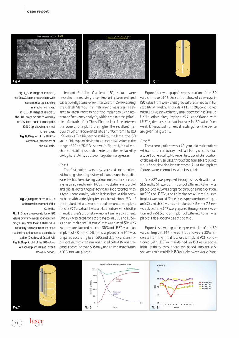
I case report
Fig. 4_SEM image of sample 2,
the Er:YAG laser-prepared site with
conventional tip, showing
minimal smear layer.
Fig. 5_SEM image of sample 3,
the SDS-prepared site followed by
Er:YAG laser irradiation using the
IO360 tip, showing minimal
smear layer.
Fig. 6_Diagram of the LEIST-v
withdrawal movement of
the IO360 tip.
Fig. 7_Diagram of the LEIST-s
withdrawal movement of the
IO360 tip.
Fig. 8_Graphic representation of ISQ
values over time as osseointegration
progresses. Note the initial decrease
in stability, followed by an increase
as the implant becomes biologically
stable. (Courtesy of Osstell AB)
Fig. 9_Graphic plot of the ISQ values
of each implant in Case I over a
12-week period.
Implant Stability Quotient (ISQ) values wererecorded immediately after implant placement andsubsequently at one-week intervals for 12 weeks, usingthe Osstell Mentor. This instrument measures resist-ance to lateral movement of the implant by using res-onance frequency analysis, which employs the princi-ples of a tuning fork. The stiffer the interface betweenthe bone and implant, the higher the resultant fre-quency, which is converted into a number from 1 to 100(ISQ value). The higher the stability, the larger the ISQvalue. This type of device has a mean ISQ value in therange of 60 to 75.37 As shown in Figure 8, initial me-chanical stability is supplemented and then replaced bybiological stability as osseointegration progresses.
Case I
The first patient was a 57-year-old male patientwith a long-standing history of diabetes and heart dis-ease. He had been taking various medications includ-ing aspirin, metformin HCl, simvastatin, metoprololand gliclazide for the past ten years. He presented witha type 3 bone quality, which is described as thin corti-cal bone with underlying dense trabecular bone.38 All ofthe implant fixtures were internal hex and the implantfor site #27 also had the Laser-Lok feature, which is themanufacturer’s proprietary implant surface treatment.Site #27 was prepared according to an SDS and LEIST-s, and an implant of 5.8 mm x 9 mm was placed. Site #26was prepared according to an SDS and LEIST-v, and animplant of 4.0 mm x 10.5 mm was placed. Site #14 wasprepared according to an SDS and LEIST-v, and an im-plant of 4.0 mm x 12 mm was placed. Site #15 was pre-pared according to an SDS only, and an implant of 4 mmx 10.5 mm was placed.
Figure 9 shows a graphic representation of the ISQvalues. Implant #15, the control, showed a decrease inISQ value from week 2 but gradually returned to initialstability at week 9. Implants #14 and 26, conditionedwith LEIST-v, showed a very small decrease in ISQ value.Unlike other sites, implant #27, conditioned with LEIST-s, demonstrated an increase in ISQ value fromweek 1. The actual numerical readings from the deviceare given in Figure 10.
Case II
The second patient was a 69-year-old male patientwith a non-contributory medical history who also hada type 3 bone quality. However, because of the locationof the maxillary sinuses, three of the four sites requiredsinus floor elevation by osteotome. All of the implantfixtures were internal hex with Laser-Lok.
Site #27 was prepared through sinus elevation, anSDS and LEIST-s, and an implant of 5.8 mm x 7.5 mm wasplaced. Site #26 was prepared through sinus elevation,an SDS and LEIST-s, and an implant of 4.5 mm x 7.5 mmimplant was placed. Site #15 was prepared according toan SDS and LEIST-v, and an implant of 4.5 mm x 7.5 mmwas placed. Site #17 was prepared through sinus eleva-tion and an SDS, and an implant of 5.8 mm x 7.5 mm wasplaced. This also served as the control.
Figure 11 shows a graphic representation of the ISQvalues. Implant #17, the control, showed a 20% in-crease from the initial ISQ value. Implant #26, condi-tioned with LEIST-v, maintained an ISQ value above initial stability throughout the period. Implant #27showed a minimal dip in ISQ value between weeks 2 and
30 I laser2_2012
Fig. 5 Fig. 6Fig. 4
Fig. 8 Fig. 9Fig. 7

case report I
I 31laser2_2012
4, but had recovered by week 5. Implant #15, condi-tioned with LEIST-v, but without sinus elevationachieved a 10% steady rise in ISQ value by week 4, whileimplants #26 and 27 required nine weeks or more toreach the 10% increase in ISQ value. The actual numer-ical readings from the device are given in Figure 12.
_Discussion
In Case I, the control showed an expected ISQ pro-gression with a decrease in week 2 and a re-establish-ment of initial stability by week 9. The 20% drop in ISQvalue was most severe at week 4, which may be relatedto the patient’s diabetic condition. Implants condi-tioned with LEIST-v did not show a significant drop inISQ value, and the LEIST-s-treated site showed a con-stant rise in ISQ value from the first day.
In Case II, the control started with an ISQ value of 57at week 1 and the stability gradually increased by 20%over six weeks. Implant #26 showed an immediate in-crease in ISQ value, while implant #27 showed a 3% dipat week 2 and returned to above the initial ISQ value atweek 5. Lai et al.39 reported good initial primary stabil-ity with sinus floor elevation and a decrease in stabilitybetween weeks 2 and 6, but ultimately a high stabilityat week 20. The authors had similar but slightly highervalues in their smaller study.
Barewal et al.40 found that the greatest change instability was between weeks 3 and 10. The control inCase I showed similar findings, but the sites conditionedwith LEIST showed very small drops in ISQ value betweenweeks 3 and 10 or immediate increases in ISQ value.Bornstein et al.41 demonstrated successful early loadingwith provisional prostheses 21 days after implant place-ment using modified-surface implants. The authorswould be interested to see whether adding the LEISTprotocol to these modified-surface implants couldachieve any improvement in stability and early loading.
_Conclusion
The results of this very small clinical study indicatethat the Er:YAG laser with a special tip design and theLEIST protocol could improve the early stability of im-
plants. The ISQ results are promising but not statisti-cally significant. Animal and histological studies withthis technique will be the next step to confirming theprogressive level of osseointegration, and more clinicalcases utilising LEIST are underway.
_Acknowledgements
The authors would like to thank elexxion AG for theirsupport and the design of the IO360 tip. Prof W. K. Leung at the Prince Philip Dental Hospital, University ofHong Kong, is acknowledged for his support with the invitrostudy. The authors would also like to thank Dr Dou-glas Chong, who performed the implant surgery forCase II. Lastly, they would like to thank Drs Frank Yungand Don Coluzzi for their guidance.
_Disclosures
The authors received no financial support fromelexxion AG, BioHorizons or Osstell AB for this study. Dr Luk is a presenter for elexxion and receives honorariafor his services. Dr Seto has no relationships to dis-close._
Editorial note: The article was first published as K. Luk & R.S.K.Seto, A novel technique of Er:YAG laser-enhanced early im-plant stability for implant dentistry: A pilot clinical case re-port, Journal of Laser Dentistry, 19/1 (2011): 162–167, and is
reprinted here with kind permission of the Academy of Laser
Dentistry. Copyright 2011 by the Academy of Laser Dentistry.
All rights reserved.
A complete list of references is available from the publisher.
Fig. 10_Numerical values of
each implant in Case I over
a 12-week period.
Fig. 11_Graphic plot of the ISQ
values of each implant in Case II over
a 12-week period.
Fig. 12_Numerical values of
each implant in Case II over
a 12-week period.
Fig. 10 Fig. 11 Fig. 12
Kenneth Luk
301, No.50 Queen's Rd Central,
Hong Kong
China
_contact laser

I case report
Figs. 1 & 2_Haemangiomas prior to
treatment.
Figs. 3 & 4_Immediately after
treatment.
_Introduction
Owing to demographic changes, eruptive or so-called senile haemangiomas have become more fre-quent among our patients. Haemangiomas are benignendothelial tumours found more often in women thanmen. Congenital capillary or cavernous haeman-giomas are characterised by remission in more than50%. Eruptive haemangiomas occur spontaneously inpatients of 60 or older, especially on the lips, but theycan also be found elsewhere. Remission of eruptivehaemangiomas is observed more seldom than in cap-illary or cavernous haemangiomas. Owing to their lo-calisation and the fact that older patients have moreserious general health problems, non-surgical andnon-invasive modalities for haemangioma treat-ments are favoured in order to minimise the risk ofprolonged bleeding and scarring. Therefore, the use oflasers is nowadays the best option for removing hae-mangiomas easily. This article demonstrates the re-moval of three oral eruptive haemangiomas using anNd:YAG laser with a wavelength of 1,064 nm.
_Case presentation
Prior to treatment, differential diagnostic aspectsmust be investigated. Lymphangiomas, retentions,
cysts and tumours of the salivary glands sometimeshave a similar appearance to haemangiomas. Evenhaematomas can be mistaken for haemangiomas. Thediagnosis is based on clinical parameters and can beconfirmed by the use of a small glass plate that rendersthe haemangioma anaemic. Aspiration can also behelpful. Angiographic investigations have no clinicalrelevance in dentistry because of their poorbenefit–cost ratio.
A 67-year-old male patient presented with threeeruptive haemangiomas. He was treated with anNd:YAG laser with a wavelength of 1,064 nm. The irra-diation time applied was two minutes and the poweroutput was 1.75 W and 15 Hz in non-contact mode.Three eruptive haemangiomas in the lower lip regions34 and 45 with a diameter of 3 mm were treated in oneappointment. The treatment was stopped when thesurface of the haemangiomas turned whitish.
Figures 1 & 2 show the haemangiomas in regions34 and 45 before the treatment. In order to enhancepatient comfort, a local anaesthetic was adminis-tered. Figures 3 & 4 give an impression of the threehaemangiomas immediately after laser treatment.One week after laser irradiation, a central indurationwith a marginal bulge was observed. This can be seen
Laser therapy of oralhaemangiomasA case report
Author_Friedrich Müller & Dr Maximilian Schmidt-Breitung, Germany
32 I laser2_2012
Fig. 2 Fig. 3Fig. 1

case report I
from figure 5. The complete removal of the haeman-giomas was followed up 11 weeks after treatment. Ascan be seen from figure 6, neither differences in thecolour of the lip mucosa occurred, nor in its texture.Although our patient reported stinging pain duringlaser irradiation, no complications or pain arose dur-ing the healing process.
_Conclusion
This case report demonstrates that the removal ofhaemangiomas can be done with very little efforteven in the dental practice. We suggest that this pro-cedure can also be used for coagulation of intra-orallesions of hereditary telangiectasis like morbus Osler-
Rendu-Weber to reduce the risk of spontaneousbleeding.
The authors declare no conflict of interest.
Fig. 5_One week after laser
irradiation.
Fig. 6_11 weeks after laser
irradiation.
I 33laser2_2012
Fig. 4 Fig. 5 Fig. 6
Friedrich Müller
Tannenring 76
65207 Wiesbaden (Auringen)
Germany
www.muellerzahnaerzte.de
_contact laser
Please contact Claudia Jahn
AD
[PIC
TUR
E: ©
SU
KIY
AKI]

I industry report
_Introduction
Both lasers and implantology are considered tobe the fashionable treatments of modern dentistry,and combining them seems to represent best prac-tice from a number of perspectives. Implantology isone of the many dental indications for the use oflasers, and it is one from which patients can benefitsignificantly.
_The ideal scenario
Ideally, the soft tissue surrounding the implantshould be prepared before starting an implant pro-cedure. The vestibular sulcus depth and frenulumshould be examined and prepared accordingly. Thiscan also be performed during the second stage ofsurgery (Fig. 1). Different laser wavelengths can beused for this kind of surgery. Er:YAG lasers cause lesspain, as they do not heat the tissue surface, unlikediode, Nd:YAG and CO2 lasers, and the patient healsmore quickly for the same reason.1, 2
_Removal of granulation tissue
One of the most important uses of lasers in im-plantology is the removal of granulation tissue anddisinfection of the surgical area after extraction.
Erbium lasers can be used for this purpose, espe-cially if there was chronic infection before the ex-traction, regardless of whether implants or bonegrafts are being placed (Fig. 2). It has even been sug-gested that implants can be placed at pre-infectedsites, although the site has to be cleaned thoroughly.3
Owing to significant differences in water content,the erbium laser can be used to only remove soft tis-sue by setting the parameters correctly (energy den-sity, pulse duration, etc.). This is done without ther-mal side-effects on the bone,4 and with greater com-fort to the patient while disinfecting the surface,5
since no force is applied, unlike with curettes. Thenon-contact or pseudo-contact procedure enablessafe cleaning of very fragile bone that may even be ina bridge form if there is a fenestration.6
Lasers in oral implantologyAuthor_Drs Ilay Maden & Zafer Kazak, Turkey
34 I laser2_2012
[BACKGROUND: ©EVV; FEMALE DENTIST: ©FRANCK BOSTON]

_Bio-modulation
After the removal of granulation tissue and su-perficial disinfection, a diode or Nd:YAG laser can beused for deep disinfection. The penetration of laserenergy of these wavelengths in hydroxyapatite andabsorption by bacterial pigments are advantageousin this case.7 These wavelengths also have a bio-modulation effect, which aids healing, leading to lessoedema and pain.8 Bio-modulation is mostly effec-tive during the initial healing phase, which is why itshould be repeated every second day for the first fewweeks.8
_Laser ablation
If the coronal part of the crest is too thin, and aplateau is desired, ablating the bone in order toachieve the desired thickness is advantageous, since,without a thermal effect, exposure of the implantneck can be avoided. This same safety benefit also ap-plies when angulated implants are placed and distalneighbouring bone needs to be removed. Also, a lasercan be used to make an indent to mark the location ofthe first drilling site because it will not slip and causeiatrogenic damage (especially helpful for novice im-plantologists). Lasers can also be used when the pro-cedure involves an osteotomy sinus lift, obtaining abone block or bone splitting (Figs. 3a & b).9, 10
_Preparation of the implant bed
The Er:YAG laser can be used for the preparation ofan implant bed in special cases, such as when the boneis very thin and soft. Thin contact tips can be used toremove the minimum amount of bone (Fig. 4), as theyare thinner than pilot drills. Then the implant bed canbe prepared using bone condensers. For other cases,it may also be beneficial to prepare the implant bedwith a laser to achieve longer stability.11 Ultimately, ifscanner systems are combined with high-poweredlasers for the purpose of shaping cavities of predeter-mined dimensions in the bone without the thermalside-effects of drills, it could be speculated that theresults would improve even further: in addition to thedisinfection of the site, the smear layer would be re-
moved from the surface, activating osteoblastic ac-tivity.12
_Uncovering the implant
The most comfortable, patient-friendly use of theEr:YAG laser is during the second stage of implantplacement: uncovering the implant (Fig. 5). Using a re-duced water spray in order to decrease bleeding, it ispossible to uncover the implant with diminished pain,even if bone is covering the implant. Using longerpulses causes haemostasis in the soft tissue, whileshorter pulses enable the removal of bone without
industry report I
I 35laser2_2012
Fig. 1_Frenectomy with a diode laser
after placement of full-arch
implant-supported prostheses.
Fig. 2_Cleaning and disinfecting the
socket with an Er:YAG laser after
extraction.
Figs. 3a & b_Bone splitting (a). Sinus
window preparation (b).
Fig. 2 Fig. 3aFig. 1

I industry report
Fig. 4_Bone ablation for the
implant bed.
Fig. 5_Uncovering the implant.
Figs. 6 a & b_De-epithelialisation of
the crestal gingival to make a
roll flap.
Figs. 7a & b_Peri-implantitis before
treatment (a). After cleaning,
disinfection and augmentation (b).
thermal side-effects. This is achieved without caus-ing pain to the patient and without raising a flap. Thisis possible, of course, if there is sufficient attachedgingiva and the crestal attached gingiva does not needto be replaced apically. When there is no harmful heat-ing of the soft tissue, there is no retraction of tissue, sothe impressions can be taken as soon as possible with-out delay. Another use is the de-epithelialisation ofthe crestal gingiva to make a roll flap in order to opti-mise vestibular soft-tissue thickness (Fig. 6).
_Peri-implantitis
The most efficient use of the Er:YAG laser is in thetreatment of peri-implantitis.14 With Er:YAG, it is pos-sible to clean the granulation tissue both on the bonesurface and implant surface (Figs. 7a & b). This is donethrough decontamination of the site, which is themain purpose of peri-implantitis treatment.15, 16 Sim-ilar to the cleaning of the surgical area after extrac-tion, it is possible to leave the highly fragile sur-rounding bone intact. The effect of the laser energyon the implant surface is dependent on the amountof energy density, power and possibly also pulse du-ration.17, 18 The parameters should be chosen cau-tiously, however. Lowering the settings may make theprocedure slower but safer for re-osseointegration.Non-surgical use of Er:YAG and diode lasers is alsopossible if the problem is not extensive.
The most prevalent reason for peri-implantitis ap-pears to be poor occlusal load distribution, with eitherprimary contacts or cantilever bridges in implant-supported prostheses. Good oral hygiene on the pa-tient’s part is mandatory. The position and design ofprostheses that are difficult to manage may limit the
effectiveness of mechanical cleaning. Once the un-derlying reason has been determined and recurrenceis prevented, the Er:YAG laser can help treat periim-plantitis. Finally, lasers are also used to enhance theaesthetics of implant-supported prostheses. Gingi-val levelling by crown lengthening and depigmenta-tion are the most common procedures performed forthis purpose.
_Conclusion
From this range of laser applications, it is evidentthat the use of lasers in modern dentistry is only lim-ited by the dentist’s imagination. Ultimately, lasersare not miracle machines that can improve the qual-ity of implantology overnight in cases in which thepractitioner has limited knowledge and experience inimplantology. However, they are indeed a major aid indoing what is needed, always enabling increasedcomfort, both for the operator and the patient. Edu-cation is certainly the key in implantology and laserdentistry in general._
Editorial note: The complete list of references is available
from the author.
36 I laser2_2012
Fig. 7a Fig. 7b
Fig. 5
Fig. 6a Fig. 6b
Fig. 3b
Dr Ilay Maden PhD, MSc, BDS
Mühürdar Caddesi 69
34710 Istanbul, Turkey
_contact laser
Fig. 4

International annualcongress of the dgzi
FAX REPLY+49 341 48474-390Please send me further information on the42nd International annual congress of the DGZI October 5–6, 2012, in Hamburg, Germany.
Office Stamp
laser 2/12
Silver Sponsor:Gold Sponsor: Bronze Sponsor:
SpeakersProf. Dr. Dr. Kai-Olaf HenkelDEProf. Dr. Dr. George Khoury/DEProf. Dr. Dr. Albert Mehl/CHProf. Dr. Herbert Deppe/DEProf. Dr. Werner Götz/DEProf. Dr. Shoji Hayashi/JPProf. Dr. Andrea Mombelli/CHProf. Dr. Dr. Frank Palm/DEProf. Dr. Suheil Boutros/USProf. Dr. Peter Rammelsberg/DEProf. Dr. Anton Sculean/CHProf. Dr. Dr. Jörg R. Strub/DE
Prof. Dr. Hans-Peter Weber/USProf. Dr. Thomas Weischer/DEPriv.-Doz. Dr. Andreas Bindl/CHDr. Tomohiro Ezaki/JPDr. Daniel Ferrari, M.Sc./DEDr. Sami Jade/LBDr. Ramy Fahmy Rezkallah/EGDr. Osamu Yamashita/JPZTM Andreas Kunz/DEZTM Tom Lassen/DEZTM Christian Müller/DEMohamed Moataz M. KhamisB.D.S., M.S., Ph.D./EG
Quality driven implantology – On the way to long term success
October 5-6, 2012//Hamburg, Germany//Elysee Hotel
Congress President//Prof. Dr. Dr. Frank Palm/DEScientific Director//Dr. Roland Hille/DE

Manufacturer News
I manufacturer _ news
38 I laser2_2012
Erchonia Europe, the provider of Low Level Laser Tech-nology (LLLT) for medical applications, has been re-named Primcogent Solutions, and now holds exclusiveNorth American and Western European distributionrights to the ZERONA® non-invasive body contouringlaser. Primcogent Solutions is a privately-held medicalcompany with offices strategically located in New YorkCity, Dallas, the United Kingdom and Germany, whoseproducts and services are backed by a clear, logicaland convincing scientific and clinical founda-tion. With extensive knowledge and success in the sales, mar-keting and financing ofhealthcare products andservices, Primcogent Solu-tions is ideally placed to fullysupport and grow the ZERONA®
provider network. Mike McDon-ald, President of Primcogent Solutions, said the new move willbenefit physicians, con-sumers and employ-ees alike: “Our pri-mary goal has al-ways been to con-tinuously evolveand provide betterproducts and services toour customers. We are proud to have taken the nextstep in the evolution of our company.” David Boris,Chairman of Primcogent Solutions, added: “We are ex-tremely excited to be able to provide the Erchonia Europe management team with the resources neededto help them grow the ZERONA provider network andadd new products into the North American and West-ern European portfolios.”
Primcogent Solutions
The Innovation Centre
99 Milton Park, Abingdon,
Oxon, OX14 4RY, United Kingdom
www.primcogent.com
Primcogent Solutions
Erchonia Europe renamed as Primcogent SolutionsFotona's latest dental laser, LightWalker, was
awarded one of the world's most distin-guished design awards, the “red dotaward: product design 2012”, beingrecognized for combining innovation,technological perfection and excellentdesign.
LightWalker's technologically ad-vanced, modern and functional de-sign offers ease-of-use and improvedergonomics: A state of the art colortouch screen with an adjustable tilt,interchangeable optics fornew technologically advancedhand pieces, and a modern,durable and lightweight sys-tem housing allow user-friendly handling. An easy-to-access water reservoir and theunique and patented OPTOflex articulated arm,which allows a full range of motion, provide further
comfort. The system enables gentle andprecise cavity preparation, sutureless soft-
tissue surgery, effective periodontal treat-ments, safe and efficient endodontic treatment
and numerous cosmetic procedures.
The red dot award is LightWalker’s thirdprestigious international quality recogni-tion. In 2011, the Pride Institute awardedthe laser system the "Best of Class Tech-nology Award", and Dentistry Today,America’s leading clinical news maga-zine for dentists, recognized LightWalkeras one of the “TOP 100 dental products of
the year”.
Fotona d.d.
Stegne 7
1000 Ljubljana, Slovenia
www.lightwalkerlaser.com
FOTONA
"red dot design award 2012" for LightWalker
Henry Schein is once again No. 1 on Fortune maga-zine's 2012 list of the most admired companies in thewholesale healthcare sector. This is the first time since2011 that the company has ranked first in this cate-gory. In the two previous years, the top position washeld by McKesson, a pharmaceutical corporationbased in San Francisco.
According to Fortune, Henry Schein not only outdidcompetitors in business-related categories like globalcompetiveness and long-term investment but alsoscored best in the social re-sponsibility category, drivenby Henry Schein Cares, a pro-gram launched in 2001 withthe aim of providing medical
supplies and logistic support to underserved commu-nities around the world. In 2011, the program providedmedical relief to the victims of Hurricane Irene in NorthAmerica and the New Zealand earthquake, among oth-ers. Nasdaq-listed Henry Schein scored record sales of$ 8.5 billion worldwide last year. In addition to its USbusinesses, the company currently maintains opera-tions in 25 countries.
Henry Schein Dental
Deutschland GmbH
Monzastraße 2a
63225 Langen, Germany
www.henryschein-dental.de
Henry Schein
Henry Schein regains top position on Fortunecompanies list

manufacturer _ news I
I 39laser2_2012
Bacterial inflammation in periodontal pockets canlead to bleeding gums, pocket formation, reforma-tion of gums with loosening and eventual loss ofteeth. Furthermore, scientific evidence indicatesthat there is an increased risk of infections develop-ing in the rest of the body, eventually resulting in vas-cular diseases in the heart and arteries.
EmunDo® PDT therapy is safe and effective for removing harmful bacteria, regardless of the Gramstain and including Gram-positive/-negative bacte-ria, as well as Gram-variable and Gram-undeter-mined species. By comparison, mechanical cleaningcannot reach and remove bacteria in all areas. Otherlaser-based therapies cannot be said to be clinicallyeffective on all types of germs.
PDT has the ability to treat exposed areas withoutthermal effect. EmunDo® has a selective, localizedeffect because it accumulates only in the inflamedareas and can be irradiated immediately withoutwaiting period. Furthermore, the bacteria containedin plaque or biofilm is less affected by antibiotics, be-cause they are shielded by the organic matrix in thefilm and may be absorbed by or adhere to the toothand epithel cells. While PDT has the advantage ofachieving excellent cosmetic results with minimalrisk of scarring, it is also a welcome alternative treat-ment for periimplantitis to save the implant by main-taining the protective function of the mucosa.
A.R.C. Laser GmbH
Bessemerstraße 14
90411 Nuremberg, Germany
www.arclaser.de
A.R.C. Laser
Healthy teeth = Healthy patients
Biolase Technology,Californian-based
dental laser manufac-turer and distributor,
has repurchased 159 of itsWaterlase MD Turbo laser systems from HenrySchein, supplier of dental and medical equipment.The equipment is expected to be used primarily as asource of parts to service the large number of in-stalled MD Turbo laser systems and for dentalschools in order to promote Waterlase technology inthe academic sector.
According to the manufacturer, the Waterlase MDTurbo, first launched in 2006, is the most success-ful all-tissue laser in the history of dental lasers, withapproximately 6,500 units sold worldwide.
Biolase purchased the MD Turbos at a very advan-tageous price and the entire purchase price was off-set by monies owed by Henry Schein to Biolase fromsales made in the normal course of business mainlyduring the first quarter of 2012 and, to a much lesser
extent, the year ended Dec. 31, 2011. None of themonies used to offset the purchase price were re-lated to the original sales of the MD Turbos to Schein.
Federico Pignatelli, Chairman and CEO of Biolase,commented, “Closing this transaction is a very im-portant step for Biolase, as it eliminates the over-hang in the marketplace that equaled approxi-mately 440 units at the end of 2010 and significantlyimpacted our sales in 2011, releases all liens on ourpatent and intellectual properties portfolio and freesus of any present and future obligations to Schein. Itis also a very advantageous transaction, as the re-maining inventory was purchased at a very conven-ient price and will mainly be used as parts to serviceour vast install base of MD Turbos.”
Biolase Europe GmbH
Paintweg 10
92685 Floss, Germany
www.biolase.de
Biolase
Biolase concludes transactionwith Henry Schein
Dental laser specialist elexxion has reported thatit has signed a new distribution agreement withGlobal Dental Supplies in Hong Kong. The five-yearcontract will give the dental distributor the exclu-sive rights to distribute elexxion’s laser technologyfor use in dentistry in several Asian countries.
Currently, the German company sells its productsthrough its subsidiaries and dealers in selectedmarkets, such as India andJapan. The distribution rightsfor Hong Kong and Macau werepreviously held by HealthcareDental, which did not renew itscontract with elexxion after2009, company officials toldDental Tribune Asia Pacific. Be-sides elexxion dental lasers,Global Dental Supplies alsodistributes products from theGerman implant companyBEGO, Sunstar, Bisco and GC, among others. “WithGlobal Dental Supplies we have a strong partnerthat gives us the opportunity to systematically ex-
pand our sales and marketing activities in Asia,”commented elexxion CEO Per Liljenqvist. He saidthat his company could particularly benefit fromthe agreement in terms of product registration andexhausting new distribution channels in the re-gion.
The latest elexxion product offering includes thedelos 3.0, a novel combined Er:YAG/diode laser in-
dicated for a wide range of dentalapplications. In addition, the com-pany distributes the pico mobilediode laser and duros, an Er:YAGdental laser claimed to facilitateefficient hard-tissue preparationand bone ablation tasks.
elexxion AG
Schützenstraße 84
78315 Radolfzell, Germany
www.elexxion.de
elexxion
elexxion signs distributor for Asian markets
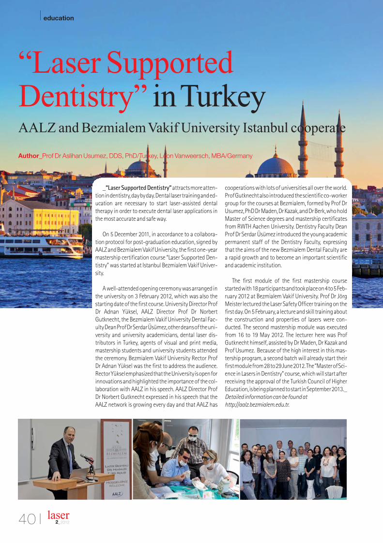
I education
_“Laser Supported Dentistry”attracts more atten-tion in dentistry, day by day. Dental laser training and ed-ucation are necessary to start laser-assisted dentaltherapy in order to execute dental laser applications inthe most accurate and safe way.
On 5 December 2011, in accordance to a collabora-tion protocol for post-graduation education, signed byAALZ and Bezmialem Vakif University, the first one-yearmastership certification course “Laser Supported Den-tistry” was started at Istanbul Bezmialem Vakif Univer-sity.
A well-attended opening ceremony was arranged inthe university on 3 February 2012, which was also thestarting date of the first course. University Director ProfDr Adnan Yüksel, AALZ Director Prof Dr NorbertGutknecht, the Bezmialem Vakif University Dental Fac-ulty Dean Prof Dr Serdar Üsümez, other deans of the uni-versity and university academicians, dental laser dis-tributors in Turkey, agents of visual and print media,mastership students and university students attendedthe ceremony. Bezmialem Vakif University Rector ProfDr Adnan Yüksel was the first to address the audience.Rector Yüksel emphasized that the University is open forinnovations and highlighted the importance of the col-laboration with AALZ in his speech. AALZ Director ProfDr Norbert Gutknecht expressed in his speech that theAALZ network is growing every day and that AALZ has
cooperations with lots of universities all over the world.Prof Gutknecht also introduced the scientific co-workergroup for the courses at Bezmialem, formed by Prof DrUsumez, PhD Dr Maden, Dr Kazak, and Dr Berk, who holdMaster of Science degrees and mastership certificatesfrom RWTH Aachen University. Dentistry Faculty DeanProf Dr Serdar Üsümez introduced the young academicpermanent staff of the Dentistry Faculty, expressingthat the aims of the new Bezmialem Dental Faculty area rapid growth and to become an important scientificand academic institution.
The first module of the first mastership coursestarted with 18 participants and took place on 4 to 5 Feb-ruary 2012 at Bezmialem Vakif University. Prof Dr JörgMeister lectured the Laser Safety Officer training on thefirst day. On 5 February, a lecture and skill training aboutthe construction and properties of lasers were con-ducted. The second mastership module was executedfrom 16 to 19 May 2012. The lecturer here was ProfGutknecht himself, assisted by Dr Maden, Dr Kazak andProf Usumez. Because of the high interest in this mas-tership program, a second batch will already start theirfirst module from 28 to 29 June 2012. The “Master of Sci-ence in Lasers in Dentistry” course, which will start afterreceiving the approval of the Turkish Council of HigherEducation, is being planned to start in September 2013._Detailed information can be found at
http://aalz.bezmialem.edu.tr.
“Laser Supported Dentistry” in TurkeyAALZ and Bezmialem Vakif University Istanbul cooperate
Author_Prof Dr Aslihan Usumez, DDS, PhD/Turkey, Leon Vanweersch, MBA/Germany
40 I laser2_2012

AEEDC_2012_A4.pdf 1AEEDC_2012_A4.pdf 1 05.03.12 14:0905.03.12 14:09
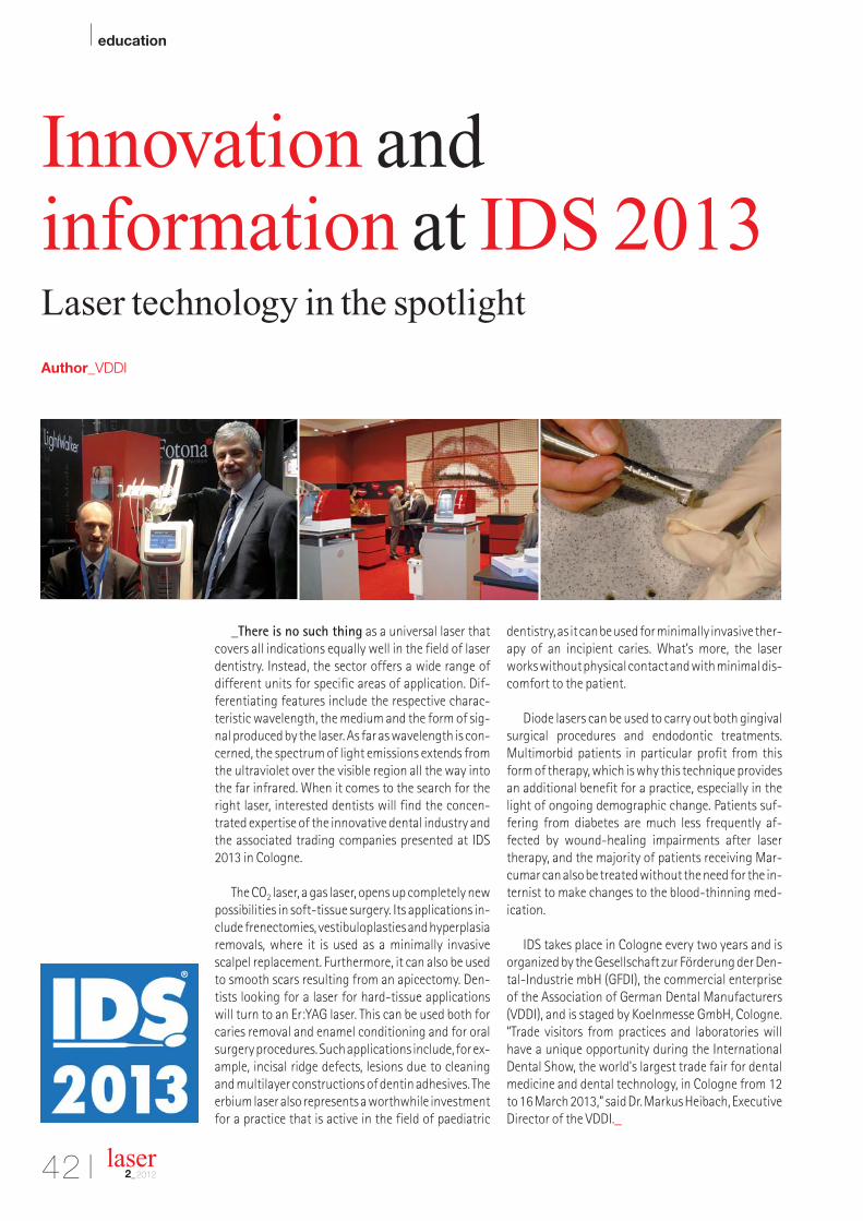
I education
_There is no such thing as a universal laser thatcovers all indications equally well in the field of laserdentistry. Instead, the sector offers a wide range ofdifferent units for specific areas of application. Dif-ferentiating features include the respective charac-teristic wavelength, the medium and the form of sig-nal produced by the laser. As far as wavelength is con-cerned, the spectrum of light emissions extends fromthe ultraviolet over the visible region all the way intothe far infrared. When it comes to the search for theright laser, interested dentists will find the concen-trated expertise of the innovative dental industry andthe associated trading companies presented at IDS2013 in Cologne.
The CO2 laser, a gas laser, opens up completely newpossibilities in soft-tissue surgery. Its applications in-clude frenectomies, vestibuloplasties and hyperplasiaremovals, where it is used as a minimally invasivescalpel replacement. Furthermore, it can also be usedto smooth scars resulting from an apicectomy. Den-tists looking for a laser for hard-tissue applicationswill turn to an Er:YAG laser. This can be used both forcaries removal and enamel conditioning and for oralsurgery procedures. Such applications include, for ex-ample, incisal ridge defects, lesions due to cleaningand multilayer constructions of dentin adhesives. Theerbium laser also represents a worthwhile investmentfor a practice that is active in the field of paediatric
dentistry, as it can be used for minimally invasive ther-apy of an incipient caries. What’s more, the laserworks without physical contact and with minimal dis-comfort to the patient.
Diode lasers can be used to carry out both gingivalsurgical procedures and endodontic treatments.Multimorbid patients in particular profit from thisform of therapy, which is why this technique providesan additional benefit for a practice, especially in thelight of ongoing demographic change. Patients suf-fering from diabetes are much less frequently af-fected by wound-healing impairments after lasertherapy, and the majority of patients receiving Mar-cumar can also be treated without the need for the in-ternist to make changes to the blood-thinning med-ication.
IDS takes place in Cologne every two years and isorganized by the Gesellschaft zur Förderung der Den-tal-Industrie mbH (GFDI), the commercial enterpriseof the Association of German Dental Manufacturers(VDDI), and is staged by Koelnmesse GmbH, Cologne.“Trade visitors from practices and laboratories willhave a unique opportunity during the InternationalDental Show, the world's largest trade fair for dentalmedicine and dental technology, in Cologne from 12to 16 March 2013,” said Dr. Markus Heibach, ExecutiveDirector of the VDDI._
Innovation and information at IDS 2013Laser technology in the spotlight
Author_VDDI
42 I laser2_2012

International events
meetings I
I 43laser2_2012
2012
Europerio 7Vienna, Austria6–12 Junewww.europerio7.com
FDI Annual World Dental ConferenceHong Kong, China29 August–1 Septemberwww.fdiworldental.org
21st DGL Annual ConferenceLeipzig, Germany7–8 Septemberwww.dgl2012.de
China Dental ShowXi’an Qujiang, China13–15 Septemberwww.ChinaDentalShow.com
32nd Dental-ExpoMoscow, Russia17–20 Septemberwww.dental-expo.com
22nd Central European Dental ExhibitionPoznan, Poland20–22 Septemberwww.cede.pl
13th Slovak Dental DaysBratislava, Slovakia 27–29 Septemberwww.incheba.sk
42nd International Congress of DGZIHamburg, Germany5–6 October www.dgzi-jahreskongress.de
20th Pragodent Dental FairPrague, Czech Republic11–13 October www.pragodent.eu
DenTech ChinaShanghai, China24–27 Octoberwww.dentech.com.cn
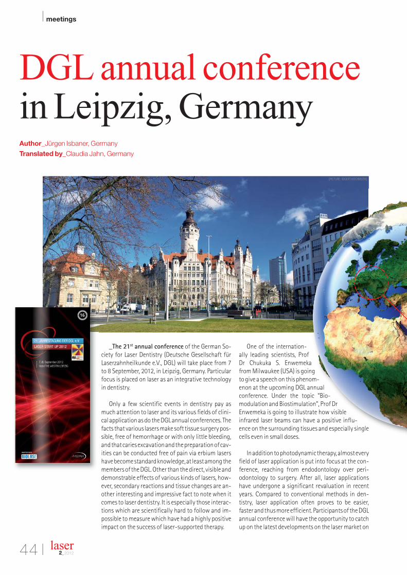
[PICTURE: ©GERT HOCHMUTH]
I meetings
DGL annual conferencein Leipzig, GermanyAuthor_Jürgen Isbaner, Germany
Translated by_Claudia Jahn, Germany
44 I laser2_2012
_The 21st annual conference of the German So-ciety for Laser Dentistry (Deutsche Gesellschaft fürLaserzahnheilkunde e.V., DGL) will take place from 7to 8 September, 2012, in Leipzig, Germany. Particularfocus is placed on laser as an integrative technologyin dentistry.
Only a few scientific events in dentistry pay asmuch attention to laser and its various fields of clini-cal application as do the DGL annual conferences. Thefacts that various lasers make soft tissue surgery pos-sible, free of hemorrhage or with only little bleeding,and that caries excavation and the preparation of cav-ities can be conducted free of pain via erbium lasershave become standard knowledge, at least among themembers of the DGL. Other than the direct, visible anddemonstrable effects of various kinds of lasers, how-ever, secondary reactions and tissue changes are an-other interesting and impressive fact to note when itcomes to laser dentistry. It is especially those interac-tions which are scientifically hard to follow and im-possible to measure which have had a highly positiveimpact on the success of laser-supported therapy.
One of the internation-ally leading scientists, ProfDr Chukuka S. Enwemekafrom Milwaukee (USA) is goingto give a speech on this phenom-enon at the upcoming DGL annualconference. Under the topic “Bio-modulation and Biostimulation”, Prof DrEnwemeka is going to illustrate how visibleinfrared laser beams can have a positive influ-ence on the surrounding tissues and especially singlecells even in small doses.
In addition to photodynamic therapy, almost everyfield of laser application is put into focus at the con-ference, reaching from endodontology over peri-odontology to surgery. After all, laser applicationshave undergone a significant revaluation in recentyears. Compared to conventional methods in den-tistry, laser application often proves to be easier,faster and thus more efficient. Participants of the DGLannual conference will have the opportunity to catchup on the latest developments on the laser market on

[EARTH: ©ANTON BALAZH]
meetings I
I 45laser2_2012
[PIC
TUR
E: ©
OR
LA]
LEIPZIG
BERLIN
the occasion of the accompanying dental trade fair inLeipzig. Parallel to the annual conference, Laser StartUp is designed to enable beginners in laser dentistryto acquire a professional knowledge base in order toenter this new field of expertise. In addition, work-shops and hands-on courses will provide practicaltraining in getting to know the various kinds of lasers.
The DGL party is the festive occasion concludingthe DGL annual conference Saturday night at THEWESTIN LEIPZIG hotel. The accomplished combina-tion of professional exchange and friendly gatheringmakes this year’s DGL annual conference an excep-tional educational event._
OEMUS MEDIA AG
Holbeinstraße 29
04229 Leipzig, Germany
Tel.: +49 341 48474-308
Fax: +49 341 48474-390
www.oemus.com
_contact laser

I meetings
_laser: Prof Dr Gutknecht, this year’s DGL annualconference takes place in early September in Leipzig,Germany. Can you tell us what the participants can ex-pect from this event? What are the highlights of theconference?
You can roughly attribute four key points to our up-coming congress in Leipzig. Firstly, we want to presentthe latest scientific discoveries to our audience in aneasily accessible way. Secondly, we aim at emphasisingthe significance of laser for a successful dental ther-apy by pointing out how laser technology has been integrated in various medical and dental therapies.
Thirdly, economic efficiency plays a crucial role in anylaser application. Therefore, another important aspectof this year’s congress is the new German Scale of Feesfor Dentists (Gebührenordnung für Zahnärzte, GOZ).And last but not least, there is the social aspect. Thesuccess of last year’s DGL anniversary party has shownonce more how important social values and friend-ships are nowadays. Our DGL party on Saturday is ded-icated to combining the scientific interest we all havein common with a friendly and relaxed atmosphere.
_How would you assess the current situation oflaser dentistry? What are the main directions its devel-opment is going to take? What do you think will be thestatus of laser dentistry in five years?
Scientifically, laser dentistry has now reached itshighest and most substantiated standard. Further-more, laser is an integrative technology highly ac-cepted by the professional organisations in medicineand dentistry. In my opinion, this shows how lasertechnology can take a decisive influence on the suc-cess of various therapies. Speaking of the main direc-tions of the development of laser dentistry, we haveto take three essential aspects into account: Firstly,there is the advancement of pico-second and femto -second lasers with regard to hard tissue applications.Secondly, laser-activated photodynamic therapy hasbecome both more specialised and developed.Thirdly, therapy concepts for already existing lasersystems are constantly being developed, which willlead to a significant growth in the sector of lasertechnology within the coming five years. This per-spective is especially relevant for periodontology, endodontology, cariology, surgery, implantology andpedodontics.
_DGL maintains a high profile, both nationally andinternationally. Can you name current activities initi-ated or supported by the DGL?
“Scientifically, laser hasnow reached its higheststandard”Author_Dajana Mischke, Germany
46 I laser2_2012
Prof Dr Norbert Gutknecht
On the occasion of the German Society for LaserDentistry (Deutsche Gesellschaft für Laserzahn-heilkunde e.V.) annual conference 2012 in Septem-ber in Leipzig, Germany, Prof Dr Norbert Gutknechtof the University Hospital of RWTH Aachen, Ger-many, commented on the current situation of laserdentistry, the key issues of the upcoming DGL an-nual conference as well as the future of laser ther-apy in dentistry and the activities of the DGL.

meetings I
Nationally, the DGL boards are intensively en-gaged in establishing statements and clinical treat-ment guidelines for the German Society for Dentaland Oral Medicne (Deutsche Gesellschaft für Zahn-,Mund- und Kieferheilkunde, DGZMK). Furthermore,subgroups of the executive board and advisoryboards are currently discussing the problem of inter-preting and billing certain laser treatment optionsaccording to the new GOZ. Internationally, DGL isrepresented both in European and international lasersocieties. Moreover, not only are members of the DGLmuch sought-for speakers, but they also are en-trusted with different additional tasks in their re-spective organisations.
_Photodynamic therapy is going to be among thekey topics of this year’s DGL conference. What is thecurrent status of photodynamic therapy in laser den-tistry and how relevant is it for the daily dental prac-tice?
In the past, photodynamic therapy only played asecondary role or the role of the outsider, both in sci-entific research and in clinical applications. However,we have noticed a grown interest in this therapeuticbranch since 2006, which has even more increased
within the past two years. By now we can rely on dif-ferent wavelengths and photosensitisers which havebeen scientifically tested and applied in clinical ther-apies. Nevertheless, the range of possible dental ap-plications of lasers is still very limited, with the high-est clinical relevance in periodontology: photody-namic therapy has proven a successful integrativemethod for the standardised procedures of peri-odontological treatment.
Prof Dr Gutknecht, thank you very much for this
interview!
I 47laser2_2012
Prof Dr Norbert Gutknecht
DGL—Deutsche Gesellschaft für Laserzahnheilkunde e.V.Pauwelsstraße 30 52074 Aachen, Germany
_contact laser

48 I laser2_2012
NEWS
The third day of lectures of the Dental Tribune StudyClub Symposia introduced on Tuesday a good varietyof speakers and topics at the Greater New York DentalMeeting. A total of six speakers of different back-grounds spoke at these well-known series of lectures,which are part of the educational arm of Dental TribuneInternational. Among them were Dr Gregori Kurtzman,
who talked about “CoreBuildups, Post & Cores andUnderstanding Ferrule”; Dr Paul Goodman, who disserted on how to “Capitalizeon the Hidden Implant Production in Your Practice”;
Dr George Freedman and Dr Fay Goldstep, who gave anexplanation of the clinical advantages of the now af-fordable laser therapy, entitled “The Diode Laser: TheEssential Soft Tissue Handpiece”.
The 4-day series of lectures of Dental Tribune StudyClub attracted a large number of dentists interested inlearning about these topics. The Tuesday lectures of-fered six hours of ADA-CEP Continuing Education cred-its.
Dr Pedro Lázaro, a Spain-based specialist in periodon-tics and dental implants, said that what is really diffi-cult is to get results that are perfect from an aesthetic
point of view, or as close to perfect as possible.
“It’s like when you see a professional tennis playerand the game looks very easy, but you know thatthere are only a few real top level champions likeRafa Nadal or Roger Federer,” said Lázaro in an in-terview after his lecture. “Not using the appropriate
technique could worsen the results of treatment,” hewarned.
From the use of lasers to aesthetic implants
by Javier Martínez de Pisón
Engineers have developed a cardboard tooth-brush that could replace not only disposabletoothbrushes in hotels and aeroplanes but alsoconventional household toothbrushes. The mate-rials used in manufacturing this toothbrush arebio-based, renewable and fully recyclable.
According to the German Federal EnvironmentalAgency, experts agree that plastic products decompose extremely slowly. Depending on environmental conditions, they estimate thatplastic bags, for instance, take up to 500 years todecompose.
In a collaborative researchproject, the Zahoransky engineering
corporation and the West Saxon University of Applied Sciences of Zwickau developed an inno-vative injection-moulding process, which made itpossible to produce a toothbrush using renewableraw materials. This invention could be an alterna-tive to disposable toothbrushes, which are alsogiven regularly to patients at dental practices.
According to Zahoransky, the new toothbrush ismanufactured from a biodegradable, fibre-filledcomposite, which is made by first pressing wastepaper fibre into free-flowing bulk solids andthereafter processing the solids into injectablegranulate using a biopolymer matrix.
Given the rising oil price, which affects the price ofsynthetic oil-based plastics, and increasing public awareness of ecologically safe productsand processes, the designers wanted to create adisposable toothbrush that is environmentallyfriendly at the same time. The production of thenewly developed toothbrush does not involve any fossil fuel because it is made solely from bio-based synthetic materials. After use, thetoothbrush can be disposed of in the compostingbin.
Compostable toothbrush
Produced by Germanmanufacturer
[PICTURE: ©MOPIC]
Middle-aged women are most likely to suffer fromfear of the dentist, a new study found. Clinical obser-vation of patients taking part in a multi-year clinicaltrial conducted at the Dental Phobia Clinic in West-mead, Sydney, has indicated that the level of dentalanxiety is highest among women in their forties.
According to the researchers, this demographic wasalso found to have perceived a traumatic dental ex-perience, including orofacial trauma, in the past andto be more prone to stress or mental disorders likedepression. The results are intended to help investi-gate the relationship between dental anxiety and the
perception of and coping with pain, as well as to de-velop strategies for managing the condition suc-cessfully.
In addition, the study could confirm findings of ear-lier research that found that cognitive processing ofdental phobia in women differs significantly fromthat in men.
Forty per cent of people in the developed world areestimated to have some form of dental anxiety. Sur-veys have shown that compared with the generalpopulation, phobic patients wait more than fivetimes longer to make a dental appointment, co-ordi-nator of the study and special needs dentist Dr AvantiKarve said. She added that the key strategy for man-aging dental phobia is to help the patient develop theskills for coping with or overcoming the condition.“Dental anxiety is very real and complex and itshould never be downplayed,” she said. The anxietystudy has been running in association with clinicalpsychologists for five years.
Australian survey finds
Dental mid-life crisis in women
“Dental anxiety
is very real and
complex and
it should never be
downplayed.”
[PICTURE: ©JUNIAL ENTERPRISES]
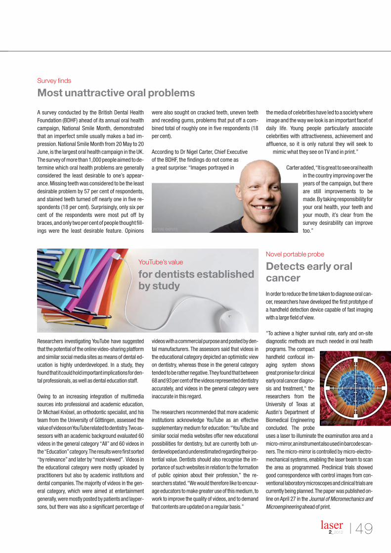
Researchers investigating YouTube have suggestedthat the potential of the online video-sharing platformand similar social media sites as means of dental ed-ucation is highly underdeveloped. In a study, theyfound that it could hold important implications for den-tal professionals, as well as dental education staff.
Owing to an increasing integration of multimediasources into professional and academic education, Dr Michael Knösel, an orthodontic specialist, and histeam from the University of Göttingen, assessed thevalue of videos on YouTube related to dentistry. Two as-sessors with an academic background evaluated 60videos in the general category “All” and 60 videos inthe “Education” category. The results were first sorted“by relevance” and later by “most viewed”. Videos inthe educational category were mostly uploaded bypractitioners but also by academic institutions anddental companies. The majority of videos in the gen-eral category, which were aimed at entertainmentgenerally, were mostly posted by patients and layper-sons, but there was also a significant percentage of
videos with a commercial purpose and posted by den-tal manufacturers. The assessors said that videos inthe educational category depicted an optimistic viewon dentistry, whereas those in the general categorytended to be rather negative. They found that between68 and 93 per cent of the videos represented dentistryaccurately, and videos in the general category wereinaccurate in this regard.
The researchers recommended that more academicinstitutions acknowledge YouTube as an effectivesupplementary medium for education: “YouTube andsimilar social media websites offer new educationalpossibilities for dentistry, but are currently both un-derdeveloped and underestimated regarding their po-tential value. Dentists should also recognise the im-portance of such websites in relation to the formationof public opinion about their profession,” the re-searchers stated. “We would therefore like to encour-age educators to make greater use of this medium, towork to improve the quality of videos, and to demandthat contents are updated on a regular basis.”
YouTube’s value
for dentists establishedby study
In order to reduce the time taken to diagnose oral can-cer, researchers have developed the first prototype ofa handheld detection device capable of fast imagingwith a large field of view.
"To achieve a higher survival rate, early and on-site diagnostic methods are much needed in oral healthprograms. The compacthandheld confocal im-aging system showsgreat promise for clinicalearly oral cancer diagno-sis and treatment," theresearchers from theUniversity of Texas atAustin's Department ofBiomedical Engineeringconcluded. The probeuses a laser to illuminate the examination area and amicro-mirror, an instrument also used in barcode scan-ners. The micro-mirror is controlled by micro-electro-mechanical systems, enabling the laser beam to scanthe area as programmed. Preclinical trials showedgood correspondence with control images from con-ventional laboratory microscopes and clinical trials arecurrently being planned. The paper was published on-line on April 27 in the Journal of Micromechanics andMicroengineering ahead of print.
Novel portable probe
Detects early oralcancer
I 49laser2_2012
A survey conducted by the British Dental HealthFoundation (BDHF) ahead of its annual oral healthcampaign, National Smile Month, demonstratedthat an imperfect smile usually makes a bad im-pression. National Smile Month from 20 May to 20June, is the largest oral health campaign in the UK.The survey of more than 1,000 people aimed to de-termine which oral health problems are generallyconsidered the least desirable to one’s appear-ance. Missing teeth was considered to be the leastdesirable problem by 57 per cent of respondents,and stained teeth turned off nearly one in five re-spondents (18 per cent). Surprisingly, only six percent of the respondents were most put off bybraces, and only two per cent of people thought fill-ings were the least desirable feature. Opinions
were also sought on cracked teeth, uneven teethand receding gums, problems that put off a com-bined total of roughly one in five respondents (18per cent).
According to Dr Nigel Carter, Chief Executiveof the BDHF, the findings do not come asa great surprise: “Images portrayed in
the media of celebrities have led to a society whereimage and the way we look is an important facet ofdaily life. Young people particularly associatecelebrities with attractiveness, achievement andaffluence, so it is only natural they will seek to
mimic what they see on TV and in print.”
Carter added, “It is great to see oral healthin the country improving over theyears of the campaign, but thereare still improvements to bemade. By taking responsibility foryour oral health, your teeth andyour mouth, it’s clear from thesurvey desirability can improvetoo.”
Survey finds
Most unattractive oral problems
[PICTURE: ©LICHTMEISTER]
[PICTURE: ©IOFOTO]
[PICTURE: ©LIGHTSPRING]

I about the publisher _ imprint
50 I laser2_2012
Copyright Regulations _laser international magazine of laser dentistry is published by Oemus Media AG and will appear in 2012 with one issue every quarter. The
magazine and all articles and illustrations therein are protected by copyright. Any utilization without the prior consent of editor and publisher is inad-mi ssible and liable to prosecution. This applies in particular to duplicate copies, translations, microfilms, and storage and processing in electronic systems.
Reproductions, including extracts, may only be made with the permission of the publisher. Given no statement to the contrary, any submissions to theeditorial department are understood to be in agreement with a full or partial publishing of said submission. The editorial department reserves the right tocheck all submitted articles for formal errors and factual authority, and to make amendments if necessary. No responsibility shall be taken for unsolicitedbooks and manuscripts. Articles bearing symbols other than that of the editorial department, or which are distinguished by the name of the author, representthe opinion of the afore-mentioned, and do not have to comply with the views of Oemus Media AG. Responsibility for such articles shall be borne by the author.Responsibility for advertisements and other specially labeled items shall not be borne by the editorial department. Likewise, no responsibility shall be assumedfor information published about associations, companies and commercial markets. All cases of consequential liability arising from inaccurate or faulty representation are excluded. General terms and conditions apply, legal venue is Leipzig, Germany.
Publisher
Torsten R. Oemus [email protected]
CEO
Ingolf Dö[email protected]
Members of the Board
Jürgen [email protected]
Lutz V. [email protected]
Chief Editorial Manager
Norbert [email protected]
Co-Editors-in-Chief
Samir NammourJean Paul Rocca
Managing Editors
Georg BachLeon Vanweersch
Division Editors
Matthias FrenzenEuropean Division
George RomanosNorth America Division
Carlos de Paula EduardoSouth America Division
Toni ZeinounMiddle East & Africa Division
Loh Hong SaiAsia & Pacific Division
Senior Editors
Aldo Brugneira JuniorYoshimitsu AbikoLynn PowellJohn FeatherstoneAdam StabholzJan TunerAnton Sculean
Editorial Board
Marcia Martins Marques, Leonardo Silberman,Emina Ibrahimi, Igor Cernavin, Daniel Heysselaer,Roeland de Moor, Julia Kamenova, T. Dostalova,Christliebe Pasini, Peter Steen Hansen, Aisha Sultan,Ahmed A Hassan, Marita Luomanen, Patrick Maher,Marie France Bertrand, Frederic Gaultier, AntonisKallis, Dimitris Strakas, Kenneth Luk, Mukul Jain,Reza Fekrazad, Sharonit Sahar-Helft, Lajos Gaspar,Paolo Vescovi, Marina Vitale, Carlo Fornaini, KenjiYoshida, Hideaki Suda, Ki-Suk Kim, Liang Ling Seow,Shaymant Singh Makhan, Enrique Trevino, AhmedKabir, Blanca de Grande, José Correia de Campos,Carmen Todea, Saleh Ghabban Stephen Hsu, AntoniEspana Tost, Josep Arnabat, Ahmed Abdullah, BorisGaspirc, Peter Fahlstedt, Claes Larsson, Michel Vock,Hsin-Cheng Liu, Sajee Sattayut, Ferda Tasar, SevilGurgan, Cem Sener, Christopher Mercer, ValentinPreve, Ali Obeidi, Anna-Maria Yannikou, SuchetanPradhan, Ryan Seto, Joyce Fong, Ingmar Ingenege-ren, Peter Kleemann, Iris Brader, Masoud Mojahedi,Gerd Volland, Gabriele Schindler, Ralf Borchers,Stefan Grümer, Joachim Schiffer, Detlef Klotz, Herbert Deppe, Friedrich Lampert, Jörg Meister,Rene Franzen, Andreas Braun, Sabine Sennhenn-Kirchner, Siegfried Jänicke, Olaf Oberhofer, Thorsten Kleinert
Editorial Office
Dajana [email protected]
Claudia [email protected]
Executive Producer
Gernot [email protected]
Designer
Sarah [email protected]
Customer Service
Marius [email protected]
Published by
OEMUS MEDIA AGHolbeinstraße 2904229 Leipzig, GermanyTel.: +49 341 48474-0Fax: +49 341 [email protected]
Printed byMessedruck Leipzig GmbHAn der Hebemärchte 604316 Leipzig, Germany
laser
international magazine of laser dentistryis published in cooperation with the World Federa-tion for Laser Dentistry (WFLD).
laserinternational magazine of laser dentistry

laserinternational magazine of laser dentistry
I would like to subscribe to laser international magazine of
laser dentistry (4 issues per year) for € 44 including shippingand VAT for German customers, € 46 including shipping and VATfor customers outside Germany, unless a written cancellation issent within 14 days of the receipt of the trial subscription. Thesubscription will be renewed automatically every year until a writ-ten cancellation is sent to OEMUS MEDIA AG, Holbeinstr. 29,04229 Leipzig, Germany, six weeks prior to the renewal date.
Last Name, First Name
Company
Street
ZIP/City/County
E-mail Signature
Signature
OEMUS MEDIA AGHolbeinstraße 29, 04229 Leipzig, Germany, Tel.: +49 341 48474-0, Fax: +49 341 48474-290, E-Mail: [email protected]
Reply via Fax +49 341 48474-290 to OEMUS MEDIA AG or per E-mail to [email protected]
Notice of revocation: I am able to revoke the subscription within 14 days after my order by sending a writtencancellation to OEMUS MEDIA AG, Holbeinstr. 29, 04229 Leipzig, Germany.
lase
r 2/1
2
" Subscribe now!
You can also subscribe via
www.oemus.com/abo
ww
w.fo
tona
.com
The universe at your fingertips.
8889
7/21
.0
Unmatched simplicity of use: Pre-sets for over 40 applications Intuitive user navigation Balanced and weightless OPTOflex arm Nd:YAG handpiece detection system Er:YAG scanner ready
Supreme clinical results in: TwinLightTM Perio Treatments (TPT) TwinLightTM Endo Treatments (TET) Effective oral implant surgery Gentle TouchWhiteTM bleaching Patient-friendly conservative dentistry
Introducing the highest technology dental laser system
Journey into a new dental experience with speed, precision and great results.Visit www.lightwalkerlaser.com today!
After endodontic laser treatment there is no smear layer around the opening of the lateral canal.w
ww
.ligh
twal
kerla
ser.c
om
88897_ad_LW_root_A4_Laser_21_0_2.pdf 188897_ad_LW_root_A4_Laser_21_0_2.pdf 1 23.04.12 14:3423.04.12 14:34