lumbar myelography with metrizamide: supplemental …delayed filming of nerve roots frustrating...
TRANSCRIPT
Hyo S. Ahn1 Arthur E. Rosenbaum 1, 2
Received April 10, 1980; accepted after revision August 22, 1980.
1 Department of Rad iology, Neuroradiology Division, Unive rsity o f Pittsburgh School of Medic ine, and Presbyteri an-University Hospital , DeSoto at O'Hara Sts., Pittsburgh, PA 152 13 . Address reprint requests to H. S. Ahn.
2Present address: Department of Radiology , Neurorad iology Division, Johns Hopkins Hospilal, Baltimore, MD 2 1 20 5.
This art ic le appears in January / February 1981 AJNR and March 1981 AJR.
AJNR 2:91-95, January / February 1981 0195 - 6 108 / 8 1 / 002 1-0091 $00.00 © American Roentgen Ray Society
Lumbar Myelography with Metrizamide: Supplemental Techniques
9 1
In a pilot group of 20 sequential patients who underwent metrizamide lumbar myelography, three modifications in technique were compared : (1) a " 30" min delayed frontal projection , (2) supine projection of the conus medullaris, and (3) horizontal beam oblique views of the lumbosacral theca . The study showed much better opacification of the lowermost lumbar and the sacral root sleeves by delayed filming in most (70% ) of the cases; that the conus medullaris , a structure difficult to visualize by the
routine method of prone positioning , could be well visualized routinely with the patient supine; and that larger numbers of lumbar nerve root sleeves could be seen with the same degree of table tilt on horizontal beam oblique than on vertical beam oblique radiography.
The earl y c linica l use of water-so luble contrast agents, Abrod il , Sk iodan, Contrast U (trade brands of methi odal sodium), afforded exce llent opacificat ion of the lumbar subarachnoid space and demonstrated more lateral extension of the respecti ve nerve root sleeves than Lipiodol [1]. The advent of the first c linica ll y tested non ionic water-soluble myelog raphic medium , metr izamide (Amipaque, Winthrop Labs. , New York, NY) obviated most of the hazards associated with prev ious water-so luble ionic myelog raphic agents [2, 3].
Despite the misc ibility of metri zamide with cerebrospinal flui d, its usuall y lower inherent radiodensity , rapid dilution, and elusiveness on displ acement have posed problems for mye log raphers recentl y exposed to water-soluble agents. Spec ial techniques have been used to overcome some of these defi c ienc ies [4-11]. We describe and demonstrate th ree more rad iographic techniques for furth er enhancing the va lue of mye log raphy with metri zamide, the prototype nonion ic agent.
Subjects and Methods
Lumbar myelog raphy with metrizamide was performed in 20 consecutive patients. There were 13 men and seven women 18- 75 years old (mean , 39 years). In each th e lumbar
subarachnoid punc ture was made at the L3 level under fluoroscopic guidance, foll owing which 12-14 ml of metrizamide (190 mg Il ml concentration) was introduced with the patient in the prone position. After slowly inject ing th e contrast material (over a 2- 3 min interval) , prone verti cal-beam oblique films were obtained with the table elevated 10°- 20° head-up, fo llowed by posteroanterior, horizontal-beam oblique, and lateral views. Then the tabletop was tilted to bring metrizamide to the lower thorac ic and upper lumbar region and a frontal view was exposed to visualize the conus medu ll ari s. Periodic fluoroscopic control
was used . The pat ient was still prone for thi s radiog raph . Immediately thereafter, the table was elevated 10° - 20 ° head-up , and the lumbar
punc ture needle was removed. Each patient was then turned supine. Under fluoroscopic control, th e cont rast medium was collec ted in the lower thorac ic and upper lumbar subarachnoid space and another frontal film was obtained centered at the conus medu llari s. Then the contrast medium was brought down to the lumbosac ral thecal sac . About 30 min
92 AHN AND ROSENBAUM AJNR:2 , January / February 1981
A B
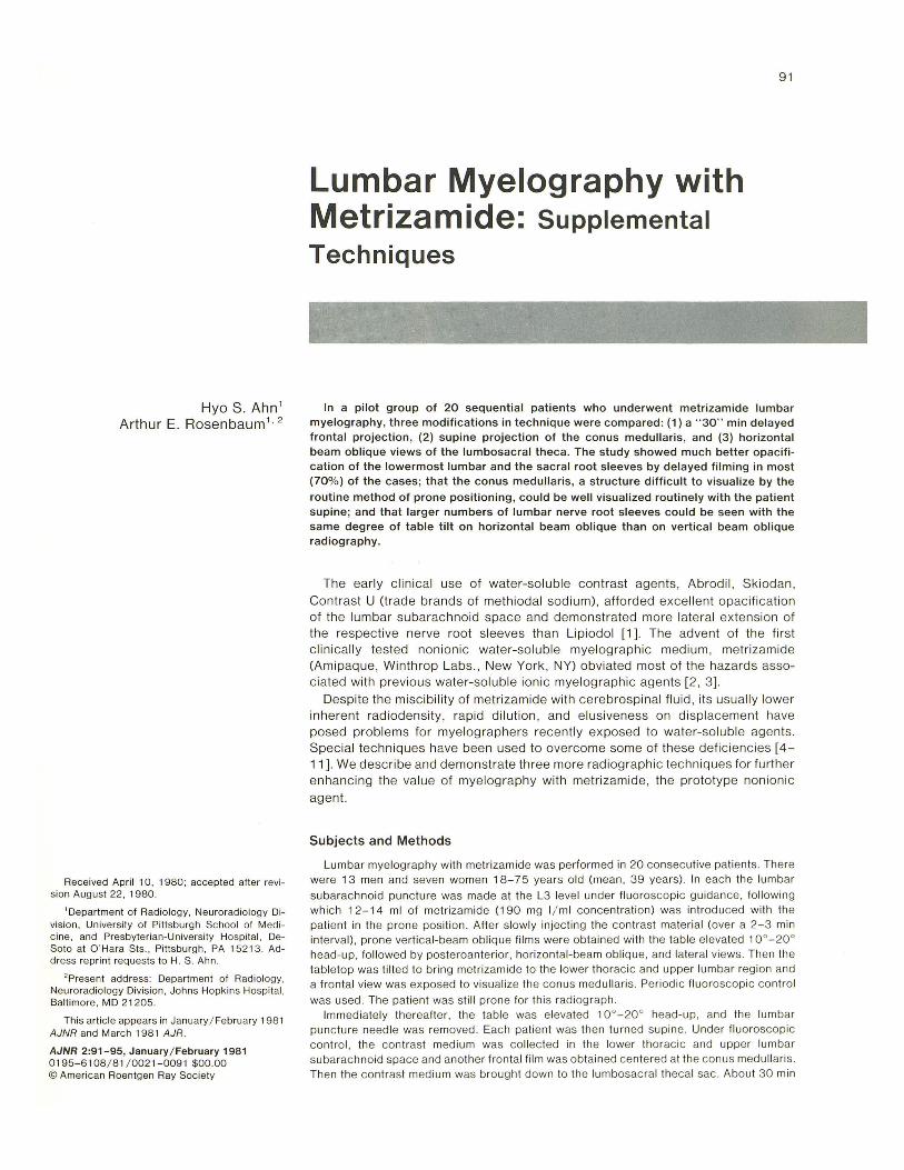
Fig. 1 .-A, Initial film after metrizamide insti llation. B, 30 min delayed film. Lower lumbar and sacral root sheaths of greater length and density (arrows). Density of contrast medium in thecal sac diminish8d .
TABLE 1: Changes in Length of Contrast-Filled Root Sleeves on Delayed Films
Length on Detayed Film (no ./% ) (n ~ 40) Nerve Roots
Increase Decrease No Change
L3 18 (45) 6 (15) 16 (40) L4 29 (73) 5 (12) 6 (15) L5 32 (80) 3 (8) 5 (12) 81 31 (77) 3 (8) 6 (15) 82 . . . . . . . . . . . 29 (73) 4 (10) 7 (17)
Total 139 (70) 21 (10) 40 (20)
after injection of metrizamide , the patient was turned prone and
ano ther posteroanterior view (delayed film) of the lumbosacral re
gion was obtained with the radiographic equ ipme nt and factors
described above.
Results
Delayed Filming
The initial and delayed frontal (posteroanterior) films were reviewed and assessed as to the degree of filling of the lumbar and sacral root sleeves. The length of the root sleeves was determined by measuring the distance from the
A B
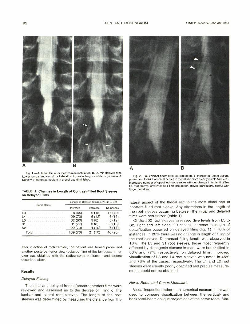
Fig . 2.-A, Vertical-beam oblique projection. B, Horizontal-beam oblique projection . Individual spinal nerves in thecal sac more clearly visible (arrows). Increased number of opacified root sleeves without change in table ti ll. (See L4 root sleeve, arrowheads.) This projection proved particu larly useful with large thecal sac.
lateral aspect of the thecal sac to the most distal part of contrast-filled root sleeve. Any alterations in the length of the root sleeves occurring between the initial and delayed films were scrutinized (table 1).
Of the 200 root sleeves assessed (five leve ls from L3 to S2 , right and left sides, 20 cases), increase in length of opacification occurred on delayed films (fig. 1) in 70% of instances. In 20% there was no change in length of fill ing of the root sleeves. Decreased filling length ' was observed in 10%. The L5 and S1 root sleeves, those most frequent ly affected by discogenic disease in man, were better filled in 80% and 77%, respectively, on delayed films . Improved visualization of L3 and L4 root sleeves was noted in 45% and 73% of the cases, respectively. The L 1 and L2 root sleeves were usually poorly opacified and precise measurements could not be obtained.
Nerve Roots and Conus Medul/aris
Visual inspection rather than numerical measurement was used to compare visualization between the vertical- and horizontal-beam oblique projections of the nerve roots . Sim-
AJNR:2, January / February 198 1 METRIZAMIDE LUMBAR MYELOGRAPHY 93
A B
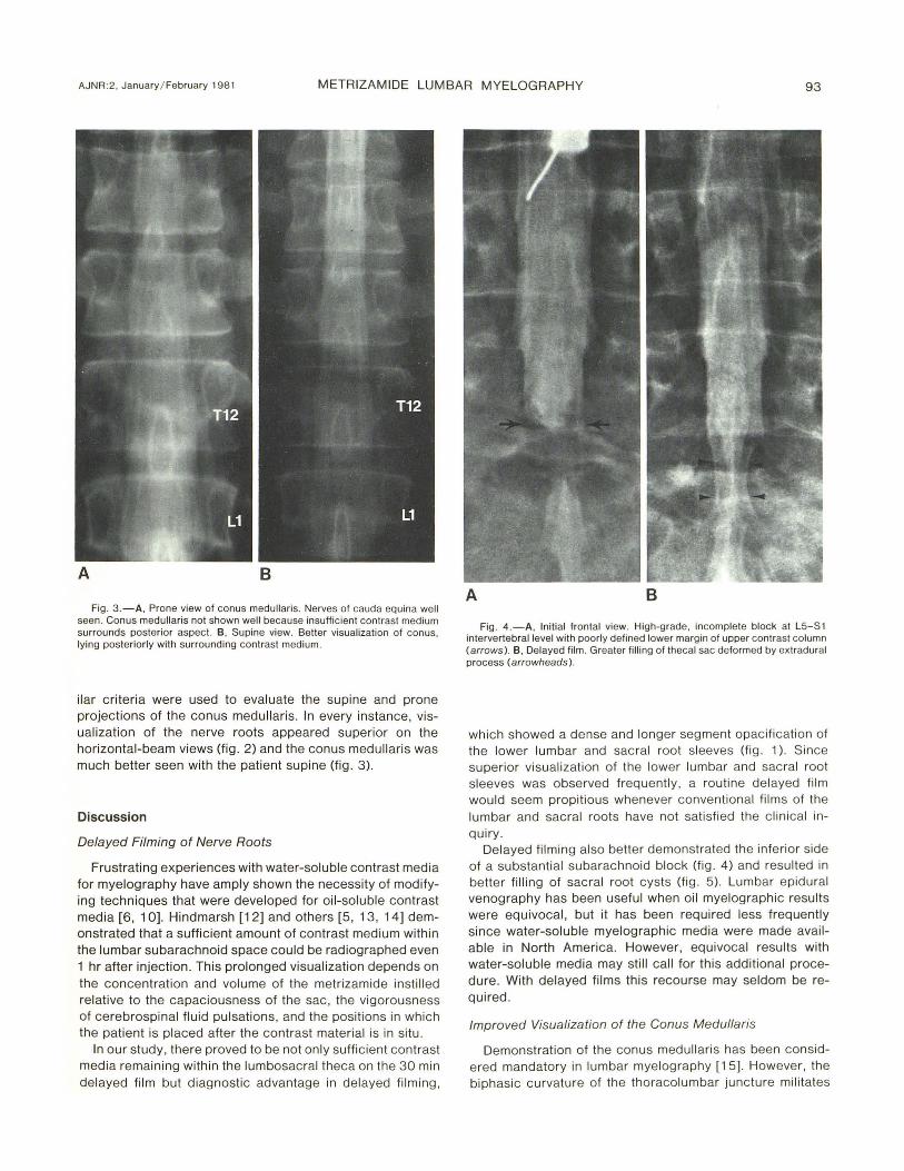
Fig. 3.-A, Prone view of conus medullaris. Nerves of cauda equina well seen. Conus medullaris not shown well because insufficient contrast medium surrounds posteri or aspect. B, Supine view. Better visualization of conus, lying posteriorly with surrounding contrast medium.
ilar criteria were used to evaluate the supine and prone projections of the conus medullaris. In every instance, visualization of the nerve roots appeared superior on the horizontal-beam views (fig . 2) and the conus medullaris was much better seen with the patient supine (fig. 3).
Discussion
Delayed Filming of Nerve Roots
Frustrating experiences with water-soluble contrast media for myelography have amply shown the necessity of modifying techniques that were developed for oil-soluble contrast media [6, 10]. Hindmarsh [12] and others [5, 13, 14] demonstrated that a sufficient amount of contrast medium within the lumbar subarachnoid space could be radiographed even 1 hr after injection. This prolonged visualization depends on the concentration and volume of the metrizamide instilled relative to the capaciousness of the sac, the vigorousness of cerebrospinal fluid pulsations, and the positions in which the patient is placed after the contrast material is in situ .
In our study, there proved to be not only sufficient contrast media remaining within the lumbosacral theca on the 30 min delayed film but diagnostic advantage in delayed filming ,
A B
Fig . 4 .-A, tnitial frontal view. High-grade, incomplete block at L5-S1 intervertebral level with poorly defined lower margin of upper contrast column (arrows ). B, Delayed film . Greater filling of thecal sac deformed by ext radural process (arrowheads).
which showed a dense and longer seg ment opacification of the lower lumbar and sacral root sleeves (fig . 1). Since superior visualization of the lower lumbar and sacral root sleeves was observed frequently, a routine delayed film would seem propitious whenever conventional film s of the lumbar and sacral roots have not sati sfied the c li nical inquiry.
Delayed filming also better demonstrated the inferior side of a substantial subarachnoid block (fig . 4) and resulted in better filling of sacral root cysts (fig . 5) . Lumbar epidural venography has been useful when oil myelographic results were equivocal, but it has been required less frequently since water-soluble myelographic media were made available in North America . However, equivocal results with water-soluble media may still call for this additional procedure . With delayed films this recourse may seldom be required .
Improved Visualization of the Conus Medullaris
Demonstration of the conus med ullaris has been considered mandatory in lumbar myelography [15]. However, the biphasic curvature of the thoracolumbar juncture mi litates

94 AHN AND ROSENBAUM AJNR:2, January / February 1981
A B
Fig . 5 .-Delayed fi ll ing of perin eural sacral root cysts. A , Ini tial film. B, 30 min delayed film. Cyst at left S2 level opaci fi ed on ly on delayed fi lm (arrows ). Marked increase in density o f another cyst at left S3 level. Another cyst ( arrowhead) con tains residual iophendylate from previous myelography.
against dependent pooling of the myelograph ic medium in the lower dorsal and upper lumbar region when the patient is in the prone position . Moreover, the conus medullaris usually lies in the dorsal aspect of the theca. These facts and the physical characte ri stics of a water-soluble contrast agent nullify the value of the prone posi tion for reg ular visualization of the conus region . However, as in iophendylate myelography, the conus is usually not seen in the absence of a block unless the patient is placed supine, with or without the needle in place (on a spec ial block) , or a high volume of iophendylate is used .
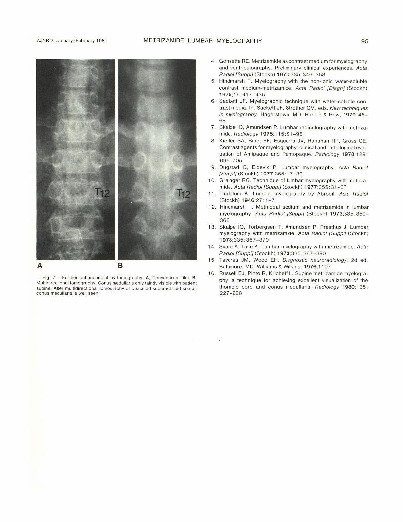
The use of fluoroscopy to ass ist accurate pooling of the metrizamide in the supine position resulted in very good visualization of the conus ' medullaris in each of our cases (figs. 3 and 6). Recently , Russell et al. [16] reported excellent visualization of the thoracic cord and conus medullaris in supine metrizamide myelography . Tomography can also be used to enhance visualization of the conus region (fig. 7).
Horizontal-Beam Oblique Projection of Lumbar Nerve Root
In North America, the lumbar nerve roots are usually radiographed by verti ca l-beam, fluoroscopi cal ly pos itioned
A B
Fig. 6. - Supine views o f conus medullari s in two cases. Excellent demonstration of lumbar enlargement. conus medullari s, and cauda equina.
spot films. Many investigators in Scandinavia [5,7,10] have stressed the value of horizontal-beam views of these lumbar nerve roots. Horizontal ob lique projections have become standard for the cervical nerve roots; the lumbar roots are also seen best by horizontal-beam oblique views.
In order to get a good horizontal-beam oblique view, the patient shou ld be in a more oblique position compared with the position needed for vertical oblique view. This steeper obliquity brings the root sleeves into dependent position , which may encourage better filling of root sleeves by gravity. Further, a larger number of root sleeves can be visualized on single film with the same degree of table tilt (fig . 2), thus reducing the frequency of extra films and minimizing table tilt ing . We believe it follows that the horizontal-beam technique should replace the vertical-beam technique for evaluation of herniated intervertebral discs .
REFERENCES
1. Arnell S, Lidstrom F. Myelog raphy with Skiodan (Abrodi l). Acta Radiol (Stock h) 1931 ;12: 287 - 289
2. Amundsen P. Myelog raphy and radiculography-development of new contrast media . Wis Med J 1977;76: 63-67
3. Metrizamide. Acta Radiol {Suppl] (Stock h) 1973;335: 1-390
AJNR :2, January / February 1981 METRIZAMIDE LUMBAR MYELOGRAPHY 9 5
A B
Fig. 7. -Further enhancement by tomog raphy. A , Conventional film. B , M ultidirec ti onal tomography. Conus medullaris only fa intly visible with patienl supine. After multidirectional tomography of opacified subarachnoid space, conus medullaris is well seen.
4 . Gonsette RE . Metri zamide as contrast medium for myelography and ventriculog raphy. Preliminary c linica l experiences. Acta Radiol [Suppl] (Stockh) 1973;335: 346-358
5. Hindmarsh T . Myelog raphy with the non-ionic water-soluble
contrast medium-metri zamide. Acta Radiol [Diagn] (Stockh) 1975;16:417-435
6 . Sackett JF. Myelographic technique with water-soluble contrast media. In: Sackett JF, Stroth er CM , eds. New techniques in myelography. Hagerstown, MD: Harper & Row, 1979 : 45-68
7. Skalpe 10, Amundsen P. Lumbar radiculography with metri zamide. Radiology 1975;115 :91-95
8. Kieffer SA, Binet EF, Esq uerra JV , Hantman RP , Gross CEo Contrast agents for myelog raphy: c linica l and rad iolog ica l evaluation of Amipaque and Pantopaque. Radio logy 1978;129: 695-705
9. Dugstad G , Eldevik P. Lumbar myelog raphy. Acta Radiol [Supp l] (Stockh) 1977;355: 1 7 - 30
10. Grainger RG . Technique of lumbar myelog raphy with metrizamide. Ac ta Radiol [Suppl] (Stockh) 1977;355: 3 1 -37
11 . Lindblom K. Lumbar myelog raphy by Abrod il. Acta Radiol (Stockh) 1946;27: 1-7
12. Hindmarsh T . Methiodal sodium and metrizamide in lumbar myelography. Acta Radiol [Suppl] (Stockh) 1973;335: 359-366
13. Skalpe 10, Torbergsen T, Amund sen P, Presthus J . Lumbar myelography with metrizamide. Acta Radiol [Suppl] (Stockh) 1973;335 : 367 - 3 79
14. Svare A, Talle K . Lumbar myelog raphy with metri zamide. Acta Radiol [Suppll (Stockh) 1973;335: 387 -390
15. Taveras JM , Wood EH . Diagnostic neuroradiology, 2d ed, Baltimore, MD: Williams & Wil k ins, 1976;11 07
16. Ru ssell EJ , Pinto R, Kricheff II. Supine metri zamide myelog ra
phy: a tech nique for achieving excellent visualization of th e thorac ic co rd and co nus medullari s. Radiology 1980; 135 : 227 - 228