the role of myelography in the management of patients with
TRANSCRIPT

(1)
ItTHE ROLE OF MYELOGRAPHY
IN
THE MANAGEMENT OF PATIENTS WITH SPINAL PATHOLOGY
AT
IfKENYATTA NATIONAL HOSPITAL
A DISSERTATION SUBMITTED IN PART FULFILMENT
FOR THE DEGREE OF MASTER OF MEDICINE IN DIAGNOSTIC RADIOLOGY
UNIVERSITY OF NAIROBI
July, 1989
Dr. Christopher J.
MEDICAL I IB R A R YUNIVERSITY OF NAIROBI
( i i )
DECLARATION
CANDIDATE: This Dissertation is my original work and has
not been presented for a Degree in any other University.
Dr. C.J. ARRUMM M.B. ChB. (Nairobi).
SUPERVISOR: This Dissertation has been submitted for examination
with my approval as the University Supervisor.
SIGNED:
Dr. M.W. WACHIRA M.B.ChB., M M e d , L e c turer,Department of Diagnostic Radiology, College of Health Sciences, University of Nairobi.
CONTENTS PAGE
TITLE & SUBMISSION (i)DECLARATION * (11)SUMMARY ............................................. 1INTRODUCTION ........................................ 2MATERIALS AND METHOD ............................... 5RESULTS ............................................. 9
(a) HISTOGRAM OF AGE & SEX DISTRIBUTION 10(b) DISTRIBUTION OF COMMON PRESENTING
SYMPTOMS 11(c) MYELOGRAPHIC PATTERN OF SPINAL PATHOLOGY 12(d) COMPARISON OF CLINICAL, MYELOGRAPHIC AND
OPERATIVE FINDINGS IN THE LUMBAR SPINE 13(e) COMPARISON OF CLINICAL, MYELOGRAPHIC AND
OPERATIVE FINDINGS IN THE THORACIC SPINE 14(f) COMPARISON OF CLINICAL, MYELOGRAPHIC AND
OPERATIVE FINDINGS IN THE CERVICAL SPINE 15(g) ILLUSTRATIONS ............................... 16
DISCUSSION ......................................... 21CONCLUSIONS ........................................ 50RECOMMENDATIONS .................................... 51REFERENCES ......................................... 53ACKNOWLEDGEMENTS ................................... 57
APPENDIX (1) Data Collection form
MEDICAL LIBRARYWNIVERS1IY OF NAIROBI

1
SUMMARY
This is a prospective study to try and determine the role of
myelography in the management of patients with spinal pathology
at the Kenyatta Rational Hospital/ (KNH) Nairobi. 60 patients
referred to the X-ray Department/ KNH between November 1987
and J a n u a r y 1989 have been included. The study has attempted
to correlate the clinical/ myelographic and surgical findings
in the patients.
The study has shown that patients in the age group 30-39 years
were referred more frequently for myelographic examination and
that the commonest spinal pathology requiring myelographic
examination is prolapsed intervertebral disc in the lumbar
spine.
Suggestions have been given at the end of the study as a guide
line for future myelographic examinations at KNH.
/ 2
2
INTRODUCTION ^
Diseases of the nervous system may limit themselves to the
spinal cord. A significant number of patients presenting with
clinical features of spinal pat’hology are seen at the Kenyatta
National Hospital/ Nairobi (KNH) - the national referral hospital.
Some of these patients require myelographic examination.
Low back pain syndromes with or without sciatica are some of
the commonest medical problems. They are responsible for a
significant number of working days lost due to ill health\
(BUIRSK G.). Other patients with spinal pathology may present
clinically with paraparesis, quadriparesis, paraplegia, etc.
The early stages of damage to nerve tissue are reversible and
some of the function may be regained. Severely damaged neurones
do not recover. It is, therefore, most important to diagnose and
treat spinal cord compression without delay. The role of the
radiologist in the management of these patients is primarily to
identify local abnormalities which will be amenable to surgical
treatment. The frequent requests for myelographic examination
of these patients prompted this study.
In Kenya the two non-invasive methods for investigating spinal
pathology (Computerised Tomography "CT" and Magnetic Resonance
Imaging "MRI") are not easily accessible to the patient. CT is
available in private medical institutions in Kenya. it is not
fully utilized because it is expensive to most patients and the
/ 3

3
Kenyan doctor community is also still poorly informed about its
capabilities. MRI is unavailable in Kenya.
At KNH myelography is the radiological examination of the spine1
after plain radiography. Myelography has evolved a lot since
the technique of negative contrast myelography was pioneered by
Dandy (in 1919 and 1925) and Jacobaeus in 1921. (WESTBERG G)
Positive contrast medium myelographic technique was introduced
in 1922 by Sicard and Forestier using Lipiodol. In 1944, Ramsey/
French and Strain introduced Pantopaque (Myodil) as an improved
contrast medium for positive contrast myelography. Due to
unsatisfactory results obtained by the above contrast media,
water soluble contrast myelographic technique was introduced.
The initial water soluble contrast media (Abrodil, Meglumine
Iothalamate (Conray 280), and Meglumine Iocarmate (Dimer X))
were all abandoned soon after introduction due to high neuro
toxicity and meningeal irritation. Amipaque (Metrizamide) was
introduced in 1974 as a watersoluble, non-ionic contrast medium
for myelography. It was found to be very unstable in solution
and has now been replaced by omnipaque (Iohexol) and Iopamiro
(Iopamidol).V ’i &’;( !
In this s t u d $ a l l the patients were examined using positive
contrast myelographic technique. The study is aimed at:-
- Determining the age and sex distribution of the patients.
- Determining their clinical presentation
- Corelating the patients' clinical findings with the plain
X-ray and myelographic findings
/ 4

4
Evaluating the subsequent management of the patients after
myelographic diagnosis has been made
Discussing the relative merits of myelography in the manage
ment of the patients and proposing a possible protocol that
could be used in the radiological investigation of patients
Uwith similar problems in future
*Yv1 ■ «'if,
■ ! *'•f'"’
1
• ’}
i'-pM
1••J

5
MATERIALS AND METHOD
Myelographic examination was done on 60 patients in this study
between October 1987 and January 1989. The patients were
referred to the X-ray Department from within KNH and other
hospitals in Kenya.
Prior to booking any patient for myelography the patient's X-ray
request form was scrutinized for details of the clinical present
ation, and the plain X-ray films reviewed for the presence of any
abnormality. In cases where there.was doubt as to whether the
examination would be useful in the management of the patient,
the clinical details of the patient were discussed with the
referring doctor prior to booking.
The referring clinicians were advised to admit the patient's on
the eve of the examination inorder tot-
la) Discuss the examination with the patients and allay their
anxiety.
(b) Ensure proper hydration of the patients so as to minimise
chances of adverse reactions to contrast media.
The patients were allowed a light breakfast on the morning
of the examination. Examinations were done in themornings to
minimize patient anxiety. No premedication was prescribed.
The radiographic equipment which was used in this study included
a standard radiography-fluoroscopy room consisting of:-
/ 6

6
(a) A tilting table able to tilt to 45° (Trendelenberg1s
position) and 90° (upright).
(b) Image intensification system.
The myelographic contrast medium was introduced in the sub
arachnoid space via lumbar puncture. Lumbar puncture was doneil
in the X-ray department/ with the patient in sitting position
or lateral decubitus position.
Lumbar puncture was done using disposable lumbar puncture
needle gauge 21 s.w.g. The disposable needles were preferred
because:-
(a) They were sharp and made approach easy.
(b) The risks of transmission of diseases such as hepatitis
and AIDS were reduced.
The puncture was considered successful if the CSF flow obtained
was an uninterrupted flow. The contrast medium was then
introduced. 58 patients out of the total of 60 patients/ were
examined using non-ionic water soluble contrast media (I0HEX0L
or IOPAMIDOL). The remaining 2 patients were examined using
myodil which had been injected by referring clinicians unaware
of the availability of the water soluble contrast media.\
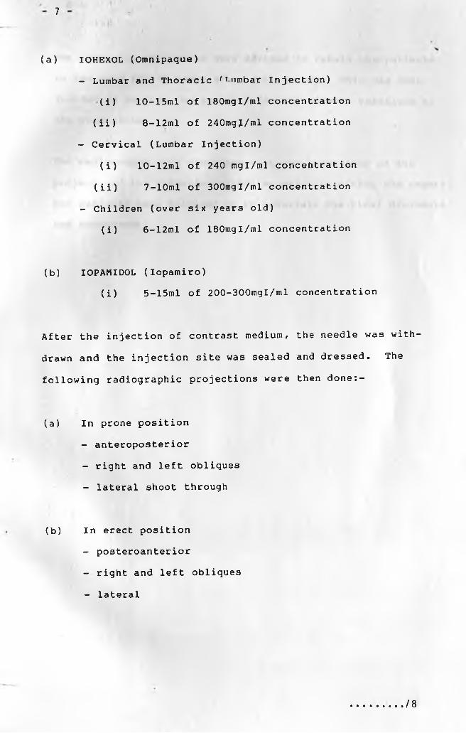
The majority of the patients in this study were adults and the
doses of contrast medium used were:-
medical librarytintvFRSITY OF NAIROBI'
/ 7
7
(a) IOHEXOL (Omnipaque)
- Lumbar and Thoracic l u m b a r Injection)
(i) 10-15ml of 180mgI/ml concentration
(ii) 8-12ml of 240mgJ/ml concentration
- Cervical (Lumbar Injection)
(i) 10-12ml of 240 mgl/ml concehtration
(ii) 7-10ml of 300mgl/ml concentration
- Children (over six years old)
(i) 6-12ml of 180mgI/ml concentration
(b) IOPAMIDOL (Iopamiro)
(i) 5-15ml of 200-300mgl/ml concentration
After the injection of contrast medium/ the needle was with
drawn and the injection site was sealed and dressed. The
following radiographic projections were then done:-
(a) In prone position
- anteroposterior
- right and left obliques
- lateral shoot through
(b) In erect position
- posteroanterior
- right and left obliques
- lateral
/ 8
8
The referring clinicians were advised to retain the patients
in the ward for 24 hours after myelography. This was done
inorder to monitor the patients for any adverse reactions to
the examination.
The radiographs were discussed with the supervisor of the
project and the referring clinician prior to giving the report.
The patients were followed up to ascertain the final diagnosis
and management.
/ 9
RESULTS
The results obtained in this study are presented in the form
of the following tables 1 - 6 .
- 9 -
/10


11
TABLE 2
DISTRIBUTION OF COMMON PRESENTING SYMPTOMS
SYMPTOM NO OF PATIENTS
Low Back Ache 38
Sciatica 22
Paraparesis 20
Paraplegia 3
Quadriplegia 1
Root pain in upper limbs 6
Sphinteric Incontinence 4
NB: (Some patients presented with more than
one symptom)
/12
12
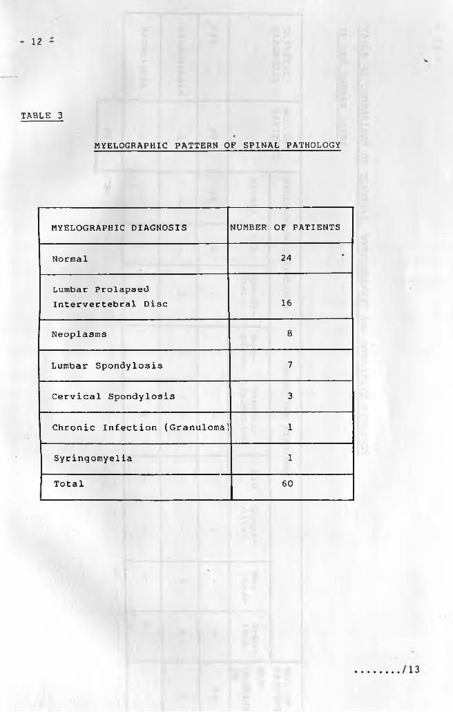
TABLE 3
MYELOGRAPHIC PATTERN OF SPINAL PATHOLOGY
MYELOGRAPHIC DIAGNOSIS NUMBER OF PATIENTS
Normal 24
Lumbar Prolapsed Intervertebral Disc 16
Neoplasms 8
Lumbar Spondylosis 7
Cervical Spondylosis 3
Chronic Infection (Granuloma' 1
Syringomyelia 1
Total 60

13
TABLE 4: COMPARISON OF CLINICAL, MYELOGRAPHIC AND OPERATIVE FINDINGS
IN THE LUMBAR SPINE
CLINICALDIAGNOSIS
NO. OF PATIENTS
MYELOGRAPHIC FINDINGS OPERATIVE FINDINGS NO. OF PATIENTS
NOTOPERATEDON
Normal PID Spondylosis
Neoplasm
ChronicInfection
PID Congenital
Neoplasm
Gran-loma
PID 39 16 16 7 - - 6 1 - - 32
Paraparesis 3 - - 3 - - - - 2 - 1
Paraplegia 1 - - - - 1 - - - 1 -
43
J

14
BLE 5: COMPARISON OF CLINICAL, MYELOGRAPHIC AND
ERATIVE FINDINGS IN THE THORACIC SPINE
INICALAGNOSIS
NO. OF PATIENTS
MYELOGRAPHIC FINDINGS OPERATIVEFINDINGS
NO.PATIENTSNOTOPERATEDONNormal Neoplasm Neoplasm Other
Findings
raparesis 8 6 2 1 - 7
raplegia 3 1 2 - - 3
11
/15
15
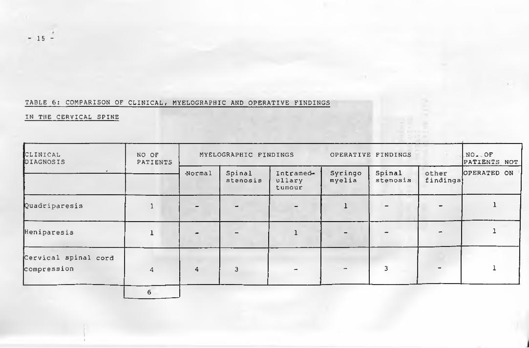
TABLE 6: COMPARISON OF CLINICAL/ MYELOGRAPHIC AND OPERATIVE FINDINGS
IN THE CERVICAL SPINE
CLINICALDIAGNOSIS
NO OF PATIENTS
MYELOGRAPHIC FINDINGS OPERATIVE FINDINGS NO ».OFPATIENTS NOT
• -Normal Spinal stenos i s
Intramedullarytumour
Syringo myeli a
Spinalstenosis
OPERATED ONfindings
Quadriparesis 1 - - - 1 - - 1
Heniparesis 1 - - 1 - - - 1
Cervical spinal cord
compression 4 4 3 - - 3 - 1
6
Ii

FIGURE 1
DEMONSTRATION OF A NORMAL LUMBAR RADICULOGRAM USING WATER-SOLUBLE CONTRAST MEDIUM - ANTEROPOSTERIOR VIEW

17
FIGURE 2
LUMBAR RADICULOGRAM ANTERO-POSTERIOR VIEW DEMONSTRATING A PROLAPSED INTERVERTEBRAL DISC AT THE DISC SPACE L4/L5
18
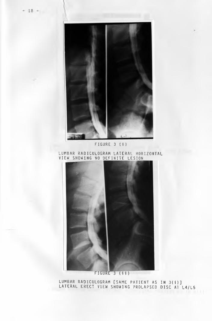
FIGURE 3 (i )
LUMBAR RADICULOGRAM LATERAL HORIZONTAL VIEW SHOWING NO DEFINITE LESION
LUMBAR RADICULOGRAM [SAME PATIENT AS IN 3(i)] LATERAL ERECT VIEW SHOWING PROLAPSED DISC AT L4/L5
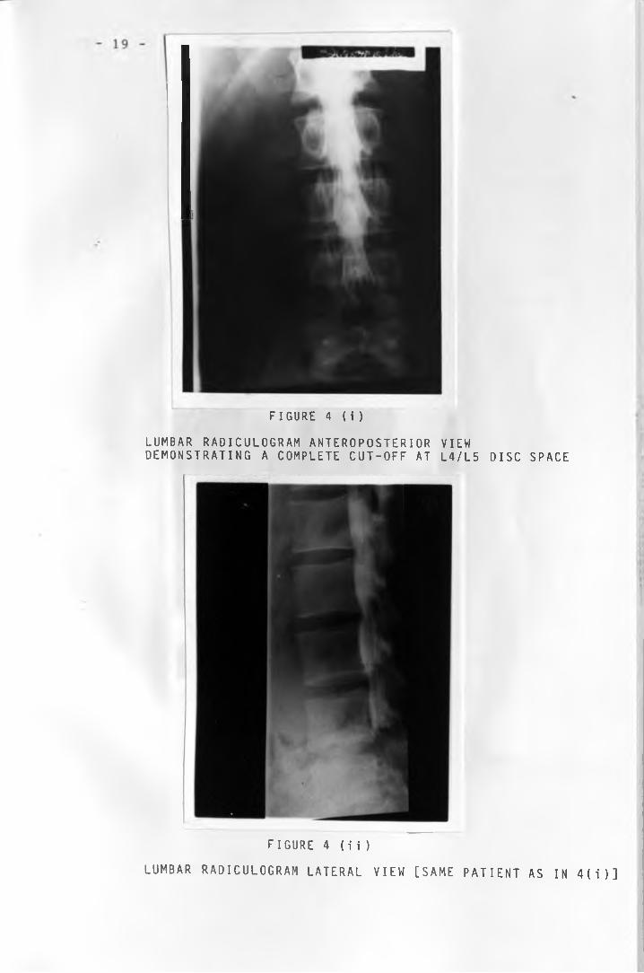
FIGURE 4 (i)
LUMBAR RADICULOGRAM ANTEROPOSTERIOR VIEW DEMONSTRATING A COMPLETE CUT-OFF AT L4/L5 DISC SPACE
FIGURE 4 (ii)
LUMBAR RADICULOGRAM LATERAL VIEW [SAME PATIENT AS IN 4(i)]
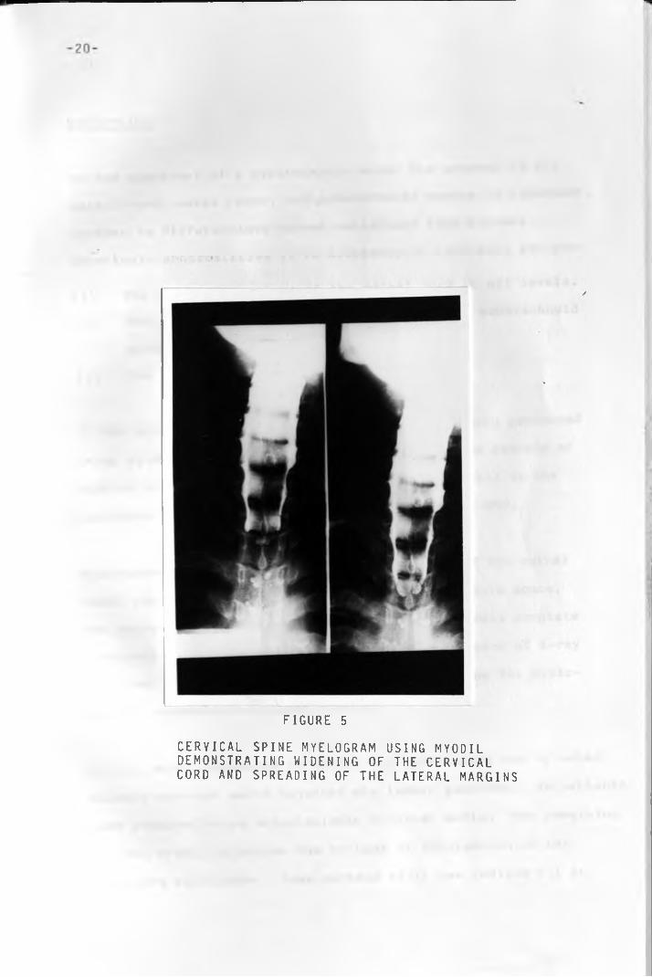
FIGURE 5
CERVICAL SPINE MYELOGRAM USING MYODIL DEMONSTRATING WIDENING OF THE CERVICAL CORD AND SPREADING OF THE LATERAL MARGINS

21
DISCUSSION
In the appraisal of a myelographic study the anatomy of the
spinal cord, nerve roots, and .subarachnoid spaces is important.
Inorder to differentiate normal variations from minimal
pathologic abnormalities it is necessary to carefully analyse:
(a) The normal dimensions of the spinal cord at all levels.
(b) The variations in the configuration of the subarachnoid
s p a c e .
(c) The size and integrity of the bony canal.
AT KNH prior to 1987 myelographic examinations were performed
using myodil as the contrast medium. No previous records or
studies are, however, available to indicate details of the
incidence and pattern of spinal cord diseases at KNH.
Myelography is the radiological visualization of the spinal
cord, cauda equina and the surrounding subarachnoid space.
The technique was originally applied to demonstrate complete
obstructions in the spinal theca. With improvement of X-ray
machines and contrast media, the demand and scope for myelo
graphic examinations became greater.
In this study themethod of choice for myelography was by water-
soluble contrast media injected via lumbar puncture. 58 patients
were examined using water-soluble contrast media. The remaining
two had myodil injection due to lack of information of the
referring clinicians. Some workers still use iodized oil in
22
patients whose clinical level of involvement is uncertain or
in whom involvement of several levels is expected. These
workers argue that since these groups of patients may require
detailed myelography of the entire spine, it may be difficult
to examine the patients with water soluble contrast medium.
This fear has been overcome, however, with more practice and
faster examination methods using water soluble contrast media.
The newer water-soluble agents have completely replaced myodil
for all cauda equina, lumbosacral radiculography, and lumbar
myelography. Water soluble media are the contrast media of
choice for most dorsal myelography and for cervical myelogram.
[WHITEHOUSE G.H. & WORTHINGTON B.S.]
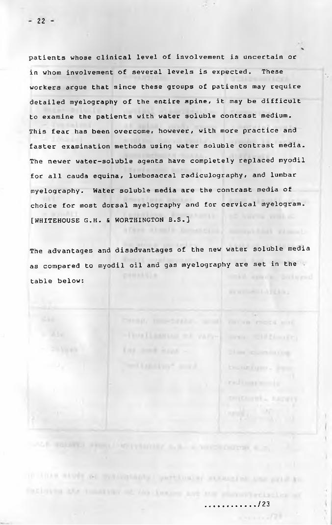
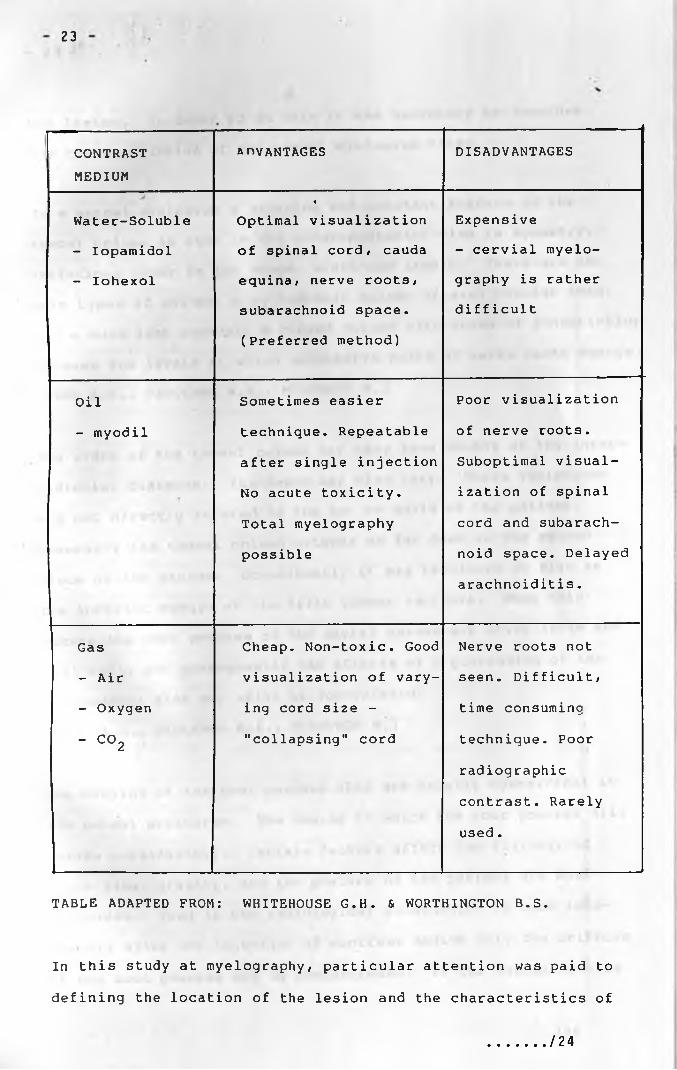
The advantages and disadvantages of the new water soluble media
as compared to myodil oil and gas myelography are set in the
table below:
/ 23
23
I CONTRAST
MEDIUM
anvANTAGES DISADVANTAGES
Water-Soluble4
Optimal visualization Expensive
- Iopamidol of spinal cord, cauda - cervial myelo-
- Iohexol equina/ nerve roots/ graphy is rather
subarachnoid space.
(Preferred method)
diff icult
Oil Sometimes easier Poor visualization
- myodil technique. Repeatable of nerve roots.
after single injection Suboptimal visual-
No acute toxicity. ization of spinal
Total myelography cord and subarach-
possible noid space. Delayed
arachnoiditis.
Gas Cheap. Non-toxic. Good Nerve roots not
- Air visualization of vary- seen. Difficult/
- Oxygen ing cord size - time consuming
- C ° 2 "collapsing" cord technique. Poor
radiographic
contrast. Rarely
u s e d .
TABLE ADAPTED FROM: WHITEHOUSE G.H. & WORTHINGTON B.S.
In this study at myelography/ particular attention was paid to
defining the location of the lesion and the characteristics of
/ 24
24 -
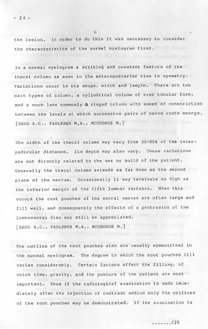
the lesion. In order to do this it was necessary to consider
the characteristics of the normal myelogram first.
In a normal myelogram a striking and constant feature of the
thecal column as seen in the anteroposterior view is symmetry.
Variations occur in its shape/ width and length. There are two
main types of column/ a cylindrical column of even tubular form/
and a much less commonly GL ringed column with zones of constriction
between the levels at which successive pairs of nerve roots emerge.
[BEGG A .C . / FAULKNER M.A., MCGEORGE M.]
The width of the thecal column may vary from 30-80% of the inter-
pedicular distance. Its depth may also vary. These variations
are not directly related to the sex or build of the patient.
Generally the thecal column extends as far down as the second
piece of the sacrum. Occasionally it may terminate as high as
the inferior margin of the fifth lumbar vertebra. When this
occurs the root pouches of the sacral nerves are often large and
fill well, and consequently the effects of a protrusion of the
lumbosacral disc may still be appreciated.
[BEGG A.C., FAULKNER M.A., MCGEORGE M.]
The outline of the root pouches also are usually symmetrical in
the normal myelogram. The degree to which the root pouches fill
varies considerably. Certain factors affect the filling, of
which time, gravity, and the posture of the patient are most
important. Thus if the radiological examination is made imme
diately after the injection of contrast medium only the orifices
of the root pouches may be demonstrated. If the examination is
/ 2 5
25
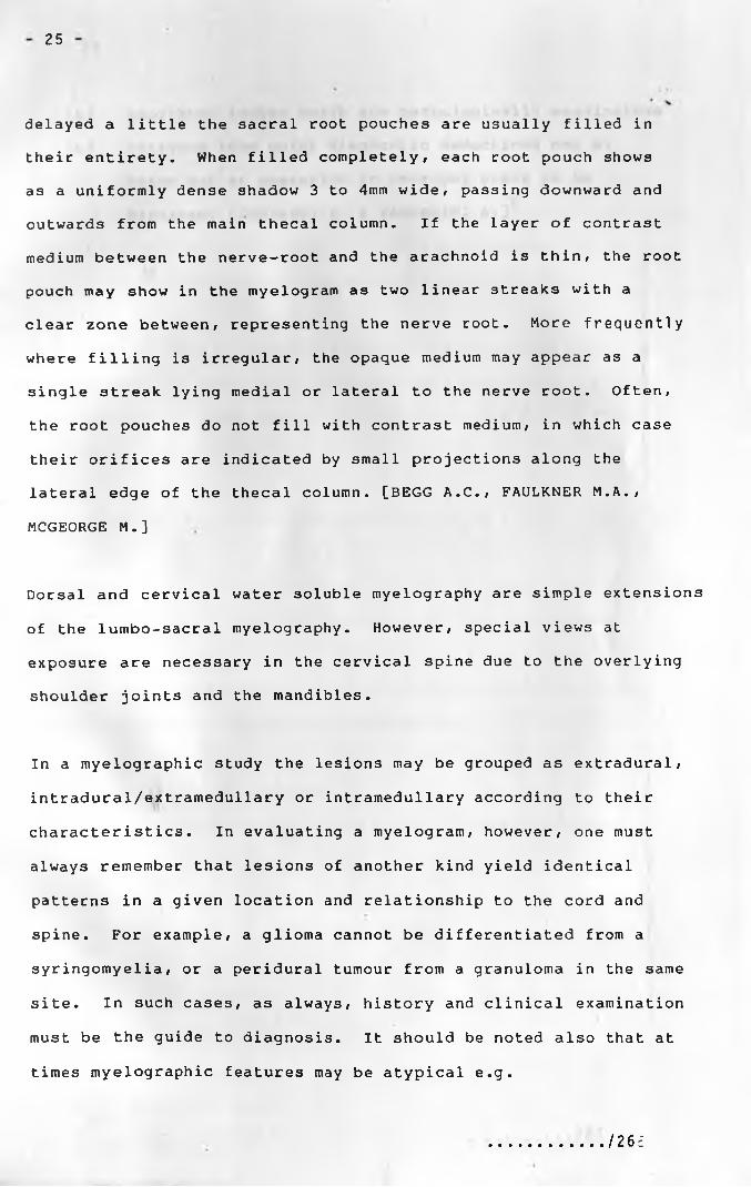
delayed a little the sacral root pouches are usually filled in
their entirety. When filled completely/ each root pouch shows
as a uniformly dense shadow 3 to 4mm wide/ passing downward and
outwards from the main thecal column. If the layer of contrast
medium between the nerve— root and the arachnoid is thin/ the root
pouch may show in the myelogram as two linear streaks with a
clear zone between/ representing the nerve root. More frequently
where filling is irregular/ the opaque medium may appear as a
single streak lying medial or lateral to the nerve root. Often#
the root pouches do not fill with contrast medium# in which case
their orifices are indicated by small projections along the
lateral edge of the thecal column. [BEGG A.C.# FAULKNER M.A.#
MCGEORGE M.]
Dorsal and cervical water soluble myelography are simple extensions
of the lumbo-sacral myelography. However# special views at
exposure are necessary in the cervical spine due to the overlying
shoulder joints and the mandibles.
In a myelographic study the lesions may be grouped as extradural#
intradural/extramedullary or intramedullary according to their
characteristics. In evaluating a myelogram# however# one must
always remember that lesions of another kind yield identical
patterns in a given location and relationship to the cord and
spine. For example, a glioma cannot be differentiated from a
syringomyelia# or a peridural tumour from a granuloma in the same
site. In such cases# as always# history and clinical examination
must be the guide to diagnosis. It should be noted also that at
times myelographic features may be atypical e.g.
..................../ 2 6 £

amorphous images which are pathologically meaningl
patterns from which diagnostic deductions may be
drawn but at operation or necropsy prove to be
1mistaken. [LOMBARDI G. & PASSERINI A.]
\
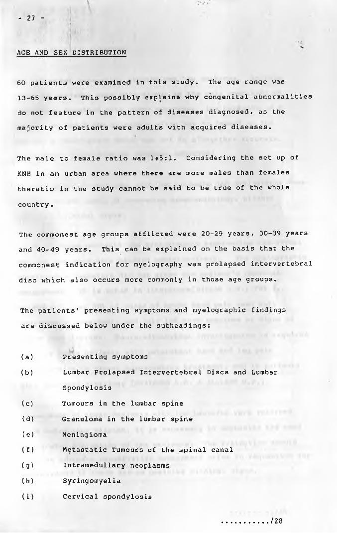
AGE AND SEX DISTRIBUTION
60 patients were examined in this study. The age range was
13-65 years. This possibly explains why congenital abnormalities
do not feature in the pattern of diseases diagnosed/ as the
majority of patients were adults with acquired diseases.
The male to female ratio was 1*5:1. Considering the set up of
KNH in an urban area where there are more males than females
theratio in the study cannot be said to be true of the whole
country.
The commonest age groups afflicted were 20-29 years < 30-39 years
and 40-49 years. This can be explained on the basis that the
commonest indication for myelography was prolapsed intervertebral
disc which also occurs more commonly in those age groups.
The patients' presenting symptoms and myelographic findings
are discussed below under the subheadings:
(a) Presenting symptoms
(b) Lumbar Prolapsed Intervertebral Discs and Lumbar
Spondylosis
(c) Tumours in the lumbar spine
(d) Granuloma in the lumbar spine
(e) Meningioma
(f) Metastatic Tumours of the spinal canal
(g) Intramedullary neoplasms
(h) Syringomyelia
(i) Cervical spondylosis
- 27 -
/ 28
28
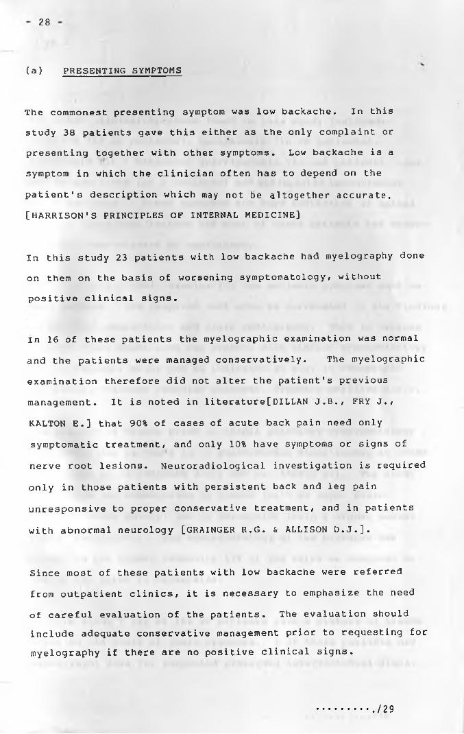
(a) PRESENTING SYMPTOMS
The commonest presenting symptom was low backache. In this
study 38 patients gave this either as the only complaint or
presenting together with other symptoms. Low backache is a
symptom in which the clinician often has to depend on the
patient's description which may not be altogether accurate.
[ H A R R I S O N ’S PRINCIPLES OF INTERNAL MEDICINE]
In this study 23 patients with low backache had myelography done
on them on the basis of worsening symptomatology, without
positive clinical signs.
In 16 of these patients the myelographic examination was normal
and the patients were managed conservatively. The myelographic
examination therefore did not alter the patient's previous
management. It is noted in literature[DILLAN J.B., FRY J.,
KALTON E.] that 90% of cases of acute back pain need only
symptomatic treatment, and only 10% have symptoms or signs of
nerve root lesions. Neuroradiological investigation is required
only in those patients with persistent back and leg pain
unresponsive to proper conservative treatment, and in patients
with abnormal neurology [GRAINGER R.G. & ALLISON D.J.].
Since most of these patients with low backache were referred
from outpatient clinics, it is necessary to emphasize the need
of careful evaluation of the patients. The evaluation should
include adequate conservative management prior to requesting for
myelography if there are no positive clinical signs.
. / 29
29
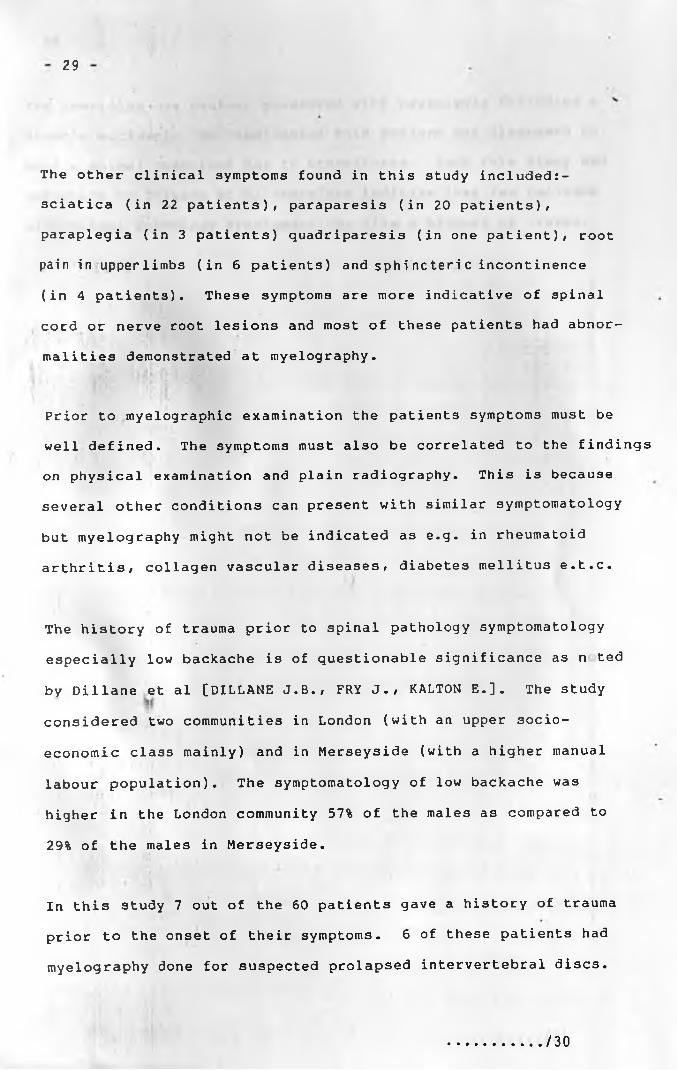
The other clinical symptoms found in this study included:-
sciatica (in 22 patients), paraparesis (in 20 patients),
paraplegia (in 3 patients) quadriparesis (in one patient), root
pain in upperlimbs (in 6 patients) and sphincteric incontinence
(in 4 patients). These symptoms are more indicative of spinal
cord or nerve root lesions and most of these patients had abnor
malities demonstrated at myelography.
Prior to myelographic examination the patients symptoms must be
well defined. The symptoms must also be correlated to the findings
on physical examination and plain radiography. This is because
several other conditions can present with similar symptomatology
but myelography might not be indicated as e.g. in rheumatoid
arthritis, collagen vascular diseases, diabetes mellitus e.t.c.
The history of trauma prior to spinal pathology symptomatology
especially low backache is of questionable significance as n ted
by Dillane et al [DILLANE J.B., FRY J., KALTON E.]. The study
considered two communities in London (with an upper socio
economic class mainly) and in Merseyside (with a higher manual
labour population). The symptomatology of low backache was
higher in the London community 57% of the males as compared to
29% of the males in Merseyside.
In this study 7 out of the 60 patients gave a history of trauma
prior to the onset of their symptoms. 6 of these patients had
myelography done for suspected prolapsed intervertebral discs.
/ 3 0

- 30 -
The remaining one patient presented with paraplegia following a
bicycle accident. On examination this patient was diagnosed to
have a spinal granuloma due to brucellosis. Both this study and
the study by Dillane et al therefore indicate that few patients
with spinal pathology symptomatology give a history of trauma.
/ 31

31
•(b) PROLAPSED INTERVERTEBRAL DISCS AND LUMBAR SPONDYLOSIS
The intervertebral discs lie be.tween each two vertebral
bodies from cervical vertebral body 2 to sacral vertebra 1.
It is composed of fibrocarti1 age, with a gelatinous central
matrix known as the nucleus pulposus. It is firmly
attached to adjoining vertebral bodies.
Degeneration of the intervertebral disc complex begi ns
early in life. It is a consequence of a variety of enviroi-
mental factors as well as normal aging. [Modic M.T. and
associates]
The sequelae of disc degeneration remains among the leading
causes of functional incapacitation in both sexes and are
an al1-too-common source of chronic disability in the
working years. [Modic M.T. et al] The diagnosis and proper
location of the affected disc(s) is important since some of
these patients can be effectively managed surgically.
Disc prolapse is the extrusion of the nucleus pulposus into
the spinal canal through a laceration of aunulus fibrosus.
In this study 39 patients were referred for lumbar myelography
with clinical diagnosis of prolapsed intervertebral disc.
Myelography demonstrated prolapsed intervertebral disc in
16 patients and lumbar spondylosis in 7 patients. Lombardi
and Passerini in a retrospective study of 701 patients with dege
nerative lumbar disease showed that the patients with
prolapsed intervertebral disc were 669 and those with lumbar
/ 32

32
spondylosis were 32. [Lombardi G. & Passerini A.]'
The results first show that clinically there are more
patients with lumbar spinal pathology requiring myelo
graphy as compared to the othe*regions of the spine.
Secondly the results show that prolapsed intervertebral disc
is the commonest lumbar pathology requiring myelography.
In this study the majority of patients with myelographic
diagnosis of prolapsed intervertebral disc either had a
posterocentral or posterolateral disc prolapse. The most
frequent site of prolapse was the L4/L5 level which is also
indicated by Holman C.B. In literature it is noted that most
disc prolapses occur backwards.[Duckworth]
The posterior ligament in the midline tends to direct the
material posterolateral1y where it may press on the nerve
root in the foramen, and cause root symptoms and signs.
Rarely a true central posterior prolapse may occur causing
pressure on the spinal cord or central roots of the cauda
equina. In these patients severe paralysis may occur, with; * ’ rub
disturbed bladder function. [Duckworth T.] Disc prolapse can• i y l * * -Cl 1 ,
occur through the endplate into the vertebral body. This may
produce acute low back pain and eventually a translucent• \ - ; •; >area close to the disc on X-ray - a so called "Schmorl's
Rode." Disc prolapse may occasionally be through the anterior
portion of the annulus fibrosus.[Hoi man C.B.]
/ 3 3

33
In this study the age range of patients with myelographic
diagnosis of prolapsed intervertebral disc was 19 years -4
57 years. The average age was 36.7 years. In literature it
is noted that intervertebral disc prolapse tends to occur in
the earlier age groups - usually between 30 - 45 years.
[Duckworth T.] Prolapsed intervertebral disc also occurs
outside these age limits not infrequently.
Myelography on patients with suspected prolapsed interver
tebral disc was done both in the prone and erect positions.
This is due to the observation that in the erect position
compression of the disc will accentuate the herniation, and
on the radiograph exposed in this position an anterior
thecal deformity will be seen. [Pilling J.R.]
Only 7 out of the patients with myelographic diagnosis of
prolapsed intervertebral disc had surgery. The majority
being managed conservatively. It is, therefore, not possible
in this study to correlate fully the myelographic and
operative findings. However, of the 7 patients who were
operated on - prolapsed intervertebral disc was confirmed in
6 patients. The other patient had a congenital abnormality
of the nerve roots [Figure^ ] Pau et al describes similar5«*
findings of spoiled and elongated nerve roots in patients with
cauda equina syndrome. [Pau A. Sehrbundt Viale E., Turtas S.,
and Viale G.L.]
/ 34

- 34
Cook P.L. and Wise K. in their study and review of
literature indicate that the accuracy of water soluble
contrast media in diagnosing prolapsed intervertebral disc
as correlated with operative findings is 90% or over.
[Cook P.L and Wise K.]
The meaning of lumbar spondylosis is not precise. It is
variably referred to by different authors as arthrosis,
osteoarthritis, osteophytosis, spondy1ochondrosis, spondy
losis deformans-, hypertrophic spondylitis, etc. There is also
confusion as to the classification, the way it is formed and
the way it acts. The fundamental features of the disease are
narrowing of one or more intervertebral discs, formation of
osteophytes and protrussion of the discs. [Lombardi G. and3Passeri ni A . ]
Multiple factors are thought to participate in the etiology
of disc degeneration including autoimmune, genetic, reabsorp
tion and biochemical. [Modic M.T. et al] Initially narrowing
of the disc, osteophytosis and protrusion may appear as
isolated, circumscribed lesions. As time goes on, the rate
of advance varying with the subject, they tend to spread to4
the whole spine. [Lombardi G. & Passerini A. ]n In this study
the age range of patients with lumbar spondylosis was
38-60 years. The average age was 52 years. Lombardi's study
showed an average age of 49 years [Lombardi G. & Passerini a .
M t ,HC \i 1 IBft ARY UNIVERSITY OF NAIROBI
/ 35

- 35
Degenerative arthritic changes in the lumbar region offer
difficulty in interpretation because the posterior
hypertrophic ridging of the margins of the vertebral
bodies indent the anterior portions of the thecal sac, at
the involved interspaces. These cause extradural deformity
similar to that produced by herniated disc. Usually the
differentiation can be made on the basis of the bilaterality
of the indentation as seen on the anteroposterior projection.
The appearance is usually that of an hour glass deformity,
with the narrow waist opposite the interspace as a conse
quence of the ridge having displaced the sac posteriorly
[Holman C.B . ]
In this study all the patients diagnosed as having lumbar
spondylosis were managed conservatively.
j
V
/ 36
36
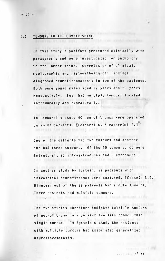
(c) TUMOURS IN THE LUMBAR SPINE
In this study 3 patients presented clinically with
paraparesis and were investigated for pathology
in the lumbar spine. Correlation of clinical,
myelographic and histopathological findings
diagnosed neurofibromatosis in two of the patients.
Both were young males aged 22 years and 25 years
respectively. Both had multiple tumours located
intradurally and extradural 1y .« .
In Lombardi's study 90 neurofibromas were operatedQ
on in 87 patients. [Lombardi G. & Passerini A.]
One of the patients had two tumours and another
one had three tumours. Of the 90 tumours, 60 were
intradural, 25 intraextradural and 5 extradural.
In another study by Epstein, 22 patients with
intraspinal neurofibromas were analysed. [Epstein B.S.]
Nineteen out of the 22 patients had single tumours.
Three patients had multiple tumours.
The two studies therefore indicate multiple tumours
of neurofibroma in a patient are less common than✓
single tumour. In Epstein's study the patients
with multiple tumours had associated generalized
neurofi bromatos is.
/ 37

37
Lombardi's study notes that neurofibromas may
develop at any age. The fifth and sixth decades
of life were the per.iods of highest frequency.
The study also showed a predominance of males
to females. It also found that the tumours were
diffusely distributed along the whole length of
the spinal column and showed no preference forg
any site. [Lombardi G. & Passerini A.]
In one of the two cases of neurofibromatosis in <his
study a dumb-bell tumour was demonstrated in one
patient. Dumb-bell tumours develop asymmetrically.
The intraspinal part may be relatively small
whereas the extraspinal part may grow to a con
siderable size. In Lombardi's study dumb-bell
tumours were found to be practically associated
with bone changes in plain X-rays e.g. widening
of intervertebral foramen. The neurological
signs may not be in proportion to the bone changes.
As a rule, this type of tumour is reported to be
limited to one vertebra. [Lombardi G. & Passerini A.]
However, two tumours may reach a large enough size
that they erode several pedicles and cause excavation
of the posterior surfaces of several vertebral bodies.
Neurofibromas may also be found at operation to
contain calcification that may not be seen on X-ray.
1 3 9

3% -
Neurofibromas are encapsulated tumours, often cystic
and usually do not make as deep a depression in
the spinal cord as do meningiomas. They are
usually attached to the posterior nerve root. They
rarely adhere to the dura. At myelography they
usually cause definitive arrest in most patients.
[Lombardi G. & Passerini A.]6
(d) GRANULOMA IN THE LUMBAR SPINE
One case of granuloma in the lumbar region was
found in this study. The patient was a male, 37
years of age. He presented at KNH with a history
of gradual difficulty in walking and weakness in
the lower limbs. The symptoms started about two
months after a bicycle accident.
At the time of admission to KNH examination showed
that the patient was paraplegic. His sensory level
was T12. The clinical diagnosis made was of para
plegia due to cord compression.
The patient's plain X-rays of the lumbar spine showed
collapsed vertebrae LI and L2. There was also a
paravertebral mass seen in the region of the L1/L2.
Myelography in this patient demonstrated a complete
block at the level of L2 vertebral body. At laminec
tomy a space occupying lesion was found extramedul1 ary
.........1 3^
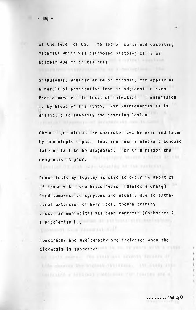
- -
at the level of L2. The lesion contained caseating
material which was diagnosed histologically as
abscess due to brucellosis.
Granulomas, whether acute or chronic, may appear as
a result of propagation from an adjacent or even
from a more remote focus of infection. Transmission
is by blood or the lymph. Not infrequently it is
difficult to identify the starting lesion.
Chronic granulomas are characterized by pain and later
by neurologic signs. They are nearly always diagnosed
late or fail to be diagnosed. For this reason the
prognosis is poor.
Brucellosis myelopathy is said to occur in about 2%
of those with bone brucellosis. [Ganado & Craig]
Cord compressive symptoms are usually due to extra
dural extension of bony foci, though primary
brucellar meningitis has been reported [Cockshott P.
& Middlemiss H.]
Tomography and myelography are indicated when the
diagnosis is suspected.
/ » 4-0

MENINGIOMA
One patient in this s'tudy had a spinal neoplasm
determined histologically as a meningioma. The
patient was a female aged 43 years. She presented
at KNH with worsening symptoms of pain and weakness
in both lower limbs over several months. Physical
examination showed that she had spastic paralysis in
the lower limbs and a sensory level at T7/T8. A
clinical diagnosis of paraparesis due to cord
compression was made.
The patient's plain X-rays of the thoracic and lumbar
spine were normal. Myelography showed a block a*; the
level of T3 with side tracking of the contrast
medium. At surgery an intradural/extramedul1 ary
tumour was found. Histology showed the tumour to be
a calcified meningioma.
Meningiomas are tumours of adult life. Lombardi
and Passeriui studied 82 patients with meningioma.
[Lombardi G. & Passerini A.]6
The average age was found to be 49 years with a range
of 13-73 years. The sixth and seventh decades of
life showing the highest incidence. The study also
indicated a distinct preference for females and a

41
more pronounced preference for the thoracic portion
of the spi nal canal .
Epstein studied 26 patients with meningiomas in the
spine [Epstein B.S.]
Twenty-one of the patients were over 50 years old.
The incidence in women exceeded that in men by 5:1.
By far most meningiomas are situated intradurally
and are extramedullary. Occasionally extradural or
an extra- and intradural meningioma is found [Brown
M.H.]
In the single case in this study the meningioma was
found to extend from T2/T3 disc level to lower margin
of T3. Lombardi notes that meningiomas are not asn.
a rule very 1arge.[Lombardi G. and Passerini A ] 1
In his study only 4 cases out of 82 had lesions
more than 4cm long. Epstein's study notes that the
lesions encountered measured from 2xl.5xlcm to
3.5x2xlcm7 mostly at the lower range of the scale.
[Epstein B.S.]
In the patient in this study, at myelography there
was a complete block at T3 level with contrast
medium terminating in a cup-like configuration.
Epstein's study showed similar result in 12 out of
^ 4 2
42
19 patients. [Epstein B.S.]
1
In Lombardi’s study 76% of the cases had complete
blockage. [Lombardi G. & Passerini A.]^
Due to this blockage of CSF pathways the tumour is
rarely completely demarcated by the contrast medium.
•: •
| >N
/ 43

43
(f) METASTATIC TUMOURS OF THE SPINAL CANAL
Three patients in this study had both plain X-rau
and myelographic features of metastatic tumours to
the spinal canal. In all the three patients the
lesions were extradural myelographically. Two of the
patients were males (Ages: 50 years and 60 years
respectively) and one was a female aged 65 years.
The primary lesion had not been determined in any
of the patients
It is noted in literature [Epstein B.S.] that a
large number of patients with metastatic tumours to
the spine usually have intraspinal extensions from
vertebral metastases. This is associated with spinal
cord and cauda equina pressure changes. The common
primary lesions include Multiple Myeloma and Carcinomas
of the breast, lung and prostate.
The thoracic spine is most commonly involved. X-rays
show visible bone destruction. Myelographic features
include blockages and moderate intrusion into the
spinal canal. [Epstein B.S.]
When the clinical and myelographic data do not agree
it may be helpful to make a second injection of
contrast medium either above or below the obstruction.
/ 4 4

44
Very extensive or multiple lesions contraindicatea
surgery. [Lombardi G. & Passeriui A.] J
Epidural metastases are found in over 75% of cancer
patients with myelopathy, and 60% of those with
radiculopathy alone. [Dillon W.P.] A patient with
cancer and neurologic symptoms of the spinal cord
or spinal root injury should, therefore, be quickly
studied with myelography, CT-myelography, or MRI.
/ 4 5

- 45 '
(g) INTRAMEDULLARY NEOPLASMS
In two patients in this study - one adult (lesion in
lower thoracic spine) and one female aged 13 years
had myelographi c features of intradural neoplasms.
The patients are still being investigated and no
surgery has been planned yet.
In the patient aged 13 years the clinical presentation
was of right hemiparesis. Myelography showed expansion
of spinal cord from T3 to Cl. Several authors have
noted that spinal tumours in children are rare and
difficult to diagnose. [Haft et al] & [Nisenson A. &
Patterson G.H.] & [Gryspeardt]
/ 4 6

46
(h) SYRINGOMYELIA
One female patient (Age: 19 years) presented
clinically with quadriparesis. Plain X-ray showed
concavity of the medial surfaces of the cervical
spine pedicles and posterior scalloping of the
cervical spine vertebral bodies. Myelography
showed widening of the cervical cord with spreading
of the lateral margins of the column of contrast
medium. CT Scan of the head and spine showed hydro
cephalus and marked dilatation of the spinal canal.
The features were consistent with syringomyelia.
Epstein studied 187 patients with spinal canal mass
lesions and encountered 2 cases of cervical syringo
myelia. [Epstei n B.S.] In one of the cases there was
widening of the sagittal diameter of the spinal canal.
Myelography on the patient showed widening of the
cervical cord and spreading of the lateral margins
of the column, indistinguishable from other intra
medullary lesions of the same size.
The clinical and symptomatologic features of syringo
myelia are fairly well defined. But the pathologic
background of the disease is not entirely uniform
because there is a wide range of variability of

47
relationship between its two fundamental components -
glia proliferation and intramedul1 ary cavities.g
[Lombardi G. & Passerini A.]
Investigation by myelography and CT would enable the
neuroradiologist to demonstrate the probable causative
pathology in most cases of syringomyelia. These
imaging modalities can also distinguish the distension
of the spinal cord with fluid from distension with
tumour. [Logue V.]
/ 48

48
[i ] CERVICAL SPONDYLOSIS
In this study 3 patients were diagnosed myelographi-
cally ab having cervical stenosis due to spondylotic
changes. The diagnosis was confirmed at surgery in
all three patients. The patients were aged 36 years,
40 years and 50 years. The youngest patient was a
female, and the other two were males.
About 35% of subjects over 50 years are affected
with symptomatic cervical spondylosis.[Lombardi G. &
Passerini A.]10 It is also noted in literature
[Epstein et al] that "any patient over 30 years of
age, no matter what symptoms or signs he presents,
is apt to have posterior cervical disc protrusion
demonstrated, if he happens to have a myelogram."
It 1s therefore important that radiologists be aware
of the lack of specifity of myelography in evaluation
of cervical disc or spondylotic root compression.
Accurate diagnosis of cervical root neuropathy is
actually made on clinical grounds, with confirmation
by electromyography in chronic cases. Myelography
in this desease can rule out the presence of other
lesions e.g. tumours.
/ 49

49
The myelographic localisation of spondylotic
changes seen in the 3 patients in this study, and the
radiological features demonstrated: protrusion of
spurs towards the canal or intervertebral foramina
and narrowing of the discal interspaces, correspond
to the findings by Epstein et al. [Epstein et al]
In his study Epstein notes that protrusion of spurs
towards the canal or the intervertebral foramina
can be deceptive. However, if therealso is develop
mental narrowing as indicated by dimunition of the
sagittal diameter, then spurring can become quite
significant. Also spondylotic processes affecting
the neural arches and articular facets indent the
dorsal aspect of the canal and can thereby signifi
cantly reduce its transverse diameter, as well as
compromise the intervertebral foramina.
50
« « H i r e
M f c D l ^ A L L io . s r to •d c i t v OF NAIROBI

50
CONCLUSIONS
(1) Spinal pathology was found affecting all age
groups including the paediatric age group.
At KNH the commonest age group requiring
myelographic investigation was the age group
30-39 years.
(2) The commonest spinal pathology for which patients
had myelographic examination was prolapsed
intervertebral disc.
(3) Myelography demonstrated pathological lesions
more often in patients with symptoms and signs
of spinal pathology than in those with symptoms
but no clinical signs.
(4) Myelography was able to detect space occupying
lesions within the spinal canal, localise the
lesions and in some cases demonstrate widespread
lesions. This was useful in determining whether
surgical intervention would be beneficial or not.
/ 51

51
RECOMMENDATIONS
In the absence of the newer non-invasive imaging modalities
at KNH, use of myelography in the radio-diagnosis of
spinal pathology is recommended.
However, myelography is an invasive examination and patient
selection must be thorough.
A protocol towards this end is suggested below:
(a) That the referring clinician should avail to the
radiologist detailed clinical data of the patient.
(b) The referring clinician and the radiologist should
review the patient's clinical data and plain films
together prior to booking the examination
(c) Due to the known side effects associated with
myelography:
The patient's should be admitted on the eve of
the examination (for physical and psychological
preparation) and be retained in the ward for 24
hours after the examination for observation.
Water soluble contrast media be used for examin
ations at all levels of the spine.
/ 52

52
(d) Patients to be examined in both prone and erect
positions for lesions in the lumbar spine.
/ 53
53
REFERENCES
(1) Banna M. & Gryspeardt Intraspinal Tumours in children Clinical Radiology Vol XXII No 1 Page 17.
(2) Begg A.C., Faulkner M.A., & McGeorge M.Myelography in lumbar intervertebral disc lesions: correlation with operative findings - British Journal of Surgery 34:141-157.
(3) Brown M.H.Intraspinal Meningiomas - Archives of Neurology &
Psychiatry 47:47:271-292
(4) Buirski G.The investigation of sciatica and Low Back Pain Syndrome: current trends - Clinical Radiology (1987) 38, 151-155.
(5) Cockshott P. & Middlemiss H.Clinical Radiology in the Tropics Page 225.
(6) Cook P.L. & Wise K.A correlation of the surgical and Radiculographic Findings in Lumbar Disc Herniation - Clinical Radiology (1979) 30, 671-682.
(7) Dillane O.B., Fry.J., & Katton E. (1966)Acute Back Syndrome: a study from general practice BMJ ii:82
(3) Dillon W.P.Emergency Myelographic Procedures - Diagnostic Radiology, University of California 1986.
(9) Duckworth T.Lecture notes in Orthopaedics and Fractures Pages 224-225.
................ 154
54
(10) Epstein B.S.Spinal Canal Mass Lesions - Radiologic Clinics of North America Vol. IV No 1
(11) Epstein B.S. et alCervical Spinal Stenosis - Radiologic Clinics of North America Vol. XV No 2 August 1977.
(12) Grainger R.G. 4 Allison D.J.Diagnostic Radiology page 1830.
(13) Gonado & Craig (1958)Brucellosis myelopathy - Journal of Bone and Joint Surgery 40A, 1380-1387.
(14) Haft H. & Shenkin H.A.Spinal Epidural Meningioma - Journal of Neurosurgery 20:801-804 (1963)
(15) Haft et al (1959)Spinal Cord Tumours in Children,Paediatrics, 23, 1152-1159
(16) Harrison's Principles of Internal Medicine 8th EditionPage 39
( 17) Hofman C.B .Roentgenologic Diagnosis of Herniated intervertebral Disc - Radiologic Clinics of North America Vol IV No 1.

55
(18) Lombardi G. & Passerini A. SPINAL CORD DISEASES[1] pages 7-8 (1964)[2] page 2[3] Page 119[4] page 120[5] Pages 120&133[6] pages 59-63[7] page 65[8] page 93[9] page 157[10] pages 119-125
(19) Logue V.14th Crookshank Lecture Clinical Radiology Vol. 22 No 1
(20) Modi c M.T. et al ( 1988)Imaging of Degenerative Disc Disease Radiology (1988) 168:177-186.
(21) Nisenson A. & Patterson G.H. (1945)Spinal Cord Tumours in Children:A study of three cases of ependymoma - Journal of Paediatrics 27, 397-406.
(22) Pau A., Sehrbundt Viale E.,Turtas S., & Vi ale G.L. Redundant Nerve Roots of Cauda EquinaSurgical Neurology 16:245-250, October 1981.
(23) Pilling J.R.Water soluble Radiculography in the Erect Posture: A Clinico-radiological study - Clinical Radiology (1979) 30, 665-570.
/ 56

56
(24) Westberg G.Gas Myelography and Percutaneous Puncture in the Diagnosis of Spinal Cord Cysts - Acta Radiologica, Supplementum 252 (1966).
(25) Whitehouse G.H. & Worthington B.S.Techniques in Diagnostic Radiology pages 264 - 280.

57
A C K N O W L E D G E M E N T SU
I.would like to thank:-*
1. DR. M.W. WACHIRA, M.B.Ch.B, M.Medwho was my Supervisor during this dissertation for the guidance that he offered me.
2. The Office of The President through the Department of Defence for the financial assistance that was granted to me to enable the study to be carried out.
3. The staff in the X-ray Department at Kenyatta National Hospital for the assistance and patience that they accorded me.
4. - Miss Mi 11i cent Karue for her fine typing.
5. My wife Alice and our two daughters Gloria and Christina for being very understanding during the the period of study.
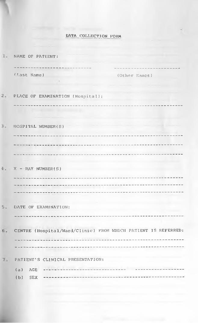
DATA COLLECTION FORM
NAME OF PATIENT:
,T.ast Name) (Other Names)
PLACE OF EXAMINATION (Hospital):
HOSPITAL N U M B E R (S )
X - RAY N U M B E R (S )
DATE OF EXAMINATION:
CENTRE (llospital/Ward/Cl inic) FROM WHICH PATIENT IS REFERRED:
PATIENT'S CLINICAL PRESENTATION:
(a) AGE
(b) SEX

7 (c) SYMPTOMS + THEIR DURATION:
(d) CLINICAL SIGNS:
(e) CLINICAL DIAGNOSIS:
8. PLAIN X - RAY REPORT:
9. MYELOGRAPHY (OR RAD1CULOGRAPHY) REPORT:
10. PATIENT'S FOLLOW UP REPORTS:
(a) POST MYELOGRAPHIC MANAGEMENT
(i) Surgical Operation ---------------------------------
(ii) Other forms of management if surgical not done:
Un iv e r s i t y o f Nairo bi
10 (b) Findings at Operations:
(c) Histological Report: