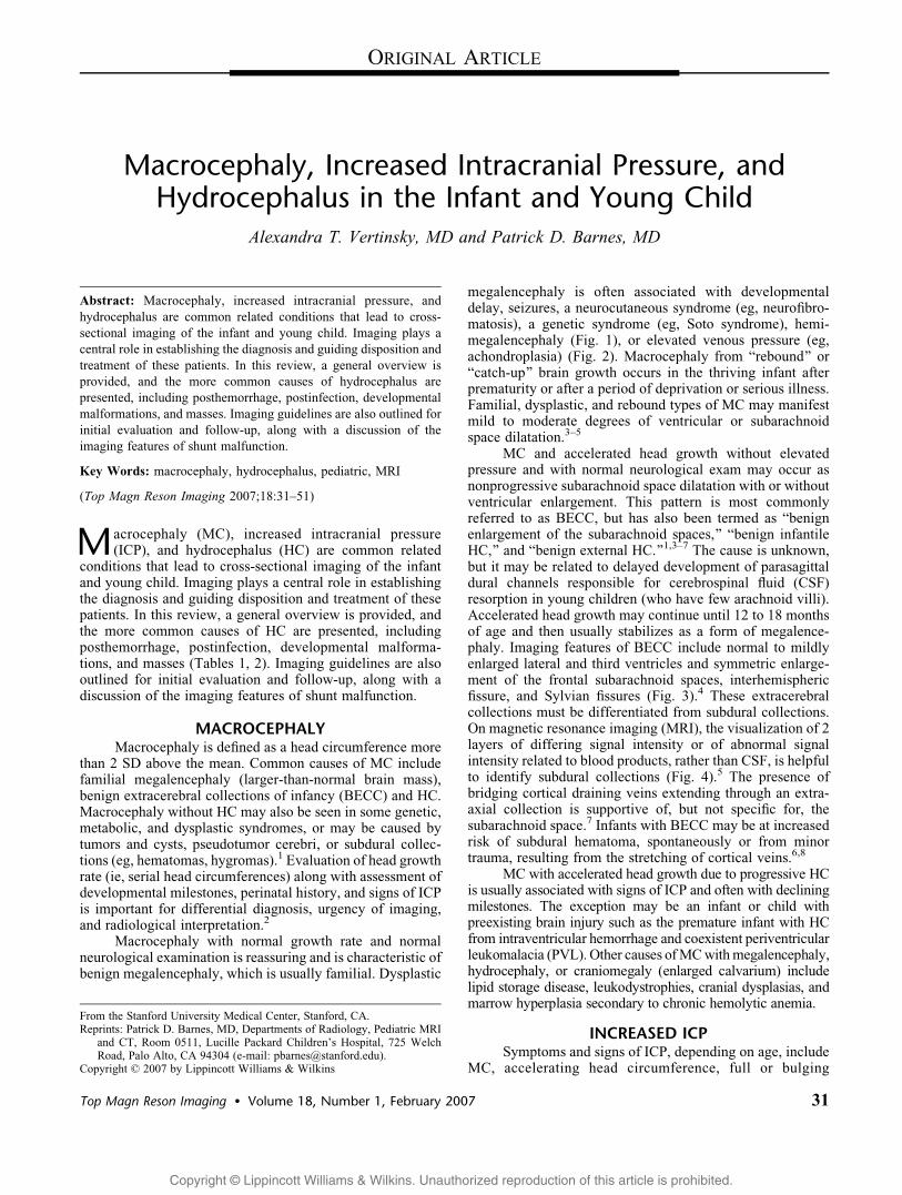
macrocephaly, increased intracranial pressure, and hydrocephalus
TRANSCRIPT
Copyright @ Lippincott Williams & Wilkins. Unauthorized reproduction of this article is prohibited.
Macrocephaly, Increased Intracranial Pressure, andHydrocephalus in the Infant and Young Child
Alexandra T. Vertinsky, MD and Patrick D. Barnes, MD
Abstract: Macrocephaly, increased intracranial pressure, and
hydrocephalus are common related conditions that lead to cross-
sectional imaging of the infant and young child. Imaging plays a
central role in establishing the diagnosis and guiding disposition and
treatment of these patients. In this review, a general overview is
provided, and the more common causes of hydrocephalus are
presented, including posthemorrhage, postinfection, developmental
malformations, and masses. Imaging guidelines are also outlined for
initial evaluation and follow-up, along with a discussion of the
imaging features of shunt malfunction.
Key Words: macrocephaly, hydrocephalus, pediatric, MRI
(Top Magn Reson Imaging 2007;18:31Y51)
Macrocephaly (MC), increased intracranial pressure(ICP), and hydrocephalus (HC) are common related
conditions that lead to cross-sectional imaging of the infantand young child. Imaging plays a central role in establishingthe diagnosis and guiding disposition and treatment of thesepatients. In this review, a general overview is provided, andthe more common causes of HC are presented, includingposthemorrhage, postinfection, developmental malforma-tions, and masses (Tables 1, 2). Imaging guidelines are alsooutlined for initial evaluation and follow-up, along with adiscussion of the imaging features of shunt malfunction.
MACROCEPHALYMacrocephaly is defined as a head circumference more
than 2 SD above the mean. Common causes of MC includefamilial megalencephaly (larger-than-normal brain mass),benign extracerebral collections of infancy (BECC) and HC.Macrocephaly without HC may also be seen in some genetic,metabolic, and dysplastic syndromes, or may be caused bytumors and cysts, pseudotumor cerebri, or subdural collec-tions (eg, hematomas, hygromas).1 Evaluation of head growthrate (ie, serial head circumferences) along with assessment ofdevelopmental milestones, perinatal history, and signs of ICPis important for differential diagnosis, urgency of imaging,and radiological interpretation.2
Macrocephaly with normal growth rate and normalneurological examination is reassuring and is characteristic ofbenign megalencephaly, which is usually familial. Dysplastic
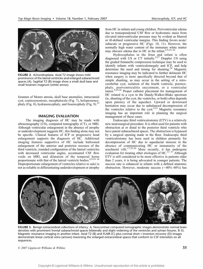
megalencephaly is often associated with developmentaldelay, seizures, a neurocutaneous syndrome (eg, neurofibro-matosis), a genetic syndrome (eg, Soto syndrome), hemi-megalencephaly (Fig. 1), or elevated venous pressure (eg,achondroplasia) (Fig. 2). Macrocephaly from Brebound[ orBcatch-up[ brain growth occurs in the thriving infant afterprematurity or after a period of deprivation or serious illness.Familial, dysplastic, and rebound types of MC may manifestmild to moderate degrees of ventricular or subarachnoidspace dilatation.3Y5
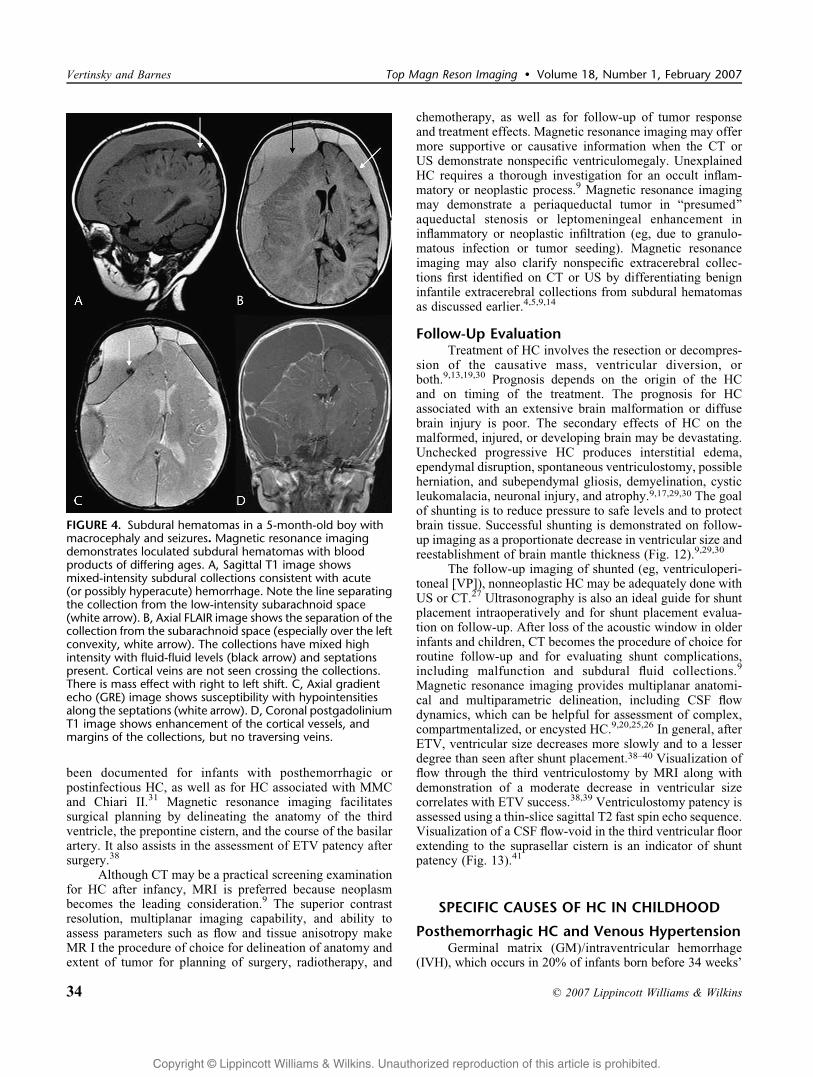
MC and accelerated head growth without elevatedpressure and with normal neurological exam may occur asnonprogressive subarachnoid space dilatation with or withoutventricular enlargement. This pattern is most commonlyreferred to as BECC, but has also been termed as Bbenignenlargement of the subarachnoid spaces,[ Bbenign infantileHC,[ and Bbenign external HC.[1,3Y7 The cause is unknown,but it may be related to delayed development of parasagittaldural channels responsible for cerebrospinal fluid (CSF)resorption in young children (who have few arachnoid villi).Accelerated head growth may continue until 12 to 18 monthsof age and then usually stabilizes as a form of megalence-phaly. Imaging features of BECC include normal to mildlyenlarged lateral and third ventricles and symmetric enlarge-ment of the frontal subarachnoid spaces, interhemisphericfissure, and Sylvian fissures (Fig. 3).4 These extracerebralcollections must be differentiated from subdural collections.On magnetic resonance imaging (MRI), the visualization of 2layers of differing signal intensity or of abnormal signalintensity related to blood products, rather than CSF, is helpfulto identify subdural collections (Fig. 4).5 The presence ofbridging cortical draining veins extending through an extra-axial collection is supportive of, but not specific for, thesubarachnoid space.7 Infants with BECC may be at increasedrisk of subdural hematoma, spontaneously or from minortrauma, resulting from the stretching of cortical veins.6,8
MC with accelerated head growth due to progressive HCis usually associated with signs of ICP and often with decliningmilestones. The exception may be an infant or child withpreexisting brain injury such as the premature infant with HCfrom intraventricular hemorrhage and coexistent periventricularleukomalacia (PVL). Other causes ofMCwithmegalencephaly,hydrocephaly, or craniomegaly (enlarged calvarium) includelipid storage disease, leukodystrophies, cranial dysplasias, andmarrow hyperplasia secondary to chronic hemolytic anemia.
INCREASED ICPSymptoms and signs of ICP, depending on age, include
MC, accelerating head circumference, full or bulging
ORIGINAL ARTICLE
Top Magn Reson Imaging & Volume 18, Number 1, February 2007 31
From the Stanford University Medical Center, Stanford, CA.Reprints: Patrick D. Barnes, MD, Departments of Radiology, Pediatric MRI
and CT, Room 0511, Lucille Packard Children_s Hospital, 725 WelchRoad, Palo Alto, CA 94304 (e-mail: [email protected]).
Copyright * 2007 by Lippincott Williams & Wilkins

Copyright @ Lippincott Williams & Wilkins. Unauthorized reproduction of this article is prohibited.
fontanelle, split sutures, poor feeding, vomiting, irritability,vision impairment, headache, lethargy, stupor, encephalo-pathy, Parinaud syndrome, sixth nerve palsy, hypertoniawith hyperreflexia, and papilledema.9,10 The causes ofICP include trauma, hemorrhage, acute hypoxic-ischemicinsult, infection, parainfectious sequela, metabolic derange-ment, HC, tumors, pseudotumor cerebri, and universalcraniosynostosis.9Y16 Imaging is indicated to define a mass,fluid collection, edema, or HC. The mass may be a cyst,neoplasm, abscess, or hematoma. The abnormal fluid collec-tion may be subdural or epidural, whether a hematoma,empyema, effusion, or hygroma. Edema may be traumatic(Fig. 5), hypoxic-ischemic (Fig. 6), toxic (eg, lead poisoning),metabolic (eg, ketoacidosis), infectious (meningitis or ence-phalitis), parainfectious (acute disseminated encephalomye-litis, Reye syndrome), or due to pseudotumor.
HYDROCEPHALUSA common cause of MC and ICP in childhood is HC.
Hydrocephalus is the state of excessive CSF volume withprogressive enlargement of the ventricles, subarachnoidspaces, or both.17Y20 Hydrocephalus may be caused by animbalance between CSF production and absorption, by ablockage of CSF flow, or from alterations in ventricularcompliance and CSF pulse pressure.17,20 Hydrocephalus due
to CSF overproduction is very rare but may occur withchoroid plexus papilloma (CPP) or villus hypertrophy.Hydrocephalus due to CSF flow block or absorptive blockmay be described as Bcommunicating[ when the block occursoutside the ventricular system (eg, basal cisterns or para-sagittal arachnoid villi) and Bnoncommunicating[ when thereis intraventricular obstruction (at or proximal to the fourthventricular outlets).9,18
Most childhood HC occurs in infancy (Table 3). Themost common cause is acquired adhesive ependymitis orarachnoiditis after hemorrhage or infection.9,17,18 Hydroce-phalus is a well-known sequela of neonatal intracranialhemorrhage especially in the preterm (PT).16 Prenatal orpostnatal infection may also lead to HC.11,12 By far the mostcommon developmental cause of HC is the Chiari IImalformation associated with myelocele/myelomeningocele(MMC). The HC often develops, or progresses, after repair ofthe spinal defect.9,21 Other common developmental causesinclude aqueductal anomalies (forking, stenosis, septation,gliosis) and the Dandy-Walker-Blake spectrum of retro-cerebellar cysts.22 Less common or rare causes of HC include
TABLE 1. Overview of Clinical Presentations
TABLE 2. Macrocephaly, ICP, and HC
FIGURE 1. Hemimegalencephaly with macrocephaly andepilepsy. Prenatal US showed ventriculomegaly. Axialnoncontrast CT at 2 days of life shows enlarged right cerebralhemisphere with a large dysplastic right lateral ventricle andthickened cortex (A). Axial T2 images (B, C) and coronal shortT inversion recovery (D) demonstrate enlargement of the righthemisphere and lateral ventricle, prominent trigone (whitearrow) and occipital horn, and thickened gyri.
Vertinsky and Barnes Top Magn Reson Imaging & Volume 18, Number 1, February 2007
32 * 2007 Lippincott Williams & Wilkins
Copyright @ Lippincott Williams & Wilkins. Unauthorized reproduction of this article is prohibited.
foramen of Monro atresia, skull base anomalies, intracranialcyst, craniosynostosis, encephalocele (Fig. 7), holoprosence-phaly (Fig. 8), hydranencephaly, and lissencephaly (Fig. 9).23
IMAGING EVALUATIONThe imaging diagnosis of HC may be made with
ultrasonography (US), computed tomography (CT), or MRI.Although ventricular enlargement in the absence of atrophyor underdevelopment suggests HC, this finding alone may notbe specific. Clinical features of ICP or progressive headenlargement supports the diagnosis of HC. Additionalimaging features supportive of HC include balloonedenlargement of the anterior and posterior recesses of thethird ventricle, rounded configuration of the lateral ventricleswith decreased ventricular angles, accentuated CSF flowvoids on MRI, and dilatation of the temporal hornsproportionate with that of the lateral ventricle bodies.6,9,24Y26
Disproportionate enlargement of ventricles relative to sulci isnot as reliable in differentiating underdevelopment or atrophy
from HC in infants and young children. Periventricular edemadue to transependymal CSF flow or hydrostatic stasis fromelevated intraventricular pressure may be evident as blurredor ill-defined ventricular margins. This finding favors acute/subacute or progressive HC (Figs. 10, 11). However, thenormally high water content of the immature white mattermay obscure edema due to HC in the infant.6,9,24Y26
Hydrocephalus in the fetus and infant is oftendiagnosed with US or CT initially.9,27 Doppler US usingthe graded fontanelle compression technique may be used toidentify infants with ventriculomegaly and ICP, and helpdetermine the need and timing for shunting.28 Magneticresonance imaging may be indicated to further delineate HCwhen surgery is more specifically directed beyond that ofsimple shunting, as may occur in the setting of a retro-cerebellar cyst, isolation of the fourth ventricle, porence-phaly, postventriculitis encystment, or a ventriculartumor.9,25,26 Proper catheter placement for management ofHC related to a cyst in the Dandy-Walker-Blake spectrum(ie, shunting of the cyst, the ventricles, or both) often dependsupon patency of the aqueduct. Upward or downwardherniation may occur due to unbalanced decompression ofthe ventricles relative to the cyst.9,22 Magnetic resonanceimaging has an important role in planning the surgicalmanagement of these cases.
Endoscopic third ventriculostomy (ETV) is a relativelynew neurosurgical procedure. It is often used for patients withobstruction at or distal to the posterior third ventricle whohave patent subarachnoid spaces. The obstruction is bypassedby a surgical opening made in the floor. Endoscopic thirdventriculostomy has been used in children primarily fordecompression of HC due to aqueductal stenosis in theabsence of communicating HC or immaturity of thearachnoid villi.17,29,30 More recently, it has undergoneevaluation for treating other etiologies of HC.31Y37 AlthoughETV is still considered to be more effective in patients olderthan 2 years, it is being advocated in younger patients. Thesuccess rate is enhanced in infants with a defined anatomicobstruction. However, moderate success (È40%Y60%) has
FIGURE 2. Achondroplasia. Axial T2 image shows mildprominence of the lateral ventricles and enlarged subarachnoidspaces (A). Sagittal T2 (B) image show a small skull base andsmall foramen magnum (white arrow).
FIGURE 3. Benign extracerebral collections of infancy. A, Noncontrast computed tomographic images demonstrate normal braindensities with prominent frontal subarachnoid spaces bilaterally and slight widening of the ventricles and sylvian fissures. BYD,Magnetic resonance imaging in another infant. Axial T2 (B) and FLAIR (C) plus coronal short T inversion recovery (D) imagesdemonstrate linear cortical veins (arrows) traversing the enlarged extracerebral spaces that conform to CSF intensities on allsequences.
Top Magn Reson Imaging & Volume 18, Number 1, February 2007 Macrocephaly, ICP, and HC
* 2007 Lippincott Williams & Wilkins 33
Copyright @ Lippincott Williams & Wilkins. Unauthorized reproduction of this article is prohibited.
been documented for infants with posthemorrhagic orpostinfectious HC, as well as for HC associated with MMCand Chiari II.31 Magnetic resonance imaging facilitatessurgical planning by delineating the anatomy of the thirdventricle, the prepontine cistern, and the course of the basilarartery. It also assists in the assessment of ETV patency aftersurgery.38
Although CT may be a practical screening examinationfor HC after infancy, MRI is preferred because neoplasmbecomes the leading consideration.9 The superior contrastresolution, multiplanar imaging capability, and ability toassess parameters such as flow and tissue anisotropy makeMR I the procedure of choice for delineation of anatomy andextent of tumor for planning of surgery, radiotherapy, and
chemotherapy, as well as for follow-up of tumor responseand treatment effects. Magnetic resonance imaging may offermore supportive or causative information when the CT orUS demonstrate nonspecific ventriculomegaly. UnexplainedHC requires a thorough investigation for an occult inflam-matory or neoplastic process.9 Magnetic resonance imagingmay demonstrate a periaqueductal tumor in Bpresumed[aqueductal stenosis or leptomeningeal enhancement ininflammatory or neoplastic infiltration (eg, due to granulo-matous infection or tumor seeding). Magnetic resonanceimaging may also clarify nonspecific extracerebral collec-tions first identified on CT or US by differentiating benigninfantile extracerebral collections from subdural hematomasas discussed earlier.4,5,9,14
Follow-Up EvaluationTreatment of HC involves the resection or decompres-
sion of the causative mass, ventricular diversion, orboth.9,13,19,30 Prognosis depends on the origin of the HCand on timing of the treatment. The prognosis for HCassociated with an extensive brain malformation or diffusebrain injury is poor. The secondary effects of HC on themalformed, injured, or developing brain may be devastating.Unchecked progressive HC produces interstitial edema,ependymal disruption, spontaneous ventriculostomy, possibleherniation, and subependymal gliosis, demyelination, cysticleukomalacia, neuronal injury, and atrophy.9,17,29,30 The goalof shunting is to reduce pressure to safe levels and to protectbrain tissue. Successful shunting is demonstrated on follow-up imaging as a proportionate decrease in ventricular size andreestablishment of brain mantle thickness (Fig. 12).9,29,30
The follow-up imaging of shunted (eg, ventriculoperi-toneal [VP]), nonneoplastic HC may be adequately done withUS or CT.27 Ultrasonography is also an ideal guide for shuntplacement intraoperatively and for shunt placement evalua-tion on follow-up. After loss of the acoustic window in olderinfants and children, CT becomes the procedure of choice forroutine follow-up and for evaluating shunt complications,including malfunction and subdural fluid collections.9
Magnetic resonance imaging provides multiplanar anatomi-cal and multiparametric delineation, including CSF flowdynamics, which can be helpful for assessment of complex,compartmentalized, or encysted HC.9,20,25,26 In general, afterETV, ventricular size decreases more slowly and to a lesserdegree than seen after shunt placement.38Y40 Visualization offlow through the third ventriculostomy by MRI along withdemonstration of a moderate decrease in ventricular sizecorrelates with ETV success.38,39 Ventriculostomy patency isassessed using a thin-slice sagittal T2 fast spin echo sequence.Visualization of a CSF flow-void in the third ventricular floorextending to the suprasellar cistern is an indicator of shuntpatency (Fig. 13).41
SPECIFIC CAUSES OF HC IN CHILDHOOD
Posthemorrhagic HC and Venous HypertensionGerminal matrix (GM)/intraventricular hemorrhage
(IVH), which occurs in 20% of infants born before 34 weeks’
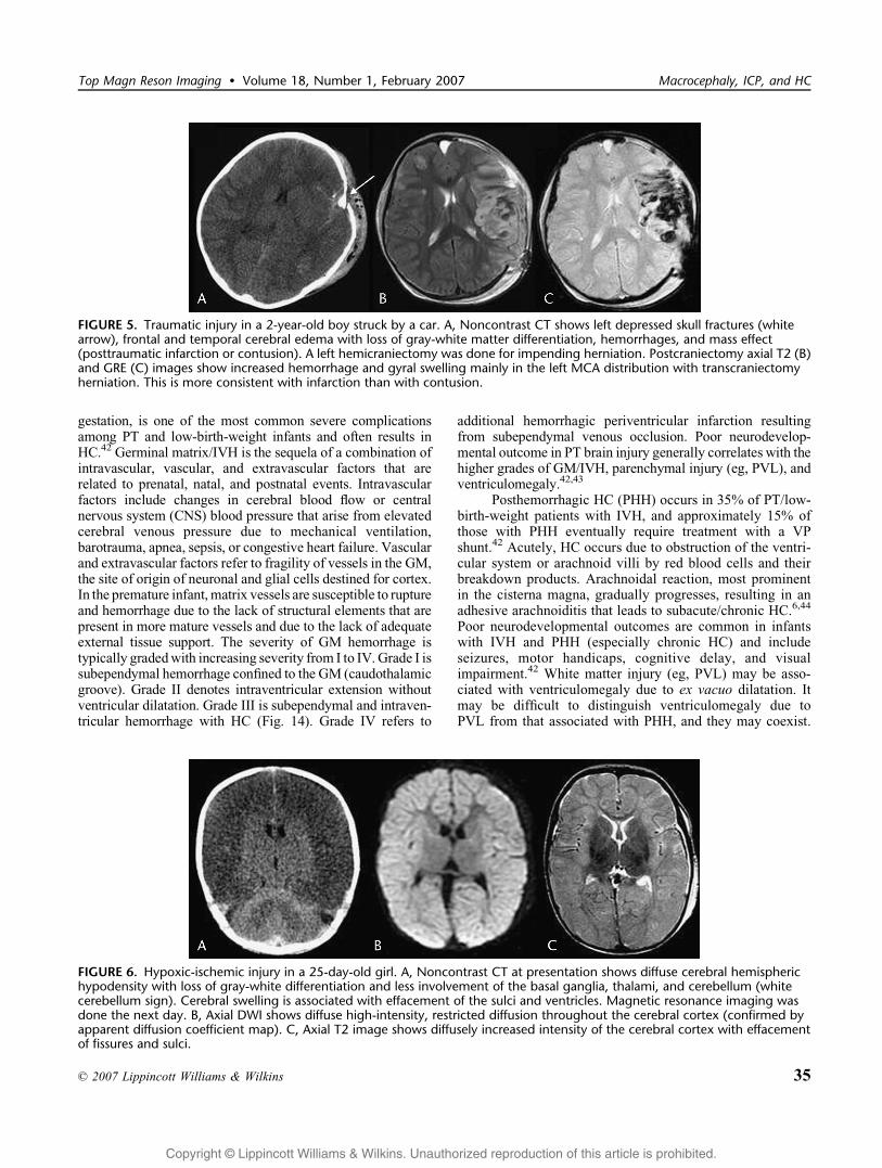
FIGURE 4. Subdural hematomas in a 5-month-old boy withmacrocephaly and seizures. Magnetic resonance imagingdemonstrates loculated subdural hematomas with bloodproducts of differing ages. A, Sagittal T1 image showsmixed-intensity subdural collections consistent with acute(or possibly hyperacute) hemorrhage. Note the line separatingthe collection from the low-intensity subarachnoid space(white arrow). B, Axial FLAIR image shows the separation of thecollection from the subarachnoid space (especially over the leftconvexity, white arrow). The collections have mixed highintensity with fluid-fluid levels (black arrow) and septationspresent. Cortical veins are not seen crossing the collections.There is mass effect with right to left shift. C, Axial gradientecho (GRE) image shows susceptibility with hypointensitiesalong the septations (white arrow). D, Coronal postgadoliniumT1 image shows enhancement of the cortical vessels, andmargins of the collections, but no traversing veins.
Vertinsky and Barnes Top Magn Reson Imaging & Volume 18, Number 1, February 2007
34 * 2007 Lippincott Williams & Wilkins
Copyright @ Lippincott Williams & Wilkins. Unauthorized reproduction of this article is prohibited.
gestation, is one of the most common severe complicationsamong PT and low-birth-weight infants and often results inHC.42 Germinal matrix/IVH is the sequela of a combination ofintravascular, vascular, and extravascular factors that arerelated to prenatal, natal, and postnatal events. Intravascularfactors include changes in cerebral blood flow or centralnervous system (CNS) blood pressure that arise from elevatedcerebral venous pressure due to mechanical ventilation,barotrauma, apnea, sepsis, or congestive heart failure. Vascularand extravascular factors refer to fragility of vessels in the GM,the site of origin of neuronal and glial cells destined for cortex.In the premature infant, matrix vessels are susceptible to ruptureand hemorrhage due to the lack of structural elements that arepresent in more mature vessels and due to the lack of adequateexternal tissue support. The severity of GM hemorrhage istypically gradedwith increasing severity from I to IV. Grade I issubependymal hemorrhage confined to the GM (caudothalamicgroove). Grade II denotes intraventricular extension withoutventricular dilatation. Grade III is subependymal and intraven-tricular hemorrhage with HC (Fig. 14). Grade IV refers to
additional hemorrhagic periventricular infarction resultingfrom subependymal venous occlusion. Poor neurodevelop-mental outcome in PT brain injury generally correlates with thehigher grades of GM/IVH, parenchymal injury (eg, PVL), andventriculomegaly.42,43
Posthemorrhagic HC (PHH) occurs in 35% of PT/low-birth-weight patients with IVH, and approximately 15% ofthose with PHH eventually require treatment with a VPshunt.42 Acutely, HC occurs due to obstruction of the ventri-cular system or arachnoid villi by red blood cells and theirbreakdown products. Arachnoidal reaction, most prominentin the cisterna magna, gradually progresses, resulting in anadhesive arachnoiditis that leads to subacute/chronic HC.6,44
Poor neurodevelopmental outcomes are common in infantswith IVH and PHH (especially chronic HC) and includeseizures, motor handicaps, cognitive delay, and visualimpairment.42 White matter injury (eg, PVL) may be asso-ciated with ventriculomegaly due to ex vacuo dilatation. Itmay be difficult to distinguish ventriculomegaly due toPVL from that associated with PHH, and they may coexist.
FIGURE 5. Traumatic injury in a 2-year-old boy struck by a car. A, Noncontrast CT shows left depressed skull fractures (whitearrow), frontal and temporal cerebral edema with loss of gray-white matter differentiation, hemorrhages, and mass effect(posttraumatic infarction or contusion). A left hemicraniectomy was done for impending herniation. Postcraniectomy axial T2 (B)and GRE (C) images show increased hemorrhage and gyral swelling mainly in the left MCA distribution with transcraniectomyherniation. This is more consistent with infarction than with contusion.
FIGURE 6. Hypoxic-ischemic injury in a 25-day-old girl. A, Noncontrast CT at presentation shows diffuse cerebral hemispherichypodensity with loss of gray-white differentiation and less involvement of the basal ganglia, thalami, and cerebellum (whitecerebellum sign). Cerebral swelling is associated with effacement of the sulci and ventricles. Magnetic resonance imaging wasdone the next day. B, Axial DWI shows diffuse high-intensity, restricted diffusion throughout the cerebral cortex (confirmed byapparent diffusion coefficient map). C, Axial T2 image shows diffusely increased intensity of the cerebral cortex with effacementof fissures and sulci.
Top Magn Reson Imaging & Volume 18, Number 1, February 2007 Macrocephaly, ICP, and HC
* 2007 Lippincott Williams & Wilkins 35

Copyright @ Lippincott Williams & Wilkins. Unauthorized reproduction of this article is prohibited.
T2/fluid-attenuated inversion recovery (FLAIR) periventri-cular white matter hyperintensity, loss of the periventricularwhite matter volume, and irregular ventricular margins arefindings characteristic of PVL (Fig. 15).45Y47
Subarachnoid and intraventricular hemorrhage in full-term infants is less common than in PT infants and may bedue to coagulopathy, dehydration, hypoxic-ischemic injury,venous thrombosis, infection, or trauma (including birth-related trauma) (Fig. 16).48 Although the mechanism of acuteand chronic PHH is similar to that for PT infant, outcome ismore variable for the term infant.6
Venous hypertension due to venous thrombosis shouldbe considered in infants presenting with unexplained seizuresand irritability or in those with hemorrhage or infarct notcorresponding to an arterial vascular territory. Venous hyper-tension may be due to developmental conditions such as
malformations of the skull base restricting venous outflow(eg, achondroplasia) or congenital heart disease or pulmonarydisease with elevated central venous pressures. It may beacquired due to thrombosis of cerebral veins or sinuses fromvarious causes such as infection, vascular malformation, orcoagulopathy (Fig. 17). In an evaluation of suspected venousthrombosis, Doppler US, contrast-enhanced CT, computedtomographic angiography, MRI with MR venogram andgadolinium enhancement, or catheter angiography may beneeded to directly demonstrate dural venous sinus thrombosis.Cortical, subependymal, or medullary venous occlusion maynot be directly demonstrated by these techniques, althoughhemorrhages or thromboses may be present in those distribu-tions. The thrombosis may appear as computed tomographichyperdensity, T1 high-intensity, T2 low-intensity, or GREhypointensity and can mimic hemorrhage. Intravenousenhancement about the thrombus may be seen as an emptyBC[ sign. Depending upon the clinical context, treatment maybe directed only to the specific cause (eg, infection) or may
TABLE 3. Causes of HC in Infancy and Childhood
Developmental Acquired
Chiari II malformation Posthemorrhage
Aqueductal anomalies Postinfection
Congenital cysts/DW malformation Posterior fossa tumors
Encephalocele Tumors about the third ventricle
Hydranencephaly Cerebral hemispheric tumors
Craniosynostosis
Skull base anomalies
Foraminal atresia
Immature arachnoid villi
Vein of Galen aneurysm
FIGURE 7. Occipital encephalocystocele in 28-week-old fetus.Sagittal T2Ysingle-shot fast spin echo image shows herniationof dysplastic occipital lobe, dura, and CSF through a smalloccipital defect (white arrow) with associatedventriculomegaly.
FIGURE 8. Holoprosencephaly in a 5-day-old baby. Axial T1(A) and midsagittal T2 (B) images show a large monoventriclewith large dorsal cyst (white arrow) alongwith a partial anteriorinterhemispheric fissure, absent corpus callosum, and smallposterior fossa.
FIGURE 9. Walker-Warburg syndrome in a 2-day-old girl.Axial T2 (A) and sagittal T1 (B) images show marked third andlateral ventriculomegaly with absent septum pellucidum andcallosal hypogenesis. There is cortical mantle thinning withan irregular gyral pattern (cobblestone lissencephaly),hypogenesis of the pons and cerebellar vermis (white arrows),and tectal dysplasia with aqueductal stenosis.
Vertinsky and Barnes Top Magn Reson Imaging & Volume 18, Number 1, February 2007
36 * 2007 Lippincott Williams & Wilkins

Copyright @ Lippincott Williams & Wilkins. Unauthorized reproduction of this article is prohibited.
also include anticoagulation or thrombolysis.49Y53 Increasedpressure within the dural sinuses creates a decreased pressuregradient across the arachnoid villi that results in decreasedCSF resorption. In the setting of venous hypertension, eitherHC or pseudotumor cerebri may occur depending on patientage and patency of the cranial sutures. In young infants (lessthan 18 months) who have open sutures, an expansilecalvarium, and soft, undermyelinated, immature white matter,HC is more likely to occur because the ventricles may expandwithout resistance. Pseudotumor is more common in olderinfants and children.6
Postinfectious HCHydrocephalus in infants may be the result of prenatal or
postnatal infection. Prenatal infections occur either byascending infection from the cervix to the amniotic fluid(usually bacteria or herpes) or via hematogenous dissemina-tion through the placenta (eg, toxoplasmosis, other infections,
rubella, cytomegalovirus [CMV] infection, and herpes sim-plex infections and other viruses). During the first 2 trimesters,infection will typically lead to malformations. In the thirdtrimester, destructive lesions occur. Ventriculomegaly is oftendue to cerebral destruction, but HC may also occur and ismost ommon in toxoplasmosis. A comprehensive reviewof prenatal infections is described elsewhere. Features ofthe 2 most common entities (CMV and toxoplasmosis) aredescribed below.6,11,53
Congenital CMV infection is a common and seriousviral infection among newborns. Depending on the timing ofthe insult, signs and symptoms include hepatosplenomegaly,microcephaly, chorioretinitis, and seizures.6,53 Affectedpatients have varying degrees of lissencephaly/polymicro-gyria, decreased cerebral white matter, astrogliosis, cerebralcalcification, delayed myelination, and cerebellar hypopla-sia.54,55 Ventriculomegaly is usually related to cerebralunderdevelopment/destruction, rather than HC (Fig. 18).Infection during early gestation tends to result in more severe
FIGURE 10. Hydrocephalus due to aqueductal stenosis. A,Sagittal T2 image demonstrates the aqueductal web (whitearrow), small fourth ventricle, and marked third ventricularenlargement with ballooning of the anterior and posteriorrecesses plus downward displacement of the floor into the sella(black arrow). B, Axial FLAIR image shows the markedly dilatedlateral ventricles with hyperintense periventricular edema(white arrow).
FIGURE 11. Cerebral underdevelopment in an infant with tetralogy of Fallot. Axial T2 images (AYC) show lateral ventricularenlargement out of proportion to the third ventricle and temporal horns. The sylvian fissures are wide. Normal hyperintensity isseen within the cerebral white matter due to immaturity. The findings suggest underdevelopment, rather than HC, although acomponent of communicating HC is always difficult to exclude and may coexist with any cause of underdevelopment or atrophy.
FIGURE 12. Communicating HC post-VP shunt in a 4-year-oldboy. A, Axial FLAIR image at presentation in early infancydemonstrates marked ventriculomegaly with thinning of thecortical mantle. B, Follow-up axial FLAIR image shows a rightparietal ventricular catheter in place (white arrow), smallventricles, and increased mantle thickness. The periventricularwhite matter hyperintensity (black arrow) likely representsundermyelination vs some leukomalacia or gliosis.
Top Magn Reson Imaging & Volume 18, Number 1, February 2007 Macrocephaly, ICP, and HC
* 2007 Lippincott Williams & Wilkins 37
Copyright @ Lippincott Williams & Wilkins. Unauthorized reproduction of this article is prohibited.
disease. Computed tomography often detects cerebral calcifi-cations (eg, periventricular). Magnetic resonance imaging bestdemonstrates the parenchymal involvement.11,54,55 Congenitaltoxoplasmosis may manifest at birth or days to weeks later.There may be generalized or predominantly CNS involvement.Calcifications are common and more random in distribution,including periventricular, cortical, and basal ganglia. Hydro-cephalus often results from the granulomatous meningeal orependymal reaction that can cause aqueductal stenosis andcommunicating HC. Ventriculomegaly may also occur sec-ondary to cerebral tissue destruction.Malformations of corticaldevelopment (eg, polymicrogyria) are uncommon.11,53,56
Postnatal meningitis may be bacterial, viral, fungal, orparasitic and caused by direct (eg, sinus or ear infection) orhematogenous spread. Common etiologies include Gram-negative bacteria (eg, Escherichia coli), group B streptococ-cus, pneumococcus, Listeria, neisseria, and tuberculosis. Inthe acute-subacute setting, meningitis can lead to HC due toclumping of purulent fluid along the CSF pathways or due toinflammation of arachnoid granulations with reduced CSFresorption. Chronically, the presence of inflammatoryexudate and blood products lead to arachnoiditis. Fungaland granulomatous meningitides are more likely to cause
clinically significant HC than bacterial and viral infections.The severity of HC is also related to the duration and severityof infection.56Y59 Normal imaging evaluation does notexclude CNS infection. Magnetic resonance imaging maysometimes demonstrate meningeal enhancement. In fungaland granulomatous infection, the meningeal enhancementand thickening often has a predilection for the basal cisterns.Magnetic resonance imaging is mainly used to evaluate thesequelae and complications of meningitis. Arachnoid locula-tions due to arachnoid scarring may occur and simulatearachnoid cysts (ACs). Ventricular dilatation may be shownby MR or CT. Other complications of meningitis, includingvenous thrombosis, infarction (arterial or venous), ventricu-litis, cerebritis, abscess, and subdural empyema, are bestdelineated with MRI (Fig. 19).56,58,59
DEVELOPMENTAL MALFORMATIONS
Chiari IIChiari II malformation accounts for about one third
of infantile HC. Almost all present at birth with a MMC.
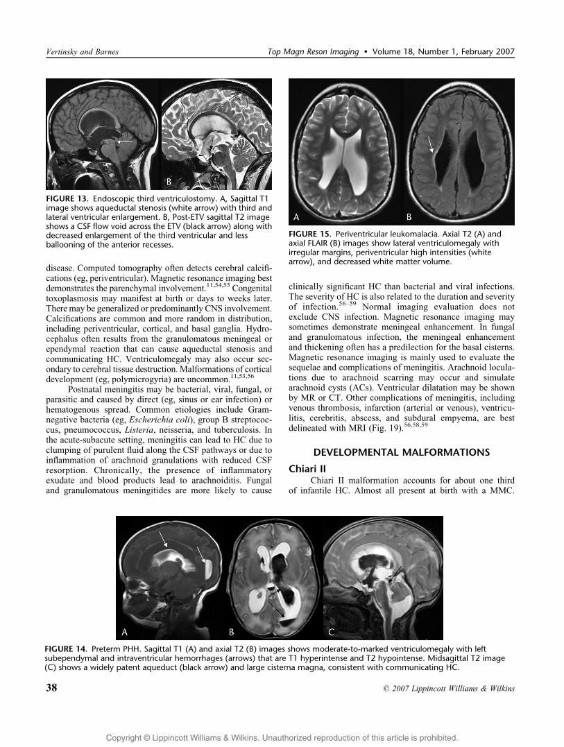
FIGURE 13. Endoscopic third ventriculostomy. A, Sagittal T1image shows aqueductal stenosis (white arrow) with third andlateral ventricular enlargement. B, Post-ETV sagittal T2 imageshows a CSF flow void across the ETV (black arrow) along withdecreased enlargement of the third ventricular and lessballooning of the anterior recesses.
FIGURE 14. Preterm PHH. Sagittal T1 (A) and axial T2 (B) images shows moderate-to-marked ventriculomegaly with leftsubependymal and intraventricular hemorrhages (arrows) that are T1 hyperintense and T2 hypointense. Midsagittal T2 image(C) shows a widely patent aqueduct (black arrow) and large cisterna magna, consistent with communicating HC.
FIGURE 15. Periventricular leukomalacia. Axial T2 (A) andaxial FLAIR (B) images show lateral ventriculomegaly withirregular margins, periventricular high intensities (whitearrow), and decreased white matter volume.
Vertinsky and Barnes Top Magn Reson Imaging & Volume 18, Number 1, February 2007
38 * 2007 Lippincott Williams & Wilkins

Copyright @ Lippincott Williams & Wilkins. Unauthorized reproduction of this article is prohibited.
Hydrocephalus usually develops after repair of the MMC.Altered CSF flow likely results from the abnormal lowposition of the fourth ventricular exit foramina below theforamen magnum, causing poor connection between thespinal and intracranial subarachnoid spaces. Hydrocephalusmanifests when the surgically closed MMC no longer acts asa pressure valve to release CSF that flows freely from theventricles into the central canal.21,60 Chiari II malformationresults from failure of neural tube closure, The cerebellarvermis herniates into the cervical spinal canal and maydegenerate. The fourth ventricle is low, vertically oriented,and narrowed. The pons is stretched inferiorly and is alsonarrowed. The medulla may extend below foramen magnumand a cervicomedullary kink may be seen. The tectum has abeaked or blunted shape. Petroclival scalloping may be seenalong with persistent or accentuated Luckenshadl (ie,
lacunar skull).21,60Y63 Other commonly associated anomaliesinclude hypogenesis of the corpus callosum, heterotopias,colpocephaly, and polygyria or stenogyria (Fig. 20).6,60Y62
The fourth ventricle may herniate behind the medulla andbelow the vermis (ie, Bencysted[). More importantly, it maybecome isolated or trapped due to poor inflow (eg, primaryaqueductal stenosis or secondary closure from shunting) andpoor outflow (exit foraminal atresia or closure). A normal-sized or large fourth ventricle in Chiari II patients mayindicate trapping or shunt malfunction. Increasing hydro-syringomyelia may also result from worsening HC, shuntmalfunction, or fourth ventricular isolation.63 After shunt-ing, the medial walls of the ventricular trigones may appeardeformed by a large CSF-containing structure. This resultsfrom Bmantle collapse[ of the dysplastic adjacent cortex andshould be differentiated from an AC or atrial diverticulum.6
Hydrocephalus (or hydrosyringomyelia) may also be seenwith other Chiari malformations (eg, I, III).
Dandy Walker Blake ContinuumPosterior fossa cystic malformations may be catego-
rized along a continuum, including the Dandy-Walkermalformation (DWM), Dandy-Walker variant (DWV),mega cisterna magna (MCM), and Blake pouch cyst (BPC)or retrocerebellar AC (RCAC). The classic Dandy-Walkermalformation is characterized by complete or partial vermianagenesis (especially inferior vermis), a large retrocerebellarcyst (ie, Bcombined fourth ventricle and cisterna magna[),
FIGURE 16. Nonpreterm, posthemorrhagic communicatingHC. Axial GRE (A) and midsagittal T2 (B) images demonstratemarked lateral, third, and fourth ventriculomegaly withill-defined ventricular margins, CSF-blood levels (black arrow),and a prominent flow-void within the patent aqueduct (whitearrow), consistent with communicating HC.
FIGURE 17. Chronic venous hypertension due to duralarteriovenous fistula and extensive venous thrombosis.Axial GRE (A) and coronal T2 (B) mages show partiallythrombosed and dilated torcula (black arrow), cerebral whitematter hyperintensity secondary to venous congestion andchronic ischemia, numerous tiny foci of hypointensity due tohemorrhage and/or thromboses, ventriculomegaly, and largesubarachnoid spaces.
FIGURE 18. Congenital CMV. Noncontrast CT demonstratesventriculomegaly with bilateral porencephaly, periventricularcalcifications (white arrow), and dysplastic cerebellum.
Top Magn Reson Imaging & Volume 18, Number 1, February 2007 Macrocephaly, ICP, and HC
* 2007 Lippincott Williams & Wilkins 39

Copyright @ Lippincott Williams & Wilkins. Unauthorized reproduction of this article is prohibited.
an enlarged posterior fossa, elevation of the torcular abovethe L, and absence of the falx cerebelli. Associated CNS andsystemic anomalies are common, including corpus callosum
hypogenesis, polymicrogyria, heterotopias, cephalocele, andholoprosencephaly (Fig. 21). Hydrocephalus occurs in mostpatients but usually does not develop until after the neonatal
FIGURE 19. Pneumococcal meningitis in a 2-month-old boy. A, Noncontrast CT demonstrates multiple cortical and white matterhypodensities (white arrows) consistent with edema, cerebritis, or multifocal infarctions. Hyperdensities are consistent withhemorrhage or thromboses. The asymmetric extracerebral low densities likely represent subdural collections. Magnetic resonanceimaging was done 1 week later. Sagittal T1 (B), axial GRE (C), coronal T2 (D), and axial postgadolinium T1 (E) images showT1-hypointense/T2-hyperintense extracerebral collections that are DWI hypointense (not shown). These likely representsubdural effusions, rather than empyemas. In addition, there is mild ventriculomegaly. Bilateral linear and nodular cerebral andextracerebral foci that are T1 hyperintense and T2/GRE hypointense (white arrows) represent hemorrhages or thromboses.Multifocal cerebral white matter and basal ganglia T2 hyperintensities likely represent edema or infarctions (curved arrows),and some show enhancement. Thick, irregular, leptomeningeal enhancement is seen over the convexities (black arrows) andalong the basal cisterms.
FIGURE 20. Chiari II in a 12-day-old boy. Axial CT (A) shows dysmorphic and enlarged lateral ventricles (a large foramen magnumwith no fourth ventricle was also observed along with other characteristic findingsVsee text). Sagittal T1 (B) and axial T2 (C)images show a small posterior fossa, low tentorium, and herniation of the cerebellar vermis into the cervical canal posterior to alow cervicomedullary junction (white arrow). The fourth ventricle is elongated and small. Tectal beaking is present along withoccipital polygyria/stenogyria (black arrow). Moderate to marked third and lateral ventriculomegaly is present withdisproportionate enlargement of the posterior horns.
Vertinsky and Barnes Top Magn Reson Imaging & Volume 18, Number 1, February 2007
40 * 2007 Lippincott Williams & Wilkins

Copyright @ Lippincott Williams & Wilkins. Unauthorized reproduction of this article is prohibited.
period. Evaluation of aqueductal patency is important beforesurgery for ventricular or cyst shunting.22,64Y66 In DWV, thecerebellar vermis is hypogenetic, the posterior fossa is usuallyof normal size, and there is separation of the fourth ventriclefrom a smaller retrocerebellar Bcyst.[ If the cerebellar vermisis completely formed and an enlarged retrocerebellar CSF
space is present, the anomaly is usually designated MCM. Anassociated wide vallecula (increased medullary-vermianangle) suggests BPC. If the retrocerebellar CSF collectionexerts mass effect on a completely formed cerebellum, thenRCAC may be diagnosed, especially if there is HC. OtherCNS or systemic anomalies are uncommon in DWV, MCM,BPC, or RCAC. These Bcystic[ posterior fossa anomalies are
FIGURE 21. Dandy-Walker malformation. Noncontrast CT(A) plus sagittal (B) and axial (CYD) MRI images show anenlarged posterior fossa with elevated torcula (black arrow),cerebellar vermis hypogenesis, and a retrocerebellar cyst thatcommunicates with the fourth ventricle. Agenesis of the corpuscallosum and lateral ventricular dysmorphia is also shown.
FIGURE 22. Aqueductal stenosis. Sagittal T1 (A) and T2 (B)images show enlarged third and lateral ventricles (thinning ofthe corpus callosum) with normal fourth ventricle. Focaldiscontinuity of the cerebral aqueduct is seen (white arrows),and there is absence of the usual CSF flow void. The tectummay be dysmorphic, but no mass is present. The thin-sectionsagittal T2 image is especially helpful for delineating anatomyof the third ventricle and basilar cisterns in anticipation of ETV.
FIGURE 23. Tectal glioma. Sagittal T1 image (A) shows thirdand lateral ventriculomegaly with a tectal mass (white arrow)and aqueductal narrowing. Axial FLAIR image (B) shows thehyperintense tectal mass (white arrow). There was minimalgadolinium enhancement (not shown). Hyperintense FLAIRCSF flow artifact is present in the anterior third ventricle.
FIGURE 24. Vein of Galen malformation, choroidal type.Axial (A) and sagittal (B) T2 images show an enlarged vein ofGalen (promesencephalic vein) (white arrows) with dilatedstraight sinus and superior sagittal sinus flow voids plusadjacent arterial feeder flow voids. Lateral and thirdventriculomegaly represents HC (aqueduct compression vsvenous hypertension). Lateral reprojected time of flightYMRangiography image (C) shows the high-intensity flow featuresof the multiple arteriovenous fistulae.
Top Magn Reson Imaging & Volume 18, Number 1, February 2007 Macrocephaly, ICP, and HC
* 2007 Lippincott Williams & Wilkins 41
Copyright @ Lippincott Williams & Wilkins. Unauthorized reproduction of this article is prohibited.
to be distinguished from cerebellar hypoplasia (formed butsmall cerebellum without cyst), pontocerebellar hypoplasia(formed but small pons and cerebellum), Joubert syndrome(superior or total vermian hypogenesis), rhombencephalosy-napsis (absent vermis with fused hemispheres), and cerebellar
atrophy or degeneration (small cerebellum with prominentfissures).22,64Y66
Aqueductal StenosisAqueductal narrowing may be primary (ie, maldeve-
lopmental) or secondary (ie, acquired, eg, adhesive ependy-mitis). It is a common cause of HC and may be isolated orassociated with other developmental or acquired conditions.Developmental narrowing may be in the form of stenosis,gliosis, forking (ie, fenestration), or a membrane (ie,aqueductal web). Hemorrhage, infection, or tumors maylead to acquired aqueductal stenosis. Onset of symptoms dueto primary aqueductal stenosis is insidious and may occur anytime from birth to adulthood.67 Computed tomography oftenshows dilatation of the third and lateral ventricles with normalor small fourth ventricle. There may be tectal dysplasia withthickening or beaking. This is to be distinguished from atectal glioma. The latter may not be apparent on CT.
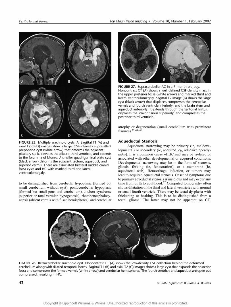
FIGURE 25. Multiple arachnoid cysts. A, Sagittal T1 (A) andaxial T2 (BYD) images show a large, CSF-intensity suprasellar/prepontine cyst (white arrow) that deforms the adjacentpituitary stalk, elevates the dilated third ventricle, and extendsto the foramina of Monro. A smaller quadrigeminal plate cyst(black arrow) deforms the adjacent tectum, aqueduct, andsuperior vermis. There are associated bilateral middle cranialfossa cysts and HC with marked third and lateralventriculomegaly.
FIGURE 26. Retrocerebellar arachnoid cyst. Noncontrast CT (A) shows the low-density CSF collection behind the deformedcerebellum along with dilated temporal horns. Sagittal T1 (B) and axial T2 (C) images show a large cyst that expands the posteriorfossa and compresses the formed vermis (white arrow) and cerebellar hemispheres. The fourth ventricle and aqueduct are open butcompressed, resulting in HC.
FIGURE 27. Supracerebellar AC in a 7-month-old boy.Noncontrast CT (A) shows a well-defined CSF-density mass inthe upper posterior fossa (white arrow) and marked third andlateral ventriculomegaly. Sagittal T2 image (B) shows the largecyst (black arrow) that displaces/compresses the cerebellarvermis and fourth ventricle inferiorly, and the brain stem andaqueduct anteriorly. It extends through the tentorial hiatus,displaces the straight sinus superiorly, and compresses theposterior third ventricle.
Vertinsky and Barnes Top Magn Reson Imaging & Volume 18, Number 1, February 2007
42 * 2007 Lippincott Williams & Wilkins

Copyright @ Lippincott Williams & Wilkins. Unauthorized reproduction of this article is prohibited.
Magnetic resonance imaging is always indicated. Sagittal T1and T2 images demonstrate the level of aqueductal stenosisand absence of a CSF flow void (Fig. 22). T2 and FLAIRimages detect tectal- and pineal-region lesions (Fig. 23).Gadolinium T1 images may show enhancement, althoughtectal gliomas enhance less often than pineal region germ celltumors (GCTs) or pineoblastomas.67Y70
MASSESMost masses of infancy are cystic or cyst-like and
include the Dandy-Walker-Blake spectrum, arachnoid orglioependymal cysts (retrocerebellar, suprasellar, intraven-tricular, quadrigeminal plate cistern), porencephaly, ence-phalocele, and the vein of Galen malformation (varix) as ablood-filled cyst (Fig. 24). Neoplasm as an expanding mass oran obstructive lesion is a rare cause of ICP or HC ininfancy.9,17,18 Beyond infancy, neoplasm becomes theleading consideration.
Arachnoid CystsArachnoid cysts of maldevelopmental origin are
congenital lesions composed of an arachnoid membrane
that secretes CSF to expand the cyst. Acquired ACs (eg,leptomeningeal cysts or arachnoid loculations) are locula-tions of CSF that are associated with arachnoid scarring. Themost common locations for AC are the Sylvian fissuresfollowed by the suprasellar cistern (Fig. 25), quadrigeminalcistern, cerebellopontine angle, and retrocerebellar space(Fig. 26). Less common sites for AC include the interhemi-spheric fissures, cerebral convexities, and anterior midlineportion of the posterior fossa (Fig. 27). Congenital ACs areusually sporadic but occur with increased incidence inpatients with autosomal dominant polycystic kidney dis-ease.6,71 Arachnoid cysts are often incidental findings onimaging for other indications. However, mechanical effectsmay result in headaches, seizures, ICP, or HC. SuprasellarAC is to be distinguished from other cystic lesions in thisregion, including Rathke cyst, craniopharyngioma, dermoid-epidermoid, teratoma, and cystic astrocytomas. Large ACs inthis location displace the third ventricle superiorly and mayobstruct the lateral ventricles at the foramina of Monro.Quadrigeminal plate ACs may displace the pineal gland andocclude the aqueduct. Posterior fossa ACs were describedearlier. Middle cranial fossa cysts may become very large andcan cause mass effect with expansion of the hemicranium.
FIGURE 28. Choroid plexus papilloma in a 7-month-old girl.Noncontrast CT (A) shows a midline intraventricular mass withcalcifications (arrow), enlarged ventricles, and a portion of theleft frontal ventricular catheter. Axial T1 (B), coronal short Tinversion recovery (C), and sagittal postgadolinium T1 (D)images show the lobulated third ventricular mass that isisointense and markedly enhancing. There is extensionthrough the foramina of Monro and marked lateralventriculomegaly.
FIGURE 29. Choroid plexus carcinoma. Sagittal T1 (A), axialand coronal T2 (BYC), and coronal postgadolinium T1 (D)images show a heterogeneous mass within the right lateralventricle. Hypervascularity, with numerous flow voids (blackarrow), is seen along with calcifications, hemorrhage (whitearrow), and marked enhancement. The mass extends into theadjacent cerebrum, and there is marked ventriculomegaly.Similar imaging features may be seen with periventricularPNET, ependymoma, or ATRT and intraventricular extension.
Top Magn Reson Imaging & Volume 18, Number 1, February 2007 Macrocephaly, ICP, and HC
* 2007 Lippincott Williams & Wilkins 43

Copyright @ Lippincott Williams & Wilkins. Unauthorized reproduction of this article is prohibited.
These cysts may also be associated with subdural andintracystic hemorrhage, or subdural hygroma. This mayoccur spontaneously, after trauma, or after surgicalfenestration.6,72Y74 On imaging, ACs are thin-walled, well-defined, cystic lesions that conform to CSF on all modalities.Ultrasonography shows a hypoechoic lesion with throughtransmission. On CT, a hypodense, nonenhancing lesion isseen with a defined wall. On MRI, the lesion follows CSFintensity on all sequences. Fluid-attenuated inversion recov-ery and diffusion-weighted imaging (DWI) differentiate AC(hypointensity) from epidermoid (high intensity), which mayhave a similar appearance on other sequences.71,75
NeoplasmsNeoplasms rarely occurring in the first 2 years of life
include some astrocytomas, choroid plexus tumors, GCTs,embryonal tumors (ie, primitive neuroectodermal tumors
[PNET]), and mesenchymal neoplasms (eg, atypical teratoidrhabdoid tumor [ATRT]).9 Tumors in children have apredilection for the midline along the ventricular pathwaysand are often associated with ICP and HC. This includes theposterior fossa along the fourth ventricle and aqueduct (eg,medulloblastoma, astrocytoma, ependymoma, ATRT) andthe supratentorial compartment about the third ventricle (eg,craniopharyngioma, astrocytoma, GCTs, PNET).9,17 Deepcerebral tumors and cerebral hemispheric tumors (eg,astrocytoma, ependymoma, choroids plexus tumors, PNET,ATRT) may produce ICP or HC by mass effect andintracranial shift or present with seizures, hemiparesis, orother focal neurological deficits.
Supratentorial Intraventricular and CerebralHemispheric Tumors
The most common intraventricular tumors in earlychildhood are choroid plexus tumors. Choroid plexus tumorsarise from epithelial cells of the choroid plexus and cause HCby either CSF overproduction or by CSF pathway obstructiondue to mass effect or associated hemorrhage or seeding.Choroid plexus tumors usually originate in the lateralventricles (most commonly in the trigone) but can ariseanywhere that choroid plexus normally is present, includingwithin the third and fourth ventricles. Choroid plexus tumorscan be divided into CPP and choroid plexus carcinomas(CPCs), which are differentiated on the basis of histologyrather than by gross pathologic finding or imaging. Theclassic appearance of a CPP on CT is that of a lobulated,isodense/hyperdense intraventricular mass that expands theventricle and enhances brightly and homogenously. On MRI,the lesions are T2 isointense/hyperintense, show markedenhancement, and may demonstrate flow voids due tohypervascularity (Fig. 28). Punctate foci of calcificationor hemorrhage may be seen within these lesions, and rarely,they may be bilateral. Aggressive CPP and CPC tend to beheterogeneous, contain cysts and hemorrhage, and mayinvade though the ventricular wall into the adjacent braininciting edema (Fig. 29).9,76Y78 Cerebrospinal fluid seedingmay also occur. In these cases, it may be difficult to distinguish
FIGURE 30. Choroid plexus papilloma postresection. A,Immediate postoperative noncontrast CT shows bifrontalcraniotomy, large extracerebral air and fluid collections, a smallamount of intraventricular hemorrhage, and moderately largelateral ventricles. A right frontal catheter (white arrow) is inplace at the site of continuity between the extracerebral spaceand ventricular system. Noncontrast CT (B) 1 month aftersurgery shows smaller ventricles but larger extracerebralcollections. The ventricular catheter was then replaced with anextracerebral (subdural) catheter.
FIGURE 31. Cerebral ATRT in a 10-month-old boy. Noncontrast CT (A) shows a heterogeneous right hemispheric mass withcalcification, cavitation (white arrow), and HC with periventricular edema. Coronal T2 (B) and sagittal postgadolinium T1(C)images show heterogeneous intensities and enhancement with cysts, necrosis, and mineralization (black arrow) plus mass effect,leftward shift, and HC with periventricular edema.
Vertinsky and Barnes Top Magn Reson Imaging & Volume 18, Number 1, February 2007
44 * 2007 Lippincott Williams & Wilkins

Copyright @ Lippincott Williams & Wilkins. Unauthorized reproduction of this article is prohibited.
CPC from periventricular PNET, ependymoma, or ATRT.Complete surgical resection of CPP results in a cure.However, the high vascularity of these tumors may preventcomplete removal, especially for CPC. In addition, infantswith complete tumor resection, whether CPP, CPC, or otherlarge tumors, often require ongoing follow-up and manage-ment of the related HC, large extracerebral collections, andshunt malfunction (Fig. 30).76,79,80 Rarely in infancy, a giantcell tumor or subependymal giant cell astrocytoma, associatedwith tuberous sclerosis, may arise at the foramen ofMonro andproduce asymmetric HC. Tumors of the cerebral hemispherescause HC when lesions are large and cause herniation withcompression of the lateral ventricles. Astrocytomas are themost common tumors of infancy and childhood and can rangefrom pilocytic astrocytomas to glioblastoma multiforme. Ininfants, ATRT, ependymoma, PNET, and desmoplasticinfantile tumors (gangliogliomas or astrocytomas) are otherdiagnostic considerations. Desmoplastic infantile tumors arelow-grade cortical neoplasms that typically present withseizures. Atypical teratoid rhabdoid tumor and PNET are
embryonal tumors and, similar to ependymoma, tend to haveheterogeneous imaging features (Fig. 31).6,9,76,81,85,89
Posterior Fossa TumorsHydrocephalus commonly occurs with posterior fossa
tumors. Tumor invasion, displacement, or compressionresults in obstruction at or below the level of the aqueductand fourth ventricle. The most common posterior fossatumors in infants and young children include medulloblas-toma, ependymoma, ATRT, and astrocytomas (cerebellar andbrain stem). Medulloblastomas are embryonal tumors that, inchildren, most commonly arise in the cerebellar vermis andgrow into the fourth ventricle (Figs. 32, 33). The othertypically midline lesion is ependymoma. Cerebrospinal fluiddissemination frequently occurs in medulloblastoma and mayalso occur in ependymoma (eg, anaplastic). Seeding may alsocause communicating HC (Fig. 34). Medulloblastomas arecellular tumors that tend to be computed tomographichyperdense, T2 isointense/hypointense, and markedly
FIGURE 32. ‘‘Typical‘‘ medulloblastoma in a 5-year-old child. Axial T2 (A), DWI (B), and sagittal postgadolinium T1 (C) imagesshow a midline posterior fossa mass (black arrows) growing into the fourth ventricle and associated with HC. The mass is T2isohypointense to gray matter and CSF, DWI hyperintense (reduced diffusion), and markedly enhances.
FIGURE 33. ‘‘Atypical‘‘ medulloblastoma in a 21-month-oldchild. Axial T2 (A) and sagittal gadolinium T1 (B) imagesshows a heterogeneously intense (multiple cavitations) andenhancing mass (black arrows) of the cerebellar vermis andfourth ventricle producing HC. Because of these imagingfeatures and young age of the patient, the differential diagnosisalso includes ependymoma, ATRT, and CPC.
FIGURE 34. Medulloblastoma seeding with HC. NoncontrastCT (A) shows ventriculomegaly and accentuated high densitieswithin the posterior fossa and along the tentorium (whitearrows). Axial gadolinium T1 image (B) shows extensiveleptomeningeal enhancement (seeding confirmed bylumbar puncture).
Top Magn Reson Imaging & Volume 18, Number 1, February 2007 Macrocephaly, ICP, and HC
* 2007 Lippincott Williams & Wilkins 45
Copyright @ Lippincott Williams & Wilkins. Unauthorized reproduction of this article is prohibited.
enhances (Fig. 31). When atypical imaging features occur, thedifferential diagnosis should also include ependymoma,ATRT, and CPC (Fig. 32). Occasionally, a medulloblastomathat arises in the cerebellar hemisphere or has a major cysticcomponent may mimic an astrocytoma. A fourth ventricularlesion that extends through the foramina of Luschka into thecerebellomedullary or cerebellopontine angle, or throughforamen magnum into the cervical spinal canal, is character-istic of ependymoma (Fig. 35). Calcifications, cysts, andhemorrhage are more common than in other posterior fossalesions and contribute to the heterogeneous density, intensity,and enhancement features of ependymoma. Atypical teratoidrhabdoid tumor may have a similar imaging appearance tomedulloblastoma or ependymoma but has a different biologicbehavior and a poorer prognosis.81Y86 Approximately 60% ofastrocytomas in pediatric patients occur in the posterior fossa(2/3 cerebellum, 1/3 brain stem). Most cerebellar astrocyto-mas are juvenile pilocytic astrocytomas (JPAs). These arelow-grade tumors that can often be totally excised and haveexcellent survival.6 Juvenile pilocytic astrocytoma typicallyarises within the hemisphere and causes HC by mass effect.The classic appearance of JPA on CT and MRI is a cystic
lesion with a brightly enhancing mural nodule (Fig. 36).However, some JPA may be cystic, solid, necrotic, orhemorrhagic. Brain stem astrocytomas may be focal ordiffuse. Diffuse tumors smoothly expand the brainstem, haveill-defined margins, and rarely enhance and infrequentlycause HC. Focal neoplasms are smaller, usually have well-defined margins or exophytic components, and often enhanceand have a much better prognosis (likely due to resectability).Tectal gliomas tend to be low-grade lesions that cause HCeven at small sizes (aqueductal stenosis) (Fig. 23). Theselesions have a good prognosis and are usually managed withshunting or ETV and serial imaging.6,9,69,81Y83
Tumors In and Around the Third VentricleTumors in and around the third ventricle that cause HC
include suprasellar lesions (chiasmatic, pituitary, andhypothalamic), thalamic lesions, and pineal region lesions.These lesions often obstruct CSF flow at the foramina ofMonro, body of the third ventricle, and cerebral aqueduct,respectively. The most common neoplasms in the suprasellarregion are chiasmatic-hypothalamic astrocytomas (pilocyticmore than fibrillary). These may present with visual changes,
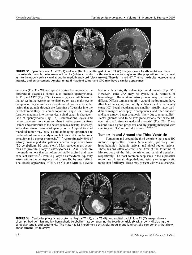
FIGURE 35. Ependymoma. Axial T2 (A) and axial (B) plus sagittal gadolinium T1 (C) images show a fourth ventricular massthat extends through the foramina of Luschka (white arrow) into both cerebellopontine angles and the prepontine cistern, as wellas into the upper cervical canal about the medulla and cord (black arrows). There is marked HC. The mass exhibits heterogeneousintensity and enhancement. Atypical teratoid rhabdoid tumor and CPC may have a similar appearance.
FIGURE 36. Cerebellar pilocytic astrocytoma. Sagittal T1 (A), axial T2 (B), and sagittal gadolinium T1 (C) images show acircumscribed vermian and left hemispheric cerebellar mass compressing the fourth ventricle (black arrows), displacing thecerebellar tonsils, and causing HC. The mass has T2-hyperintense cystic plus nodular and laminar solid components that showenhancement (white arrow).
Vertinsky and Barnes Top Magn Reson Imaging & Volume 18, Number 1, February 2007
46 * 2007 Lippincott Williams & Wilkins
Copyright @ Lippincott Williams & Wilkins. Unauthorized reproduction of this article is prohibited.
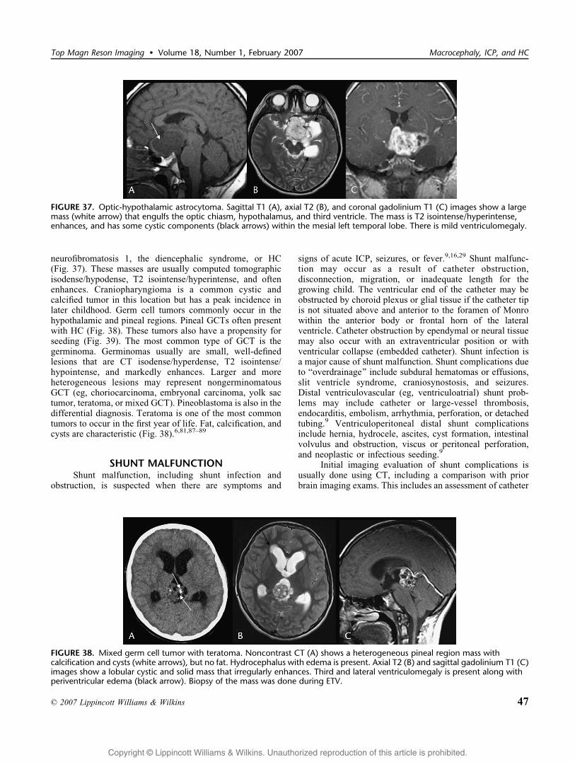
neurofibromatosis 1, the diencephalic syndrome, or HC(Fig. 37). These masses are usually computed tomographicisodense/hypodense, T2 isointense/hyperintense, and oftenenhances. Craniopharyngioma is a common cystic andcalcified tumor in this location but has a peak incidence inlater childhood. Germ cell tumors commonly occur in thehypothalamic and pineal regions. Pineal GCTs often presentwith HC (Fig. 38). These tumors also have a propensity forseeding (Fig. 39). The most common type of GCT is thegerminoma. Germinomas usually are small, well-definedlesions that are CT isodense/hyperdense, T2 isointense/hypointense, and markedly enhances. Larger and moreheterogeneous lesions may represent nongerminomatousGCT (eg, choriocarcinoma, embryonal carcinoma, yolk sactumor, teratoma, or mixed GCT). Pineoblastoma is also in thedifferential diagnosis. Teratoma is one of the most commontumors to occur in the first year of life. Fat, calcification, andcysts are characteristic (Fig. 38).6,81,87Y89
SHUNT MALFUNCTIONShunt malfunction, including shunt infection and
obstruction, is suspected when there are symptoms and
signs of acute ICP, seizures, or fever.9,16,29 Shunt malfunc-tion may occur as a result of catheter obstruction,disconnection, migration, or inadequate length for thegrowing child. The ventricular end of the catheter may beobstructed by choroid plexus or glial tissue if the catheter tipis not situated above and anterior to the foramen of Monrowithin the anterior body or frontal horn of the lateralventricle. Catheter obstruction by ependymal or neural tissuemay also occur with an extraventricular position or withventricular collapse (embedded catheter). Shunt infection isa major cause of shunt malfunction. Shunt complications dueto Boverdrainage[ include subdural hematomas or effusions,slit ventricle syndrome, craniosynostosis, and seizures.Distal ventriculovascular (eg, ventriculoatrial) shunt prob-lems may include catheter or large-vessel thrombosis,endocarditis, embolism, arrhythmia, perforation, or detachedtubing.9 Ventriculoperitoneal distal shunt complicationsinclude hernia, hydrocele, ascites, cyst formation, intestinalvolvulus and obstruction, viscus or peritoneal perforation,and neoplastic or infectious seeding.9
Initial imaging evaluation of shunt complications isusually done using CT, including a comparison with priorbrain imaging exams. This includes an assessment of catheter
FIGURE 37. Optic-hypothalamic astrocytoma. Sagittal T1 (A), axial T2 (B), and coronal gadolinium T1 (C) images show a largemass (white arrow) that engulfs the optic chiasm, hypothalamus, and third ventricle. The mass is T2 isointense/hyperintense,enhances, and has some cystic components (black arrows) within the mesial left temporal lobe. There is mild ventriculomegaly.
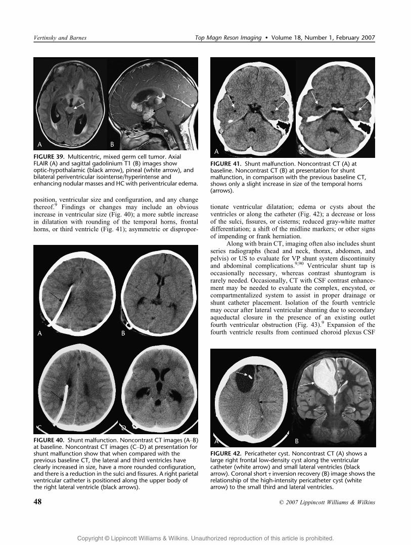
FIGURE 38. Mixed germ cell tumor with teratoma. Noncontrast CT (A) shows a heterogeneous pineal region mass withcalcification and cysts (white arrows), but no fat. Hydrocephalus with edema is present. Axial T2 (B) and sagittal gadolinium T1 (C)images show a lobular cystic and solid mass that irregularly enhances. Third and lateral ventriculomegaly is present along withperiventricular edema (black arrow). Biopsy of the mass was done during ETV.
Top Magn Reson Imaging & Volume 18, Number 1, February 2007 Macrocephaly, ICP, and HC
* 2007 Lippincott Williams & Wilkins 47
Copyright @ Lippincott Williams & Wilkins. Unauthorized reproduction of this article is prohibited.
position, ventricular size and configuration, and any changethereof.9 Findings or changes may include an obviousincrease in ventricular size (Fig. 40); a more subtle increasein dilatation with rounding of the temporal horns, frontalhorns, or third ventricle (Fig. 41); asymmetric or dispropor-
tionate ventricular dilatation; edema or cysts about theventricles or along the catheter (Fig. 42); a decrease or lossof the sulci, fissures, or cisterns; reduced gray-white matterdifferentiation; a shift of the midline markers; or other signsof impending or frank herniation.
Along with brain CT, imaging often also includes shuntseries radiographs (head and neck, thorax, abdomen, andpelvis) or US to evaluate for VP shunt system discontinuityand abdominal complications.9,90 Ventricular shunt tap isoccasionally necessary, whereas contrast shuntogram israrely needed. Occasionally, CT with CSF contrast enhance-ment may be needed to evaluate the complex, encysted, orcompartmentalized system to assist in proper drainage orshunt catheter placement. Isolation of the fourth ventriclemay occur after lateral ventricular shunting due to secondaryaqueductal closure in the presence of an existing outletfourth ventricular obstruction (Fig. 43).9 Expansion of thefourth ventricle results from continued choroid plexus CSF
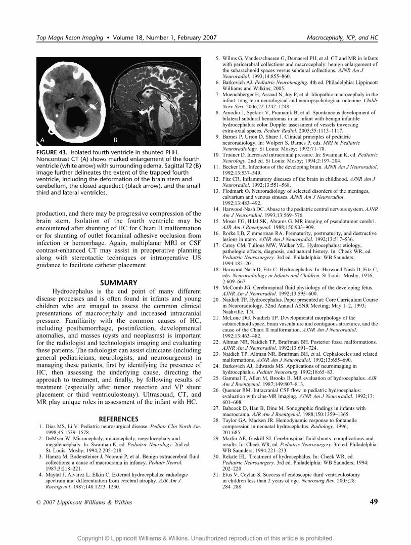
FIGURE 41. Shunt malfunction. Noncontrast CT (A) atbaseline. Noncontrast CT (B) at presentation for shuntmalfunction, in comparison with the previous baseline CT,shows only a slight increase in size of the temporal horns(arrows).
FIGURE 39. Multicentric, mixed germ cell tumor. AxialFLAIR (A) and sagittal gadolinium T1 (B) images showoptic-hypothalamic (black arrow), pineal (white arrow), andbilateral periventricular isointense/hyperintense andenhancing nodular masses and HC with periventricular edema.
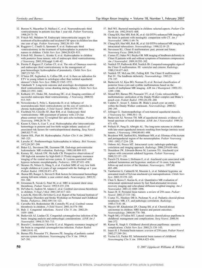
FIGURE 40. Shunt malfunction. Noncontrast CT images (AYB)at baseline. Noncontrast CT images (CYD) at presentation forshunt malfunction show that when compared with theprevious baseline CT, the lateral and third ventricles haveclearly increased in size, have a more rounded configuration,and there is a reduction in the sulci and fissures. A right parietalventricular catheter is positioned along the upper body ofthe right lateral ventricle (black arrows).
FIGURE 42. Pericatheter cyst. Noncontrast CT (A) shows alarge right frontal low-density cyst along the ventricularcatheter (white arrow) and small lateral ventricles (blackarrow). Coronal short T inversion recovery (B) image shows therelationship of the high-intensity pericatheter cyst (whitearrow) to the small third and lateral ventricles.
Vertinsky and Barnes Top Magn Reson Imaging & Volume 18, Number 1, February 2007
48 * 2007 Lippincott Williams & Wilkins
Copyright @ Lippincott Williams & Wilkins. Unauthorized reproduction of this article is prohibited.
production, and there may be progressive compression of thebrain stem. Isolation of the fourth ventricle may beencountered after shunting of HC for Chiari II malformationor for shunting of outlet foraminal adhesive occlusion frominfection or hemorrhage. Again, multiplanar MRI or CSFcontrast-enhanced CT may assist in preoperative planningalong with stereotactic techniques or intraoperative USguidance to facilitate catheter placement.
SUMMARYHydrocephalus is the end point of many different
disease processes and is often found in infants and youngchildren who are imaged to assess the common clinicalpresentations of macrocephaly and increased intracranialpressure. Familiarity with the common causes of HC,including posthemorrhage, postinfection, developmentalanomalies, and masses (cysts and neoplasms) is importantfor the radiologist and technologists imaging and evaluatingthese patients. The radiologist can assist clinicians (includinggeneral pediatricians, neurologists, and neurosurgeons) inmanaging these patients, first by identifying the presence ofHC, then assessing the underlying cause, directing theapproach to treatment, and finally, by following results oftreatment (especially after tumor resection and VP shuntplacement or third ventriculostomy). Ultrasound, CT, andMR play unique roles in assessment of the infant with HC.
REFERENCES1. Dias MS, Li V. Pediatric neurosurgical disease. Pediatr Clin North Am.
1998;45:1539Y1578.2. DeMyer W. Microcephaly, microcephaly, megalocephaly and
megalencephaly. In: Swaiman K, ed. Pediatric Neurology. 2nd ed.St. Louis: Mosby; 1994;2:205Y218.
3. Hamza M, Bodensteiner J, Noorani P, et al. Benign extracerebral fluidcollections: a cause of macrocrania in infancy. Pediatr Neurol.1987;3:218Y221.
4. Maytal J, Alvarez L, Elkin C. External hydrocephalus: radiologicspectrum and differentiation from cerebral atrophy. AJR Am JRoentgenol. 1987;148:1223Y1230.
5. Wilms G, Vanderschueren G, Demaerel PH, et al. CT and MR in infantswith pericerebral collections and macrocephaly: benign enlargement ofthe subarachnoid spaces versus subdural collections. AJNR Am JNeuroradiol. 1993;14:855Y860.
6. Barkovich AJ. Pediatric Neuroimaging. 4th ed. Philadelphia: LippincottWilliams and Wilkins; 2005.
7. Muenchberger H, Assaad N, Joy P, et al. Idiopathic macrocephaly in theinfant: long-term neurological and neuropsychological outcome. ChildsNerv Syst. 2006;22:1242Y1248.
8. Amodio J, Spektor V, Pramanik B, et al. Spontaneous development ofbilateral subdural hematomas in an infant with benign infantilehydrocephalus: color Doppler assessment of vessels traversingextra-axial spaces. Pediatr Radiol. 2005;35:1113Y1117.
9. Barnes P, Urion D, Share J. Clinical principles of pediatricneuroradiology. In: Wolpert S, Barnes P, eds. MRI in PediatricNeuroradiology. St Louis: Mosby; 1992:71Y78.
10. Trauner D. Increased intracranial pressure. In: Swaiman K, ed. PediatricNeurology. 2nd ed. St Louis: Mosby; 1994;2:197Y204.
11. Becker LE. Infections of the developing brain. AJNR Am J Neuroradiol.1992;13:537Y549.
12. Fitz CR. Inflammatory diseases of the brain in childhood. AJNR Am JNeuroradiol. 1992;13:551Y568.
13. Flodmark O. Neuroradiology of selected disorders of the meninges,calvarium and venous sinuses. AJNR Am J Neuroradiol.1992;13:483Y492.
14. Harwood-Nash DC. Abuse to the pediatric central nervous system. AJNRAm J Neuroradiol. 1993;13:569Y576.
15. Moser FG, Hilal SK, Abrams G. MR imaging of pseudotumor cerebri.AJR Am J Roentgenol. 1988;150:903Y909.
16. Rorke LB, Zimmerman RA. Prematurity, postmaturity, and destructivelesions in utero. AJNR Am J Neuroradiol. 1992;13:517Y536.
17. Carey CM, Tullous MW, Walker ML. Hydrocephalus: etiology,pathologic effects, diagnosis, and natural history. In: Cheek WR, ed.Pediatric Neurosurgery. 3rd ed. Philadelphia: WB Saunders;1994:185Y201.
18. Harwood-Nash D, Fitz C. Hydrocephalus. In: Harwood-Nash D, Fitz C,eds. Neuroradiology in Infants and Children. St Louis: Mosby; 1976;2:609Y667.
19. McComb JG. Cerebrospinal fluid physiology of the developing fetus.AJNR Am J Neuroradiol. 1992;13:595Y600.
20. Naidich TP. Hydrocephalus. Paper presented at: Core Curriculum Coursein Neuroradiology, 32nd Annual ASNR Meeting; May 1Y2, 1993;Nashville, TN.
21. McLone DG, Naidich TP. Developmental morphology of thesubarachnoid space, brain vasculature and contiguous structures, and thecause of the Chiari II malformation. AJNR Am J Neuroradiol.1992;13:463Y482.
22. Altman NR, Naidich TP, Braffman BH. Posterior fossa malformations.AJNR Am J Neuroradiol. 1992;13:691Y724.
23. Naidich TP, Altman NR, Braffman BH, et al. Cephaloceles and relatedmalformations. AJNR Am J Neuroradiol. 1992;13:655Y690.
24. Barkovich AJ, Edwards MS. Applications of neuroimaging inhydrocephalus. Pediatr Neurosurg. 1992;18:65Y83.
25. Gammal T, Allen M, Brooks B. MR evaluation of hydrocephalus. AJRAm J Roentgenol. 1987;149:807Y813.
26. Quencer RM. Intracranial CSF flow in pediatric hydrocephalus:evaluation with cine-MR imaging. AJNR Am J Neuroradiol. 1992;13:601Y608.
27. Babcock D, Han B, Dine M. Sonographic findings in infants withmacrocrania. AJR Am J Roentgenol. 1988;150:1359Y1365.
28. Taylor GA, Madsen JR. Hemodynamic response to fontanellecompression in neonatal hydrocephalus. Radiology. 1996;201:685.
29. Marlin AE, Gaskill SJ. Cerebrospinal fluid shunts: complications andresults. In: Cheek WR, ed. Pediatric Neurosurgery. 3rd ed. Philadelphia:WB Saunders; 1994:221Y233.
30. Rekate HL. Treatment of hydrocephalus. In: Cheek WR, ed.Pediatric Neurosurgery. 3rd ed. Philadelphia: WB Saunders; 1994:202Y220.
31. Etus V, Ceylan S. Success of endoscopic third ventriculostomyin children less than 2 years of age. Neurosurg Rev. 2005;28:284Y288.
FIGURE 43. Isolated fourth ventricle in shunted PHH.Noncontrast CT (A) shows marked enlargement of the fourthventricle (white arrow) with surrounding edema. Sagittal T2 (B)image further delineates the extent of the trapped fourthventricle, including the deformation of the brain stem andcerebellum, the closed aqueduct (black arrow), and the smallthird and lateral ventricles.
Top Magn Reson Imaging & Volume 18, Number 1, February 2007 Macrocephaly, ICP, and HC
* 2007 Lippincott Williams & Wilkins 49
Copyright @ Lippincott Williams & Wilkins. Unauthorized reproduction of this article is prohibited.
32. Buxton N, Macarthur D, Mallucci C, et al. Neuroendoscopic thirdventriculostomy in patients less than 1 year old. Pediatr Neurosurg.1998;29:73Y76.
33. Fritsch MJ, Mehdorn M. Endoscopic intraventricular surgery fortreatment of hydrocephalus and loculated CSF space in children less thanone year of age. Pediatr Neurosurg. 2002;36:183Y188.
34. Ruggiero C, Cinalli G, Spennato P, et al. Endoscopic thirdventriculostomy in the treatment of hydrocephalus in posterior fossatumors in children. Childs Nerv Syst. 2004;20:828Y833.
35. Fritsch MJ, Doerner L, Kienke S, et al. Hydrocephalus in childrenwith posterior fossa tumors: role of endoscopic third ventriculostomy.J Neurosurg. 2005;103(suppl 1):40Y42.
36. Peretta P, Ragazzi P, Carlino CF, et al. The role of Ommaya reservoirand endoscopic third ventriculostomy in the management ofpost-hemorrhagic hydrocephalus of prematurity. Childs Nerv Syst.2007;23:765Y771.
37. O’brien DF, Seghedoni A, Collins DR, et al. Is there an indication forETV in young infants in aetiologies other than isolated aqueductalstenosis? Childs Nerv Syst. 2006;22:1565Y1572.
38. Takahashi Y. Long-term outcome and neurologic development afterthird ventriculostomy versus shunting during infancy. Childs Nerv Syst.2006;22:1591Y1602.
39. Kulkarni AV, Drake JM, Armstrong DC, et al. Imaging correlates ofsuccessful endoscopic third ventriculostomy. J Neurosurg. 2000;92:915Y919.
40. Nowoslawska E, Polis L, Kaniewska D, et al. Influence ofneuroendoscopic third ventriculostomy on the size of ventricles inchronic hydrocephalus. J Child Neurol. 2004;19:579Y587.
41. Fischbein NJ, Ciricillo SF, Barr RM, et al. Endoscopic thirdventriculostomy: MR assessment of patency with 2-D cinephase-contrast versus T2-weighted fast spin echo technique. PediatrNeurosurg. 1998;28:70Y78.
42. Kazan S, Gura A, Ucar T, et al. Hydrocephalus after intraventricularhemorrhage in preterm and low-birth weight infants: analysis ofassociated risk factors for ventriculoperitoneal shunting. Surg Neurol.2005;S2:77Y81.
43. Garton HJL, Piatt JH. Hydrocephalus. Pediatr Clin N Am. 2004;51:305Y325.
44. Laroche JC. Posthemorrhagic hydrocephalus in infancy. Biol Neonate.1972;20:287Y299.
45. Baker LL, Stevenson DK, Enzmann DR. End-stage periventricularleukomalacia: MR evaluation. Radiology. 1988;168:809Y815.
46. Keeney SE, Adcock EW, McArdle CB. Prospective observations of100 high-risk neonates by high-field (1.5Tesla) magnetic resonanceimaging of the central nervous system. II. Lesions associated withhypoxic-ischemic encephalopathy. Pediatrics. 1991;87:431Y438.
47. Skranes JS, Nilsen G, Olaug S, et al. Cerebral MRI of very low birthweight children at 6 years of age compared with the findings at 1 year.Pediatr Radiol. 1998;28:471Y475.
48. Jhawar BS, Ranger A, Steven D. Risk factors for intracranial hemorrhageamong full-term infants: a case control study. Neurosurgery. 2003;52:581Y588.
49. Grossman R, Novak G, Patel M, et al. MRI in neonatal dural sinusthrombosis. Pediatr Neurol. 1993;9:235Y238.
50. DeVeber G, Andrew M, Adams C, et al. Cerebral sinovenous thrombosisin children. N Engl J Med. 2001;345:417Y423.
51. Lynch JK, Hirtz DG, DeVeber G, et al. Report of the National Institute ofNeurologic Disorders and Stroke Workshop on Perinatal and ChildhoodStroke. Pediatrics. 2002;109:116Y123.
52. Carvalho KS, Bodensteiner JB, Connolly PJ, et al. Cerebral venousthrombosis in children. J Child Neurol. 2001;16:574Y580.
53. Bale J. Congenital infections. Neurol Clin N Am. 2002;20:1039Y1060.
54. Barkovich AJ, Lindan CE. Congenital cytomegalovirus infection of thebrain: imaging analysis and embryologic considerations. AJNR Am JNeuroradiol. 1994;15:703Y715.
55. Boesch C, Issakainen J, Kewitz G, et al. Magnetic resonance imaging ofthe brain in congenital cytomegalovirus infection. Pediatr Radiol.1989;19:91Y93.
56. Barnes PD, Poussaint TY, Burrows PE: Imaging of pediatric centralnervous system infections. Neuroimaging Clin N Am. 1994;4:367Y391.
57. Bell WE. Bacterial meningitis in children: selected aspects. Pediatr ClinNorth Am. 1992;39:651Y668.
58. Chang KH, Han MH, Roh JK, et al. Gd-DTPA-enhanced MR imaging ofthe brain in patients with meningitis: comparison with CT. Am JNeuroradiol. 1990;11:69Y76.
59. Chang KH, Han MH, Roh JK, et al. Gd-DTPA-enhanced MR imaging inintracranial tuberculosis. Neuroradiology. 1990;32:19Y25.
60. Stevenson KL. Chiari II malformation: past, present and future.Neurosurg Focus. 2004;16:E5.
61. Curnes JT, OakesWJ, Boyko OB. MR imaging of hindbrain deformity inChiari II patients with and without symptoms of brainstem compression.Am J Neuroradiol. 1989;10:293Y302.
62. Naidich TP, Pudlowski RM, Naidich JB. Computed tomographic signs ofthe Chiari II malformation. III: ventricles and cisterns. Radiology.1980;134:657Y663.
63. Naidich TP, McLone DG, Fulling KH. The Chiari II malformation:Part IV. The hindbrain deformity. Neuroradiology. 1983;25:179Y197.
64. Barkovich AJ, Kjos BO, Norman D, et al. Revised classification ofposterior fossa cysts and cystlike malformations based on theresults of multiplanar MR imaging. AJR Am J Roentgenol. 1989;153:1289Y1300.
65. Strand RD, Barnes PD, Poussaint TY, et al. Cystic retrocerebellarmalformations: unification of the Dandy-Walker complex and Blake’spouch cyst. Pediatr Radiol. 1993;28:258Y260.
66. Calabro F, Arcuri T, Jinkins JR. Blake’s pouch cyst: an entitywithin the Dandy-Walker continuum. Neuroradiology. 2000;42:290Y295.
67. Jellinger G. Anatomopathology of non-tumoral aqueductal stenosis.J Neurosurg Sci. 1986;30:1Y16.
68. Barkovich AJ, Newton TH. MR of aqueductal stenosis: evidence of abroad spectrum of tectal distortion. AJNR Am J Neuroradiol. 1989;10:471Y476.
69. Pollack IF, Pang D, Albright AL. The long-term outcome in childrenwith late-onset aqueductal stenosis resulting from benign intrinsic tectaltumors. J Neurosurg. 1994;80:681Y688.
70. Boydston WR, Sanford RA, Muhlbauer MS, et al. Gliomas of the tectumand periaqueductal region of the mesencephalon. Pediatr Neurosurg.1991Y1992;17:234Y238.
71. Osborn AG, Preece MT. Intracranial cysts: radiologic-pathologiccorrelation and imaging approach. Radiology. 2006;239:650Y664.
72. Donaldson JW, Edwards-Brown M, Luerssen TG. Arachnoid cystrupture with concurrent subdural hygroma. Pediatr Neurosurg.2000;32:137Y139.
73. Parsch CS, Krauss J, Hofmann E, et al. Arachnoid cysts associated withsubdural hematomas and hygromas: analysis of 16 cases, long-termfollow-up and review of the literature. Neurosurgery. 1997;40:483Y490.
74. Tamburrini G, Caldarelli M, Massimi L, et al. Subdural hygroma: anunwanted result of Sylvian arachnoid cyst marsupialization. Childs NervSyst. 2003;19:159Y165.
75. Chen S, Ikawa F, Kurisu K, et al. Quantitative MR evaluation ofintracranial epidermoid tumors by fast fluid-attenuated inversionrecovery imaging and echo-planar diffusion-weighted imaging. Am JNeuroradiol. 2001;22:1089Y1096.
76. Isaacs H. II. Perinatal brain tumors: a review of 250 cases. PediatrNeurol. 2002;27:333Y342.
77. Coates TL, Hinshaw DB Jr, Peckman N, et al. Pediatric choroid plexusneoplasms: MR, CT, and pathologic correlation. Radiology.1989;173:81Y88.
78. Meyers SP, Khademian ZP, Chuang SH, et al. Choroid plexuscarcinomas in children: MRI features and patient outcomes.Neuroradiology. 2004;46:770Y780.
79. Nagib MG, O’Fallon MT. Lateral ventricle choroid plexus papilloma inchildhood: management and complications. Surg Neurol. 2000;54:366Y372.
80. Kumar R, Singh S. Childhood choroid plexus papillomas: operativecomplications. Childs Nerv Syst. 2005;21:138Y143.
81. Isaacs H. I. Perinatal brain tumors: a review of 250 cases. Pediatr Neurol.2002;27:249Y261.
82. Vezina LG, Packer RJ. Infratentorial brain tumors of childhood.Neuroimaging Clin N Am. 1994;4:423Y436.
Vertinsky and Barnes Top Magn Reson Imaging & Volume 18, Number 1, February 2007
50 * 2007 Lippincott Williams & Wilkins
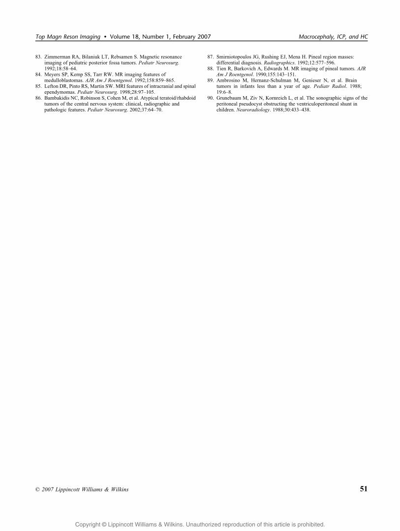
Copyright @ Lippincott Williams & Wilkins. Unauthorized reproduction of this article is prohibited.
83. Zimmerman RA, Bilaniuk LT, Rebsamen S. Magnetic resonanceimaging of pediatric posterior fossa tumors. Pediatr Neurosurg.1992;18:58Y64.
84. Meyers SP, Kemp SS, Tarr RW. MR imaging features ofmedulloblastomas. AJR Am J Roentgenol. 1992;158:859Y865.
85. Lefton DR, Pinto RS, Martin SW. MRI features of intracranial and spinalependymomas. Pediatr Neurosurg. 1998;28:97Y105.
86. Bambakidis NC, Robinson S, Cohen M, et al. Atypical teratoid/rhabdoidtumors of the central nervous system: clinical, radiographic andpathologic features. Pediatr Neurosurg. 2002;37:64Y70.
87. Smirniotopoulos JG, Rushing EJ, Mena H. Pineal region masses:differential diagnosis. Radiographics. 1992;12:577Y596.
88. Tien R, Barkovich A, Edwards M. MR imaging of pineal tumors. AJRAm J Roentgenol. 1990;155:143Y151.
89. Ambrosino M, Hernanz-Schulman M, Genieser N, et al. Braintumors in infants less than a year of age. Pediatr Radiol. 1988;19:6Y8.
90. Grunebaum M, Ziv N, Kornreich L, et al. The sonographic signs of theperitoneal pseudocyst obstructing the ventriculoperitoneal shunt inchildren. Neuroradiology. 1988;30:433Y438.
Top Magn Reson Imaging & Volume 18, Number 1, February 2007 Macrocephaly, ICP, and HC
* 2007 Lippincott Williams & Wilkins 51